Translating Protocols Between Scanner Manufacturer and Model
|
|
|
- June Charles
- 6 years ago
- Views:
Transcription

1 Translating Protocols Between Scanner Manufacturer and Model Robert J. Pizzutiello, MS, FAAPM, FACMP Sr. Vice-President, Global Physics Solutions President, Upstate Medical Physics
2 Objectives Understand the complexities and pitfalls of translating protocols between manufacturers and models Use the CT Protocol Tools on the AAPM Web to translate protocols Apply the tools to real life examples
3 Outline Basic CT Parameters common to all manufacturers Proprietary names, manufacturer specific The AAPM Lexicon website Expected results CTDI vol within range Time related factors (motion, ma, effective mas, pitch, etc.) Thickness/resolution and noise requirements in the reconstructed images Retro recon capabilities/limitations? Start with most common exams, with ACR MAP data Adult head, abdomen, ped abdomen Chest protocol examples (Mayo) Example using AAPM Web site for Routine Head scan (WIP) Balance specific features of different manufacturers
4 Single manufacturer, multiple scanners Seems easy right?... Different scanner characteristics Max ma Tube rotation time Detector configuration Two approaches Standardization: All scanners same protocols Easier for Radiologists to compare Optimize using max capabilities of each scanner Select patients/exams for optimum clinical benefit
5 Multiple manufacturers, multiple scanners This gets complex in a hurry Different scanner characteristics Max ma Tube rotation time Detector configuration Two approaches (Standardization Optimization) Make optimal use of features Smartphone, texting, etc. Nomenclature, nom, nombre, Namen haben
6 When is a rose not a rose? Names are different Challenge for staff Big challenge for medical physicists Concern discussed at 2010 CT Dose Summit, and in many venues by leaders within AAPM AAPM jumped into action!
7 Working Group on Standardization of CT Nomenclature and Protocols (WG) 1. To develop consensus protocols for frequently performed CT examinations, summarizing the basic requirements of the exam and giving several model-specific examples of scan and reconstruction parameters. General comments on contrast administration may be included, as appropriate. 2. To develop by consensus a set of standardized terms for use on CT scanners, including all parameters that control the scan acquisition or reconstruction that are programmed by the user, displayed on the final image or included in a DICOM-specified tag, or described a fundamental CT principle (such as a beam-shaping filter). With AAPM leadership, we will seek support of the ACR and ASRT. Also work with MITA so that the standardized terms are eventually adopted by the IEC
 Dianna Cody (Co-chair) Dustin")

8 WG Members AAPM ACR ASRT FDA MITA GE Hitachi Philips Siemens Toshiba AAPM: ACR: ASRT: DICOM: FDA: MITA: GE: Hitachi: Philips: Siemens: Toshiba: Biweekly conference calls since RSNA 2010 Members: Cynthia McCollough (Chair) Dianna Cody (Co-chair) Dustin Gress James Kofler Michael McNitt-Gray Robert Pizzutiello Mark Armstrong Theresa Branham Priscilla Butler Virginia Lester David Clunie Kevin O'Donnell Thalia Mills Gail Rodriguez John Jaeckle Mark Silverman Mark Olszewski Christianne Leidecker Richard Mather
9 First deliverable: The Lexicon CT scan parameters: Translation of terms for different manufacturers Introduction For the CT technologist who operates multiple scanner models, perhaps from multiple manufacturers, the variability in names for important scan acquisition and reconstruction parameters can lead to confusion, reduced comfort and an increased potential for error. The intent of this CT terminology lexicon is to allow users to translate important CT acquisition and reconstruction terms between different manufacturers' systems.
10 This website will be updated as the terminology standardization work progresses. The generic descriptions or terms in the first column are intended to orient the user to the relevant concepts; they are not consensus "preferred terms." The generic descriptions are not based on any single existing or pending terminology standard; however the references cited below were consulted in developing the generic descriptions. Future efforts of this Working Group include making recommendations for standardized terminology.
11 A number of individuals and groups have advocated for terminology standardization in CT, including at a March 30-31, 2010 FDA public meeting entitled "Device Improvements to Reduce Unnecessary Radiation Exposure from Medical Imaging" (transcripts available at: ts/workshopsconferences/ucm pdf; see p ). Participants proposed a cooperative effort among professional organizations (AAPM, ASRT, ACR, etc.), industry, FDA, and standards organizations to accomplish this task, as is now being undertaken by this Working Group.
12 First deliverable: The Lexicon This represents a first step in the terminology standardization effort undertaken by this working group. Phase 2 of our work will: 1. Identify relevant terms from established standard lexicons (e.g. RadLex and DICOM) and other relevant literature and publish an expanded lexicon including these terms. 2. Form consensus recommendations on preferred terms.
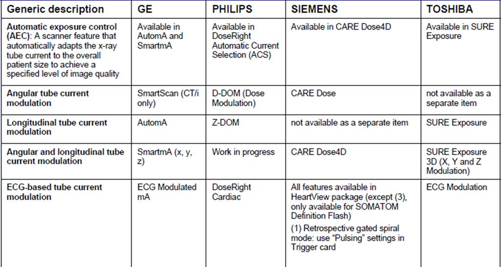
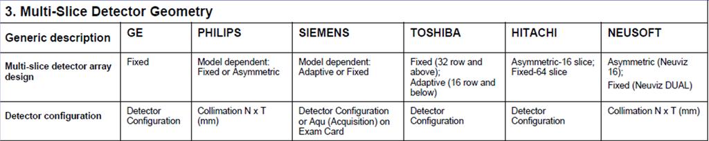
13 1. Scan acquisition and user interface basics 2. Dose modulation and reduction tools 3. Multi-Slice Detector Geometry 4. Image Reconstruction and Display 5. Contrast Media Tools 6. Multi-planar formats and 3-D Processing 7. Service and Application Tools 8. Workflow
14
15

16 AAPM 2011 Summit on CT Dose


17 AAPM 2011 Summit on CT Dose


18 AAPM 2011 Summit on CT Dose

19
20

21 Courtesy C. McCollough

22 AAPM 2011 Summit on CT Dose
23 Noise, Image Thickness and Pitch Fundamental relationship Noise increases as fewer photons form the image In spiral CT, image noise is dependent on pitch mas must be changed as pitch is changed Relationship is linear on some systems, but not all Siemens Effective mas = mas/pitch Review how manufacturers handle noise
: 5 Rel.")
: 100 % 2.")

 Increased image noise")
24 Image Thickness Noise 1 # Photons Image (mm): 5 Rel. Noise: 100 % Req. mas (for = noise): 100 % % 200 % % 400 % % 800 % Better z-resolution (less partial vol. averaging) Increased image noise Potential for increased radiation dose Courtesy J. Koefler
25 Let s look at some specific protocols Chest Examples from Mayo Clinic Protocols Chosen to make optimal use of each scanners capabilities Courtesy C. McCollough
26 Routine Chest Courtesy C. McCollough
27 Routine Chest Courtesy C. McCullough
28 Routine Chest Courtesy C. McCollough

29 Routine Chest Courtesy C. McCollough

30 Routine Chest Courtesy C. McCollough

31 AAPM 2011 Summit on CT Dose Routine Chest Courtesy C. McCollough

32 Routine Chest Courtesy C. McCollough
33
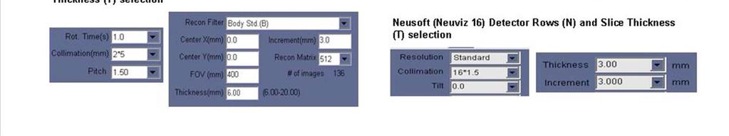
34 Practical Example Routine Adult Head Work in Progress Start by defining expected results D t N CTDI vol within range Time related factors (motion, ma, effective mas, pitch, etc.) Thickness/resolution and noise requirements in the reconstructed images How is Noise reference applied, by mfr? First recon? Retro recon capabilities/limitations? Check CT Protocols on AAPM web site In Routine Head example, look at these parameters
35 ROUTINE HEAD (BRAIN) - Indications A. Acute head trauma. Partial List B. Suspected acute intracranial hemorrhage. C. Immediate postoperative evaluation following brain surgery D. Suspected shunt malfunctions, or shunt revisions. E. Mental status change. F. Increased intracranial pressure. G. Headache. H. Etc.
36 Diagnostic Task Use these to guide discussions of image quality requirements (thickness, noise, etc.) Detect collections of blood Identify brain masses Detect brain edema or ischemia Identify shift in the normal locations of the brain structures including cephalad or caudal directions Evaluate the location of shunt hardware and the size of the ventricles Evaluate the size of the sulci and relative changes in symmetry
37 Radiation Dose Management D Tube Current Modulation (or Automatic Exposure Control) may be used, but is often turned off. According to ACR CT Accreditation Program guidelines: the reference level CTDIvol is 75 mgy the pass/fail limit is 80 mgy. These values are for a routine head and may be significantly different (higher or lower) for a given patient with unique indications, etc. NOTE: All CTDIvol are for 16 cm diameter phantom
38 t General Scan Instruction Suggestions Table height at External Auditory Meatus (EAM). PATIENT POSITIONING: Patient supine, head first, head in head-holder. D To reduce or avoid ocular lens exposure, the scan angle should be parallel to a line created by the supraorbital ridge and the inner table of the posterior margin of the foramen magnum. This may be accomplished by either by head tuck or gantry tilt in most situations.
39 EXAMPLE PROTOCOLS of both AXIAL/SEQUENTIAL and HELICAL scans are provided. There are advantages and disadvantages to using either axial or helical scans for routine heads. The best choice varies by patient, by indication and by scanner. Users of this document should consider the following and consult with both the manufacturer and a medical physicist to assist in determining which mode to use and when.
40 EXAMPLE PROTOCOLS of both AXIAL/SEQUENTIAL and HELICAL scans are provided. AXIAL SCANS generally have less artifact, but the scan takes slightly longer HELICAN SCANS may have more image artifact, especially for scanners with < 16 detector rows, but can give close to or equivalent performance for scanners with 64 detector rows.
41 HEAD ROUTINE (SEQUENTIAL): SELECTED SIEMENS SCANNERS D Topogram: Lateral, 256 mm. Patient positioning: Patient lying in supine position, arms resting along the body, secure head well in the head holder, support lower legs. Gantry tilt is available for sequence scanning, not for spiral scanning. Gantry tilt is not available for dual source scanners.
42 HEAD ROUTINE (SEQUENTIAL): SELECTED SIEMENS SCANNERS For all head studies, it is very important for image quality to position the patient in the center of the scan field. Use the lateral laser beam to make sure that the patient is positioned in the center. In order to optimize image quality versus radiation dose, scans are provided within a maximum scan field of 300 mm with respect to the iso-center. No recon job with a field of view exceeding those limits will be possible. Therefore, patient positioning has to be performed accurately to ensure a centered location of the skull.
43 HEAD ROUTINE (SEQUENTIAL): SELECTED SIEMENS SCANNERS Sensation 16 Sensation 64 Definition (dual source, 64 slices) Definition AS (128 slices) Parameter Software version VB30 VB30 VA34 VA27 VA34 Scan mode seq seq seq seq seq Tube voltage / kv Effective mas / Qual ref 270/ mas* Base/Cerebrum Rotation time / s Collimation / mm / Base/Cerebrum Pitch n.a. n.a. n.a. n.a. n.a. Dose modulation n.a. n.a. n.a. n.a. n.a. Scan area head head head head head Scan length / mm 40.5/ Base/Cerebrum Scan time / s 1.0/ Base/Cerebrum CTDIvol (16 cm phantom) 60.5/59.5 Base/Cerebrum Reconstruction I Kernel H31s H31s H31s H31s H31s Slice / mm 4.5/ Base/Cerebrum Slice increment / mm n.a. n.a. n.a. n.a. n.a. Definition Flash (dual source, 128 slices)
44 HEAD ROUTINE (SPIRAL): SELECTED SIEMENS SCANNERS Gantry tilt is available for sequence scanning, not for spiral scanning. Gantry tilt is not available for dual source scanners. For all head studies, it is very important for image quality to position the patient in the center of the scan field. Use the lateral laser beam to make sure that the patient is positioned in the center.
45 HEAD ROUTINE (SPIRAL): SELECTED SIEMENS SCANNERS In order to optimize image quality versus radiation dose, scans are provided within a maximum scan field of 300 mm with respect to the iso-center. No recon job with a field of view exceeding those limits will be possible. Therefore, patient positioning has to be performed accurately to ensure a centered location of the skull. TOPOGRAM: Lateral, 256, 120 kv, 50 ma, direction is craniocaudal.
46 HEAD ROUTINE (SPIRAL): SELECTED SIEMENS SCANNERS Parameter Sensation 64 Definition (dual source, 64 slices) Definition AS (128 slices) Software version VB30 VA34 VA27 VA34 Scan mode spi spi spi spi Tube voltage / kv Effective mas / Qual ref mas* Rotation time / s Collimation / mm Pitch Definition Flash (dual source, 128 slices) Dose modulation CARE Dose CARE Dose CARE Dose CARE Dose CTDIvol Reconstruction Recon Start Top of Frontal Sinus Top of Frontal Sinus Top of Frontal Sinus Top of Frontal Sinus Recon End Vertex Vertex Vertex Vertex Kernel H31s H31s H31s H31s Slice / mm Slice increment / mm
47 GE Recon Algorithms Soft Standard Detail Lung Bone Edge Bone Plus Courtesy C. McCollough
48 Siemens Recon Kernels B10 B90 Body (90 is sharpest) H10 H90 Head U30 U90 Ultra High Resolution T20 T81 Topogram Lower number smoother Higher number sharper Multiples of 10 are the basic kernels In between values are special kernels Courtesy C. McCollough
49 Review Basic CT Parameters common to all manufacturers The AAPM Lexicon website Expected results CTDI vol within range Time related factors (motion, ma, effective mas, pitch, etc.) Thickness/resolution and noise requirements in the reconstructed images Retro recon capabilities/limitations? Start with most common exams, with ACR MAP data Adult head, abdomen, ped abdomen Chest protocol examples (Mayo) Example using AAPM Web site for Routine Head scan (WIP) Balance specific benefits of features for each manufacturer
Automated dose control in multi-slice CT. Nicholas Keat Formerly ImPACT, St George's Hospital, London
 Automated dose control in multi-slice CT Nicholas Keat Formerly ImPACT, St George's Hospital, London Introduction to presentation CT contributes ~50+ % of all medical radiation dose Ideally all patients
TOPICS: CT Protocol Optimization over the Range of Patient Age & Size and for Different CT Scanner Types: Recommendations & Misconceptions
 CT Protocol Optimization over the Range of Patient Age & Size and for Different CT Scanner Types: Recommendations & Misconceptions TOPICS: Computed Tomography Quick Overview CT Dosimetry Effects of CT
Research Support. Dual-Source CT: What is it and How Do I Test it? Cynthia H. McCollough, Ph.D.
 Dual-Source CT: What is it and How Do I Test it? Cynthia H. McCollough, Ph.D. CT Clinical Innovation Center Department of Radiology Mayo Clinic College of Medicine Rochester, MN Research Support National
diagnostic examination
 RADIOLOGICAL PHYSICS 2011 Raphex diagnostic examination Adel A. Mustafa, Ph.D., Editor PUBLISHED FOR: RAMPS (Radiological and Medical Physics Society of New York) preface The RAPHEX Diagnostic exam 2011
QC by the MPE in Belgium
 Acceptance testing of state-of-the-art CT scanners using a new national protocol: first experience on a large number of scanners of different make and model the working group Radiology of the Belgian Hospital
1. Patient size AEC. Large Patient High ma. Small Patient Low ma
 Comparison of the function and performance of CT AEC systems CTUG meeting by Emily Field Trainee clinical scientist 14 th th Breakdown CT Automatic Exposure Control (AEC) Background Project Description
Image Quality and Dose. Image Quality and Dose. Image Quality and Dose Issues in MSCT. Scanner parameters affecting IQ and Dose
 Image Quality and Dose Issues in MSCT Image Quality and Dose Image quality Image noise Spatial resolution Contrast Artefacts Speckle and sharpness S. Edyvean St. George s Hospital London SW17 0QT Radiation
Pitfalls and Remedies of MDCT Scanners as Quantitative Instruments
 intensity m(e) m (/cm) 000 00 0 0. 0 50 0 50 Pitfalls and Remedies of MDCT Scanners as Jiang Hsieh, PhD GE Healthcare Technology University of Wisconsin-Madison Root-Causes of CT Number Inaccuracies Nature
TORNIER BLUEPRINT. 3D Planning + PSI SCAN PROTOCOL
 TORNIER BLUEPRINT 3D Planning + PSI SCAN PROTOCOL Contents 3 Introduction 3 Patient preparation 3 Scanning instructions 4 Image instructions 5 Scanning parameters 6 Technical instructions 2 BLUEPRINT 3D
HISTORY. CT Physics with an Emphasis on Application in Thoracic and Cardiac Imaging SUNDAY. Shawn D. Teague, MD
 CT Physics with an Emphasis on Application in Thoracic and Cardiac Imaging Shawn D. Teague, MD DISCLOSURES 3DR- advisory committee CT PHYSICS WITH AN EMPHASIS ON APPLICATION IN THORACIC AND CARDIAC IMAGING
160-slice CT SCANNER / New Standard for the Future
 TECHNOLOGY HISTORY For over 130 years, Toshiba has been a world leader in developing technology to improve the quality of life. Our 50,000 global patents demonstrate a long, rich history of leading innovation.
12/21/2016. Siemens Medical Systems Research Agreement Philips Healthcare Research Agreement AAN and ASN Committees
 Joseph V. Fritz, PhD Nandor Pintor, MD Dent Neurologic Institute ASN 2017 Friday, January 20, 2017 Siemens Medical Systems Research Agreement Philips Healthcare Research Agreement AAN and ASN Committees
Slide 1. Slide 2. Slide 3 ACR CT Accreditation. Multi-Slice CT Artifacts and Quality Control. What are the rules or recommendations for CT QC?
 Slide 1 Multi-Slice CT Artifacts and Quality Control Dianna Cody, Ph.D. Chief, Radiologic Physics UT MD Anderson Cancer Center Houston, TX Slide 2 What are the rules or recommendations for CT QC? AAPM
The disclaimer on page 1 is an integral part of this document. Copyright November 30, 2017 by AAPM. All rights reserved.
 DISCLAIMER: TO THE EXTENT ALLOWED BY LOCAL LAW, THIS INFORMATION IS PROVIDED TO YOU BY THE AMERICAN ASSOCIATION OF PHYSICISTS IN MEDICINE, A NON-PROFIT ORGANIZATION ORGANIZED TO PROMOTE THE APPLICATION
abc MHRA Philips Mx8000 IDT CT scanner technical evaluation September 2004 Best choice best practice nww.medical-devices.nhs.
 abc September 2004 MHRA 04099 Philips Mx8000 IDT CT scanner technical evaluation Best choice best practice www.mhra.gov.uk nww.medical-devices.nhs.uk About MHRA evaluation reports. What you can expect.
Clinical Experience Using the Open Bore Multislice CT System Supria (16 slice CT) MEDIX VOL. 61 P.8 P.11
 MEDIX VOL. 61 P.8 P.11") Clinical Experience Using the Open Bore Multislice CT System Supria (16 slice CT) Hiroki Kadoya Yukiko Kitagawa MEDIX VOL. 61 P.8 P.11 Clinical Experience Using the Open Bore Multislice CT System Supria
Clinical Experience Using the Open Bore Multislice CT System Supria (16 slice CT) Hiroki Kadoya Yukiko Kitagawa MEDIX VOL. 61 P.8 P.11 Clinical Experience Using the Open Bore Multislice CT System Supria
Acceptance Testing of a Digital Breast Tomosynthesis Unit
 Acceptance Testing of a Digital Breast Tomosynthesis Unit 2012 AAPM Spring Clinical Meeting Jessica Clements, M.S., DABR Objectives Review of technology and clinical advantages Acceptance Testing Procedures
Focal Spot Blooming in CT: We Didn t Know We Had a Problem Until We Had a Solution
 Focal Spot Blooming in CT: We Didn t Know We Had a Problem Until We Had a Solution Cynthia H. McCollough, PhD, DABR, FAAPM, FACR Director, CT Clinical Innovation Center Professor of Medical Physics and
Diagnostic X-Ray Shielding
 Diagnostic X-Ray Shielding Multi-Slice CT Scanners Using NCRP 147 Methodology Melissa C. Martin, M.S., FAAPM, FACR Therapy Physics Inc., Bellflower, CA AAPM Annual Meeting, Orlando, FL FL Refresher Course
Wide beam CT dosimetry. Elly Castellano
 Wide beam CT dosimetry Elly Castellano Outline revision: CT dose indices wide-beam CT: the end of the road for CTDI? the IEC rescue plan for CTDI 100 the american way AAPM report 111 better estimates of
Maximum Performance, Minimum Space
 TECHNOLOGY HISTORY For over 130 years, Toshiba has been a world leader in developing technology to improve the quality of life. Our 50,000 global patents demonstrate a long, rich history of leading innovation.
COMPUTED TOMOGRAPHY 1
 COMPUTED TOMOGRAPHY 1 Why CT? Conventional X ray picture of a chest 2 Introduction Why CT? In a normal X-ray picture, most soft tissue doesn't show up clearly. To focus in on organs, or to examine the
NeuViz 16 Computed Tomography. Elevating routine imaging for exceptional results
 NeuViz 16 Computed Tomography Elevating routine imaging for exceptional results Essence NeuViz 16 Raising the bar on clinical utility in routine imaging. Get more. More clinical information for patients.
QC Testing for Computed Tomography (CT) Scanner
 Scanner") QC Testing for Computed Tomography (CT) Scanner QA - Quality Assurance All planned and systematic actions needed to provide confidence on a structure, system or component. all-encompassing program, including
QC Testing for Computed Tomography (CT) Scanner QA - Quality Assurance All planned and systematic actions needed to provide confidence on a structure, system or component. all-encompassing program, including
Software and Hardware in CCTA. Elly Castellano PhD
 Software and Hardware in CCTA Elly Castellano PhD Outline technical requirements for coronary CTA the modern cardiac CT scanner ECG-gating technology image reconstruction algorithms 2 Technical requirements
Computed Tomography. The Fundamentals of... THE FUNDAMENTALS OF... Jason H. Launders, MSc. Current Technology
 The Fundamentals of... Computed Tomography Computed Tomography (CT) systems use x-rays to produce images of slices through a patient s anatomy. Despite having lower spatial resolution than other x-ray
2D, 3D CT Intervention, and CT Fluoroscopy
 2D, 3D CT Intervention, and CT Fluoroscopy SOMATOM Definition, Definition AS, Definition Flash Answers for life. Siemens CT Vision Siemens CT Vision The justification for the existence of the entire medical
Dose Reduction and Image Preservation After the Introduction of a 0.1 mm Cu Filter into the LODOX Statscan unit above 110 kvp
 Dose Reduction and Image Preservation After the Introduction of a into the LODOX Statscan unit above 110 kvp Abstract: CJ Trauernicht 1, C Rall 1, T Perks 2, G Maree 1, E Hering 1, S Steiner 3 1) Division
Iterative Reconstruction
 RECENT ADVANCES IN CT RADIATION DOSE REDUCTION TECHNIQUES Iterative Reconstruction Kalpana Kanal, PhD, FSCBTMR, FACR, FAAPM Professor and Director, Diagnostic Physics Section University of Washington Seattle,
Iterative Reconstruction in Image Space. Answers for life.
 Iterative Reconstruction in Image Space Answers for life. Iterative Reconstruction in Image Space * (IRIS) * Please note: IRIS is used as an abbreviation for Iterative Reconstruction in Image Space throughout
Influence of different iteration levels in fourth generation iterative reconstruction technique on image noise in CT examinations of the neck
 Influence of different iteration levels in fourth generation iterative reconstruction technique on image noise in CT examinations of the neck Poster No.: C-2205 Congress: ECR 2012 Type: Scientific Paper
Exposure in Dental Radiology: A Comparison Between Intra-oral, Panoramic and Tomographic Examinations
 Exposure in Dental Radiology: A Comparison Between Intra-oral, Panoramic and Tomographic Examinations S. Baechler 1, P. Monnin 1, A. Aroua 1, J.F. Valley 1, M. Perrier, P. Trueb 3, F.R. Verdun 1 1 University
Breast Tomosynthesis. Bob Liu, Ph.D. Department of Radiology Massachusetts General Hospital And Harvard Medical School
 Breast Tomosynthesis Bob Liu, Ph.D. Department of Radiology Massachusetts General Hospital And Harvard Medical School Outline Physics aspects of breast tomosynthesis Quality control of breast tomosynthesis
Detector technology in simultaneous spectral imaging
 Computed tomography Detector technology in simultaneous spectral imaging Philips IQon Spectral CT Z. Romman, I. Uman, Y. Yagil, D. Finzi, N. Wainer, D. Milstein; Philips Healthcare While CT has become
Authors: Cabral, Ricardo 1 ; Carvoeiras, Pedro 2 ; Fatana, João 2, ; Alves, Rita 1. 1 Centro Hospitalar Lisboa Norte - Hospital de Santa Maria; 2
 Authors: Cabral, Ricardo 1 ; Carvoeiras, Pedro 2 ; Fatana, João 2, ; Alves, Rita 1. 1 Centro Hospitalar Lisboa Norte - Hospital de Santa Maria; 2 Medical Consult, SA; Establish a method to correlate image
Electronic Noise in CT Detectors: Impact on Image Noise and Artifacts
 Medical Physics and Informatics Original Research Duan et al. Electronic Noise in CT Detectors Medical Physics and Informatics Original Research Xinhui Duan 1 Jia Wang 1,2 Shuai Leng 1 ernhard Schmidt
1. Queries are issued to the image archive for information about computed tomographic (CT)
") Appendix E1 Exposure Extraction Method examinations. 1. Queries are issued to the image archive for information about computed tomographic (CT) 2. Potential dose report screen captures (hereafter, dose
Appendix E1 Exposure Extraction Method examinations. 1. Queries are issued to the image archive for information about computed tomographic (CT) 2. Potential dose report screen captures (hereafter, dose
Maximizing clinical outcomes
 Maximizing clinical outcomes Digital Tomosynthesis Dual Energy Subtraction Automated Long Length Imaging Improved image quality at a low dose Xray Xray Patented ISS capture technology promotes high sensitivity
Surveying and QC of Stereotactic Breast Biopsy Units for ACR Accreditation
 Surveying and QC of Stereotactic Breast Biopsy Units for ACR Accreditation AAPM Annual Clinical Meeting Indianapolis, IN August 5, 2013 Learning Objectives Become familiar with the recommendations and
Optimized CT metal artifact reduction using the Metal Deletion Technique (MDT)
") Optimized CT metal artifact reduction using the Metal Deletion Technique (MDT) F Edward Boas, Roland Bammer, and Dominik Fleischmann Extended abstract for RSNA 2012 Purpose CT metal streak artifacts are
Optimized CT metal artifact reduction using the Metal Deletion Technique (MDT) F Edward Boas, Roland Bammer, and Dominik Fleischmann Extended abstract for RSNA 2012 Purpose CT metal streak artifacts are
Aquilion Precision Ultra-High Resolution CT: Quantifying diagnostic image quality
 Aquilion Precision Ultra-High CT: Quantifying diagnostic image quality Kirsten Boedeker, PhD, DABR Senior Manager, Quantitative Image Quality Canon Medical Systems Corporation Introduction Over the last
Reducing Radiation Exposure from Survey CT Scans
 Reducing Survey CT Scan Exposure Pediatric Imaging Original Research Jennifer C. O Daniel 1 Donna M. Stevens 2 Dianna D. Cody 2 O Daniel JC, Stevens DM, Cody DD Received July 28, 2004; accepted after revision
Test Equipment for Radiology and CT Quality Control Contents
 Test Equipment for Radiology and CT Quality Control Contents Quality Control Testing...2 Photometers for Digital Clinical Display QC...3 Primary Workstations...3 Secondary Workstations...3 Testing of workstations...3
SOMATOM Esprit A Bundle of Energy
 SOMATOM Esprit A Bundle of Energy DATA SOMATOM Esprit An economical CT scanner designed for...... Excellent spiral image quality... A wide range of clinical applications... Value performance and reliabilty
Practical Aspects of Medical Physics Surveys of Mammography Equipment and Facilities
 Practical Aspects of Medical Physics Surveys of Mammography Equipment and Facilities Melissa Martin, M.S., FAAPM, FACR, FACMP AAPM Annual Meeting - Philadelphia July 19, 2010 MO-B-204C-1 Educational Objectives
Overview of Safety Code 35
 Common Quality Control Procedures for All s Quality Control Procedures Film All s Daily Quality Control Tests Equipment Warm-up (D1) According to manufacturers instructions Can include auto calibration(d1)
CT Basics: Image Quality Module 6
 Module 6 For educational and institutional use. This transcript is licensed for noncommercial, educational inhouse or online educational course use only in educational and corporate institutions. Any broadcast,
CT Basics: Data Acquisition Module 3
 Module 3 Transcript For educational and institutional use. This transcript is licensed for noncommercial, educational inhouse or online educational course use only in educational and corporate institutions.
COCIR SELF-REGULATORY INITIATIVE FOR MEDICAL IMAGING EQUIPMENT COMPUTED TOMOGRAPHY MEASUREMENT OF ENERGY CONSUMPTION
 COCIR SELF-REGULATORY INITIATIVE FOR MEDICAL IMAGING EQUIPMENT COMPUTED TOMOGRAPHY MEASUREMENT OF ENERGY CONSUMPTION Revision: 1 Date: June 2015 Approved: June 2015 TABLE OF CONTENT 1. INTRODUCTION...
While digital techniques have the potential to reduce patient doses, they also have the potential to significantly increase them.
 In press 2004 1 2 Guest Editorial (F. Mettler, H. Ringertz and E. Vano) Guest Editorial (F. Mettler, H. Ringertz and E. Vano) Digital radiology An appropriate analogy that is easy for most people to understand
Radiation Dose Index monitoring (RDIM) systems and establishment of local DRLs
 systems and establishment of local DRLs") IAEA RER/9/135 COURSE ON OPTIMIZATION IN COMPUTED TOMOGRAPHY Sofia, Bulgaria, 21017 Radiation Dose Index monitoring (RDIM) systems and establishment of local DRLs Dean Pekarovič UMC Ljubljana, Institute
IAEA RER/9/135 COURSE ON OPTIMIZATION IN COMPUTED TOMOGRAPHY Sofia, Bulgaria, 21017 Radiation Dose Index monitoring (RDIM) systems and establishment of local DRLs Dean Pekarovič UMC Ljubljana, Institute
Digital radiography (DR) post processing techniques for pediatric radiology
 post processing techniques for pediatric radiology") Digital radiography (DR) post processing techniques for pediatric radiology St Jude Children s Research Hospital Samuel Brady, MS PhD DABR samuel.brady@stjude.org Purpose Review common issues and solutions
Digital radiography (DR) post processing techniques for pediatric radiology St Jude Children s Research Hospital Samuel Brady, MS PhD DABR samuel.brady@stjude.org Purpose Review common issues and solutions
Outline. Digital Radiography. Understanding Digital Modalities: Image Quality and Dose. Image Quality. Dose Control
 Understanding Digital Modalities: Image Quality and Dose S. Jeff Shepard, M.S. University of Texas M. D. Anderson Cancer Center Houston, Texas Special Acknowledgement: Stephen K. Thompson, M.S. William
X-RAYS - NO UNAUTHORISED ENTRY
 Licencing of premises Premises Refer Guidelines A radiation warning sign and warning notice, X-RAYS - NO UNAUTHORISED ENTRY must be displayed at all entrances leading to the rooms where x-ray units are
SAFIRE. Sinogram Affirmed Iterative Reconstruction. Answers for life.
 Neuro Thoracic Abdominal Abdominal Cardiovascular Pediatric SAFIRE Sinogram Affirmed Iterative Reconstruction Answers for life. SAFIRE * (Sinogram Affirmed Iterative Reconstruction) * The information
Suppression of metal artifacts using image-based monoenergetic DECT imaging
 Suppression of metal artifacts using image-based monoenergetic DECT imaging Poster No.: C-0519 Congress: ECR 2011 Type: Scientific Paper Authors: B. Krauss, B. Schmidt, M. Sedlmair, T. Flohr; Forchheim/DE
FOREWORD. Acknowledgements
 ΠΑΝΕΠΙΣΗΜΙΟ ΠΑΣΡΩΝ Διαημημαηικό Πρόγραμμα Μεηαπηστιακών ποσδών ζηην Ιαηρική Φσζική Διπλωμαηική εργαζία «ΔΟΙΜΕΣΡΙΑ ΑΘΕΝΩΝ Ε ΕΞΕΣΑΕΙ ΤΠΟΛΟΓΙΣΙΚΗ ΣΟΜΟΓΡΑΦΙΑ ΠΟΛΛΑΠΛΩΝ ΣΟΜΩΝ» ηέλλα Γ. Θαλαζζινού Α.Μ : 1575
T h e P h a n t o m L a b o r a t o r y
 T h e P h a n t o m L a b o r a t o r y 1 CCT228 ATCM Phantom Manual Copyright 2017 WARRANTY THE PHANTOM LABORATORY INCORPORATED ( Seller ) warrants that this product shall remain in good working order
Industry Breakthrough
 Industry Breakthrough Dynamic SPECT Acquisition Quantifying Myocardial Blood Flow D-S P EC T Cardiac Imaging System Nuclear Cardiology in the 21st Century In the 21st century, most nuclear cameras are
Wide-Detector CT for TAVR Planning:
 Wide-Detector CT for TAVR Planning: Impact on Iodine Dose, Radiation Dose, and Image Quality SCBTMR 2015 Annual Course Thursday, October 8 William P. Shuman MD FSCBTMR Department of Radiology University
SONIALVISION G4 Multi-purpose Digital R/F System C506-E075
 SONIALVISION G4 Multi-purpose Digital R/F System C506-E075 Selecting the best for every examination environment BEST in CLASS Multi-purpose Digital R/F System 2 With the Sonialvision G4, Shimadzu offers
SONIALVISION G4 Multi-purpose Digital R/F System C506-E075
 SONIALVISION G4 Multi-purpose Digital R/F System C506-E075 Selecting the best for every examination environment BEST in CLASS Multi-purpose Digital R/F System With the Sonialvision G4, Shimadzu offers
Redefining Ergonomics
 Samsung Electronics Co., Ltd. inspires the world and shapes the future with transformative ideas and technologies, redefining the worlds of TVs, smartphones, wearable devices, tablets, cameras, digital
of sufficient quality and quantity
 of sufficient quality and quantity The patient s body attenuates the beam as it passes though the body More energy is deposited in organs located near the entry of the beam than near the exit of the beam
Quality Control of Full Field Digital Mammography Units
 Quality Control of Full Field Digital Mammography Units Melissa C. Martin, M.S., FACMP, FACR, FAAPM Melissa@TherapyPhysics.com 310-612-8127 ACMP Annual Meeting Virginia Beach, VA May 2, 2009 History of
Measurement of table feed speed in modern CT
 JOURNAL OF APPLIED CLINICAL MEDICAL PHYSICS, VOLUME 15, NUMBER 3, 2014 Measurement of table feed speed in modern CT Atsushi Fukuda, 1,2a Pei-Jan P. Lin, 3 Kosuke Matsubara, 2 Tosiaki Miyati 2 Department
Digital Imaging and Communications in Medicine (DICOM) Supplement 188: Multi-energy CT Images
 Supplement 188: Multi-energy CT Images") Supplement 188: Multi-energy CT Images Page 1 2 4 6 Digital Imaging and Communications in Medicine (DICOM) 8 Supplement 188: Multi-energy CT Images 10 12 14 16 18 20 Prepared by: 22 DICOM Standards Committee,
Supplement 188: Multi-energy CT Images Page 1 2 4 6 Digital Imaging and Communications in Medicine (DICOM) 8 Supplement 188: Multi-energy CT Images 10 12 14 16 18 20 Prepared by: 22 DICOM Standards Committee,
29 CP Define CT Reconstruction Diameter more precisely and correct Enhanced CT illustration Page 1
 29 CP-1569 - Define CT Reconstruction Diameter more precisely and correct Enhanced CT illustration Page 1 1 Status Final Text 2 Date of Last Update 2016/09/08 3 Person Assigned David Clunie 4 mailto:dclunie@dclunie.com
Digital Imaging started in the 1972 with Digital subtraction angiography Clinical digital imaging was employed from the 1980 ~ 37 years ago Amount of
 Digital Imaging started in the 1972 with Digital subtraction angiography Clinical digital imaging was employed from the 1980 ~ 37 years ago Amount of radiation to the population due to Medical Imaging
CT Basics: Equipment and Instrumentation Module 2
 Module 2 Transcript For educational and institutional use. This transcript is licensed for noncommercial, educational in-house or online educational course use only in educational and corporate institutions.
Digital Imaging and Communications in Medicine (DICOM) Supplement 188: Multi-energy CT Images
 Supplement 188: Multi-energy CT Images") Supplement 188: Multi-energy CT Images Page 1 2 4 6 Digital Imaging and Communications in Medicine (DICOM) 8 Supplement 188: Multi-energy CT Images 10 12 14 16 18 20 Prepared by: 22 DICOM Standards Committee,
Supplement 188: Multi-energy CT Images Page 1 2 4 6 Digital Imaging and Communications in Medicine (DICOM) 8 Supplement 188: Multi-energy CT Images 10 12 14 16 18 20 Prepared by: 22 DICOM Standards Committee,
Multi-Access Biplane Lab
 Multi-Access Biplane Lab Advanced technolo gies deliver optimized biplane imaging Designed in concert with leading physicians, the Infinix VF-i/BP provides advanced, versatile patient access to meet the
Quality Control for Stereotactic Breast Biopsy. Robert J. Pizzutiello, Jr., F.A.C.M.P. Upstate Medical Physics, Inc
 Quality Control for Stereotactic Breast Biopsy Robert J. Pizzutiello, Jr., F.A.C.M.P. Upstate Medical Physics, Inc. 716-924-0350 Methods of Imaging Guided Breast Biopsy Ultrasound guided, hand-held needle
INNOVATION BY DESIGN. Toshiba A History of Leadership REMOTE CONTROL R/F SYSTEM
 INNOVATION BY DESIGN For over 130 years, Toshiba has led the world in developing technology to improve the quality of life. This Made for Life TM commitment is reflected in our family of leading-edge imaging
SONIALVISION G4 Multi-purpose Digital R/F System
 C506-E075A SONIALVISION G4 Multi-purpose Digital R/F System Founded in 1875, Shimadzu Corporation, a leader in the development of advanced technologies, has a distinguished history of innovation built
Ludlum Medical Physics
 Ludlum Medical Physics Medical Imaging Radiology QA Test Tools NEW LUDLUM PRODUCT LINE Medical Physics Products Medical Physics Products What are they? Products used to measure radiation output and to
Radiation Dose Modulation. the Multidetector CT Era: From Basics to Practice 1
 Note: This copy is for your personal non-commercial use only. To order presentation-ready copies for distribution to your colleagues or clients, contact us at www.rsna.org/rsnarights. EDUCATION EXHIBIT
Q3D. Speak to a 3D Specialist. CBCT 3D / Panoramic Imaging GENERAL DIMENSIONS. Suni Imaging Product Lines GET.
 GENERAL Q3D Q3D Ceph Exposure Time FOV Voxel Size Focal Spot Target Angle Tube Voltage Tube Current Line Voltage Warranty Panoramic CT 9 to 17 sec 9 to 17 sec 4 to 12 sec 7.7/14.5 sec 7.7/14.5 sec 4 x
Improvement of CT image quality with iterative reconstruction idose4
 Improvement of CT image quality with iterative reconstruction idose4 Poster No.: C-0387 Congress: ECR 2014 Type: Scientific Exhibit Authors: M.-L. Olsson, K. Norrgren, M. Söderberg; Malmö/SE Keywords:
Multislice HELICAL CT SCANNER
 Multislice HELICAL CT SCANNER Product Data No. MPDCT0237EAE APPLICATION The Asteion TM is a multislice Helical CT scanner that supports whole-body scanning. The system generates 5.3 slices per second using
Dose Reduction in Helical CT: Dynamically Adjustable z-axis X-Ray Beam Collimation
 Medical Physics and Informatics Original Research Christner et al. CT Dose Reduction Medical Physics and Informatics Original Research Downloaded from www.ajronline.org by 8.243.133.8 on 2/26/18 from IP
CHAPTER 2 COMMISSIONING OF KILO-VOLTAGE CONE BEAM COMPUTED TOMOGRAPHY FOR IMAGE-GUIDED RADIOTHERAPY
 14 CHAPTER 2 COMMISSIONING OF KILO-VOLTAGE CONE BEAM COMPUTED TOMOGRAPHY FOR IMAGE-GUIDED RADIOTHERAPY 2.1 INTRODUCTION kv-cbct integrated with linear accelerators as a tool for IGRT, was developed to
Instant DR in Jordan
 Hashemite University leads the way with first Instant DR in Jordan DR Retrofit supports research and education goals of the Faculty of Allied Health Sciences, while enhancing care for staff and students
The future of nuclear imaging is clear
 Cardius X-ACT The future of nuclear imaging is clear Increased regulations, growing competition, and concerns about radiation exposure are just a sampling of the current challenges facing the nuclear medicine
Radionuclide Imaging MII Single Photon Emission Computed Tomography (SPECT)
") Radionuclide Imaging MII 3073 Single Photon Emission Computed Tomography (SPECT) Single Photon Emission Computed Tomography (SPECT) The successful application of computer algorithms to x-ray imaging in
Radionuclide Imaging MII 3073 Single Photon Emission Computed Tomography (SPECT) Single Photon Emission Computed Tomography (SPECT) The successful application of computer algorithms to x-ray imaging in
MUSICA Nerve Center. Artificial Intelligence. Intelligent tools for your Digital Radiography workflow. Fluoroscopy. Workflow Optimization
 Image Quality Bariatric Abdomen Pediatric Imaging Diagnostic Confidence Fluoroscopy Neonatal Imaging Scatter Suppression Dental Full Leg Full Spine Exposure Control Index Artificial Intelligence General
DIAGNOSTIC ACCREDITATION PROGRAM. Radiology and CT Quality Control Procedures Workbook
 DIAGNOSTIC ACCREDITATION PROGRAM Radiology and CT Quality Control Procedures Workbook Quality Control Procedures Radiography/CR/DR Safety Code 35 Summary For more detail about each quality control (QC)
Model Based Iterative Reconstructions represent a paradigm shift - Imaging with almost no noise
 Model Based Iterative Reconstructions represent a paradigm shift - Imaging with almost no noise Jonas Rydberg, M.D. Professor of Radiology Indiana University School of Medicine Indianapolis, Indiana Medical
Invisible sophistication. Visible simplicity. CS Welcome to the simplicity of compact panoramic imaging
 Invisible sophistication. Visible simplicity. CS 8100 Welcome to the simplicity of compact panoramic imaging Introducing the CS 8100 The Carestream Dental Factor Humanized technology We keep our technology
Clinical Experiences with a Patient Skin Dose Monitoring and Tracking Program
 Clinical Experiences with a Patient Skin Dose Monitoring and Tracking Program Allen R. Goode, MS, DABR Chief Diagnostic Medical Physicist Department of Radiology & Medical Imaging University of Virginia
Simultaneous Multi-Slice (Slice Accelerated) Diffusion EPI
 Diffusion EPI") Simultaneous Multi-Slice (Slice Accelerated) Diffusion EPI Val M. Runge, MD Institute for Diagnostic and Interventional Radiology Clinics for Neuroradiology and Nuclear Medicine University Hospital Zurich
Simultaneous Multi-Slice (Slice Accelerated) Diffusion EPI Val M. Runge, MD Institute for Diagnostic and Interventional Radiology Clinics for Neuroradiology and Nuclear Medicine University Hospital Zurich
Image Quality, Artifacts and Hazards in Imaging. Laura Gruber, MBA, RT(R), RDMS, RVT Sr. Director Medical Imaging
, RDMS, RVT Sr. Director Medical Imaging") Image Quality, Artifacts and Hazards in Imaging Laura Gruber, MBA, RT(R), RDMS, RVT Sr. Director Medical Imaging Case 1 2 Case 1 What is the image quality issue in this picture? A.) Improper exposure/technique
Image Quality, Artifacts and Hazards in Imaging Laura Gruber, MBA, RT(R), RDMS, RVT Sr. Director Medical Imaging Case 1 2 Case 1 What is the image quality issue in this picture? A.) Improper exposure/technique
NEMA XR X-ray Equipment for Interventional Procedures User Quality Control Mode
 NEMA XR 27-2012 X-ray Equipment for Interventional Procedures User Quality Control Mode Published by: National Electrical Manufacturers Association 1300 North 17th Street, Suite 1752 Rosslyn, Virginia
CR Basics and FAQ. Overview. Historical Perspective
 Page: 1 of 6 CR Basics and FAQ Overview Computed Radiography is a term used to describe a system that electronically records a radiographic image. Computed Radiographic systems use unique image receptors
Quantitation of clinical feedback on image quality differences between two CT scanner models
 Received: 4 August 2016 Revised: 4 November 2016 Accepted: 12 December 2016 DOI: 10.1002/acm2.12050 MEDICAL IMAGING Quantitation of clinical feedback on image quality differences between two CT scanner
GE Healthcare. Senographe 2000D Full-field digital mammography system
 GE Healthcare Senographe 2000D Full-field digital mammography system Digital has arrived. The Senographe 2000D Full-Field Digital Mammography (FFDM) system gives you a unique competitive advantage. That
Inside Biograph mct.
 Inside Biograph mct The technologies behind the world s first molecular CT. www.siemens.com/mi Large 78 cm bore helps reduce claustrophobia and provides more room for RTP positioning devices. 227 kg (500
Data. microcat +SPECT
 Data microcat +SPECT microcat at a Glance Designed to meet the throughput, resolution and image quality requirements of academic and pharmaceutical research, the Siemens microcat sets the standard for
NEWTOM GO 2D GREAT.VISION
 CEFLA s.c. Via Selice Provinciale 23/a 40026 Imola Italy t. +39 045 8202727 045 583500 info@newtom.it newtom.it 05/2018 NGO2GB181S00 According to the standards in force, in extra-eu areas the availability
PET: New Technologies & Applications, Including Oncology
 PET: New Technologies & Applications, Including Oncology, PhD, FIEEE Imaging Research Laboratory Department of Radiology University of Washington, Seattle, WA Disclosures Research Contract, GE Healthcare