of sufficient quality and quantity
|
|
|
- Bertram Wilkins
- 5 years ago
- Views:
Transcription


1

2

3 of sufficient quality and quantity
4 The patient s body attenuates the beam as it passes though the body More energy is deposited in organs located near the entry of the beam than near the exit of the beam Exposure-to-dose conversion factor for breasts is 14X higher for AP vs. PA Chest view (Tables 23-24, HHS Pub ) Wolbarst Physics of Radiology 2 nd Edition
5 Exposure-to-dose conversion factor for breasts is 14X higher for AP vs. PA Chest view (Tables 23-24, HHS Pub ) The field of view (FOV) affects patient dose in two ways Organs within the FOV are irradiated unless shielded The larger the FOV, the more scatter is produced increasing the depth dose for the same entrance exposure About 10% higher for 35x35cm vs. 15x15cm (Table B.8 NCRP 102) Wolbarst Physics of Radiology 2 nd Edition
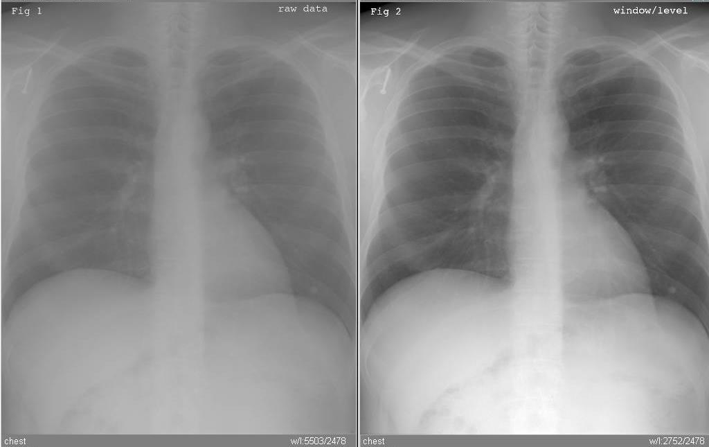
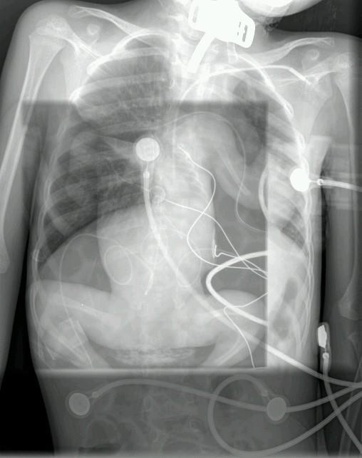
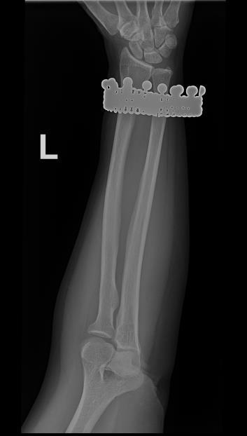
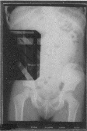
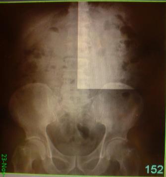
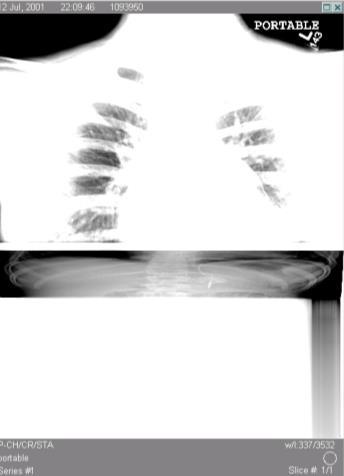
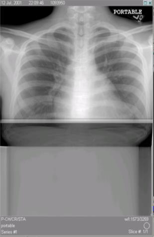
=>loss of contrast Effective dose for image on right is more than 2 x higher!")
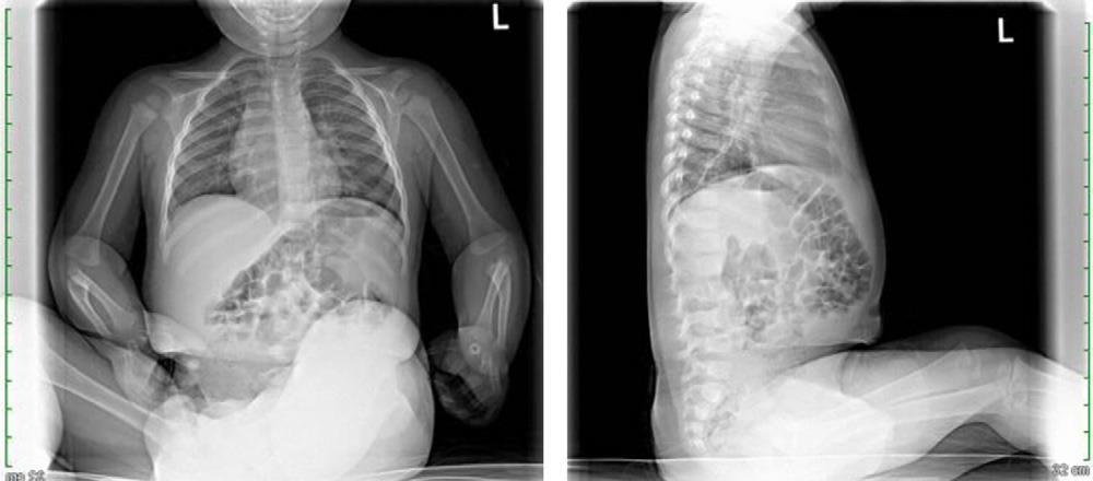
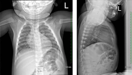
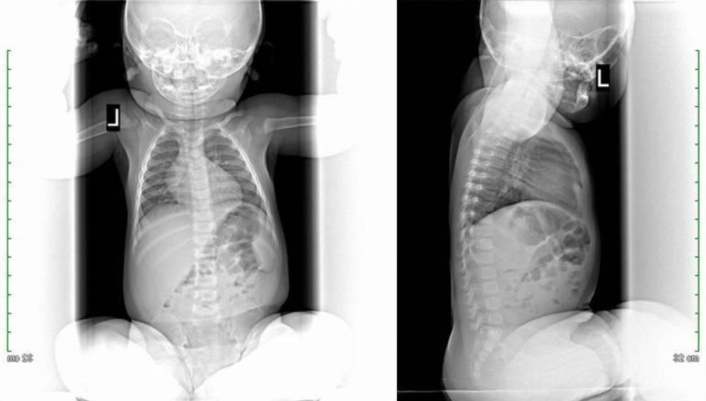
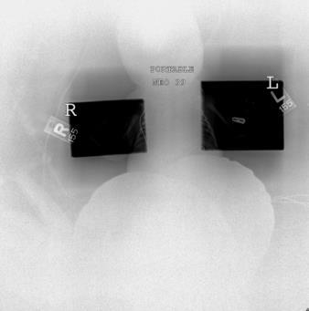
6 Dense objects (mandible, hardware, shields)=>loss of contrast Too much uncovered area (under-collimation, boundary not detected)=>loss of contrast Effective dose for image on right is more than 2 x higher! Centering, multiple FOV s, boundaries of FOV
7 Rotating polygon mirror Analog-to-Digital Converter Laser fast scan Photomultiplier tube Digital Image Light guide Amplifier Latent Image slow scan Imaging plate

8 conversion layer (courtesy J. A. Rowlands and Wei Zhao)

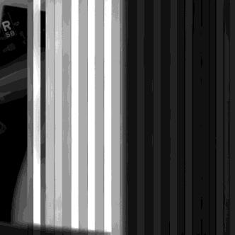
9 This correction is only as good as the uniformity of the x-ray field during calibration and the present condition of the scanner and imaging plate.


10 This correction is only as good as the uniformity of the x-ray field during calibration and the present condition of the scanner and imaging plate.
 and amplifiers as well as corrections for nonfunctional ( dead )")
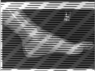
11 In DR, corrections must be applied for differences in gain and offset among individual detector elements (dels) and amplifiers as well as corrections for nonfunctional ( dead ) dels.

12 These corrections are applied in two dimensions.

13 These corrections are applied in two dimensions. Like CR, the correction is only as good as the uniformity of the x-ray field during calibration and the present condition of the detector.

14 Like CR, the correction is only as good as the uniformity of the x-ray field during calibration and the present condition of the detector.

15 Courtesy Laurence Parr, Naval Medical Center Portsmouth, VA Like CR, the correction is only as good as the uniformity of the x-ray field during calibration and the present condition of the detector.

16 Like CR, the correction is only as good as the uniformity of the x-ray field during calibration and the present condition of the detector.
17 Like CR, the correction is only as good as the uniformity of the x-ray field during calibration and the present condition of the detector.

18 Like CR, the correction is only as good as the uniformity of the x-ray field during calibration and the present condition of the detector.
19
20 Courtesy Eric Gingold, Thomas Jefferson University Hospital

21 Courtesy Eric Gingold, Thomas Jefferson University Hospital

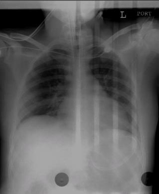
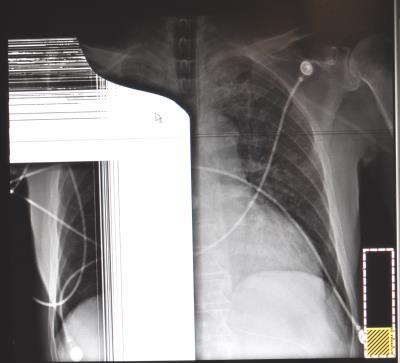
22 Hamlet Previous CR Chest image modified by non-linear LUT and edge enhanced
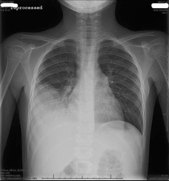
23 Hamlet


24 Hamlet AP 110 kvp 5.8 mas AP 120 kvp 3.1 mas PA 100 kvp 17.3 mas Technique chart calls for PA 115 kvp 2.5 mas (180 cm SID w/ non-removable grid)
25 Only enough SNR is needed to visualize important clinical features Agfa Fuji Kodak/CSH Konica/Minolta GE Philips Canon Swissray IDC lgm S# EI S# DEI EI_S REX DI F#
26 The variety and inconsistency of traditional Exposure Indicators created a problem for technologists who work with different CR and DR systems. Specific target values have yet to be established
27 The objective data is there for you to establish your own local standards for target values and action levels


28 Beware segmentation errors! DI = DI =

29 plus Even when the display is properly GSDF calibrated, the PACS may not display the image so that it looks the same as it did to the technologist. Acquisition Station PACS Display
30 plus Even when the display is properly GSDF calibrated, the PACS may not display the image so that it looks the same as it did to the technologist. The PACS may not display all of the information that is necessary to understand how the image was acquired.


31 plus Even when the display is properly GSDF calibrated, the PACS may not display the image so that it looks the same as it did to the technologist. The PACS may not display all of the information that is necessary to understand how the image was acquired.
32
33
34

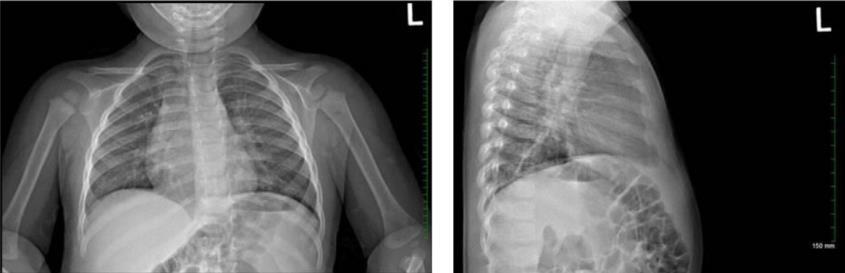
35 Processed Unprocessed


36 Take-away points from all this chaos CR, DR, and PACS are wonderful technologies fully capable of acquiring high quality radiographic projection images and displaying them for primary interpretation. In order to evaluate interval change in a patient, the radiologist may need to look beyond the visual appearance of the image and consider how comparison images were acquired or digitally processed. CR, DR, and PACS vendors have made great strides in providing objective information about how each image was acquired and processed for technologist feedback, radiologist oversight, medical physicist troubleshooting, and for incorporation into a QI Program. SPR was instrumental in catalyzing improvements in CR, DR, and PACS technology, especially for managing patient radiation dose. This is not a solved problem. Marvin s responses (the manically depressed robot) CR, DR, and PACS are fully capable of producing inconsistent and nondiagnostic radiographic projection images with unnecessary radiation doses to patients. Here I am, brain the size of a planet, and they ask me to pick up a piece of paper. I ve seen it. It s rubbish. Well I wish you d just tell me rather than try to engage my enthusiasm. I d give you advice, but you wouldn t listen. No one ever does. The highest level of quality that the medical physicist and the imaging operation is going to be able to provide is the lowest level of quality that the radiologist is willing to accept. I m not getting you down at all am I?

37
Outline. Digital Radiography. Understanding Digital Modalities: Image Quality and Dose. Image Quality. Dose Control
 Understanding Digital Modalities: Image Quality and Dose S. Jeff Shepard, M.S. University of Texas M. D. Anderson Cancer Center Houston, Texas Special Acknowledgement: Stephen K. Thompson, M.S. William
Digital Imaging Considerations Computed Radiography
 Digital Imaging Considerations Digital Radiography Computed Radiography o Cassette based Direct or Indirect Digital Radiography o Cassetteless Computed Radiography 1 CR Image Acquisition Most like conventional
COMPUTED RADIOGRAPHY CHAPTER 4 EFFECTIVE USE OF CR
 This presentation is a professional collaboration of development time prepared by: Rex Christensen Terri Jurkiewicz and Diane Kawamura New Technology https://www.youtube.com/watch?v=ptkzznazb 7U COMPUTED
History of digital imaging
 CR/QA RADCHEX History of digital imaging Early, crude digital detectors were developed in the 1970 s Image quality was problematic Processing time of digital images was untenable Viewing, transfer and
A Practical Overview of the Clinical and Operational Impact of Computed Radiography(CR) Implementations. Shirley Weddle, RT(R)(M), CIIP, BBA
 Implementations. Shirley Weddle, RT(R)(M), CIIP, BBA") A Practical Overview of the Clinical and Operational Impact of Computed Radiography(CR) Implementations Shirley Weddle, RT(R)(M), CIIP, BBA OBJECTIVES Define Computed Radiography (CR) Discuss CR vendor
A Practical Overview of the Clinical and Operational Impact of Computed Radiography(CR) Implementations Shirley Weddle, RT(R)(M), CIIP, BBA OBJECTIVES Define Computed Radiography (CR) Discuss CR vendor
Digital radiography (DR) post processing techniques for pediatric radiology
 post processing techniques for pediatric radiology") Digital radiography (DR) post processing techniques for pediatric radiology St Jude Children s Research Hospital Samuel Brady, MS PhD DABR samuel.brady@stjude.org Purpose Review common issues and solutions
Digital radiography (DR) post processing techniques for pediatric radiology St Jude Children s Research Hospital Samuel Brady, MS PhD DABR samuel.brady@stjude.org Purpose Review common issues and solutions
Teaching Digital Radiography and Fluoroscopic Radiation Protection
 Teaching Digital Radiography and Fluoroscopic Radiation Protection WCEC 20 th Student Educator Radiographer Conference Dennis Bowman, RT(R), CRT (R)(F) Community Hospital of the Monterey Peninsula (CHOMP)
Acquisition, Processing and Display
 Acquisition, Processing and Display Terri L. Fauber, R.T. (R)(M) Department of Radiation Sciences School of Allied Health Professions Virginia Commonwealth University Topics Image Characteristics Image
CR Basics and FAQ. Overview. Historical Perspective
 Page: 1 of 6 CR Basics and FAQ Overview Computed Radiography is a term used to describe a system that electronically records a radiographic image. Computed Radiographic systems use unique image receptors
The Evaluation of Collimator Alignment of Diagnostic X-ray Tube Using Computed Radiography System
 The Evaluation of Collimator Alignment of Diagnostic X-ray Tube Using Computed Radiography System The Evaluation of Collimator Alignment of Diagnostic X-ray Tube Using Computed Radiography System Manus
Ask EuroSafe Imaging Tips & Tricks. Paediatric Imaging Working Group. Dose Management in Digital Radiography
 Ask EuroSafe Imaging Tips & Tricks Paediatric Imaging Working Group Dose Management in Digital Radiography Raija Seuri (HUS Medical Imaging Center, FI) Cristina Almeida (Centro Hospitalar de Lisboa Central,
Exposure Indices and Target Values in Radiography: What Are They and How Can You Use Them?
 Exposure Indices and Target Values in Radiography: What Are They and How Can You Use Them? Definition and Validation of Exposure Indices Ingrid Reiser, PhD DABR Department of Radiology University of Chicago
3/31/2011. Objectives. Emory University. Historical Development. Historical Development. Historical Development
 Teaching Radiographic Technique in a Digital Imaging Paradigm Objectives 1. Discuss the historical development of digital imaging. Dawn Couch Moore, M.M.Sc., RT(R) Assistant Professor and Director Emory
Disclosures. Outline 7/31/2017. Current Implementation Status of IEC Standard : Exposure Index (EI) for Digital Radiography
 for Digital Radiography") Current Implementation Status of IEC Standard 62494-1: Exposure Index (EI) for Digital Radiography July 31, 2017 Ryan Fisher, PhD, DABR Katie Hulme, MS, DABR None Disclosures Outline Review of IEC Standard
Current Implementation Status of IEC Standard 62494-1: Exposure Index (EI) for Digital Radiography July 31, 2017 Ryan Fisher, PhD, DABR Katie Hulme, MS, DABR None Disclosures Outline Review of IEC Standard
Acceptance Testing of a Digital Breast Tomosynthesis Unit
 Acceptance Testing of a Digital Breast Tomosynthesis Unit 2012 AAPM Spring Clinical Meeting Jessica Clements, M.S., DABR Objectives Review of technology and clinical advantages Acceptance Testing Procedures
Learning Objectives: What s my motivation? (unknown screen actor) Workshop Overview
 Workshop Overview") Practical Medical Physics Adapting Traditional Clinical Medical Physics to Digital Radiography Charles E. Willis, Ph.D., DABR Associate Professor Department of Imaging Physics The University of Texas M.D.
Practical Medical Physics Adapting Traditional Clinical Medical Physics to Digital Radiography Charles E. Willis, Ph.D., DABR Associate Professor Department of Imaging Physics The University of Texas M.D.
SYLLABUS. TITLE: Equipment Operation I. DEPARTMENT: Radiologic Technology
 CODE: RADT 156 INSTITUTE: Health Science TITLE: Equipment Operation I DEPARTMENT: Radiologic Technology COURSE DESCRIPTION: This course covers the principles of equipment operation and maintenance of radiographic
2217 US Highway 70 East Garner, NC Main: Fax:
 Viztek is committed to providing the highest image quality possible in our CR & DR product lines. There are several factors that directly affect the overall quality of CR & DR based images. The eposure
Test Equipment for Radiology and CT Quality Control Contents
 Test Equipment for Radiology and CT Quality Control Contents Quality Control Testing...2 Photometers for Digital Clinical Display QC...3 Primary Workstations...3 Secondary Workstations...3 Testing of workstations...3
- KiloVoltage. Technique 101: Getting Back to Basics
 Why do I need to know technique? Technique 101: Getting Back to Basics Presented by: Thomas G. Sandridge, M.S., M.Ed., R.T.(R) Program Director Northwestern Memorial Hospital School of Radiography Chicago,
Image Quality Artifacts in Digital Imaging
 MAHIDOL UNIVERSITY Wisdom of the Land Image Quality Artifacts in Digital Imaging Napapong Pongnapang, Ph.D. Department of Radiological Technology Faculty of Medical Technology Mahidol University, Bangkok,
Beam-Restricting Devices
 Beam-Restricting Devices Three factors contribute to an increase in scatter radiation: Increased kvp Increased Field Size Increased Patient or Body Part Size. X-ray Interactions a some interact with the
Y11-DR Digital Radiography (DR) Image Quality
 Image Quality") Y11-DR Digital Radiography (DR) Image Quality Image quality is stressed for all systems in Safety Code 35. In the relevant sections Health Canada s advice is the manufacturer s recommended test procedures
Y11-DR Digital Radiography (DR) Image Quality Image quality is stressed for all systems in Safety Code 35. In the relevant sections Health Canada s advice is the manufacturer s recommended test procedures
Practical Medical Physics Session: TG-151 Dose Monitoring. August 5, 2013 Katie Hulme, M.S.
 Practical Medical Physics Session: TG-151 Dose Monitoring August 5, 2013 Katie Hulme, M.S. Digital Imaging and Dose Creep Images courtesy of Agfa Healthcare Under-Exposed Over-Exposed Freedman et al.,
Overview. Professor Roentgen was a Physicist!!! The Physics of Radiation Oncology X-ray Imaging
 The Physics of Radiation Oncology X-ray Imaging Charles E. Willis, Ph.D. DABR Associate Professor Department of Imaging Physics The University of Texas M.D. Anderson Cancer Center Houston, Texas Overview
Image Display and Perception
 Image Display and Perception J. Anthony Seibert, Ph.D. Department of Radiology UC Davis Medical Center Sacramento, California, USA Image acquisition, display, & interpretation X-rays kvp mas Tube filtration
Mammography is a radiographic procedure specially designed for detecting breast pathology Approximately 1 woman in 8 will develop breast cancer over
 Mammography is a radiographic procedure specially designed for detecting breast pathology Approximately 1 woman in 8 will develop breast cancer over a lifetime Breast cancer screening programs rely on
Digital Imaging started in the 1972 with Digital subtraction angiography Clinical digital imaging was employed from the 1980 ~ 37 years ago Amount of
 Digital Imaging started in the 1972 with Digital subtraction angiography Clinical digital imaging was employed from the 1980 ~ 37 years ago Amount of radiation to the population due to Medical Imaging
DISC QC/QA Program for Digital Imaging Systems using the DR Radchex Plus Meter
 DISC QC/QA Program for Digital Imaging Systems using the DR Radchex Plus Meter Revision Date: January 5th, 2017 www.disc-imaging.com Table of Contents Section A: Preliminary Setup Requirements... 4 Tools
STEREOTACTIC BREAST BIOPSY EQUIPMENT SURVEYS
 STEREOTACTIC BREAST BIOPSY EQUIPMENT SURVEYS JAMES A. TOMLINSON, M.S. Diagnostic Radiological Physicist American Board of Radiology Certified Medical Physics Consultants, Inc. Bio 28 yrs experience 100%
X-RAYS - NO UNAUTHORISED ENTRY
 Licencing of premises Premises Refer Guidelines A radiation warning sign and warning notice, X-RAYS - NO UNAUTHORISED ENTRY must be displayed at all entrances leading to the rooms where x-ray units are
Exposure System Selection
 Principles of Imaging Science II (RAD120) Exposure Systems Exposure System Selection Radiographic exposure is a very complex process Best technique systems manipulate one variable while holding others
1. Patient size AEC. Large Patient High ma. Small Patient Low ma
 Comparison of the function and performance of CT AEC systems CTUG meeting by Emily Field Trainee clinical scientist 14 th th Breakdown CT Automatic Exposure Control (AEC) Background Project Description
Automated dose control in multi-slice CT. Nicholas Keat Formerly ImPACT, St George's Hospital, London
 Automated dose control in multi-slice CT Nicholas Keat Formerly ImPACT, St George's Hospital, London Introduction to presentation CT contributes ~50+ % of all medical radiation dose Ideally all patients
Quality Control for Stereotactic Breast Biopsy. Robert J. Pizzutiello, Jr., F.A.C.M.P. Upstate Medical Physics, Inc
 Quality Control for Stereotactic Breast Biopsy Robert J. Pizzutiello, Jr., F.A.C.M.P. Upstate Medical Physics, Inc. 716-924-0350 Methods of Imaging Guided Breast Biopsy Ultrasound guided, hand-held needle
I. PERFORMANCE OF X-RAY PRODUCTION COMPONENTS FLUOROSCOPIC ACCEPTANCE TESTING: TEST PROCEDURES & PERFORMANCE CRITERIA
 FLUOROSCOPIC ACCEPTANCE TESTING: TEST PROCEDURES & PERFORMANCE CRITERIA EDWARD L. NICKOLOFF DEPARTMENT OF RADIOLOGY COLUMBIA UNIVERSITY NEW YORK, NY ACCEPTANCE TESTING GOALS PRIOR TO 1st CLINICAL USAGE
8/2/2017. Radiologist Responsibilities. Radiologist Responsibilities. Medical Physicist Mammography Equipment Evaluation and Annual Survey
 Implementation of the 2016 ACR Digital Mammography QC Manual Medical Physicist Mammography Equipment Evaluation and Annual Survey Eric A Berns, PhD, FACR Radiologist Responsibilities Radiologist Responsibilities
COMPUTED TOMOGRAPHY 1
 COMPUTED TOMOGRAPHY 1 Why CT? Conventional X ray picture of a chest 2 Introduction Why CT? In a normal X-ray picture, most soft tissue doesn't show up clearly. To focus in on organs, or to examine the
RADIOGRAPHIC EXPOSURE
 RADIOGRAPHIC EXPOSURE Receptor Exposure Receptor Exposure the that interacts with the receptor. Computed Radiography ( ) requires a. Direct Digital Radiography (DR) requires a. Exposure Indicators Exposure
10/3/2012. Study Harder
 This presentation is a professional collaboration of development time prepared by: Rex Christensen Terri Jurkiewicz and Diane Kawamura Study Harder CR detection is inefficient, inferior to film screen
Quality assurance: a comparison study of radiographic exposure for neonatal chest radiographs at 4 academic hospitals
 DOI 10.1007/s00247-011-2290-1 ORIGINAL ARTICLE Quality assurance: a comparison study of radiographic exposure for neonatal chest radiographs at 4 academic hospitals Mervyn D. Cohen & Richard Markowitz
Fire CR Calibration Guide
 1 Fire CR Calibration Guide This reference guide will guide you through the steps to complete the calibration for the Fire CR.. Getting Started: 1. Click on the Opal Icon on the Desktop. Figure 1 2. Once
Features and Weaknesses of Phantoms for CR/DR System Testing
 Physics testing of image detectors Parameters to test Features and Weaknesses of Phantoms for CR/DR System Testing Spatial resolution Contrast resolution Uniformity/geometric distortion Dose response/signal
Practical Aspects of Medical Physics Surveys of Mammography Equipment and Facilities
 Practical Aspects of Medical Physics Surveys of Mammography Equipment and Facilities Melissa Martin, M.S., FAAPM, FACR, FACMP AAPM Annual Meeting - Philadelphia July 19, 2010 MO-B-204C-1 Educational Objectives
10/26/2015. Study Harder
 This presentation is a professional collaboration of development time prepared by: Rex Christensen Terri Jurkiewicz and Diane Kawamura Study Harder CR detection is inefficient, inferior to film screen
Outline ASRT Changes Impact on current curriculum Potential new courses WECM Changes Last update Resources and needs
 Change nd Annual Blinn College 2 nd Educator s Workshop For Radiologic Sciences July 28, 2007 Christi Carter, MSRS, RT(R) Outline ASRT Changes Impact on current curriculum Potential new courses WECM Changes
Breast Tomosynthesis. Bob Liu, Ph.D. Department of Radiology Massachusetts General Hospital And Harvard Medical School
 Breast Tomosynthesis Bob Liu, Ph.D. Department of Radiology Massachusetts General Hospital And Harvard Medical School Outline Physics aspects of breast tomosynthesis Quality control of breast tomosynthesis
Quality Control of Full Field Digital Mammography Units
 Quality Control of Full Field Digital Mammography Units Melissa C. Martin, M.S., FACMP, FACR, FAAPM Melissa@TherapyPhysics.com 310-612-8127 ACMP Annual Meeting Virginia Beach, VA May 2, 2009 History of
Collimation Assessment Using GAFCHROMIC XR-M2
 Collimation Assessment Using GAFCHROMIC XR-M2 I. Introduction A method of collimation assessment for GE Senographe full-field digital mammography (FFDM) systems is described that uses a self-developing
radiography detector
 Clinical evaluation of a full field digital projection radiography detector Gary S. Shaber'1, Denny L. Leeb, Jeffrey Belib, Gregory Poweii1', Andrew D.A. Maidment'1 a Thomas Jefferson University Hospital,
TESTING FLAT-PANEL IMAGING SYSTEMS: What the Medical Physicist Needs to Know. JAMES A. TOMLINSON, M.S., D.A.B.R. Diagnostic Radiological Physicist
 TESTING FLAT-PANEL IMAGING SYSTEMS: What the Medical Physicist Needs to Know JAMES A. TOMLINSON, M.S., D.A.B.R. Diagnostic Radiological Physicist Topics Image Uniformity and Artifacts Image Quality - Detail
Essentials of Digital Imaging
 Essentials of Digital Imaging Module 2 Transcript 2016 ASRT. All rights reserved. Essentials of Digital Imaging Module 2 Processing 1. ASRT Animation 2. Welcome Welcome to Essentials of Digital Imaging
While digital techniques have the potential to reduce patient doses, they also have the potential to significantly increase them.
 In press 2004 1 2 Guest Editorial (F. Mettler, H. Ringertz and E. Vano) Guest Editorial (F. Mettler, H. Ringertz and E. Vano) Digital radiology An appropriate analogy that is easy for most people to understand
QC by the MPE in Belgium
 Acceptance testing of state-of-the-art CT scanners using a new national protocol: first experience on a large number of scanners of different make and model the working group Radiology of the Belgian Hospital
Mammography: Physics of Imaging
 Mammography: Physics of Imaging Robert G. Gould, Sc.D. Professor and Vice Chair Department of Radiology and Biomedical Imaging University of California San Francisco, California Mammographic Imaging: Uniqueness
X-ray Imaging. PHYS Lecture. Carlos Vinhais. Departamento de Física Instituto Superior de Engenharia do Porto
 X-ray Imaging PHYS Lecture Carlos Vinhais Departamento de Física Instituto Superior de Engenharia do Porto cav@isep.ipp.pt Overview Projection Radiography Anode Angle Focal Spot Magnification Blurring
T h e P h a n t o m L a b o r a t o r y
 T h e P h a n t o m L a b o r a t o r y 1 CCT228 ATCM Phantom Manual Copyright 2017 WARRANTY THE PHANTOM LABORATORY INCORPORATED ( Seller ) warrants that this product shall remain in good working order
Ludlum Medical Physics
 Ludlum Medical Physics Medical Imaging Radiology QA Test Tools NEW LUDLUM PRODUCT LINE Medical Physics Products Medical Physics Products What are they? Products used to measure radiation output and to
Title: A COMPARISON OF Cs-137 AND X-RAY SOURCES AS CALIBRATION REFERENCES FOR THERMOLUMINESCENT DOSIMETER CHIPS
 Title: A COMPARISON OF Cs-137 AND X-RAY SOURCES AS CALIBRATION REFERENCES FOR THERMOLUMINESCENT DOSIMETER CHIPS By Aravind Ravichandran arr192@mail.usask.ca University of Saskatchewan Address: 2424 Cumberland
Nuclear Associates
 Nuclear Associates 07-706 Patient Phantom/Penetrometer System Users Manual March 2005 Manual No. 07-706-1 Rev. 2 2004, 2005 Fluke Corporation, All rights reserved. Printed in U.S.A. All product names are
Overview of Safety Code 35
 Common Quality Control Procedures for All s Quality Control Procedures Film All s Daily Quality Control Tests Equipment Warm-up (D1) According to manufacturers instructions Can include auto calibration(d1)
HISTORY. CT Physics with an Emphasis on Application in Thoracic and Cardiac Imaging SUNDAY. Shawn D. Teague, MD
 CT Physics with an Emphasis on Application in Thoracic and Cardiac Imaging Shawn D. Teague, MD DISCLOSURES 3DR- advisory committee CT PHYSICS WITH AN EMPHASIS ON APPLICATION IN THORACIC AND CARDIAC IMAGING
IBEX TECHNOLOGY APPLIED TO DIGITAL RADIOGRAPHY
 WHITE PAPER: IBEX TECHNOLOGY APPLIED TO DIGITAL RADIOGRAPHY IBEX Innovations Ltd. Registered in England and Wales: 07208355 Address: Discovery 2, NETPark, William Armstrong Way, Sedgefield, UK Patents:
Clinical Experiences with a Patient Skin Dose Monitoring and Tracking Program
 Clinical Experiences with a Patient Skin Dose Monitoring and Tracking Program Allen R. Goode, MS, DABR Chief Diagnostic Medical Physicist Department of Radiology & Medical Imaging University of Virginia
ddr Compact Series Setting a new benchmark in digital radiography.
 ddr Compact Series Setting a new benchmark in digital radiography. ddrcompact When productivity and exceptional value come together. With the introduction of its newest DR system, the ddrcompact, Swissray
Nuclear Associates
 Nuclear Associates 07-644 Grid Alignment Test Tool Users Manual March 2005 Manual No. 07-644-1 Rev. 2 2004, 2005 Fluke Corporation, All rights reserved. Printed in U.S.A. All product names are trademarks
Half value layer and AEC receptor dose compliance survey in Estonia
 Half value layer and AEC receptor dose compliance survey in Estonia K. Kepler, A. Vladimirov Training Centre of Medical Physics, University of Tartu Testing Centre of the University of Tartu, Estonia E-mail:
Computed Tomography. The Fundamentals of... THE FUNDAMENTALS OF... Jason H. Launders, MSc. Current Technology
 The Fundamentals of... Computed Tomography Computed Tomography (CT) systems use x-rays to produce images of slices through a patient s anatomy. Despite having lower spatial resolution than other x-ray
RAD 150 RADIOLOGIC EXPOSURE TECHNIQUE II
 RAD 150 RADIOLOGIC EXPOSURE TECHNIQUE II APPROVED 12/O2/2011 EFFECTIVE SPRING 2013-14 Prefix & Number RAD 150 Course Title: Radiologic Exposure Technique II & Lab Purpose of this submission: New Change/Updated
Comparison of computed radiography and filmõscreen combination using a contrast-detail phantom
 JOURNAL OF APPLIED CLINICAL MEDICAL PHYSICS, VOLUME 4, NUMBER 1, WINTER 2003 Comparison of computed radiography and filmõscreen combination using a contrast-detail phantom Z. F. Lu,* E. L. Nickoloff, J.
Wide beam CT dosimetry. Elly Castellano
 Wide beam CT dosimetry Elly Castellano Outline revision: CT dose indices wide-beam CT: the end of the road for CTDI? the IEC rescue plan for CTDI 100 the american way AAPM report 111 better estimates of
Hardware for High Energy Applications 30 October 2009
 Paper No. 003 09 Hardware for High Energy Applications 30 October 2009 This document was created by the Federal Working Group on Industrial Digital Radiography. Reproduction is authorized. Federal Working
A study of exposure index value fluctuations in computed radiography and direct digital radiography using multiple manufacturers
 A study of exposure index value fluctuations in computed radiography and direct digital radiography using multiple manufacturers Poster No.: C-3011 Congress: ECR 2010 Type: Topic: Authors: Scientific Exhibit
DIGITAL RADIOGRAPHY ARTIFACTS
 IMAGING LAB MPHY 487 DIGITAL RADIOGRAPHY ARTIFACTS Mohammad Esmael Alsulimane B.Sc, M.Sc Medical Physics Lecturer - Physics Department All Rights Reserved: Some information and figures in this presentation
New Exposure Indicators for Digital Radiography Simplified for Radiologists and Technologists
 Medical Physics and Informatics Technical Innovation Don et al. New Simplified Exposure Indicators Medical Physics and Informatics Technical Innovation Steven Don 1 ruce R. Whiting 2 Lois Jo Rutz 3 ruce
Unit thickness. Unit area. σ = NΔX = ΔI / I 0
 Unit thickness I 0 ΔI I σ = ΔI I 0 NΔX = ΔI / I 0 NΔX Unit area Δx Average probability of reaction with atom for the incident photons at unit area with the thickness of Delta-X Atom number at unit area
Current technology in digital image production (CR/DR and other modalities) Jaroonroj Wongnil 25 Mar 2016
 Jaroonroj Wongnil 25 Mar 2016") Current technology in digital image production (CR/DR and other modalities) Jaroonroj Wongnil 25 Mar 2016 Current technology in digital image production (CR/DR and other modalities) 2/ Overview Digital
Current technology in digital image production (CR/DR and other modalities) Jaroonroj Wongnil 25 Mar 2016 Current technology in digital image production (CR/DR and other modalities) 2/ Overview Digital
QC Testing for Computed Tomography (CT) Scanner
 Scanner") QC Testing for Computed Tomography (CT) Scanner QA - Quality Assurance All planned and systematic actions needed to provide confidence on a structure, system or component. all-encompassing program, including
QC Testing for Computed Tomography (CT) Scanner QA - Quality Assurance All planned and systematic actions needed to provide confidence on a structure, system or component. all-encompassing program, including
DICOM Implementations for Digital Radiography
 The Medicine Behind the Image DICOM Implementations for Digital Radiography David A. Clunie Princeton Radiology Pharmaceutical Research Disclosure & Acknowledgements CTO RadPharm Proprietor of PixelMed
Think Digital. ddr Modulaire A quantum leap in radiography workflow and efficiency.
 Think Digital. ddr Modulaire A quantum leap in radiography workflow and efficiency. ddrmodulaire When performance is decisive. For the most demanding requirements. The ddrmodulaire is a space efficient,
Do you have any other questions? Please call us at (Toll Free) or , or
 or , or") INSTRUCTIONS Read the appropriate course/ textbook. This is an open book test. A score of 75% or higher is needed to receive CE credit. You will have a maximum of three attempts to pass this course. Please
INSTRUCTIONS Read the appropriate course/ textbook. This is an open book test. A score of 75% or higher is needed to receive CE credit. You will have a maximum of three attempts to pass this course. Please
X-RAY MEDICAL EQUIPMENT
 X-RAY MEDICAL EQUIPMENT CHEST RADIOGRAPHY GENERAL RADIOGRAPHY & FLUOROSCOPY RADIOTHERAPY MOBILE HEALTHCARE MAMMOGRAPHY MAMMOSCAN FULL FIELD DIGITAL MAMMOGRAPHY SYSTEM Biopsy Attachment џ MAMMOSCAN an ADANI
Essentials of Digital Imaging
 Essentials of Digital Imaging Module 6 Transcript 2016 ASRT. All rights reserved. Essentials of Digital Imaging Module 6 Dose Reduction and Patient Safety 1. ASRT Animation 2. Welcome Welcome to Essentials
Moving from film to digital: A study of digital x-ray benefits, challenges and best practices
 Moving from film to digital: A study of digital x-ray benefits, challenges and best practices H.U. Pöhler 1 and N. D Ademo 2 DÜRR NDT GmbH & Co. KG, Höpfigheimer Straße 22, Bietigheim-Bissingen, 74321,
1. Carlton, Richard R., and Arlene M. Adler. Principles of Radiographic Imaging: An Art and a Science, 5th edition (2013).
.") CODE: RADT 151 INSTITUTE: Health Science TITLE: Radiographic Exposure DEPARTMENT: Radiologic Technology COURSE DESCRIPTION: This course covers the principles of radiographic exposure selection and manipulation
CODE: RADT 151 INSTITUTE: Health Science TITLE: Radiographic Exposure DEPARTMENT: Radiologic Technology COURSE DESCRIPTION: This course covers the principles of radiographic exposure selection and manipulation
Investigation of the line-pair pattern method for evaluating mammographic focal spot performance
 Investigation of the line-pair pattern method for evaluating mammographic focal spot performance Mitchell M. Goodsitt, a) Heang-Ping Chan, and Bob Liu Department of Radiology, University of Michigan, Ann
Pitfalls and Remedies of MDCT Scanners as Quantitative Instruments
 intensity m(e) m (/cm) 000 00 0 0. 0 50 0 50 Pitfalls and Remedies of MDCT Scanners as Jiang Hsieh, PhD GE Healthcare Technology University of Wisconsin-Madison Root-Causes of CT Number Inaccuracies Nature
Reducing Radiation Exposure from Survey CT Scans
 Reducing Survey CT Scan Exposure Pediatric Imaging Original Research Jennifer C. O Daniel 1 Donna M. Stevens 2 Dianna D. Cody 2 O Daniel JC, Stevens DM, Cody DD Received July 28, 2004; accepted after revision
Setting up digital imaging department!
 Outline Setting up digital imaging department! From screen/film to digital radiography PACS/Tele radiology Setting up digital department Digital Imaging Napapong Pongnapang, Ph.D. Department of Radiological
Artefacts found in computed radiography
 The British Journal of Radiology, 74 (2001), 195 202 E 2001 The British Institute of Radiology Pictorial review Artefacts found in computed radiography L J CESAR, RT(R)(QM), B A SCHUELER, PhD, F E ZINK,
Minnesota Rules, Chapter 4732 X-ray Revision
 Minnesota Rules, Chapter 4732 X-ray Revision DRAFT FLUOROSCOPIC X-RAY SYSTEMS, 1.0 Subpart 1. Applicability. Subpart 2. Limitation of the useful beam. Subpart 3. Measuring compliance; primary protective
Wide-Detector CT for TAVR Planning:
 Wide-Detector CT for TAVR Planning: Impact on Iodine Dose, Radiation Dose, and Image Quality SCBTMR 2015 Annual Course Thursday, October 8 William P. Shuman MD FSCBTMR Department of Radiology University
Visibility of Detail
 Visibility of Detail Radiographic Quality Quality radiographic images represents the, and information is for diagnosis. The of the anatomic structures and the accuracy of their ( ) determine the overall
Instant DR in Jordan
 Hashemite University leads the way with first Instant DR in Jordan DR Retrofit supports research and education goals of the Faculty of Allied Health Sciences, while enhancing care for staff and students
Get more from your images with Symphony Image Processing
 DIRECT RADIOGRAPHY The user-friendly DelWorks image acquisition and processing software possesses a wide range of tools for a variety of image manipulations. Its user interface simplifies every step of
9/10/2012. Computed Radiography Chapter 3 Physics and Technology. What is Computed Radiography?
 Computed Radiography Chapter 3 Physics and Technology This presentation is a professional collaboration of development time prepared by: Rex Christensen Terri Jurkiewicz and Diane Kawamura Today s Humor:
KODAK DIRECTVIEW CR Mammography Feature User s Guide
 KODAK DIRECTVIEW CR Mammography Feature User s Guide 17 September 2010 9G3741 Version 1.0 Carestream Health, Inc. 150 Verona Street Rochester, NY 14608 CARESTREAM, DIRECTVIEW, and DRYVIEW are trademarks
NJDEP Medical Physicist s Radiographic QC Survey Registration Number:
 Facility Name NJDEP ID # NJDEP Medical Physicist s Radiographic QC Survey PLEASE PRINT Facility Information Unit Information Manufacturer Model Console Model # Console serial # Tube serial # Location (room)
Joint ICTP/IAEA Advanced School on Dosimetry in Diagnostic Radiology and its Clinical Implementation May 2009
 2033-6 Joint ICTP/IAEA Advanced School on Dosimetry in Diagnostic Radiology and its Clinical Implementation 11-15 May 2009 Dosimetry for Fluoroscopy Basics Renato Padovani EFOMP Joint ICTP-IAEA Advanced
Digital Mammography Quality Control for the Mammographic Technologist
 Ontario Breast Screening Program Digital Mammography Quality Control for the Mammographic Technologist Authors: G.E. Mawdsley, A.K. Bloomquist, M.J. Yaffe October 2011 Revision 3.1 Mammographic Physics