DIGITAL RADIOGRAPHY ARTIFACTS
|
|
|
- Scott Blankenship
- 5 years ago
- Views:
Transcription

1 IMAGING LAB MPHY 487 DIGITAL RADIOGRAPHY ARTIFACTS Mohammad Esmael Alsulimane B.Sc, M.Sc Medical Physics Lecturer - Physics Department
2 All Rights Reserved: Some information and figures in this presentation are collection from presentations who's name are listed below. Artifacts in Digital Radiography (DDR & CR) By Leung Chuen Yung - RAD II, QEH General Radiographic Image Artifacts, The Art of the Image: The Identification and Remediation of Image Artifacts in Projection Radiography By Alisa Walz-Flannigan, Ph.D. DABR - Mayo Clinic, Rochester, Minnesota Please Note: The first two references it is very important and useful for thus who are works as Medical Physicists related to Medical Imaging fields.

3 Contents Causes and Sources of Artifacts in Digital Medical Radiography
4 Artifacts Any undesirable objects OR structures recorded on the radiography image cause degraded image quality. Produced from : - Patients such as motion, poor preparations - Technologists such as less knowledge, less training - Machines, there are various artifacts from CR and DR radiography machines.


5 Noisy Detector Power Supply Artifacts
6 Noisy Detector Power Supply Artifacts Appearance: Vertical lines, which are symmetrical around the center of the image. Cause: Caused by a noisy detector power supply Solution: Replace power supply

7 Loose Cone Artifacts
8 Loose Cone Artifacts Appearance: White edges. Cause: Cone has fallen out of the x-ray tube port and is blocking the collimator from opening. Solution: Remove the collimator and re-attach the cone to the tube port

9 Bar code Artifacts DR
10 Bar code Artifacts Appearance: Barcode appear on screen Cause: Cause by a failure in a data module or the detector Solution: Perform bad pixel calibration, if calibration fail then replace detector


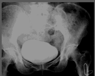
11 Double Exposure CR Artifacts
12 Double Exposure Artifacts Appearance: Duplication of images Causes: Two subsequent exposure on same imaging plate Solution: Proper knowledge of using of X-ray equipment

13 Poor Collimation Artifacts
14 Poor Collimation Artifacts Appearance: Unsharp images Causes: Improper collimation Solution: Proper collimation in accordance with cassette size and body part
15 Effect of FOV


16 Scatter and Collimation


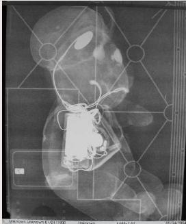
17 Exposure Through CR Back of Cassette
18 Exposure Through Back of Cassette Appearance: Various patterns of image according to cassette design Causes: Poor basic knowledge of construction of cassettes Solution: Proper education of radiographers in handling of cassettes


19 Improper Exposure KV Artifacts
20 Improper Exposure KV Artifacts Appearance: Darkening or whitening of image Causes: Improper exposure setting Solution: Proper exposure factors to be used based on body part and patient size


21 Moire Pattern Artifacts

22 Moire Pattern Artifacts Appearance: Different types of moire pattern Causes: Improper Grid usage with low grid frequencies Solution: Usage of grids with 60 lines/cm or more; grid lines should run perpendicular to plate reader s laser scan lines


23 Grid (Bucky) Gustav Bucky who discover and event the Bucky. Notice the effect of Compton scattering and how the image degrades. Designed a lead grid to absorb the scattered radiation. The problem appears the lead grid on screen film. Hollis Potter he solve this problem by events the movable grid, its using till today.
")
24 Grid (Bucky)


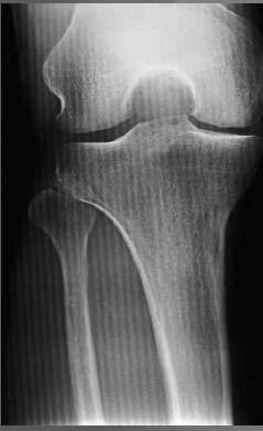
25 Importance of Scatter The two images shown above were obtained using no scatter removal grid and using the identical radiographic techniques 65 kv and 3 mas. The only difference between these two images is that the one on the left had blocks of Plexiglas added adjacent to the knee phantom; this additional material produces a large amount of Compton scatter, which markedly degrades the image contrast seen in the knee region.

26 Scatter Removal Grids KV = 75 kv mas = 3 No Scatter Removal Grid KV = 75 kv mas = 25 Scatter Removal Grid


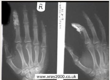
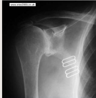
27 Scratches Artifacts This artifact will may cause wrong diagnosis (stone) CR
28 Scratches Artifacts Appearance: Kink marks on the image Causes: Mishandling of imaging plate during cleaning process Solution: Cassettes and image plates should be handled with care

29 Light Bulb Artifacts
30 Light Bulb Artifacts Appearance: Darkening of lower and outer portions of an image Causes: High exposure, back scattered radiation entering imaging plate from patient s bed due to increased exposure for obese patients or due to uncollimated x ray Solution: Reduce back scatter by lowering the KV or proper collimation.

31 Dust Artifacts CR
.")
32 Dust Artifacts Appearance: Focal radiopacities Causes: Dust particles wedged over imaging plate Solution: Regular cleaning of imaging plates with proper cleaner (EthylAlcohol). Paper towels or gauze should not be used because they leave fibers on the plate, the use of lint-free cloth is advisable.

33 CR Disparity Artifact
34 Disparity Artifact Appearance: Defective scanning resulting in alteration in image contrast, lower half of it was exposed to laser beam for longer time, which resulted in brighter image output Causes: Malfunctioning of rollers in CR reader Solution: Periodic cleaning of roller in CR reader by the supplier Optimal image

35 Ruler Damage Artifacts CR This artifact will may cause wrong diagnosis (granulomas in lung)
36 Damage of imaging plate due to rollers Appearance: Chest radiograph shows radiopacities (arrows) along right lateral chest wall, focal linear radiopacities Causes: Mechanical damaging of Imaging Plate during transport through rollers Solution: Replace that part of roller

37 Deodorant Artifacts


38 Hair Bun

39 Clothes Ribbing


40 Jewellery

41 Lighter in the pocket


42 Umbilical Ring
43 Plaster

44 Strap


45 CR Finger Marks

46 Air in the Ventricles Air head Is this ARTIFACT??
Artefacts found in computed radiography
 The British Journal of Radiology, 74 (2001), 195 202 E 2001 The British Institute of Radiology Pictorial review Artefacts found in computed radiography L J CESAR, RT(R)(QM), B A SCHUELER, PhD, F E ZINK,
THE ART OF THE IMAGE: IDENTIFICATION AND REMEDIATION OF IMAGE ARTIFACTS IN MAMMOGRAPHY
 THE ART OF THE IMAGE: IDENTIFICATION AND REMEDIATION OF IMAGE ARTIFACTS IN MAMMOGRAPHY William Geiser, MS DABR Senior Medical Physicist MD Anderson Cancer Center Houston, Texas wgeiser@mdanderson.org INTRODUCTION
COMPUTED RADIOGRAPHY CHAPTER 4 EFFECTIVE USE OF CR
 This presentation is a professional collaboration of development time prepared by: Rex Christensen Terri Jurkiewicz and Diane Kawamura New Technology https://www.youtube.com/watch?v=ptkzznazb 7U COMPUTED
Beam-Restricting Devices
 Beam-Restricting Devices Three factors contribute to an increase in scatter radiation: Increased kvp Increased Field Size Increased Patient or Body Part Size. X-ray Interactions a some interact with the
LECTURE 1 The Radiographic Image
 LECTURE 1 The Radiographic Image Prepared by:- KAMARUL AMIN ABDULLAH @ ABU BAKAR UiTM Faculty of Health Sciences Medical Imaging Department 11/23/2011 KAMARUL AMIN (C) 1 Lesson Objectives At the end of
RADIOGRAPHIC EXPOSURE
 RADIOGRAPHIC EXPOSURE Receptor Exposure Receptor Exposure the that interacts with the receptor. Computed Radiography ( ) requires a. Direct Digital Radiography (DR) requires a. Exposure Indicators Exposure
Fabrício Sampaio Péres Kury Federal University of Rio de Janeiro Medical School
 Fabrício Sampaio Péres Kury Federal University of Rio de Janeiro Medical School Harvard Medical School Exchange Clerkship Program Primary Care Radiology Clerkship Gillian Lieberman, M. D. Monday, September
3/31/2011. Objectives. Emory University. Historical Development. Historical Development. Historical Development
 Teaching Radiographic Technique in a Digital Imaging Paradigm Objectives 1. Discuss the historical development of digital imaging. Dawn Couch Moore, M.M.Sc., RT(R) Assistant Professor and Director Emory
STUDENT REVIEW QUESTION SET K CR/DR CONTENT AREA
 STUDENT REVIEW QUESTION SET K CR/DR CONTENT AREA RADT 2913 COMPREHENSIVE REVIEW 1 The CR cassette is backed by aluminum that: A. reflects x-rays B. absorbs x-rays C. captures the image D. transmits x-rays
CR Basics and FAQ. Overview. Historical Perspective
 Page: 1 of 6 CR Basics and FAQ Overview Computed Radiography is a term used to describe a system that electronically records a radiographic image. Computed Radiographic systems use unique image receptors
Multiple Choice Identify the letter of the choice that best completes the statement or answers the question.
 RA110 test 3 Multiple Choice Identify the letter of the choice that best completes the statement or answers the question. 1. An object 35 cm in width is radiographed at 100 cm SID and at a 50 cm SOD. What
10/3/2012. Study Harder
 This presentation is a professional collaboration of development time prepared by: Rex Christensen Terri Jurkiewicz and Diane Kawamura Study Harder CR detection is inefficient, inferior to film screen
10/26/2015. Study Harder
 This presentation is a professional collaboration of development time prepared by: Rex Christensen Terri Jurkiewicz and Diane Kawamura Study Harder CR detection is inefficient, inferior to film screen
X-ray Imaging. PHYS Lecture. Carlos Vinhais. Departamento de Física Instituto Superior de Engenharia do Porto
 X-ray Imaging PHYS Lecture Carlos Vinhais Departamento de Física Instituto Superior de Engenharia do Porto cav@isep.ipp.pt Overview Projection Radiography Anode Angle Focal Spot Magnification Blurring
NON-DESTRUCTIVE EVALUATION UTILIZING IMAGING PLATES FOR FIELD RADIOGRAPHY APPLICATIONS
 19 th World Conference on Non-Destructive Testing 2016 NON-DESTRUCTIVE EVALUATION UTILIZING IMAGING PLATES FOR FIELD RADIOGRAPHY APPLICATIONS Brian S. WHITE 1 1 Carestream NDT, 1049 Ridge Road West, Rochester,
Exposure Indices and Target Values in Radiography: What Are They and How Can You Use Them?
 Exposure Indices and Target Values in Radiography: What Are They and How Can You Use Them? Definition and Validation of Exposure Indices Ingrid Reiser, PhD DABR Department of Radiology University of Chicago
Exposure System Selection
 Principles of Imaging Science II (RAD120) Exposure Systems Exposure System Selection Radiographic exposure is a very complex process Best technique systems manipulate one variable while holding others
Veterinary Science Preparatory Training for the Veterinary Assistant. Floron C. Faries, Jr., DVM, MS
 Veterinary Science Preparatory Training for the Veterinary Assistant Floron C. Faries, Jr., DVM, MS Radiology Floron C. Faries, Jr., DVM, MS Objectives Determine the appropriate machine settings for making
Image Quality Artifacts in Digital Imaging
 MAHIDOL UNIVERSITY Wisdom of the Land Image Quality Artifacts in Digital Imaging Napapong Pongnapang, Ph.D. Department of Radiological Technology Faculty of Medical Technology Mahidol University, Bangkok,
Digital radiography: Practical advantages of Digital Radiography. Practical Advantages in image quality
 Digital radiography: Digital radiography is set to become the most common form of processing radiographic images in the next 10 years. This is due to a number of practical and image quality issues. Practical
Nuclear Associates , , , , , ,
 Nuclear Associates 57-411, 57-412, 57-413 57-426, 57-431, 57-432 57-433, 57-435, 57-436 CLEAR-Pb Transparent X-Ray Compensation Filters Users Manual March 2005 Manual No. 57-XXX-1 Rev. 2 2003, 2005 Fluke
Teaching Digital Radiography and Fluoroscopic Radiation Protection
 Teaching Digital Radiography and Fluoroscopic Radiation Protection WCEC 20 th Student Educator Radiographer Conference Dennis Bowman, RT(R), CRT (R)(F) Community Hospital of the Monterey Peninsula (CHOMP)
RAD 150 RADIOLOGIC EXPOSURE TECHNIQUE II
 RAD 150 RADIOLOGIC EXPOSURE TECHNIQUE II APPROVED 12/O2/2011 EFFECTIVE SPRING 2013-14 Prefix & Number RAD 150 Course Title: Radiologic Exposure Technique II & Lab Purpose of this submission: New Change/Updated
Digital Imaging Considerations Computed Radiography
 Digital Imaging Considerations Digital Radiography Computed Radiography o Cassette based Direct or Indirect Digital Radiography o Cassetteless Computed Radiography 1 CR Image Acquisition Most like conventional
X-RAYS - NO UNAUTHORISED ENTRY
 Licencing of premises Premises Refer Guidelines A radiation warning sign and warning notice, X-RAYS - NO UNAUTHORISED ENTRY must be displayed at all entrances leading to the rooms where x-ray units are
A Practical Overview of the Clinical and Operational Impact of Computed Radiography(CR) Implementations. Shirley Weddle, RT(R)(M), CIIP, BBA
 Implementations. Shirley Weddle, RT(R)(M), CIIP, BBA") A Practical Overview of the Clinical and Operational Impact of Computed Radiography(CR) Implementations Shirley Weddle, RT(R)(M), CIIP, BBA OBJECTIVES Define Computed Radiography (CR) Discuss CR vendor
A Practical Overview of the Clinical and Operational Impact of Computed Radiography(CR) Implementations Shirley Weddle, RT(R)(M), CIIP, BBA OBJECTIVES Define Computed Radiography (CR) Discuss CR vendor
Visibility of Detail
 Visibility of Detail Radiographic Quality Quality radiographic images represents the, and information is for diagnosis. The of the anatomic structures and the accuracy of their ( ) determine the overall
History of digital imaging
 CR/QA RADCHEX History of digital imaging Early, crude digital detectors were developed in the 1970 s Image quality was problematic Processing time of digital images was untenable Viewing, transfer and
PD233: Design of Biomedical Devices and Systems
 PD233: Design of Biomedical Devices and Systems (Lecture-8 Medical Imaging Systems) (Imaging Systems Basics, X-ray and CT) Dr. Manish Arora CPDM, IISc Course Website: http://cpdm.iisc.ac.in/utsaah/courses/
An Activity in Computed Tomography
 Pre-lab Discussion An Activity in Computed Tomography X-rays X-rays are high energy electromagnetic radiation with wavelengths smaller than those in the visible spectrum (0.01-10nm and 4000-800nm respectively).
SYLLABUS. TITLE: Equipment Operation I. DEPARTMENT: Radiologic Technology
 CODE: RADT 156 INSTITUTE: Health Science TITLE: Equipment Operation I DEPARTMENT: Radiologic Technology COURSE DESCRIPTION: This course covers the principles of equipment operation and maintenance of radiographic
Calibration Aids for Metron
 Calibration Aids for Metron Equine 877-638-3868 metron@dvmconnexx.com www.dvmconnexx.com www.metron-imaging.com 2009-2013 DVMCONNEXX. All rights reserved. Calibration Aids for Metron Equine YOUR METRON
X-RAY MEDICAL EQUIPMENT
 X-RAY MEDICAL EQUIPMENT CHEST RADIOGRAPHY GENERAL RADIOGRAPHY & FLUOROSCOPY RADIOTHERAPY MOBILE HEALTHCARE MAMMOGRAPHY MAMMOSCAN FULL FIELD DIGITAL MAMMOGRAPHY SYSTEM Biopsy Attachment џ MAMMOSCAN an ADANI
Overview. Professor Roentgen was a Physicist!!! The Physics of Radiation Oncology X-ray Imaging
 The Physics of Radiation Oncology X-ray Imaging Charles E. Willis, Ph.D. DABR Associate Professor Department of Imaging Physics The University of Texas M.D. Anderson Cancer Center Houston, Texas Overview
The effect of compensating filter on image quality in lateral projection of thoraco lumbar radiography
 Journal of Physics: Conference Series OPEN ACCESS The effect of compensating filter on image quality in lateral projection of thoraco lumbar radiography To cite this article: N A A Daud et al 2014 J. Phys.:
ISO INTERNATIONAL STANDARD
 INTERNATIONAL STANDARD ISO 16371-1 First edition 2011-10-01 Non-destructive testing Industrial computed radiography with storage phosphor imaging plates Part 1: Classification of systems Essais non destructifs
of sufficient quality and quantity
 of sufficient quality and quantity The patient s body attenuates the beam as it passes though the body More energy is deposited in organs located near the entry of the beam than near the exit of the beam
Effect of Backscattered Radiation on X-Ray Image Contrast
 Applied Physics Research; Vol. 9, No. 1; 2017 ISSN 1916-9639 E-ISSN 1916-9647 Published by Canadian Center of Science and Education Effect of Backscattered Radiation on X-Ray Image Contrast A. T. Naji
The Evaluation of Collimator Alignment of Diagnostic X-ray Tube Using Computed Radiography System
 The Evaluation of Collimator Alignment of Diagnostic X-ray Tube Using Computed Radiography System The Evaluation of Collimator Alignment of Diagnostic X-ray Tube Using Computed Radiography System Manus
Investigation of the line-pair pattern method for evaluating mammographic focal spot performance
 Investigation of the line-pair pattern method for evaluating mammographic focal spot performance Mitchell M. Goodsitt, a) Heang-Ping Chan, and Bob Liu Department of Radiology, University of Michigan, Ann
9/10/2012. Computed Radiography Chapter 3 Physics and Technology. What is Computed Radiography?
 Computed Radiography Chapter 3 Physics and Technology This presentation is a professional collaboration of development time prepared by: Rex Christensen Terri Jurkiewicz and Diane Kawamura Today s Humor:
Do you have any other questions? Please call us at (Toll Free) or , or
 or , or") INSTRUCTIONS Read the appropriate course/ textbook. This is an open book test. A score of 75% or higher is needed to receive CE credit. You will have a maximum of three attempts to pass this course. Please
INSTRUCTIONS Read the appropriate course/ textbook. This is an open book test. A score of 75% or higher is needed to receive CE credit. You will have a maximum of three attempts to pass this course. Please
Digital radiography (DR) post processing techniques for pediatric radiology
 post processing techniques for pediatric radiology") Digital radiography (DR) post processing techniques for pediatric radiology St Jude Children s Research Hospital Samuel Brady, MS PhD DABR samuel.brady@stjude.org Purpose Review common issues and solutions
Digital radiography (DR) post processing techniques for pediatric radiology St Jude Children s Research Hospital Samuel Brady, MS PhD DABR samuel.brady@stjude.org Purpose Review common issues and solutions
Nuclear Associates
 Nuclear Associates 07-591 Focal Spot Test Tool Users Manual February 2005 Manual No. 07-591-1 Rev. 2 2004, 2005 Fluke Corporation, All rights reserved. Printed in U.S.A. All product names are trademarks
Computed Radiography Image Artifacts Revisited
 Medical Physics and Informatics Review Shetty et al. Computed Radiography Image rtifacts Medical Physics and Informatics Review Chandrakant Manmath Shetty 1 shita arthur vinash Kambadakone Nilna Narayanan
X-RAY. Lecture No.4. Image Characteristics:
 Lecture No.4 X-RAY أ.م.د. اسامة مراد ابراهيم Image Characteristics: *Radiographic density: It s the degree of blackness of the film. when a film is exposed by an x-ray beam (or by light in case of screenfilm
STEREOTACTIC BREAST BIOPSY EQUIPMENT SURVEYS
 STEREOTACTIC BREAST BIOPSY EQUIPMENT SURVEYS JAMES A. TOMLINSON, M.S. Diagnostic Radiological Physicist American Board of Radiology Certified Medical Physics Consultants, Inc. Bio 28 yrs experience 100%
Ludlum Medical Physics
 Ludlum Medical Physics Medical Imaging Radiology QA Test Tools NEW LUDLUM PRODUCT LINE Medical Physics Products Medical Physics Products What are they? Products used to measure radiation output and to
Nuclear Associates
 Nuclear Associates 07-644 Grid Alignment Test Tool Users Manual March 2005 Manual No. 07-644-1 Rev. 2 2004, 2005 Fluke Corporation, All rights reserved. Printed in U.S.A. All product names are trademarks
Computed Radiography of Resistance Temperature Sensor for Indian PHWR
 National Seminar & Exhibition on Non-Destructive Evaluation, NDE 2014, Pune, December 4-6, 2014 (NDE-India 2014) Vol.20 No.6 (June 2015) - The e-journal of Nondestructive Testing - ISSN 1435-4934 www.ndt.net/?id=17831
Mammography is a radiographic procedure specially designed for detecting breast pathology Approximately 1 woman in 8 will develop breast cancer over
 Mammography is a radiographic procedure specially designed for detecting breast pathology Approximately 1 woman in 8 will develop breast cancer over a lifetime Breast cancer screening programs rely on
Photomultiplier Tube
 Nuclear Medicine Uses a device known as a Gamma Camera. Also known as a Scintillation or Anger Camera. Detects the release of gamma rays from Radionuclide. The radionuclide can be injected, inhaled or
Learning Objectives: What s my motivation? (unknown screen actor) Workshop Overview
 Workshop Overview") Practical Medical Physics Adapting Traditional Clinical Medical Physics to Digital Radiography Charles E. Willis, Ph.D., DABR Associate Professor Department of Imaging Physics The University of Texas M.D.
Practical Medical Physics Adapting Traditional Clinical Medical Physics to Digital Radiography Charles E. Willis, Ph.D., DABR Associate Professor Department of Imaging Physics The University of Texas M.D.
China Resources Wandong Medical Equipment Co., Ltd. High Frequency 50kW, 150kV Radiography System - HF50-R
 China Resources Wandong Medical Equipment Co., Ltd. High Frequency 50kW, 150kV Radiography System - HF50-R Building 3, No.9, Jiuxianqiaodong Road, Chaoyang District, Beijing 100015, P.R. China E-mail:
Practical Aspects of Medical Physics Surveys of Mammography Equipment and Facilities
 Practical Aspects of Medical Physics Surveys of Mammography Equipment and Facilities Melissa Martin, M.S., FAAPM, FACR, FACMP AAPM Annual Meeting - Philadelphia July 19, 2010 MO-B-204C-1 Educational Objectives
Digital Imaging started in the 1972 with Digital subtraction angiography Clinical digital imaging was employed from the 1980 ~ 37 years ago Amount of
 Digital Imaging started in the 1972 with Digital subtraction angiography Clinical digital imaging was employed from the 1980 ~ 37 years ago Amount of radiation to the population due to Medical Imaging
Overview of Safety Code 35
 Common Quality Control Procedures for All s Quality Control Procedures Film All s Daily Quality Control Tests Equipment Warm-up (D1) According to manufacturers instructions Can include auto calibration(d1)
Unfors EDD-30 Radiation Protection in Fluoroscopy
 Unfors EDD-30 Radiation Protection in Fluoroscopy Immediate Warning Decrease Your Dose Interventional radiology procedures are considered to be essential to medical diagnosis and treatment. It is recognized,
SPRINGFIELD TECHNICAL COMMUNITY COLLEGE ACADEMIC AFFAIRS
 SPRINGFIELD TECHNICAL COMMUNITY COLLEGE ACADEMIC AFFAIRS Course Number: RADG 112 Department: Radiography Course Title: Image Production & Eval. Semester: Spring Year: 1997 Objectives/ Unit One: Introduction
Y11-DR Digital Radiography (DR) Image Quality
 Image Quality") Y11-DR Digital Radiography (DR) Image Quality Image quality is stressed for all systems in Safety Code 35. In the relevant sections Health Canada s advice is the manufacturer s recommended test procedures
Y11-DR Digital Radiography (DR) Image Quality Image quality is stressed for all systems in Safety Code 35. In the relevant sections Health Canada s advice is the manufacturer s recommended test procedures
Fig.2: Scanner VistaScan for image plates
 RADIOGRAPHIC INSPECTION OF WELDINGS BY DIGITAL SENSORS H. Thiele, H.-J. Friemel RADIS GmbH, Johanniskirchen, Germany Abstract: The newly available digital sensors for radiographic inspection are suitable
7/20/2014. Outline. Outline. Disclosures. Learning Objectives. SBB: Practical Aspects of ACR Accreditation, QC and ACR On Site Surveys
 Outline SBB: Practical Aspects of ACR Accreditation, QC and ACR On Site Surveys Robert J. Pizzutiello, MS, FACR, FAAPM, FAC Residency Program Director, Upstate Medical Physics, PC Senior Vice President,
While digital techniques have the potential to reduce patient doses, they also have the potential to significantly increase them.
 In press 2004 1 2 Guest Editorial (F. Mettler, H. Ringertz and E. Vano) Guest Editorial (F. Mettler, H. Ringertz and E. Vano) Digital radiology An appropriate analogy that is easy for most people to understand
CARESTREAM INDUSTREX Digital Imaging Plates
 TECHNICAL DATA / Non-Destructive Testing February 2011 TI-2632 CARESTREAM INDUSTREX Digital Imaging Plates CARESTREAM INDUSTREX Flex XL, GP and HR Digital Imaging Plates (IPs) are designed for computed
PRACTICAL CONSIDERATIONS AND EFFECTS OF METALLIC SCREEN FLUORESCENCE AND BACKSCATTER CONTROL IN GAMMA COMPUTED RADIOGRAPHY
 19 th World Conference on Non-Destructive Testing 2016 PRACTICAL CONSIDERATIONS AND EFFECTS OF METALLIC SCREEN FLUORESCENCE AND BACKSCATTER CONTROL IN GAMMA COMPUTED RADIOGRAPHY Steven MANGO 1 1 Carestream
Evaluation of a quality control phantom for digital chest radiography
 JOURNAL OF APPLIED CLINICAL MEDICAL PHYSICS, VOLUME 2, NUMBER 2, SPRING 2001 Evaluation of a quality control phantom for digital chest radiography Eugene Mah* Department of Radiology, Medical University
Radiology Physics Lectures: Digital Radiography. Digital Radiography. D. J. Hall, Ph.D. x20893
 Digital Radiography D. J. Hall, Ph.D. x20893 djhall@ucsd.edu Background Common Digital Modalities Digital Chest Radiograph - 4096 x 4096 x 12 bit CT - 512 x 512 x 12 bit SPECT - 128 x 128 x 8 bit MRI -
Assessment of Beam Alignment, Collimation and Half Value Layer of Some Selected X-Ray Machines in Plateau State, Nigeria
 International Journal of Innovative Scientific & Engineering Technologies Research 5(4):-5, Oct.-Dec., 07 SEAHI PUBLICATIONS, 07 www.seahipaj.org ISSN: 60-896X Assessment of Beam Alignment, Collimation
TESTING FLAT-PANEL IMAGING SYSTEMS: What the Medical Physicist Needs to Know. JAMES A. TOMLINSON, M.S., D.A.B.R. Diagnostic Radiological Physicist
 TESTING FLAT-PANEL IMAGING SYSTEMS: What the Medical Physicist Needs to Know JAMES A. TOMLINSON, M.S., D.A.B.R. Diagnostic Radiological Physicist Topics Image Uniformity and Artifacts Image Quality - Detail
MAMMOGRAPHY - HIGH LEVEL TROUBLESHOOTING
 MAMMOGRAPHY - HIGH LEVEL TROUBLESHOOTING Maynard High New York Medical College SS2001-M.High 1 Objectives: Review MQSA and ACR annual QC tests as opportunities for troubleshooting before a significant
Ask EuroSafe Imaging Tips & Tricks. Paediatric Imaging Working Group. Dose Management in Digital Radiography
 Ask EuroSafe Imaging Tips & Tricks Paediatric Imaging Working Group Dose Management in Digital Radiography Raija Seuri (HUS Medical Imaging Center, FI) Cristina Almeida (Centro Hospitalar de Lisboa Central,
Quality Control for Stereotactic Breast Biopsy. Robert J. Pizzutiello, Jr., F.A.C.M.P. Upstate Medical Physics, Inc
 Quality Control for Stereotactic Breast Biopsy Robert J. Pizzutiello, Jr., F.A.C.M.P. Upstate Medical Physics, Inc. 716-924-0350 Methods of Imaging Guided Breast Biopsy Ultrasound guided, hand-held needle
Implementation of the Theory of Terri L. Fauber on Various Tube Voltage (KV) and Current (mas) on Radiographic Density
 and Current (mas) on Radiographic Density") Implementation of the Theory of Terri L. Fauber on Various Tube Voltage (KV) and Current (mas) on Radiographic I. Musdalifah 1,*, S. Suryani 2, N. Rauf 2 1 Academy of Radiodiagnostic Engineering and Radiotherapy
Implementation of the Theory of Terri L. Fauber on Various Tube Voltage (KV) and Current (mas) on Radiographic I. Musdalifah 1,*, S. Suryani 2, N. Rauf 2 1 Academy of Radiodiagnostic Engineering and Radiotherapy
AeroDR X60. Motorized X-ray System
 AeroDR X60 Motorized X-ray System AeroDR X60 2 MOTORIZED X-RAY SYSTEM The AeroDR X60 is a high quality, cost effective, compact motorized floor mounted X-ray system, built around Konica Minolta s renowned
Contrast. Contrast: the difference in density on adjacent areas of a radiograph or other image receptor. Subjective. Long Scale (Low Contrast)
") Contrast Contrast: the difference in density on adjacent areas of a radiograph or other image receptor. Subject Subjective Radiographic Long Scale (Low Contrast) Short Scale (High Contrast) Factors affecting
Contrast Contrast: the difference in density on adjacent areas of a radiograph or other image receptor. Subject Subjective Radiographic Long Scale (Low Contrast) Short Scale (High Contrast) Factors affecting
1-1. GENERAL 1-2. DISCOVERY OF X-RAYS
 1-1. GENERAL Radiography is a highly technical field, indispensable to the modern dental practice, but presenting many potential hazards. The dental radiographic specialist must be thoroughly familiar
QC Testing for Computed Tomography (CT) Scanner
 Scanner") QC Testing for Computed Tomography (CT) Scanner QA - Quality Assurance All planned and systematic actions needed to provide confidence on a structure, system or component. all-encompassing program, including
QC Testing for Computed Tomography (CT) Scanner QA - Quality Assurance All planned and systematic actions needed to provide confidence on a structure, system or component. all-encompassing program, including
2017 West Coast Educators Conference Orlando. Projection Geometry. 1. Review hierarchy of image qualities (amplified version):
:") Spatial Resolution in the Digital Age: NOTES Quinn B. Carroll, MEd, RT 2017 West Coast Educators Conference Orlando Projection Geometry 1. Review hierarchy of image qualities (amplified version): a. Maximum
Spatial Resolution in the Digital Age: NOTES Quinn B. Carroll, MEd, RT 2017 West Coast Educators Conference Orlando Projection Geometry 1. Review hierarchy of image qualities (amplified version): a. Maximum
ADC COMPACT FULL-LEG/FULL-SPINE APPLICATION SOFTWARE USER MANUAL
 ADC COMPACT FULL-LEG/FULL-SPINE APPLICATION SOFTWARE USER MANUAL TABLE OF CONTENTS Full-leg/full-spine image stitching: a new and accurate CR-based imaging software 1 The conventional approach 1 CR and
SYLLABUS. 1. Identification of Subject:
 SYLLABUS Date/ Revision : 30 January 2017/1 Faculty : Life Sciences Approval : Dean, Faculty of Life Sciences SUBJECT : Biophysics 1. Identification of Subject: Name of Subject : Biophysics Code of Subject
DIGITAL RADIOGRAPHY. Digital radiography is a film-less technology used to record radiographic images.
 DIGITAL RADIOGRAPHY Digital radiography is a film-less technology used to record radiographic images. 1 The purpose of digital imaging is to generate images that can be used in the diagnosis and assessment
Appropriate Inspection Distance of Digital X-Ray Imaging Equipment for Diagnosis
 Indian Journal of Science and Technology Vol 8(S8), 380-386, April 2015 ISSN (Print) : 0974-6846 ISSN (Online) : 0974-5645 DOI: 10.17485/ijst/2015/v8iS8/70528 Appropriate Inspection Distance of Digital
Practix. Mobile Radiography. Changing its point of view comes naturally. Practix 300
 Practix 300 Specifications Practix Mobile Radiography Changing its point of view comes naturally Practix 300 With its swiveling telescopic tube arm and rotatable column, the Practix 300 adds a new and
Objective Evaluation of Radiographic Contrast- Enhancement Masks
 Chapter 8 Objective Evaluation of Radiographic Contrast- Enhancement Masks The development and application of radiographic contrast-enhancement masks (RCMs) in digital radiography (DR) were discussed in
Acquisition, Processing and Display
 Acquisition, Processing and Display Terri L. Fauber, R.T. (R)(M) Department of Radiation Sciences School of Allied Health Professions Virginia Commonwealth University Topics Image Characteristics Image
Acceptance Testing of a Digital Breast Tomosynthesis Unit
 Acceptance Testing of a Digital Breast Tomosynthesis Unit 2012 AAPM Spring Clinical Meeting Jessica Clements, M.S., DABR Objectives Review of technology and clinical advantages Acceptance Testing Procedures
Essentials of Digital Imaging
 Essentials of Digital Imaging Module 4 Transcript 2016 ASRT. All rights reserved. Essentials of Digital Imaging Module 4 Image Analysis 1. ASRT Animation 2. Welcome Welcome to Essentials of Digital Imaging:
DIGITAL IMAGING Recognise the importance of quality assurance
 DIGITAL IMAGING Recognise the importance of quality assurance There are two types of digital image receptor both of which capture a two dimensional image of the three dimensional patient. These are Computed
RADIOGRAPHY TERMS TO KNOW SELF STUDY DENTALELLE TUTORING
 RADIOGRAPHY TERMS TO KNOW SELF STUDY DENTALELLE TUTORING PLEASE NOTE You DO NOT need to study these for the board exam if this is why you bought our Radiography course, however if you come across any terms
Ultimate DR flexibility
 Ultimate DR flexibility to fit your room, workflow, and budget KODAK DIRECTVIEW DR 7500 System KODAK DIRECTVIEW DR 7500 System YOU HAVE NEVER SEEN A DIGITAL RADIOGRAPHY SYSTEM LIKE THIS! Your radiography
QUALITY CONTROL TESTS IN SOME DIAGNOSTIC X-RAY UNITS IN BANGLADESH
 Bangladesh Journal of Medical Physics Vol. 4, No.1, 2011 QUALITY CONTROL TESTS IN SOME DIAGNOSTIC X-RAY UNITS IN BANGLADESH M. Begum 1, A. S. Mollah 2, M. A. Zaman 3 and A. K. M. M. Rahman 4 1 Health Physics
Maximizing clinical outcomes
 Maximizing clinical outcomes Digital Tomosynthesis Dual Energy Subtraction Automated Long Length Imaging Improved image quality at a low dose Xray Xray Patented ISS capture technology promotes high sensitivity
SPRINGFIELD TECHNICAL COMMUNITY COLLEGE ACADEMIC AFFAIRS
 SPRINGFIELD TECHNICAL COMMUNITY COLLEGE ACADEMIC AFFAIRS Course Number: RADG 212 Department: Radiography Course Title: Equip. Operation & Maint. Semester: Spring Year: 1997 Objectives/ Unit One: The X-ray
Essentials of Digital Imaging
 Essentials of Digital Imaging Module 7 Transcript 2016 ASRT. All rights reserved. Essentials of Digital Imaging Module 7 Quality 1. ASRT Animation 2. Welcome Welcome to the Essentials of Digital Imaging:
Studies on reduction of exposure dose using digital scattered X-ray removal processing
 Studies on reduction of exposure dose using digital scattered X-ray removal processing Poster No.: C-1834 Congress: ECR 2015 Type: Scientific Exhibit Authors: K. Kashiyama, M. Funahashi, T. Nakaoka, T.
An Activity in Computed Tomography
 Pre-lab Discussion An Activity in Computed Tomography X-rays X-rays are high energy electromagnetic radiation with wavelengths smaller than those in the visible spectrum (0.01-10nm and 4000-800nm respectively).
Studies on reduction of exposure dose using digital scattered X-ray removal processing
 Studies on reduction of exposure dose using digital scattered X-ray removal processing Poster No.: C-1834 Congress: ECR 2015 Type: Scientific Exhibit Authors: K. Kashiyama, M. Funahashi, T. Nakaoka, T.
Recommended Training Curriculum For Digital Radiography Personnel (Level II)
") Paper No. 005-11 Recommended Training Curriculum For Digital Radiography Personnel (Level II) 1 December 2011 This document was created by the Federal Working Group on Industrial Digital Radiography. Reproduction
Paper No. 005-11 Recommended Training Curriculum For Digital Radiography Personnel (Level II) 1 December 2011 This document was created by the Federal Working Group on Industrial Digital Radiography. Reproduction
Hardware for High Energy Applications 30 October 2009
 Paper No. 003 09 Hardware for High Energy Applications 30 October 2009 This document was created by the Federal Working Group on Industrial Digital Radiography. Reproduction is authorized. Federal Working
Film Replacement in Radiographic Weld Inspection The New ISO Standard
 BAM Berlin Film Replacement in Radiographic Weld Inspection The New ISO Standard 17636-2 Uwe Ewert, Uwe Zscherpel, Mirko Jechow Requests and information to: uwez@bam.de 1 Outline - The 3 essential parameters