Digital Imaging started in the 1972 with Digital subtraction angiography Clinical digital imaging was employed from the 1980 ~ 37 years ago Amount of
|
|
|
- Emil Jenkins
- 5 years ago
- Views:
Transcription

1
2 Digital Imaging started in the 1972 with Digital subtraction angiography Clinical digital imaging was employed from the 1980 ~ 37 years ago Amount of radiation to the population due to Medical Imaging has increased sixfold from 1980~2006
3 Number of diagnostic and fluoroscopic studies has sky rocked from 25millions in 1950 to 293 million in 2006 The benefit from medical radiation have remain clear for more than 100 year despite the risk Radiographers play a critical role in ensuring patient radiation safety during Medical Imaging procedures

4 Time Saving Wider Exposure Latitude Ability to incorporate images into already existing PACS, RIS, HIS Post Processing Capabilities Lower Radiation Dose compared to Film Screen systems.
5 Dose Creep: This refer to the tendency to use or give more radiation than necessary
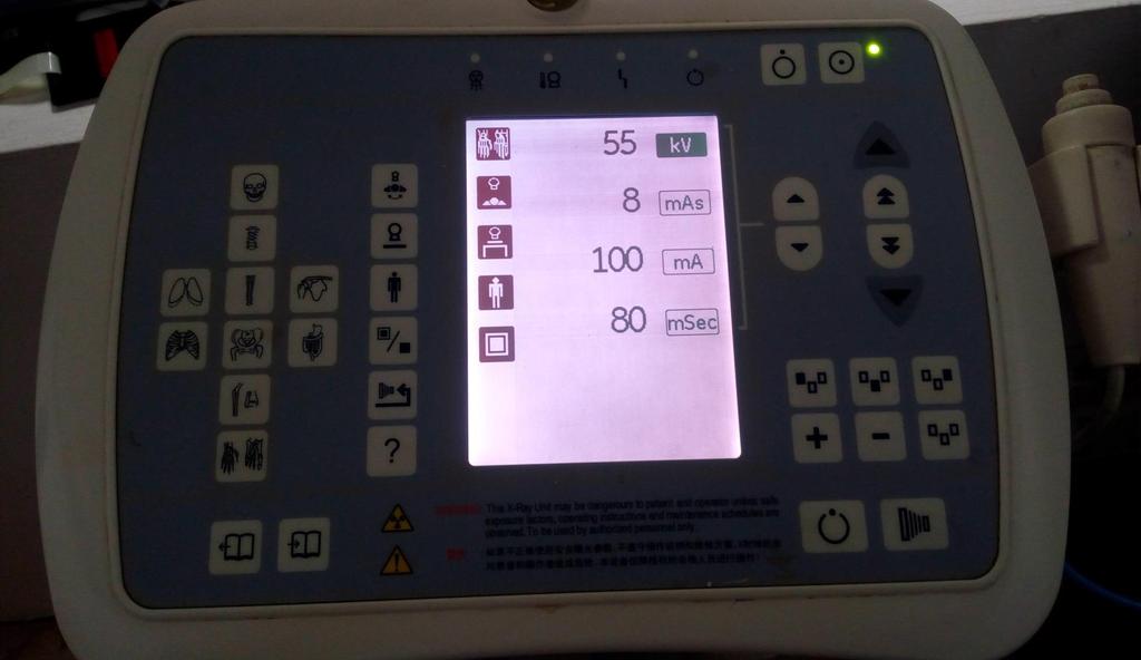
6 Dose. ALARA principle still holds Radiation exposure used in conventional system is based largely on certain conditions Specific Screen-Film system Type of screen Type of Film Condition used to processed the film State of Chemicals
7 In digital Imaging, image acquisition, Postprocessing and display are all separated into unique stages. Due to these factors, the tendency for an image to be OVER EXPOSED or UNDEREXPOSED is high. Adjustment to compensate this over/under exposure are mostly done in the display process.
8 Best Practice The Best practice is to select the appropriate exposure techniques factors for the patient size and condition based on planned exposure system designed with the radiologist to determine adequate image quality Do we do this???
9 Image Contrast Image contrast in Digital Imaging depends on two parameters Subject Contrast Display Contrast
10 Subject Contrast depends on various tissue composition Tissue density eg Bone and Air Body thickness KVp Absorption of radiation by tissue Subject Contrast cannot be controlled or adjusted by POSTPROCESSING
11 Display Contrast.. This is dependent on Post processing software-smart computers The monitor windowing The screen resolution eg. DICOM Screen(Barco Screens- High Resolution Screens)
12 Auto adjustment of an image contrast through powerful post processing software can even ensure that an image is of diagnostic quality even if its over exposed. In Digital Imaging excess exposure doesn't affect image quality except in extreme level of exposure
13 Due to this radiographers might be inclined to adjust exposure techniques to slightly increase the amount of radiation which subsequently.. increase patient dose ~DOSE CREEP Decrease image noise Least resistance and queries from Radiologist Avoid Repeats
14 The ability to adjust the image contrast of a digital image does affect the radiographer s attention to the primary principle of radiation protection optimal image quality with minimal patient exposure Best Practice.. As expert on exposure in the radiology team,radiographers should be proactive in remaining up-to-date on the basics of radiation protection and new technologies.
15 Procedure Validity.. 12% of request from doctors are unnecessary therefore. Radiographers: Should be the last medical profession to potentially screen for appropriateness before a radiological exam Needs to be a patient advocate and protect them against unnecessary exposure
16 should consult radiologist or referring doctors request for additional information that can further indicate the correct procedure to be performed when there is a suspicion of an inappropriate exam order. tracking of previous examination to prevent duplication of procedures eg RIS, HIS, PACS
17 Best Practices it is best practice in digital radiography for the radiographer to carefully review the examination ordered to prevent potential duplication and to ensure appropriateness as related to the patient s history.
18 Departmental Protocols.. Required for department accreditation It should be made accessible to practicing radiographers Done with consultation with radiologist Eg Exposure Chart KVp mas AEC Grid
19 Screening for Pregnancy.. Verification of pregnancy is department dependent ~ department policy Child bearing age women need to be asked of the possibility of pregnancy watch out for possible physical signs and lead-up questions Professional communication can help put Patient and RAD as ease.
20 If Pregnant. There should be a departmental policy Written document-consent form Discus with Radiologist if exam is necessary or any alternate procedure can be used to avoid exposure Best Practice The screening of patient for potential pregnancy is an essential best practice for radiation safety in digital imaging
21 Appropriate and consistent use of exposure technique chart and adequate kvp and AEC is essential to procedure diagnostic image while minimize patient radiation exposure kvp. Controls the energy of radiation reacting with the image receptor. kvp should be used in the same way as in film-screen system
22 High KVp and low mas is favored by most radiographers for Adult imaging 15% increase in average KVp and a corresponding 15% reduction in mas will produce good image contrast as well as reduce patient exposure increase in KVp will increase scatter hence the need for GRIDS to improve image contrast.
23 Best Practice. A best practice in digital imaging is to use the highest kvp within the optimal range for the position and part coupled with the lowest amount of mas needed to provide an adequate exposure to the image receptor.
24 AECs. Controls exposure times hence total mas Works as same as in film screen systems. Requires frequent and systematic calibrations Modern AEC have more than 3 ionization chambers mostly 5 Needs proper positioning of anatomical part over ionizing chamber
25 Underexposure from AEC will occur if the area of anatomy is too small to cover at least one AEC cell Over Exposure on AEC.eg Metallic implant on the AEC cell Best Practice. A best practice in digital radiography is to use AEC when indicated and to use AEC that has been calibrated to the type of image receptor to provide a consistent exposure to the image receptor.
26 Anatomic Program Radiography/Exposure Chart APR- A system of programmed exposure technique setting that is organized by position and procedure and set through the control panel at the radiographers unit APR gives options for various body size(small, Medium and Large) A combination of AEC(Ionization chamber selection) Allows for Manual adjustment of parameters.
27
28 Exposure Charts: It s a requirement in all radiography unit Should be accessible to all radiographers Eliminates the confusion concerning mas, kvp, SID, Grid, focal spot size etc It provide an acceptable levels of radiation that provides diagnostic image quality within the ALARA principle Drawn with typical/average patient attributes.
29 Exposure charts needs continuous monitoring and revision to ensure exposure techniques does produce diagnostic images within ALARA principle EXPOSURE CHART does not replace RADIOGRAPHERS as selection of exposures is dependent on patient condition, pathology and prevailing circumstances.
30 Best Practice: A best practice in digital radiography is to use exposure technique that are continuously improve and applicable to a wide range of patient sizes
31 Collimation/Electronic Masking Adjustment of the radiation beam to a determined area of interest. Effects: Minimize patient exposure Minimize Scatter radiation to the image receptor Improve image quality Prevent errors during postprocessing of the images
32 Masking:- Masking is an electronic software that recognizes the boarders of the exposed area on the image receptor. The area of unexposed side generally appear very bright. The mask will reduce the bright area by applying an overlay between the boarder of the exposed and unexposed areas on the receptor during post processing. Masking improve image viewing. Masking shouldn t occur within or over an anatomy of interest.
33
34 Masking and cropping shouldn t be use as a REPLACEMENT for physical collimation. Physical CROPPING has a negative effect on image contrast

35
36 In Digital Imaging, image receptors are more sensitive to low levels of radiations (Scatter) and the resulting digital image might demonstrate REDUCED IMAGE CONTRAST because of the excess scatter striking the receptor.
37 Collimation and Image Plate Multiple exposure can be made on an Image plate but depends on Type of Receptor used in the Image Plate Departmental protocol General Rule: Beam field should be well aligned No overlapping of exposures The need for a flexible PB covering of the unexposed/exposed side of the plate REGARDLESS of plate technology. Improve image quality

38
39 Best Practices: A best practice in digital radiography is to collimate the x-ray beam to the anatomical area appropriate for the procedure. Electronic masking to improve image viewing conditions should be applied in a manner that demonstrates the actual exposure field edge to document appropriate collimation. Masking must not be applied over anatomy that was contained in the exposure field at the time of exposure.
40 Shielding A fundamental radiation safety practice in digital imaging. Lack of shielding increase the radiation exposure dose to the patient Anatomical areas close to the exposure field needs shielding It shouldn t interfere with the ROI
41 There should be a departmental protocol At a minimum, gonadal shielding should be done if ROI is within 5cm of the edge of collimation. Improper shield can interfere in image postprocessing and optimum display for ROI if the shielding material is included as part of data processing Best Practice: A best practice in digital radiography is the use of lead shielding for anatomic parts that are adjacent to the x-ray field
42
43 Anatomical Markers Radiogrpahers need to use PB markers placed on the image receptor before exposure. Electronic Annotations of anatomical markers is not acceptable and not a substitue for PB marker. Failure to use PB marker has LEGAL issues There should be a departmental protocol for the site of placement of PB marker per each exams.
44
45 ACR and therefore GSR re-emphasis the use of PB markers as part of the digital imaging guidelines in Radiography Best Practices A best practice in digital radiography is the consistent use of lead anatomical side markers captured on the original image during x-ray exposure.
46 Thank You
COMPUTED RADIOGRAPHY CHAPTER 4 EFFECTIVE USE OF CR
 This presentation is a professional collaboration of development time prepared by: Rex Christensen Terri Jurkiewicz and Diane Kawamura New Technology https://www.youtube.com/watch?v=ptkzznazb 7U COMPUTED
X-RAYS - NO UNAUTHORISED ENTRY
 Licencing of premises Premises Refer Guidelines A radiation warning sign and warning notice, X-RAYS - NO UNAUTHORISED ENTRY must be displayed at all entrances leading to the rooms where x-ray units are
RAD 150 RADIOLOGIC EXPOSURE TECHNIQUE II
 RAD 150 RADIOLOGIC EXPOSURE TECHNIQUE II APPROVED 12/O2/2011 EFFECTIVE SPRING 2013-14 Prefix & Number RAD 150 Course Title: Radiologic Exposure Technique II & Lab Purpose of this submission: New Change/Updated
Exposure Indices and Target Values in Radiography: What Are They and How Can You Use Them?
 Exposure Indices and Target Values in Radiography: What Are They and How Can You Use Them? Definition and Validation of Exposure Indices Ingrid Reiser, PhD DABR Department of Radiology University of Chicago
Digital radiography (DR) post processing techniques for pediatric radiology
 post processing techniques for pediatric radiology") Digital radiography (DR) post processing techniques for pediatric radiology St Jude Children s Research Hospital Samuel Brady, MS PhD DABR samuel.brady@stjude.org Purpose Review common issues and solutions
Digital radiography (DR) post processing techniques for pediatric radiology St Jude Children s Research Hospital Samuel Brady, MS PhD DABR samuel.brady@stjude.org Purpose Review common issues and solutions
Beam-Restricting Devices
 Beam-Restricting Devices Three factors contribute to an increase in scatter radiation: Increased kvp Increased Field Size Increased Patient or Body Part Size. X-ray Interactions a some interact with the
Teaching Digital Radiography and Fluoroscopic Radiation Protection
 Teaching Digital Radiography and Fluoroscopic Radiation Protection WCEC 20 th Student Educator Radiographer Conference Dennis Bowman, RT(R), CRT (R)(F) Community Hospital of the Monterey Peninsula (CHOMP)
Exposure System Selection
 Principles of Imaging Science II (RAD120) Exposure Systems Exposure System Selection Radiographic exposure is a very complex process Best technique systems manipulate one variable while holding others
Multiple Choice Identify the letter of the choice that best completes the statement or answers the question.
 RA110 test 3 Multiple Choice Identify the letter of the choice that best completes the statement or answers the question. 1. An object 35 cm in width is radiographed at 100 cm SID and at a 50 cm SOD. What
CR Basics and FAQ. Overview. Historical Perspective
 Page: 1 of 6 CR Basics and FAQ Overview Computed Radiography is a term used to describe a system that electronically records a radiographic image. Computed Radiographic systems use unique image receptors
3/31/2011. Objectives. Emory University. Historical Development. Historical Development. Historical Development
 Teaching Radiographic Technique in a Digital Imaging Paradigm Objectives 1. Discuss the historical development of digital imaging. Dawn Couch Moore, M.M.Sc., RT(R) Assistant Professor and Director Emory
Mammography is a radiographic procedure specially designed for detecting breast pathology Approximately 1 woman in 8 will develop breast cancer over
 Mammography is a radiographic procedure specially designed for detecting breast pathology Approximately 1 woman in 8 will develop breast cancer over a lifetime Breast cancer screening programs rely on
LECTURE 1 The Radiographic Image
 LECTURE 1 The Radiographic Image Prepared by:- KAMARUL AMIN ABDULLAH @ ABU BAKAR UiTM Faculty of Health Sciences Medical Imaging Department 11/23/2011 KAMARUL AMIN (C) 1 Lesson Objectives At the end of
While digital techniques have the potential to reduce patient doses, they also have the potential to significantly increase them.
 In press 2004 1 2 Guest Editorial (F. Mettler, H. Ringertz and E. Vano) Guest Editorial (F. Mettler, H. Ringertz and E. Vano) Digital radiology An appropriate analogy that is easy for most people to understand
SYLLABUS. TITLE: Equipment Operation I. DEPARTMENT: Radiologic Technology
 CODE: RADT 156 INSTITUTE: Health Science TITLE: Equipment Operation I DEPARTMENT: Radiologic Technology COURSE DESCRIPTION: This course covers the principles of equipment operation and maintenance of radiographic
Acquisition, Processing and Display
 Acquisition, Processing and Display Terri L. Fauber, R.T. (R)(M) Department of Radiation Sciences School of Allied Health Professions Virginia Commonwealth University Topics Image Characteristics Image
Outline ASRT Changes Impact on current curriculum Potential new courses WECM Changes Last update Resources and needs
 Change nd Annual Blinn College 2 nd Educator s Workshop For Radiologic Sciences July 28, 2007 Christi Carter, MSRS, RT(R) Outline ASRT Changes Impact on current curriculum Potential new courses WECM Changes
Essentials of Digital Imaging
 Essentials of Digital Imaging Module 6 Transcript 2016 ASRT. All rights reserved. Essentials of Digital Imaging Module 6 Dose Reduction and Patient Safety 1. ASRT Animation 2. Welcome Welcome to Essentials
DISC QC/QA Program for Digital Imaging Systems using the DR Radchex Plus Meter
 DISC QC/QA Program for Digital Imaging Systems using the DR Radchex Plus Meter Revision Date: January 5th, 2017 www.disc-imaging.com Table of Contents Section A: Preliminary Setup Requirements... 4 Tools
RADIOGRAPHIC EXPOSURE
 RADIOGRAPHIC EXPOSURE Receptor Exposure Receptor Exposure the that interacts with the receptor. Computed Radiography ( ) requires a. Direct Digital Radiography (DR) requires a. Exposure Indicators Exposure
SECTION I - CHAPTER 1 DIGITAL RADIOGRAPHY: AN OVERVIEW OF THE TEXT. Exam Content Specifications 8/22/2012 RADT 3463 COMPUTERIZED IMAGING
 RADT 3463 - COMPUTERIZED IMAGING Section I: Chapter 1 RADT 3463 Computerized Imaging 1 SECTION I - CHAPTER 1 DIGITAL RADIOGRAPHY: AN OVERVIEW OF THE TEXT RADT 3463 COMPUTERIZED IMAGING Section I: Chapter
The Evaluation of Collimator Alignment of Diagnostic X-ray Tube Using Computed Radiography System
 The Evaluation of Collimator Alignment of Diagnostic X-ray Tube Using Computed Radiography System The Evaluation of Collimator Alignment of Diagnostic X-ray Tube Using Computed Radiography System Manus
Test Equipment for Radiology and CT Quality Control Contents
 Test Equipment for Radiology and CT Quality Control Contents Quality Control Testing...2 Photometers for Digital Clinical Display QC...3 Primary Workstations...3 Secondary Workstations...3 Testing of workstations...3
Do you have any other questions? Please call us at (Toll Free) or , or
 or , or") INSTRUCTIONS Read the appropriate course/ textbook. This is an open book test. A score of 75% or higher is needed to receive CE credit. You will have a maximum of three attempts to pass this course. Please
INSTRUCTIONS Read the appropriate course/ textbook. This is an open book test. A score of 75% or higher is needed to receive CE credit. You will have a maximum of three attempts to pass this course. Please
4. Contrast is the. There must The function of contrast is to:. The types of contrast are.
 RADIOGRAPHIC VISIBILITY OF DETAIL STUDY QUESTIONS 1. What is visibility of detail? It is controlled by properties. What are the two factors that affect it? 2. What is sharpness of detail? It is controlled
Visibility of Detail
 Visibility of Detail Radiographic Quality Quality radiographic images represents the, and information is for diagnosis. The of the anatomic structures and the accuracy of their ( ) determine the overall
10/3/2012. Study Harder
 This presentation is a professional collaboration of development time prepared by: Rex Christensen Terri Jurkiewicz and Diane Kawamura Study Harder CR detection is inefficient, inferior to film screen
THE ART OF THE IMAGE: IDENTIFICATION AND REMEDIATION OF IMAGE ARTIFACTS IN MAMMOGRAPHY
 THE ART OF THE IMAGE: IDENTIFICATION AND REMEDIATION OF IMAGE ARTIFACTS IN MAMMOGRAPHY William Geiser, MS DABR Senior Medical Physicist MD Anderson Cancer Center Houston, Texas wgeiser@mdanderson.org INTRODUCTION
Digital Imaging Considerations Computed Radiography
 Digital Imaging Considerations Digital Radiography Computed Radiography o Cassette based Direct or Indirect Digital Radiography o Cassetteless Computed Radiography 1 CR Image Acquisition Most like conventional
10/26/2015. Study Harder
 This presentation is a professional collaboration of development time prepared by: Rex Christensen Terri Jurkiewicz and Diane Kawamura Study Harder CR detection is inefficient, inferior to film screen
Acceptance Testing of a Digital Breast Tomosynthesis Unit
 Acceptance Testing of a Digital Breast Tomosynthesis Unit 2012 AAPM Spring Clinical Meeting Jessica Clements, M.S., DABR Objectives Review of technology and clinical advantages Acceptance Testing Procedures
R&F X-ray systsem. Savings With Every Exposure
 R&F X-ray systsem Savings With Every Exposure Savings with every exposure Versatility meets value Dependable performance User-friendly interface Expandable imaging capabilities Excellent image quality
BENNETT RENAISSANCE SERIES. Dedicated to Quality, Innovation and the Chiropractic Profession
 BENNETT RENAISSANCE SERIES TM Dedicated to Quality, Innovation and the Chiropractic Profession About HCMI, Inc. For over 20 years, HCMI, Inc. has been providing quality equipment for the chiropractic profession.
NJDEP Medical Physicist s Radiographic QC Survey Registration Number:
 Facility Name NJDEP ID # NJDEP Medical Physicist s Radiographic QC Survey PLEASE PRINT Facility Information Unit Information Manufacturer Model Console Model # Console serial # Tube serial # Location (room)
STEREOTACTIC BREAST BIOPSY EQUIPMENT SURVEYS
 STEREOTACTIC BREAST BIOPSY EQUIPMENT SURVEYS JAMES A. TOMLINSON, M.S. Diagnostic Radiological Physicist American Board of Radiology Certified Medical Physics Consultants, Inc. Bio 28 yrs experience 100%
1. Carlton, Richard R., and Arlene M. Adler. Principles of Radiographic Imaging: An Art and a Science, 5th edition (2013).
.") CODE: RADT 151 INSTITUTE: Health Science TITLE: Radiographic Exposure DEPARTMENT: Radiologic Technology COURSE DESCRIPTION: This course covers the principles of radiographic exposure selection and manipulation
CODE: RADT 151 INSTITUTE: Health Science TITLE: Radiographic Exposure DEPARTMENT: Radiologic Technology COURSE DESCRIPTION: This course covers the principles of radiographic exposure selection and manipulation
I. PERFORMANCE OF X-RAY PRODUCTION COMPONENTS FLUOROSCOPIC ACCEPTANCE TESTING: TEST PROCEDURES & PERFORMANCE CRITERIA
 FLUOROSCOPIC ACCEPTANCE TESTING: TEST PROCEDURES & PERFORMANCE CRITERIA EDWARD L. NICKOLOFF DEPARTMENT OF RADIOLOGY COLUMBIA UNIVERSITY NEW YORK, NY ACCEPTANCE TESTING GOALS PRIOR TO 1st CLINICAL USAGE
Automated dose control in multi-slice CT. Nicholas Keat Formerly ImPACT, St George's Hospital, London
 Automated dose control in multi-slice CT Nicholas Keat Formerly ImPACT, St George's Hospital, London Introduction to presentation CT contributes ~50+ % of all medical radiation dose Ideally all patients
X-RAY IMAGING EE 472 F2017. Prof. Yasser Mostafa Kadah
 X-RAY IMAGING EE 472 F2017 Prof. Yasser Mostafa Kadah www.k-space.org Recommended Textbook Stewart C. Bushong, Radiologic Science for Technologists: Physics, Biology, and Protection, 10 th ed., Mosby,
Outline. Digital Radiography. Understanding Digital Modalities: Image Quality and Dose. Image Quality. Dose Control
 Understanding Digital Modalities: Image Quality and Dose S. Jeff Shepard, M.S. University of Texas M. D. Anderson Cancer Center Houston, Texas Special Acknowledgement: Stephen K. Thompson, M.S. William
Nuclear Associates
 Nuclear Associates 07-644 Grid Alignment Test Tool Users Manual March 2005 Manual No. 07-644-1 Rev. 2 2004, 2005 Fluke Corporation, All rights reserved. Printed in U.S.A. All product names are trademarks
Truly flexible to meet your clinical needs
 Truly flexible to meet your clinical needs 2 Adapting to meet your needs Flexible Fast and responsive Excellent image quality Designed with ergonomic efficiency Equipped with dose management tools 3 Three
INTRODUCTION TO FLEXIBLE BRONCHOSCOPY. Fluoroscopy Synopsis HENRI G COLT MD SECOND EDITION THE BRONCHOSCOPY EDUCATION PROJECT SERIES
 SECOND EDITION INTRODUCTION TO FLEXIBLE BRONCHOSCOPY Fluoroscopy Synopsis HENRI G COLT MD With contributions from Dr. S. Murgu THE BRONCHOSCOPY EDUCATION PROJECT SERIES FLUOROSCOPY SYNOPSIS The purpose
Redefining Ergonomics
 Samsung Electronics Co., Ltd. inspires the world and shapes the future with transformative ideas and technologies, redefining the worlds of TVs, smartphones, wearable devices, tablets, cameras, digital
Contrast. Contrast: the difference in density on adjacent areas of a radiograph or other image receptor. Subjective. Long Scale (Low Contrast)
") Contrast Contrast: the difference in density on adjacent areas of a radiograph or other image receptor. Subject Subjective Radiographic Long Scale (Low Contrast) Short Scale (High Contrast) Factors affecting
Contrast Contrast: the difference in density on adjacent areas of a radiograph or other image receptor. Subject Subjective Radiographic Long Scale (Low Contrast) Short Scale (High Contrast) Factors affecting
X-ray Imaging. PHYS Lecture. Carlos Vinhais. Departamento de Física Instituto Superior de Engenharia do Porto
 X-ray Imaging PHYS Lecture Carlos Vinhais Departamento de Física Instituto Superior de Engenharia do Porto cav@isep.ipp.pt Overview Projection Radiography Anode Angle Focal Spot Magnification Blurring
Nuclear Associates
 Nuclear Associates 07-649 CDRH Fluoroscopic Phantom Users Manual March 2005 Manual No. 07-649-1 Rev. 2 2004, 2005 Fluke Corporation, All rights reserved. Printed in U.S.A. All product names are trademarks
Precision Performance Power
 ODYSSEY HF SERIES ULTRA High Frequency X-Ray Technology Precision Performance Power Innovations in Digital Imaging. TM STEP 1 Select anatomical region STEP 2 Select anatomical view STEP 3 Ready for exposure
SPRINGFIELD TECHNICAL COMMUNITY COLLEGE ACADEMIC AFFAIRS
 SPRINGFIELD TECHNICAL COMMUNITY COLLEGE ACADEMIC AFFAIRS Course Number: RADG 112 Department: Radiography Course Title: Image Production & Eval. Semester: Spring Year: 1997 Objectives/ Unit One: Introduction
Nuclear Associates
 Nuclear Associates 07-647 R/F QC Phantom Operators Manual March 2005 Manual No. 07-647-1 Rev. 2 2004, 2005 Fluke Corporation, All rights reserved. All product names are trademarks of their respective companies
ODYSSEY HF SERIES. ULTRA High Frequency X-Ray Technology. Precision... Performance... Power. Innovations in Digital Imaging.
 ODYSSEY HF SERIES ULTRA High Frequency X-Ray Technology Precision... Performance... Power Innovations in Digital Imaging. TM STEP 1 Select anatomical region STEP 2 Select anatomical view STEP 3 Ready for
Nuclear Associates EZ CR-DIN Phantoms
 Nuclear Associates 07-605-7777 EZ CR-DIN Phantoms Users Manual August 2006 Manual No. 07-605-7777-1 Rev. 4 2006 Fluke Corporation, All rights reserved. Printed in U.S.A. All product names are trademarks
Y11-DR Digital Radiography (DR) Image Quality
 Image Quality") Y11-DR Digital Radiography (DR) Image Quality Image quality is stressed for all systems in Safety Code 35. In the relevant sections Health Canada s advice is the manufacturer s recommended test procedures
Y11-DR Digital Radiography (DR) Image Quality Image quality is stressed for all systems in Safety Code 35. In the relevant sections Health Canada s advice is the manufacturer s recommended test procedures
KODAK DIRECTVIEW CR Mammography Feature User s Guide
 KODAK DIRECTVIEW CR Mammography Feature User s Guide 17 September 2010 9G3741 Version 1.0 Carestream Health, Inc. 150 Verona Street Rochester, NY 14608 CARESTREAM, DIRECTVIEW, and DRYVIEW are trademarks
Learning Objectives: What s my motivation? (unknown screen actor) Workshop Overview
 Workshop Overview") Practical Medical Physics Adapting Traditional Clinical Medical Physics to Digital Radiography Charles E. Willis, Ph.D., DABR Associate Professor Department of Imaging Physics The University of Texas M.D.
Practical Medical Physics Adapting Traditional Clinical Medical Physics to Digital Radiography Charles E. Willis, Ph.D., DABR Associate Professor Department of Imaging Physics The University of Texas M.D.
ECOVIEW 9 / ECOVIEW 9 PLUS Digital Radiographic System
 ECORAY Co., LTD. 3F, Urbanlight B/D, 630, Eonju-ro, Gangnam-gu, Seoul, Korea 135-832 Factory at 621-14, Dochun-dong, Gwangsan-gu, Gwangju, Korea 506-301 TEL. : +82-70-7510-3400 FAX. : +82-70-8630-3420
FMT18 FLOOR MOUNTED SYSTEM
 mas Time AEC 320 kvp 64 mas 320 ma 320 ma 320 DEN 0.0 mm Cu 17 in X 17 in 72.0 in FMT18 FLOOR MOUNTED SYSTEM with Synchronized Tracking System Overview Clinical Efficiency The FMT18 System was designed
PLD5600A High Frequency Digital Gastrointestinal &DR System(630mA)
") PLD5600A High Frequency Digital Gastrointestinal &DR System(630mA) Application: Full support perspective, gastrointestinal spot film, GI (barium meal, barium enema), orthopedic photography, pediatrics
PLD5600A High Frequency Digital Gastrointestinal &DR System(630mA) Application: Full support perspective, gastrointestinal spot film, GI (barium meal, barium enema), orthopedic photography, pediatrics
Nuclear Associates
 Nuclear Associates 07-706 Patient Phantom/Penetrometer System Users Manual March 2005 Manual No. 07-706-1 Rev. 2 2004, 2005 Fluke Corporation, All rights reserved. Printed in U.S.A. All product names are
Digital radiography. bucky table and wall stand as a dual detector or wireless system. Amadeo DR Systems
 Amadeo DR Systems with dicom PACS DX-R Software X-ray Systems for the Future Digital radiography with Amadeo R-DR including bucky table and wall stand as a dual detector or wireless system High contrast
ECOVIEW 9 / ECOVIEW 9 PLUS Digital Radiographic System
 Co., LTD. 3F, Urbanlight B/D, 630, Eonju-ro, Gangnam-gu, Seoul, Korea 135-832 Factory at 621-14, Dochun-dong, Gwangsan-gu, Gwangju, Korea 506-301 TEL. : +82-70-7510-3400 FAX. : +82-70-8630-3420 sales@ecoray.kr
A Practical Overview of the Clinical and Operational Impact of Computed Radiography(CR) Implementations. Shirley Weddle, RT(R)(M), CIIP, BBA
 Implementations. Shirley Weddle, RT(R)(M), CIIP, BBA") A Practical Overview of the Clinical and Operational Impact of Computed Radiography(CR) Implementations Shirley Weddle, RT(R)(M), CIIP, BBA OBJECTIVES Define Computed Radiography (CR) Discuss CR vendor
A Practical Overview of the Clinical and Operational Impact of Computed Radiography(CR) Implementations Shirley Weddle, RT(R)(M), CIIP, BBA OBJECTIVES Define Computed Radiography (CR) Discuss CR vendor
COMPUTED RADIOGRAPHY (CR)
") COMPUTED RADIOGRAPHY (CR) Moving with the time Avi Avner BVSc BSc CVR DVDI MRCVS CR-Basics A five step process: 1. X-ray image received on phosphor plate 2. Image extracted from phosphor plate by Laser
COMPUTED RADIOGRAPHY (CR) Moving with the time Avi Avner BVSc BSc CVR DVDI MRCVS CR-Basics A five step process: 1. X-ray image received on phosphor plate 2. Image extracted from phosphor plate by Laser
of sufficient quality and quantity
 of sufficient quality and quantity The patient s body attenuates the beam as it passes though the body More energy is deposited in organs located near the entry of the beam than near the exit of the beam
X o- System. Chiro- Systems. Chiropractic X-Ray Systems and Digital Solutions. Digital Radiography Computerized Radiography Chiropractic Systems
 X o- System Chiropractic X-Ray Systems and Digital Solutions Chiro- Systems Digital Radiography Computerized Radiography Chiropractic Systems Quantum s Chiropractic Imaging Solutions Digital Work Flow
Investigation of the line-pair pattern method for evaluating mammographic focal spot performance
 Investigation of the line-pair pattern method for evaluating mammographic focal spot performance Mitchell M. Goodsitt, a) Heang-Ping Chan, and Bob Liu Department of Radiology, University of Michigan, Ann
Studies on reduction of exposure dose using digital scattered X-ray removal processing
 Studies on reduction of exposure dose using digital scattered X-ray removal processing Poster No.: C-1834 Congress: ECR 2015 Type: Scientific Exhibit Authors: K. Kashiyama, M. Funahashi, T. Nakaoka, T.
Studies on reduction of exposure dose using digital scattered X-ray removal processing
 Studies on reduction of exposure dose using digital scattered X-ray removal processing Poster No.: C-1834 Congress: ECR 2015 Type: Scientific Exhibit Authors: K. Kashiyama, M. Funahashi, T. Nakaoka, T.
Assessment of Beam Alignment, Collimation and Half Value Layer of Some Selected X-Ray Machines in Plateau State, Nigeria
 International Journal of Innovative Scientific & Engineering Technologies Research 5(4):-5, Oct.-Dec., 07 SEAHI PUBLICATIONS, 07 www.seahipaj.org ISSN: 60-896X Assessment of Beam Alignment, Collimation
- KiloVoltage. Technique 101: Getting Back to Basics
 Why do I need to know technique? Technique 101: Getting Back to Basics Presented by: Thomas G. Sandridge, M.S., M.Ed., R.T.(R) Program Director Northwestern Memorial Hospital School of Radiography Chicago,
China Resources Wandong Medical Equipment Co., Ltd. High Frequency 50kW, 150kV Radiography System - HF50-R
 China Resources Wandong Medical Equipment Co., Ltd. High Frequency 50kW, 150kV Radiography System - HF50-R Building 3, No.9, Jiuxianqiaodong Road, Chaoyang District, Beijing 100015, P.R. China E-mail:
X-rays. X-rays are produced when electrons are accelerated and collide with a target. X-rays are sometimes characterized by the generating voltage
 X-rays Ouch! 1 X-rays X-rays are produced when electrons are accelerated and collide with a target Bremsstrahlung x-rays Characteristic x-rays X-rays are sometimes characterized by the generating voltage
Dedicated Veterinary Imaging Solutions Digital, CR and Analog Imaging Solutions for any size patient and any size budget.
 by Dedicated Veterinary Imaging Solutions Digital, CR and Analog Imaging Solutions for any size patient and any size budget. Serving the Veterinary Profession for Over 75 Years. ... We See Things Differently
MUSICA Nerve Center. Artificial Intelligence. Intelligent tools for your Digital Radiography workflow. Fluoroscopy. Workflow Optimization
 Image Quality Bariatric Abdomen Pediatric Imaging Diagnostic Confidence Fluoroscopy Neonatal Imaging Scatter Suppression Dental Full Leg Full Spine Exposure Control Index Artificial Intelligence General
CONTRASTING VIEWS DIGITAL VS CONVENTIONAL RADIOGRAPHY
 Vet Times The website for the veterinary profession https://www.vettimes.co.uk CONTRASTING VIEWS DIGITAL VS CONVENTIONAL RADIOGRAPHY Author : PETRA AGTHE Categories : Vets Date : April 7, 2008 PETRA AGTHE
RAD. Experiences Using the RADspeed Pro EDGE Package. 1. Hospital Description. 2. Background of Adoption. Hirohito Tanaka
 RAD Experiences Using the RADspeed Pro EDGE Package Hirohito Tanaka, R.T. Department of Radiology, Chibune General Hospital Hirohito Tanaka 1. Hospital Description Our hospital was originally established
Ludlum Medical Physics
 Ludlum Medical Physics Medical Imaging Radiology QA Test Tools NEW LUDLUM PRODUCT LINE Medical Physics Products Medical Physics Products What are they? Products used to measure radiation output and to
Veterinary Science Preparatory Training for the Veterinary Assistant. Floron C. Faries, Jr., DVM, MS
 Veterinary Science Preparatory Training for the Veterinary Assistant Floron C. Faries, Jr., DVM, MS Radiology Floron C. Faries, Jr., DVM, MS Objectives Determine the appropriate machine settings for making
Get more from your images with Symphony Image Processing
 DIRECT RADIOGRAPHY The user-friendly DelWorks image acquisition and processing software possesses a wide range of tools for a variety of image manipulations. Its user interface simplifies every step of
DR _ solutions. We understand that customers don t need just products, they want. solutions
 DR _ solutions We understand that customers don t need just products, they want solutions index company profile 1974-2005 2006-2007 - 2008 ITALRAY Srl was founded in 1974 as the production branch of Marzocchi
Get more from your images with Symphony Image Processing
 DIRECT RADIOGRAPHY The user-friendly DelWorks image acquisition and processing software provides a wide range of tools for a variety of image enhancements. Its user interface simplifies every step of the
MaxRay Handheld X-ray Systems Operator Training Exam
 MaxRay Handheld X-ray Systems Operator Training Exam Employee: Instructor: ate: Score: Instructions Read each question carefully and choose the best answer. 1) LR is 2) 3) 4) a. a safety principle meant
Management of pediatric radiation dose using Philips fluoroscopy systems DoseWise: perfect image, perfect sense
 Pediatr Radiol (2006) 36 (Suppl 2): 216 220 DOI 10.1007/s00247-006-0216-0 ALARA Dick Stueve Management of pediatric radiation dose using Philips fluoroscopy systems DoseWise: perfect image, perfect sense
SAMSUNG SMART DIGITAL RADIOGRAPHY. XGEO GU60A series REDEFINING ERGONOMICS
 SAMSUNG SMART DIGITAL RADIOGRAPHY XGEO GU60A series REDEFINING ERGONOMICS U-arm Positioner (Fully Automated) The XGEO GU60A/60A-65 is a universal, fully motorized system. Its unique U-arm rotates +120
Ask EuroSafe Imaging Tips & Tricks. Paediatric Imaging Working Group. Dose Management in Digital Radiography
 Ask EuroSafe Imaging Tips & Tricks Paediatric Imaging Working Group Dose Management in Digital Radiography Raija Seuri (HUS Medical Imaging Center, FI) Cristina Almeida (Centro Hospitalar de Lisboa Central,
MXHF-1500RF is controlled by Digital key panel console that displays KV, ma and mas with APR menu programmed.
 R/F TV X-RAY SYSTEM DIAGNOSTIC RADIOGRAPHIC FLUOROSCOPIC TV SYSTEM MXHF-1500RF SYSTEM OUTLINE Product Data No. 041021-01 MXHF-1500RF is controlled by Digital key panel console that displays KV, ma and
Performance and care. all in one
 Performance and care all in one INNOVATION IS WHAT DRIVES US THINKING ABOUT THE FUTURE Preventive diagnostics remains an essential weapon in defeating breast cancer. Metaltronica s forward-thinking design
SPRINGFIELD TECHNICAL COMMUNITY COLLEGE ACADEMIC AFFAIRS
 SPRINGFIELD TECHNICAL COMMUNITY COLLEGE ACADEMIC AFFAIRS Course Number: RADG 212 Department: Radiography Course Title: Equip. Operation & Maint. Semester: Spring Year: 1997 Objectives/ Unit One: The X-ray
Multi-Access Biplane Lab
 Multi-Access Biplane Lab Advanced technolo gies deliver optimized biplane imaging Designed in concert with leading physicians, the Infinix VF-i/BP provides advanced, versatile patient access to meet the
The effect of compensating filter on image quality in lateral projection of thoraco lumbar radiography
 Journal of Physics: Conference Series OPEN ACCESS The effect of compensating filter on image quality in lateral projection of thoraco lumbar radiography To cite this article: N A A Daud et al 2014 J. Phys.:
MAMMOGRAPHY - HIGH LEVEL TROUBLESHOOTING
 MAMMOGRAPHY - HIGH LEVEL TROUBLESHOOTING Maynard High New York Medical College SS2001-M.High 1 Objectives: Review MQSA and ACR annual QC tests as opportunities for troubleshooting before a significant
Disclosures. Outline 7/31/2017. Current Implementation Status of IEC Standard : Exposure Index (EI) for Digital Radiography
 for Digital Radiography") Current Implementation Status of IEC Standard 62494-1: Exposure Index (EI) for Digital Radiography July 31, 2017 Ryan Fisher, PhD, DABR Katie Hulme, MS, DABR None Disclosures Outline Review of IEC Standard
Current Implementation Status of IEC Standard 62494-1: Exposure Index (EI) for Digital Radiography July 31, 2017 Ryan Fisher, PhD, DABR Katie Hulme, MS, DABR None Disclosures Outline Review of IEC Standard
P R E S E N T E D B Y. K A M A R U L A M I N A B D U L L A H Dip. MED. IMG., BSc. MED. IMG. (UiTM)
") + - P R E S E N T E D B Y K A M A R U L A M I N A B D U L L A H Dip. MED. IMG., BSc. MED. IMG. (UiTM) 1 I N T R O D U C T I O N : An x-ray generator is a device that Supplies electrical power to x-ray
+ - P R E S E N T E D B Y K A M A R U L A M I N A B D U L L A H Dip. MED. IMG., BSc. MED. IMG. (UiTM) 1 I N T R O D U C T I O N : An x-ray generator is a device that Supplies electrical power to x-ray
Patient-based radiographic exposure factor selection: a systematic review
 REVIEW ARTICLE Patient-based radiographic exposure factor selection: a systematic review William Ching, BAppSc(Hons), John Robinson, BAppSc, & Mark McEntee, PhD Discipline of Medical Radiation Sciences,
SYLLABUS. 1. Identification of Subject:
 SYLLABUS Date/ Revision : 30 January 2017/1 Faculty : Life Sciences Approval : Dean, Faculty of Life Sciences SUBJECT : Biophysics 1. Identification of Subject: Name of Subject : Biophysics Code of Subject
OTC18 OVERHEAD TUBE CRANE SYSTEM
 OTC18 OVERHEAD TUBE CRANE SYSTEM System Overview Clinical Performance Versatile and intuitive, the OTC18M System delivers enhanced patient comfort and optimized workflow. Precisely designed to withstand
1-1. GENERAL 1-2. DISCOVERY OF X-RAYS
 1-1. GENERAL Radiography is a highly technical field, indispensable to the modern dental practice, but presenting many potential hazards. The dental radiographic specialist must be thoroughly familiar
2217 US Highway 70 East Garner, NC Main: Fax:
 Viztek is committed to providing the highest image quality possible in our CR & DR product lines. There are several factors that directly affect the overall quality of CR & DR based images. The eposure
Open. the Digitized world. Fuji Computed Radiography
 Open the Digitized world Fuji Computed Radiography If just one of these applies to you... Managing developing fluid is hard and darkroom work is a hassle... Images are not stable... Isn t digitalization
COURSE SYLLABUS. Instructor Information. Course Description. Prerequisites/Corequisites SCANS. End-of-Course Outcomes/Objectives
 COURSE SYLLABUS Department: Course Title: Radiologic Technology Principles of Radiographic Imaging I Section Name: RADR 1313 Start Date: 01/17/2012 End Date: 05/14/2012 Modality: FACE-TO-FACE Credits: