Acquisition, Processing and Display
|
|
|
- Lydia Patterson
- 6 years ago
- Views:
Transcription

1 Acquisition, Processing and Display Terri L. Fauber, R.T. (R)(M) Department of Radiation Sciences School of Allied Health Professions Virginia Commonwealth University
2 Topics Image Characteristics Image Receptors Image Acquisition Digital Image Processing & Display Exposure Technique Factors
3 Image Production Primary beam attenuated by tissues. Exit or remnant radiation composed of varying intensities. Image receptor receives or captures the remnant radiation and Creates a latent or invisible image that needs processing.
4 Image Formation Image Formation for digital imaging differs from filmscreen. Image receptors respond differently to the remnant radiation. Digital image stored and displayed as computer data visible on a monitor as a range of brightness levels Film image is processed to display a range of densities on a polyester sheet
5 Image Receptors Types Film Film-screen Digital Computed radiography (CR) Direct radiography (DR)

6 Digital vs. Film- Screen Radiography Film-Screen Limitations Superimposition of anatomical structures Difficult to visualize widely varying types of tissue Poor soft tissue differentiation Delay in viewing image No changes in image following processing Lack of quantitative analysis of attenuation characteristics Storage and retrieval costs Digital Advantages Post-processing can alter the image to remove superimposing anatomical structures Allows visualization of widely varying types of tissue Excellent soft tissue differentiation Minimal delay in viewing image Allows manipulation of image postprocessing Provides quantitative data analysis of tissue attenuation characteristics No hard copy storage and retrieval costs

7 Image Characteristics Visualize the area of interest Brightness/Density Differentiate among the anatomic details Contrast Resolution/Contrast Maximize sharpness Spatial Resolution/Recorded Detail Minimize distortion Minimize Quantum Noise
8 Density/Brightness The range of image densities is created by the variation in x-ray absorption and transmission as the x-ray beam passes through anatomic tissues. Low density = High brightness High density = Low brightness

9 Digital Image Characteristics A digital image is recorded as a combination of rows and columns known as matrix The smallest component of the matrix is the pixel (picture element); measured in microns (0.001 mm); and recorded as a single numerical value The location of the pixel within the image matrix corresponds to an area within the patient or volume of tissue referred to as voxel

10 Matrix Size For a given field of view (FOV), a larger matrix size includes a greater number of smaller pixels. A matrix size of 1024 x 1024 has 1,048,576 individual pixels; a matrix size of 2048 x 2048 has 4,194,304 pixels.

11 Image Characteristics Each pixel is assigned a numeric value (quantization) that represents a shade of gray or brightness level based on the attenuation characteristics of the volume of tissue imaged When displayed on a computer monitor, high density is low brightness and low density is high brightness
12 Pixel Bit Depth The number of bits determines the number of shades of gray the digital system is capable of displaying (2 n ). 12- and 14- bit pixel can display 4096 and shades of gray, respectively. Increasing pixel bit depth improves image quality

13 Signal-to-Noise Ratio (SNR) SNR is a method of describing the strength of the radiation exposure compared with the amount of noise apparent in a digital image. Because the photon intensities are converted to an electronic signal that is digitized, the term signal refers to the strength or amount of radiation exposure captured by the IR to create the image SNR Image Quality

14 Noise Quantum Noise- low photon energy used to record the image. Electronic or system noise- random effect that degrades the quality of the image. A result of the use of electronic components in digital imaging. When the digital image displays increased noise, regardless of the source, anatomic details have decreased visibility.
15 Digital Imaging Process Unlike film-screen, where acquisition, processing and display are all in one (film), digital imaging separates these three processes: Image Acquisition Image Processing Image Display
16 Digital Radiography Computed Radiography (CR) X-ray photons exiting pt. Photostimulable phosphor Reader (laser) Digital data Memory storage Display monitor Laser printer Direct Radiography (DR) Memory storage X-ray photons exiting pt. Digital data Flat panel detector Display monitor Laser printer


17 Digital Image Receptors (DR) Flat Panel Detectors Exit radiation absorbed by phosphor Laser extracts stored energy as light Light convert to electric signal Electric signal digitized Exit radiation converted to light then electrons or directly to electrons Electric signal digitized CR and DR differ in the method of image acquisition Seibert, AJ. Digital Radiography: Image Quality and Radiation Dose. Health Physics, 2008;95(5):

18 Cross Section of PSP screen The CR IR includes a cassette that houses the imaging plate (IP). The radiation exiting the patient interacts with the IP, where the photon intensities are absorbed by the phosphor. The phosphor layer is composed of barium fluorohalide crystals doped with europium, referred to as the photostimulable phosphor (PSP). This type of phosphor emits visible light when stimulated by a high-intensity laser beam, called photostimulable luminescence.

19 PSP Latent Image Formation CR emits light first during x- ray exposure Carroll, Q Practical Radiographic Imaging, 8 th ed, 2007 The Latent image is formed when some of the freed electrons become trapped Following laser light stimulation, the trapped electrons return to normal state and release visible light.
20 CR Image Acquisition Exit radiation interacts with imaging plate composed of barium fluoride bromide crystals coated with europium; Absorbed energy stored in photostimulable phosphor material. Some energy is released as visible light, but most results in electrons released in phosphor layer due to photoelectric interactions Electrons are trapped in phosphor layer until light energy is released during laser scanning (electrons are trapped in an excited state)
21 CR Latent Image Digitization Three stages Scanning Sampling Quantization

22 CR Image Acquisition: Scanning A continuous pattern of light intensities are sent to the Photomultiplier Tube (PMT) The PMT collects, amplifies, and converts the visible light to an electrical signal proportional to the range of energies stored in the IP Electrical signal is directed to the ADC for sampling and quantization
23 CR Image Acquisition: Sampling Analog to Digital Conversion accomplished by sampling Sampling Frequency for CR Image Receptors Increasing the sampling frequency will increase the spatial resolution
24 Sampling Frequency Nyquist Theorem: you must sample for each pixel at least 2 times to achieve the desired level of spatial resolution. Increasing the sampling frequency will more accurately reflect the analog signal, increase spatial resolution, and improve the quality of the digital image.
25 Sampling & Spatial Resolution In computed radiography (CR) some manufacturers fix the sampling frequency to maintain a fixed spatial resolution while others vary the sampling frequency to maintain a fixed matrix size. If the spatial resolution is fixed, the image matrix size is simply proportional to the imaging plate (IP) size. A larger IP will have a larger matrix to maintain spatial resolution If, on the other hand, the matrix size is fixed, changing the size of the IP will affect the spatial resolution of the digital image.

26 Fixed Sampling Frequency In CR, increasing the IP size for a fixed sampling frequency will maintain the pixel size for a fixed spatial resolution (larger matrix).

27 Fixed Matrix Size In CR, increasing the IP size for a fixed matrix size will increase the pixel size and decrease spatial resolution.
28 Image Acquisition: Quantization During quantization, each pixel, representing a brightness value, is assigned a numerical value that reflects the precision with which each sampled point is recorded. The pixel bit depth determines the system s ability to display a range of shades of gray to represent anatomic tissues. Pixel bit depth (2 n ) is fixed by the choice of ADC and digital systems manufactured with greater bit depth can display more shades of gray which improves the contrast resolution of the digital image.
29 Direct Radiography Flat Panel Detectors (FPD) are solid-state IRs employing a large area active matrix (array) of electronic components ranging in size from 17 x 14 inches to 17 x 17 inches. FPDs are constructed with layers in order to receive the x-ray photons and convert them to electrical charges for storage and readout. Signal storage, signal readout, and digitizing electronics are integrated into the FPD.


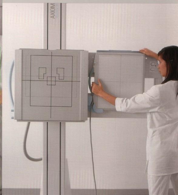
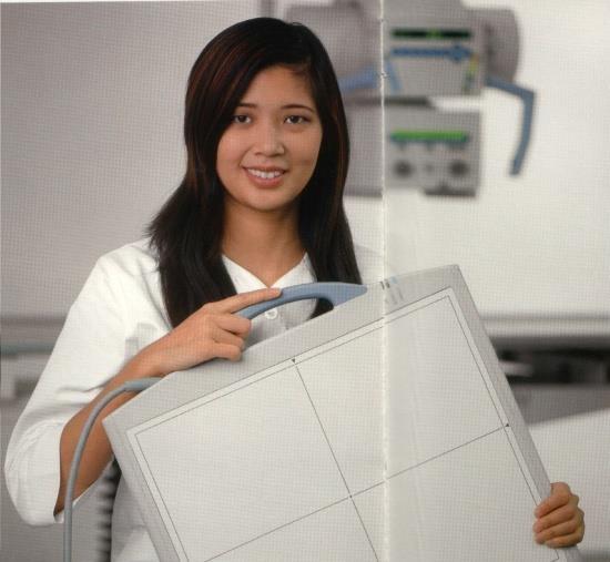
30 Flat Panel Detector- Mobile
31 DR Image Acquisition Indirect conversion Flat panel detector array exposed to exit radiation Scintillator converts x-rays to light Light energy converted to electric signals Electric signals digitized Direct conversion Flat panel detector array exposed to exit radiation Selenium converts x-rays to electric signal Electric signals digitized


32 Direct Radiography Image Receptors Indirect Conversion Direct Conversion
33 Flat Panel Detector Systems Indirect Conversion Scintillator Direct Conversion Electronic charge Visible light Electronic charge Digital Image Digital Image

34 Detector Element Size (DEL) Because the pixel detector is built into the DR flat panel type of image receptor, the size and pitch of the pixel is determined by the DEL and fixed. The spatial resolution is limited to the DEL. A system that uses a smaller DEL size will have improved spatial resolution.

35 Sampling Pitch/ Pixel Pitch The distance between the midpoint of adjacent pixels (pixel pitch) will also affect the spatial resolution of the digital image. Decreasing the pixel or sampling pitch will increase spatial resolution and image quality.
36 Film-Screen Image Receptors Prior to digital radiography, film was used to acquire, process, and display the radiographic image. An emulsion layer is adhered to a polyester sheet and serves as the radiation-sensitive and light sensitive layer of the film. Before the development of intensifying screens, film was exposed to the primary beam and then hand processed to produce the desired image. In an effort to reduce patient exposure, film is placed in a cassette that houses two intensifying screens. The film is exposed to the light emitted from the screen in proportion to the amount of radiation exposure.

37 Latent Image Formation-Film Crystals in the emulsion absorb radiant energy Gurney Mott Theory Sensitivity speck entraps free electrons Silver ions collect at speck Chemical processing converts exposed crystals to black metallic silver and viewed as densities
38 Dynamic Range The range of exposure intensities an image receptor can accurately detect. A- Film-screen receptors of narrow dynamic range (latitude) B- Digital image receptors have wide dynamic range (latitude)
39 Dynamic Range Wide Dynamic Range Digital image receptors can detect a greater range of x-ray intensities than film. Dynamic Range Low and high x-ray intensities exiting the patient can be detected and displayed.

40 Film-Screen Image Receptor Exposure Optimum Exposure Insufficient Exposure Excessive Exposure

41 A 14 pixel bit depth system has 16,384 shades of gray or brightness levels available to display the wide range of radiation exposures the image receptor captures.


42 Digital Image Receptor Exposure Insufficient Exposure Optimum Exposure Excessive Exposure

43 Digital Image Processing-CR/DR Following acquisition and digitization of the latent image, various computer manipulations are applied to the digital data for the purpose of optimizing the appearance of the image. Histogram Analysis Values of interest Automatic rescaling Exposure indicator Lookup Tables
44 Processing: Histogram Analysis Graphic representation of a data set or all the pixel values within the exposed image. The histogram represents the number of digital pixel values versus the relative prevalence of the pixel values in the image. X-axis: amount of exposure Y-axis: the incidence of pixels for each exposure level The computer software has stored histogram models, each having a shape characteristic of the selected anatomic region and projection. These stored histogram models have values of interest (VOI), which determine what range of the histogram data set should be included in the displayed image.

45 Processing: Histogram Analysis A histogram is the graphic display of the distribution of pixel values. Each image has its own histogram and is compared to the preestablished histograms for each anatomic projection
46 Processing: Histogram Analysis Histogram analysis is also employed to maintain consistent image brightness despite overexposure or underexposure of the IR, known as automatic rescaling The computer rescales the image based on the comparison of the histogram, which is actually a process of mapping the grayscale to the VOI to present a specific display of brightness
47 Processing: Exposure Indicator During digital processing and as a result of the histogram analysis a numerical value is displayed to indicate the level of x-ray exposure received to the image receptor. Computed Radiography (CR) Exposure Indicators are vendor specific and not currently standardized. Vendor Exposure Indicator Value = 1 mr exposure 2 x Exposure 1/2 Exposure Fuji and Konica Sensitivity (S) Carestream (Kodak) Exposure Index (EI) Agfa Log Median Value (lgm)

48 Processing: Automatic Rescaling
49 Standard Exposure Indicator Exposure Index (EI) an index of the exposure at the detector in the relevant image region Target Exposure Index (EI T ) the target reference obtained when the image receptor is exposed properly Deviation Index (DI) measures how far the actual EI value deviates from the projection-specific EI T DI provides immediate feedback to the radiographer regarding proper exposure Important to note that these values do not provide information about patient exposure and dose.
50 Processing: Look Up Tables (LUTs) Following the histogram analysis, LUTs are a method of altering the image to change the display of the digital image in a variety of ways. Because digital IRs have a linear exposure response and a very large dynamic range, raw data images exhibit low contrast and must be altered to improve visibility of anatomic structures. Image brightness and grayscale can be manipulated to alter how the area of interest is visualized and thus improving the quality of the image.
 B- Digital image receptors have wide dynamic range")
51 Dynamic Range The range of exposure intensities an image receptor can accurately detect or capture. A- Film-screen receptors of narrow dynamic range (latitude) B- Digital image receptors have wide dynamic range (latitude)

52 Processing: Look Up Tables Graphic display comparing original pixel values and pixel values of a processed image. Straight line indicates the raw data has not been altered. A 14 pixel bit depth system has 16,384 shades of gray or brightness levels available to display the wide range of radiation exposures the image receptor captures.
53 Processing: Look Up Tables Altering the raw data from the original image and comparing the resultant graph resembles a characteristic curve.


54 Processing: Look Up Tables New pixel values applied to the original pixel values can change the contrast of the displayed image.
55 Image Display Soft copy viewing refers to the display of the digital image at a computer workstation. The quality of the digital image is also affected by important features of the display monitor. Primary display monitors used by radiologists for diagnostic interpretation must be of higher quality than those used for routine image review (secondary). Display monitors used for diagnostic interpretation are typically monochrome high-resolution monitors and can be formatted as portrait or landscape and configured with one, two, or four monitors.
 recommended Post-Processing Capabilities Electronic Collimation Window Width Window Level Edge")
56 Image Display Display Monitors Cathode Ray tube (CRT) Liquid Crystal Display (LCD) Viewing conditions Luminance- amount of light emitted from the monitor Ambient lighting- room illumination, natural light, fluorescent, High Resolution 5-megapixel (2,048 x 2,560 pixels) recommended Post-Processing Capabilities Electronic Collimation Window Width Window Level Edge Enhancement
57 Viewing Conditions Placement of display monitors Positioning away from any direct light source will reduce the amount of reflection on the faceplate of the monitor. Ambient lighting: level of light in room Maintaining a low level of ambient lighting can help to enhance the viewer s perception of the image brightness and contrast displayed on the monitor CRTs reflect more ambient lighting due to thicker faceplate
58 Luminance A measurement of the light intensity emitted from the surface of the monitor. The amount of brightness emitted from the display monitor. Luminance affects the quality of the displayed image. Measured in units of candela per square meter (cd/m 2 ).
59 Luminance A ratio of the maximum to minimum luminance is evaluated as a part of display monitor quality control. A digital system capable of displaying 16,384 shades of gray (14-bit) requires a monitor capable of displaying a great grayscale range. Monitors that have a higher luminance ratio are capable of displaying a greater grayscale range.
60 Image Display: Post Processing Electronic Masking Once the image is processed, regions viewed on the image can be altered, also known as shuttering or electronic collimation Remove the increased brightness surrounding the radiation exposed field Remove regions within the radiation exposed field that provide no useful information Electronic masking has no effect on overall image quality or patient exposure
61 Image Display: Brightness Changing the window level will change the image brightness viewed in the area of interest
62 Image Display: Contrast A narrow window width (decreased) will increase image contrast A wide window width will decrease image contrast. An image with increased contrast resolution, when viewed optimally, increases the visibility of very subtle anatomic features.

63 Image Display: Image Visibility The center or midpoint of the window level and the width of the window will demonstrate density and contrast of the displayed image.


64 Contrast Enhancement Post Processing Adjustment


65 Post Processing A major advantage of digital imaging is the ability to alter image quality after the image is processed. In addition to altering the density and contrast, a variety of image enhancements can be performed such as edge enhancement smoothing
66 Exposure Technique Digital imaging systems are capable of producing optimum brightness regardless of exposure errors Radiographers are responsible for selecting exposure techniques that maintain patient exposure As Low AS Reasonable Achievable (ALARA) Exposure Dose Creep
67 Exposure Techniques mas & kvp Both affect exposure to the image receptor. Higher kvp can be used with lower mas to maintain adequate exposure. Scatter control Digital image receptors are sensitive to scatter radiation. The use of grids and appropriate collimation are important. Speed class Digital image receptors can respond to low and high exposures due to their dynamic range and the computer can rescale the brightness of the image.
68 Exposure Techniques Judiciously evaluate APR exposure techniques. Select exposure techniques that reduce patient exposure while maintaining diagnostic image quality. Higher kvp /lower mas; Manually collimate; Shield Monitor exposure factors and image quality for each patient. Use AEC consistently and accurately. Develop and use exposure technique charts.
69 Never underestimate the role of the radiographer in reducing patient radiation exposures. As the complexity of equipment increases, radiographers must be properly educated and trained. It is very easy to relinquish control to the automation of the equipment.
Digital Imaging Considerations Computed Radiography
 Digital Imaging Considerations Digital Radiography Computed Radiography o Cassette based Direct or Indirect Digital Radiography o Cassetteless Computed Radiography 1 CR Image Acquisition Most like conventional
CR Basics and FAQ. Overview. Historical Perspective
 Page: 1 of 6 CR Basics and FAQ Overview Computed Radiography is a term used to describe a system that electronically records a radiographic image. Computed Radiographic systems use unique image receptors
COMPUTED RADIOGRAPHY CHAPTER 4 EFFECTIVE USE OF CR
 This presentation is a professional collaboration of development time prepared by: Rex Christensen Terri Jurkiewicz and Diane Kawamura New Technology https://www.youtube.com/watch?v=ptkzznazb 7U COMPUTED
3/31/2011. Objectives. Emory University. Historical Development. Historical Development. Historical Development
 Teaching Radiographic Technique in a Digital Imaging Paradigm Objectives 1. Discuss the historical development of digital imaging. Dawn Couch Moore, M.M.Sc., RT(R) Assistant Professor and Director Emory
10/26/2015. Study Harder
 This presentation is a professional collaboration of development time prepared by: Rex Christensen Terri Jurkiewicz and Diane Kawamura Study Harder CR detection is inefficient, inferior to film screen
SYLLABUS. TITLE: Equipment Operation I. DEPARTMENT: Radiologic Technology
 CODE: RADT 156 INSTITUTE: Health Science TITLE: Equipment Operation I DEPARTMENT: Radiologic Technology COURSE DESCRIPTION: This course covers the principles of equipment operation and maintenance of radiographic
10/3/2012. Study Harder
 This presentation is a professional collaboration of development time prepared by: Rex Christensen Terri Jurkiewicz and Diane Kawamura Study Harder CR detection is inefficient, inferior to film screen
Digital radiography: Practical advantages of Digital Radiography. Practical Advantages in image quality
 Digital radiography: Digital radiography is set to become the most common form of processing radiographic images in the next 10 years. This is due to a number of practical and image quality issues. Practical
Essentials of Digital Imaging
 Essentials of Digital Imaging Module 1 Transcript 2016 ASRT. All rights reserved. Essentials of Digital Imaging Module 1 Fundamentals 1. ASRT Animation 2. Welcome Welcome to Essentials of Digital Imaging
Essentials of Digital Imaging
 Essentials of Digital Imaging Module 2 Transcript 2016 ASRT. All rights reserved. Essentials of Digital Imaging Module 2 Processing 1. ASRT Animation 2. Welcome Welcome to Essentials of Digital Imaging
9/10/2012. Computed Radiography Chapter 3 Physics and Technology. What is Computed Radiography?
 Computed Radiography Chapter 3 Physics and Technology This presentation is a professional collaboration of development time prepared by: Rex Christensen Terri Jurkiewicz and Diane Kawamura Today s Humor:
STUDENT REVIEW QUESTION SET K CR/DR CONTENT AREA
 STUDENT REVIEW QUESTION SET K CR/DR CONTENT AREA RADT 2913 COMPREHENSIVE REVIEW 1 The CR cassette is backed by aluminum that: A. reflects x-rays B. absorbs x-rays C. captures the image D. transmits x-rays
Current technology in digital image production (CR/DR and other modalities) Jaroonroj Wongnil 25 Mar 2016
 Jaroonroj Wongnil 25 Mar 2016") Current technology in digital image production (CR/DR and other modalities) Jaroonroj Wongnil 25 Mar 2016 Current technology in digital image production (CR/DR and other modalities) 2/ Overview Digital
Current technology in digital image production (CR/DR and other modalities) Jaroonroj Wongnil 25 Mar 2016 Current technology in digital image production (CR/DR and other modalities) 2/ Overview Digital
SECTION I - CHAPTER 2 DIGITAL IMAGING PROCESSING CONCEPTS
 RADT 3463 - COMPUTERIZED IMAGING Section I: Chapter 2 RADT 3463 Computerized Imaging 1 SECTION I - CHAPTER 2 DIGITAL IMAGING PROCESSING CONCEPTS RADT 3463 COMPUTERIZED IMAGING Section I: Chapter 2 RADT
A Comprehensive Review of Image Production
 A Comprehensive Review of Image Production Presented by: John Fleming, M.Ed., RT(R)(MR)(CT) St. Petersburg College Office: (727) 341-3758 E-mail: flemingj@spcollege.edu Lesson Objectives: ARRT Content
COMPUTED RADIOGRAPHY (CR)
") COMPUTED RADIOGRAPHY (CR) Moving with the time Avi Avner BVSc BSc CVR DVDI MRCVS CR-Basics A five step process: 1. X-ray image received on phosphor plate 2. Image extracted from phosphor plate by Laser
COMPUTED RADIOGRAPHY (CR) Moving with the time Avi Avner BVSc BSc CVR DVDI MRCVS CR-Basics A five step process: 1. X-ray image received on phosphor plate 2. Image extracted from phosphor plate by Laser
DIGITAL RADIOGRAPHY. Digital radiography is a film-less technology used to record radiographic images.
 DIGITAL RADIOGRAPHY Digital radiography is a film-less technology used to record radiographic images. 1 The purpose of digital imaging is to generate images that can be used in the diagnosis and assessment
RADIOGRAPHIC EXPOSURE
 RADIOGRAPHIC EXPOSURE Receptor Exposure Receptor Exposure the that interacts with the receptor. Computed Radiography ( ) requires a. Direct Digital Radiography (DR) requires a. Exposure Indicators Exposure
Setting up digital imaging department!
 Outline Setting up digital imaging department! From screen/film to digital radiography PACS/Tele radiology Setting up digital department Digital Imaging Napapong Pongnapang, Ph.D. Department of Radiological
Small Animal Radiographic Techniques and Positioning COPYRIGHTED MATERIAL
 Small Animal Radiographic Techniques and Positioning COPYRIGHTED MATERIAL Section 1 Theory and Equipment 1 Introduction to Digital Imaging Small animal radiography has changed dramatically in the past
LECTURE 1 The Radiographic Image
 LECTURE 1 The Radiographic Image Prepared by:- KAMARUL AMIN ABDULLAH @ ABU BAKAR UiTM Faculty of Health Sciences Medical Imaging Department 11/23/2011 KAMARUL AMIN (C) 1 Lesson Objectives At the end of
SECTION I - CHAPTER 1 DIGITAL RADIOGRAPHY: AN OVERVIEW OF THE TEXT. Exam Content Specifications 8/22/2012 RADT 3463 COMPUTERIZED IMAGING
 RADT 3463 - COMPUTERIZED IMAGING Section I: Chapter 1 RADT 3463 Computerized Imaging 1 SECTION I - CHAPTER 1 DIGITAL RADIOGRAPHY: AN OVERVIEW OF THE TEXT RADT 3463 COMPUTERIZED IMAGING Section I: Chapter
Dental Radiography. One of the problems of dental radiography is having different dimensions than normal.
 The prototype receptor (the recording medium) most commonly used in dental radiography is the radiographic film. However, there are many other new more efficient receptors than the formed one that can
Moving from film to digital: A study of digital x-ray benefits, challenges and best practices
 Moving from film to digital: A study of digital x-ray benefits, challenges and best practices H.U. Pöhler 1 and N. D Ademo 2 DÜRR NDT GmbH & Co. KG, Höpfigheimer Straße 22, Bietigheim-Bissingen, 74321,
Digital radiography (DR) post processing techniques for pediatric radiology
 post processing techniques for pediatric radiology") Digital radiography (DR) post processing techniques for pediatric radiology St Jude Children s Research Hospital Samuel Brady, MS PhD DABR samuel.brady@stjude.org Purpose Review common issues and solutions
Digital radiography (DR) post processing techniques for pediatric radiology St Jude Children s Research Hospital Samuel Brady, MS PhD DABR samuel.brady@stjude.org Purpose Review common issues and solutions
Visibility of Detail
 Visibility of Detail Radiographic Quality Quality radiographic images represents the, and information is for diagnosis. The of the anatomic structures and the accuracy of their ( ) determine the overall
Do you have any other questions? Please call us at (Toll Free) or , or
 or , or") INSTRUCTIONS Read the appropriate course/ textbook. This is an open book test. A score of 75% or higher is needed to receive CE credit. You will have a maximum of three attempts to pass this course. Please
INSTRUCTIONS Read the appropriate course/ textbook. This is an open book test. A score of 75% or higher is needed to receive CE credit. You will have a maximum of three attempts to pass this course. Please
- KiloVoltage. Technique 101: Getting Back to Basics
 Why do I need to know technique? Technique 101: Getting Back to Basics Presented by: Thomas G. Sandridge, M.S., M.Ed., R.T.(R) Program Director Northwestern Memorial Hospital School of Radiography Chicago,
Amorphous Selenium Direct Radiography for Industrial Imaging
 DGZfP Proceedings BB 67-CD Paper 22 Computerized Tomography for Industrial Applications and Image Processing in Radiology March 15-17, 1999, Berlin, Germany Amorphous Selenium Direct Radiography for Industrial
Outline ASRT Changes Impact on current curriculum Potential new courses WECM Changes Last update Resources and needs
 Change nd Annual Blinn College 2 nd Educator s Workshop For Radiologic Sciences July 28, 2007 Christi Carter, MSRS, RT(R) Outline ASRT Changes Impact on current curriculum Potential new courses WECM Changes
Examination of Pipe Welds by Image Plate Based Computed Radiography System
 Examination of Pipe Welds by Image Plate Based Computed Radiography System Sanjoy Das, M.S.Rana, Benny Sebastian, D. Mukherjee and K.K. Abdulla Atomic Fuels Division Bhabha Atomic Research Centre Mumbai
Introduction. Chapter 16 Diagnostic Radiology. Primary radiological image. Primary radiological image
 Introduction Chapter 16 Diagnostic Radiology Radiation Dosimetry I Text: H.E Johns and J.R. Cunningham, The physics of radiology, 4 th ed. http://www.utoledo.edu/med/depts/radther In diagnostic radiology
A Practical Overview of the Clinical and Operational Impact of Computed Radiography(CR) Implementations. Shirley Weddle, RT(R)(M), CIIP, BBA
 Implementations. Shirley Weddle, RT(R)(M), CIIP, BBA") A Practical Overview of the Clinical and Operational Impact of Computed Radiography(CR) Implementations Shirley Weddle, RT(R)(M), CIIP, BBA OBJECTIVES Define Computed Radiography (CR) Discuss CR vendor
A Practical Overview of the Clinical and Operational Impact of Computed Radiography(CR) Implementations Shirley Weddle, RT(R)(M), CIIP, BBA OBJECTIVES Define Computed Radiography (CR) Discuss CR vendor
X-ray Imaging. PHYS Lecture. Carlos Vinhais. Departamento de Física Instituto Superior de Engenharia do Porto
 X-ray Imaging PHYS Lecture Carlos Vinhais Departamento de Física Instituto Superior de Engenharia do Porto cav@isep.ipp.pt Overview Projection Radiography Anode Angle Focal Spot Magnification Blurring
RADIOGRAPHY TERMS TO KNOW SELF STUDY DENTALELLE TUTORING
 RADIOGRAPHY TERMS TO KNOW SELF STUDY DENTALELLE TUTORING PLEASE NOTE You DO NOT need to study these for the board exam if this is why you bought our Radiography course, however if you come across any terms
Digital Imaging started in the 1972 with Digital subtraction angiography Clinical digital imaging was employed from the 1980 ~ 37 years ago Amount of
 Digital Imaging started in the 1972 with Digital subtraction angiography Clinical digital imaging was employed from the 1980 ~ 37 years ago Amount of radiation to the population due to Medical Imaging
RAD 150 RADIOLOGIC EXPOSURE TECHNIQUE II
 RAD 150 RADIOLOGIC EXPOSURE TECHNIQUE II APPROVED 12/O2/2011 EFFECTIVE SPRING 2013-14 Prefix & Number RAD 150 Course Title: Radiologic Exposure Technique II & Lab Purpose of this submission: New Change/Updated
Radiology Physics Lectures: Digital Radiography. Digital Radiography. D. J. Hall, Ph.D. x20893
 Digital Radiography D. J. Hall, Ph.D. x20893 djhall@ucsd.edu Background Common Digital Modalities Digital Chest Radiograph - 4096 x 4096 x 12 bit CT - 512 x 512 x 12 bit SPECT - 128 x 128 x 8 bit MRI -
of sufficient quality and quantity
 of sufficient quality and quantity The patient s body attenuates the beam as it passes though the body More energy is deposited in organs located near the entry of the beam than near the exit of the beam
Exposure Indices and Target Values in Radiography: What Are They and How Can You Use Them?
 Exposure Indices and Target Values in Radiography: What Are They and How Can You Use Them? Definition and Validation of Exposure Indices Ingrid Reiser, PhD DABR Department of Radiology University of Chicago
Unit thickness. Unit area. σ = NΔX = ΔI / I 0
 Unit thickness I 0 ΔI I σ = ΔI I 0 NΔX = ΔI / I 0 NΔX Unit area Δx Average probability of reaction with atom for the incident photons at unit area with the thickness of Delta-X Atom number at unit area
Image Display and Perception
 Image Display and Perception J. Anthony Seibert, Ph.D. Department of Radiology UC Davis Medical Center Sacramento, California, USA Image acquisition, display, & interpretation X-rays kvp mas Tube filtration
Radiology. Radiograph: Is the image of an object made with use of X- ray instead of light.
 Radiology د. اريج Lec. 3 X Ray Films Radiograph: Is the image of an object made with use of X- ray instead of light. Dental x- ray film: Is a recording media on which image of the object was made by exposing
Seminar 8. Radiology S8 1
 Seminar 8 Radiology Medical imaging. X-ray image formation. Energizing and controlling the X-ray tube. Image detectors. The acquisition of analog and digital images. Digital image processing. Selected
Outline. Digital Radiography. Understanding Digital Modalities: Image Quality and Dose. Image Quality. Dose Control
 Understanding Digital Modalities: Image Quality and Dose S. Jeff Shepard, M.S. University of Texas M. D. Anderson Cancer Center Houston, Texas Special Acknowledgement: Stephen K. Thompson, M.S. William
2017 West Coast Educators Conference Orlando. Projection Geometry. 1. Review hierarchy of image qualities (amplified version):
:") Spatial Resolution in the Digital Age: NOTES Quinn B. Carroll, MEd, RT 2017 West Coast Educators Conference Orlando Projection Geometry 1. Review hierarchy of image qualities (amplified version): a. Maximum
Spatial Resolution in the Digital Age: NOTES Quinn B. Carroll, MEd, RT 2017 West Coast Educators Conference Orlando Projection Geometry 1. Review hierarchy of image qualities (amplified version): a. Maximum
Comparison of computed radiography and filmõscreen combination using a contrast-detail phantom
 JOURNAL OF APPLIED CLINICAL MEDICAL PHYSICS, VOLUME 4, NUMBER 1, WINTER 2003 Comparison of computed radiography and filmõscreen combination using a contrast-detail phantom Z. F. Lu,* E. L. Nickoloff, J.
Photostimulable phosphor plates (PSPs)
") DIGITAL IMAGING Digital imaging Photostimulable phosphor plates (PSPs) Indirect digital PSPs are composed of a polyester base with a phosphor layer (europium activated barium fluorohalide) on one side.
DIGITAL IMAGING Digital imaging Photostimulable phosphor plates (PSPs) Indirect digital PSPs are composed of a polyester base with a phosphor layer (europium activated barium fluorohalide) on one side.
Exposure System Selection
 Principles of Imaging Science II (RAD120) Exposure Systems Exposure System Selection Radiographic exposure is a very complex process Best technique systems manipulate one variable while holding others
Features and Weaknesses of Phantoms for CR/DR System Testing
 Physics testing of image detectors Parameters to test Features and Weaknesses of Phantoms for CR/DR System Testing Spatial resolution Contrast resolution Uniformity/geometric distortion Dose response/signal
History of digital imaging
 CR/QA RADCHEX History of digital imaging Early, crude digital detectors were developed in the 1970 s Image quality was problematic Processing time of digital images was untenable Viewing, transfer and
The Evaluation of Collimator Alignment of Diagnostic X-ray Tube Using Computed Radiography System
 The Evaluation of Collimator Alignment of Diagnostic X-ray Tube Using Computed Radiography System The Evaluation of Collimator Alignment of Diagnostic X-ray Tube Using Computed Radiography System Manus
radiography detector
 Clinical evaluation of a full field digital projection radiography detector Gary S. Shaber'1, Denny L. Leeb, Jeffrey Belib, Gregory Poweii1', Andrew D.A. Maidment'1 a Thomas Jefferson University Hospital,
PERFORMANCE CHARACTERIZATION OF AMORPHOUS SILICON DIGITAL DETECTOR ARRAYS FOR GAMMA RADIOGRAPHY
 12 th A-PCNDT 2006 Asia-Pacific Conference on NDT, 5 th 10 th Nov 2006, Auckland, New Zealand PERFORMANCE CHARACTERIZATION OF AMORPHOUS SILICON DIGITAL DETECTOR ARRAYS FOR GAMMA RADIOGRAPHY Rajashekar
CARESTREAM INDUSTREX Digital Imaging Plates
 2016-09-29 TI-2632 CARESTREAM INDUSTREX Digital Imaging Plates CARESTREAM INDUSTREX Flex Digital Imaging Plates (IPs) are designed for computed radiography in nondestructive testing applications. These
NOTES Teaching Digital Processing and Display Quinn B Carroll, MEd, RT WCEC Conference, 2015
 NOTES Teaching Digital Processing and Display Quinn B Carroll, MEd, RT WCEC Conference, 2015 1. Teaching Pitfalls: Accuracy Vs. Simplicity 2. Teaching Decisions: 1. Is it relevant? (Not: Is it in my comfort
Veterinary Science Preparatory Training for the Veterinary Assistant. Floron C. Faries, Jr., DVM, MS
 Veterinary Science Preparatory Training for the Veterinary Assistant Floron C. Faries, Jr., DVM, MS Radiology Floron C. Faries, Jr., DVM, MS Objectives Determine the appropriate machine settings for making
Quality Control of Full Field Digital Mammography Units
 Quality Control of Full Field Digital Mammography Units Melissa C. Martin, M.S., FACMP, FACR, FAAPM Melissa@TherapyPhysics.com 310-612-8127 ACMP Annual Meeting Virginia Beach, VA May 2, 2009 History of
CARESTREAM INDUSTREX Digital Imaging Plates
 TECHNICAL DATA / Non-Destructive Testing February 2011 TI-2632 CARESTREAM INDUSTREX Digital Imaging Plates CARESTREAM INDUSTREX Flex XL, GP and HR Digital Imaging Plates (IPs) are designed for computed
Beam-Restricting Devices
 Beam-Restricting Devices Three factors contribute to an increase in scatter radiation: Increased kvp Increased Field Size Increased Patient or Body Part Size. X-ray Interactions a some interact with the
ACQUISITION HARDWARE FOR DIGITAL IMAGING
 ACQUISITION HARDWARE FOR DIGITAL IMAGING WILLIAM R. WIDMER Use of digital radiography is growing rapidly in veterinary medicine. Two basic digital imaging systems are available, computed radiography (CR)
UNDERSTANDING THE CURRENT CAPABILITIES AND LIMITATIONS OF DIGITAL INDUSTRIAL RADIOGRAPHY
 UNDERSTANDING THE CURRENT CAPABILITIES AND LIMITATIONS OF DIGITAL INDUSTRIAL RADIOGRAPHY A DISCUSSION REGARDING THE CURRENT STATE OF THE TECHNOLOGY AND THE REASONS BEHIND THE SLOW TRANSITION FROM FILM
2217 US Highway 70 East Garner, NC Main: Fax:
 Viztek is committed to providing the highest image quality possible in our CR & DR product lines. There are several factors that directly affect the overall quality of CR & DR based images. The eposure
SPRINGFIELD TECHNICAL COMMUNITY COLLEGE ACADEMIC AFFAIRS
 SPRINGFIELD TECHNICAL COMMUNITY COLLEGE ACADEMIC AFFAIRS Course Number: RADG 112 Department: Radiography Course Title: Image Production & Eval. Semester: Spring Year: 1997 Objectives/ Unit One: Introduction
Disclosures. Outline 7/31/2017. Current Implementation Status of IEC Standard : Exposure Index (EI) for Digital Radiography
 for Digital Radiography") Current Implementation Status of IEC Standard 62494-1: Exposure Index (EI) for Digital Radiography July 31, 2017 Ryan Fisher, PhD, DABR Katie Hulme, MS, DABR None Disclosures Outline Review of IEC Standard
Current Implementation Status of IEC Standard 62494-1: Exposure Index (EI) for Digital Radiography July 31, 2017 Ryan Fisher, PhD, DABR Katie Hulme, MS, DABR None Disclosures Outline Review of IEC Standard
Overview. Professor Roentgen was a Physicist!!! The Physics of Radiation Oncology X-ray Imaging
 The Physics of Radiation Oncology X-ray Imaging Charles E. Willis, Ph.D. DABR Associate Professor Department of Imaging Physics The University of Texas M.D. Anderson Cancer Center Houston, Texas Overview
Multiple Choice Identify the letter of the choice that best completes the statement or answers the question.
 RA110 test 3 Multiple Choice Identify the letter of the choice that best completes the statement or answers the question. 1. An object 35 cm in width is radiographed at 100 cm SID and at a 50 cm SOD. What
Essentials of Digital Imaging
 Essentials of Digital Imaging Module 4 Transcript 2016 ASRT. All rights reserved. Essentials of Digital Imaging Module 4 Image Analysis 1. ASRT Animation 2. Welcome Welcome to Essentials of Digital Imaging:
Conversion to Digital Radiography from Film Radiography
 Conversion to Digital Radiography from Film Radiography Steve Mango Worldwide Technical Manager Carestream NDT Rochester, NY Overview: Overview of digital Basic computed radiography (CR) Basic digital
CONTRASTING VIEWS DIGITAL VS CONVENTIONAL RADIOGRAPHY
 Vet Times The website for the veterinary profession https://www.vettimes.co.uk CONTRASTING VIEWS DIGITAL VS CONVENTIONAL RADIOGRAPHY Author : PETRA AGTHE Categories : Vets Date : April 7, 2008 PETRA AGTHE
10/15/2012 SECTION III - CHAPTER 6 DIGITAL FLUOROSCOPY RADT 3463 COMPUTERIZED IMAGING
 RADT 3463 - COMPUTERIZED IMAGING Section III: Chapter 6 RADT 3463 Computerized Imaging 1 SECTION III - CHAPTER 6 DIGITAL FLUOROSCOPY RADT 3463 COMPUTERIZED IMAGING Section III: Chapter 6 RADT 3463 Computerized
Evaluation of a quality control phantom for digital chest radiography
 JOURNAL OF APPLIED CLINICAL MEDICAL PHYSICS, VOLUME 2, NUMBER 2, SPRING 2001 Evaluation of a quality control phantom for digital chest radiography Eugene Mah* Department of Radiology, Medical University
X-RAY IMAGING EE 472 F2017. Prof. Yasser Mostafa Kadah
 X-RAY IMAGING EE 472 F2017 Prof. Yasser Mostafa Kadah www.k-space.org Recommended Textbook Stewart C. Bushong, Radiologic Science for Technologists: Physics, Biology, and Protection, 10 th ed., Mosby,
DIGITAL IMAGE PROCESSING IN X-RAY IMAGING
 DIGITAL IMAGE PROCESSING IN X-RAY IMAGING Shalini Kumari 1, Bachan Prasad 2,Aliya Nasim 3 Department of Electronics And Communication Engineering R.V.S College of Engineering & Technology, Jamshedpur,
Welcome to your selected S.T.A.R.S. developed continuing education home study!
 9.5 Dear GXMO/LSO/LSR participant, Welcome to your selected S.T.A.R.S. developed continuing education home study! In the spring of 2013, The Ohio Department of Health (ODH) approved ALL of them for GXMO
Essentials of Digital Imaging
 Essentials of Digital Imaging Module 7 Transcript 2016 ASRT. All rights reserved. Essentials of Digital Imaging Module 7 Quality 1. ASRT Animation 2. Welcome Welcome to the Essentials of Digital Imaging:
Experiences of users in Digital Radiography
 Computed Radiography Products & Applications Experiences of users in Digital Radiography Jimmy Opdekamp May Jimmy 2006Opdekamp Global Product Manager CR Int l Workshop Imaging NDT Chennai, 25-28 April
DIGITAL IMAGING Recognise the importance of quality assurance
 DIGITAL IMAGING Recognise the importance of quality assurance There are two types of digital image receptor both of which capture a two dimensional image of the three dimensional patient. These are Computed
PRACTICAL CONSIDERATIONS AND EFFECTS OF METALLIC SCREEN FLUORESCENCE AND BACKSCATTER CONTROL IN GAMMA COMPUTED RADIOGRAPHY
 19 th World Conference on Non-Destructive Testing 2016 PRACTICAL CONSIDERATIONS AND EFFECTS OF METALLIC SCREEN FLUORESCENCE AND BACKSCATTER CONTROL IN GAMMA COMPUTED RADIOGRAPHY Steven MANGO 1 1 Carestream
Ask EuroSafe Imaging Tips & Tricks. Paediatric Imaging Working Group. Dose Management in Digital Radiography
 Ask EuroSafe Imaging Tips & Tricks Paediatric Imaging Working Group Dose Management in Digital Radiography Raija Seuri (HUS Medical Imaging Center, FI) Cristina Almeida (Centro Hospitalar de Lisboa Central,
Photomultiplier Tube
 Nuclear Medicine Uses a device known as a Gamma Camera. Also known as a Scintillation or Anger Camera. Detects the release of gamma rays from Radionuclide. The radionuclide can be injected, inhaled or
Gamex CR 2.0 Program description and operating manual
 Gamex CR 2.0 Program description and operating manual Issue No. : 2.0 Date of Issue : Jan. 2013 Z.U.T. NDT SOFT http://www.ndtsoft.eu Copyright (c) 2013 by Z.U.T. NDT SOFT All Rights Reserved Disclaimer
Computed Radiography
 BAM Berlin Computed Radiography --INDE 2007, Kalpakkam, India -- Uwe Zscherpel, Uwe Ewert BAM Berlin, Division VIII.3 Requests Requests and and information information to: to: Dr. Dr. U. U. Zscherpel Zscherpel
X-ray Tube and Generator Basic principles and construction
 X-ray Tube and Generator Basic principles and construction Dr Slavik Tabakov - Production of X-rays OBJECTIVES - X-ray tube construction - Anode - types, efficiency - X-ray tube working characteristics
4. Contrast is the. There must The function of contrast is to:. The types of contrast are.
 RADIOGRAPHIC VISIBILITY OF DETAIL STUDY QUESTIONS 1. What is visibility of detail? It is controlled by properties. What are the two factors that affect it? 2. What is sharpness of detail? It is controlled
X-ray Tube and Generator Basic principles and construction
 X-ray Tube and Generator Basic principles and construction Dr Slavik Tabakov - Production of X-rays and Patient Dose OBJECTIVES - X-ray tube construction - Anode - types, efficiency - Classical X-ray generator
Digital Imaging Rochester Institute of Technology
 Digital Imaging 1999 Rochester Institute of Technology So Far... camera AgX film processing image AgX photographic film captures image formed by the optical elements (lens). Unfortunately, the processing
Studies on reduction of exposure dose using digital scattered X-ray removal processing
 Studies on reduction of exposure dose using digital scattered X-ray removal processing Poster No.: C-1834 Congress: ECR 2015 Type: Scientific Exhibit Authors: K. Kashiyama, M. Funahashi, T. Nakaoka, T.
Studies on reduction of exposure dose using digital scattered X-ray removal processing
 Studies on reduction of exposure dose using digital scattered X-ray removal processing Poster No.: C-1834 Congress: ECR 2015 Type: Scientific Exhibit Authors: K. Kashiyama, M. Funahashi, T. Nakaoka, T.
Teaching Digital Radiography and Fluoroscopic Radiation Protection
 Teaching Digital Radiography and Fluoroscopic Radiation Protection WCEC 20 th Student Educator Radiographer Conference Dennis Bowman, RT(R), CRT (R)(F) Community Hospital of the Monterey Peninsula (CHOMP)
Mammography: Physics of Imaging
 Mammography: Physics of Imaging Robert G. Gould, Sc.D. Professor and Vice Chair Department of Radiology and Biomedical Imaging University of California San Francisco, California Mammographic Imaging: Uniqueness
Breast Imaging Basics: Module 10 Digital Mammography
 Module 10 Transcript For educational and institutional use. This test bank is licensed for noncommercial, educational inhouse or online educational course use only in educational and corporate institutions.
AUTOMATED AND QUANTITATIVE METHOD FOR QUALITY ASSURANCE OF DIGITAL RADIOGRAPHY IMAGING SYSTEMS
 International Workshop SMART MATERIALS, STRUCTURES & NDT in AEROSPACE Conference NDT in Canada 2011 2-4 November 2011, Montreal, Quebec, Canada AUTOMATED AND QUANTITATIVE METHOD FOR QUALITY ASSURANCE OF
Mammography is a radiographic procedure specially designed for detecting breast pathology Approximately 1 woman in 8 will develop breast cancer over
 Mammography is a radiographic procedure specially designed for detecting breast pathology Approximately 1 woman in 8 will develop breast cancer over a lifetime Breast cancer screening programs rely on
Computed Radiography of Resistance Temperature Sensor for Indian PHWR
 National Seminar & Exhibition on Non-Destructive Evaluation, NDE 2014, Pune, December 4-6, 2014 (NDE-India 2014) Vol.20 No.6 (June 2015) - The e-journal of Nondestructive Testing - ISSN 1435-4934 www.ndt.net/?id=17831
NDE SOLUTIONS RADIOGRAPHY COURSE OUTLINE
 NDE SOLUTIONS RADIOGRAPHY COURSE OUTLINE 80 Hour Course Length 1.0 NDT Qualification and Introduction (3 Hours) 1.1 NDT Introduction 1.2 NDT Qualification and Certification 1.2.1 NAS 410 1.2.2 SNT-TC-1A
Practical Aspects of Medical Physics Surveys of Mammography Equipment and Facilities
 Practical Aspects of Medical Physics Surveys of Mammography Equipment and Facilities Melissa Martin, M.S., FAAPM, FACR, FACMP AAPM Annual Meeting - Philadelphia July 19, 2010 MO-B-204C-1 Educational Objectives
PRACTICE GUIDELINE FOR DETERMINANTS OF IMAGE QUALITY IN DIGITAL MAMMOGRAPHY
 The American College of Radiology, with more than 30,000 members, is the principal organization of radiologists, radiation oncologists, and clinical medical physicists in the United States. The College
Basis of Computed Radiography & PACS
 Basis of Computed Radiography & PACS Slavik Tabakov Computed Radiography (CR) refers to new types of X-ray detectors (i.e. replaces the X-ray Film) The CR output media is a digital image, which can be
Objective Evaluation of Radiographic Contrast- Enhancement Masks
 Chapter 8 Objective Evaluation of Radiographic Contrast- Enhancement Masks The development and application of radiographic contrast-enhancement masks (RCMs) in digital radiography (DR) were discussed in