X-ray Imaging. PHYS Lecture. Carlos Vinhais. Departamento de Física Instituto Superior de Engenharia do Porto
|
|
|
- Philippa Bruce
- 5 years ago
- Views:
Transcription
1 X-ray Imaging PHYS Lecture Carlos Vinhais Departamento de Física Instituto Superior de Engenharia do Porto
2 Overview Projection Radiography Anode Angle Focal Spot Magnification Blurring Image Receptors Screen-Film Cassette Intensifying Screens Quantum Noise Optical Density HD Curve Contrast Sensitivity or Speed Latitude Screen-Film System Contrast and Dose Effect of kv on OD Effect of mas on OD Scattered Radiation Anti-scatter Grid Grid Artifacts Air Gap Carlos Vinhais 2


3 Projection Radiography X-ray PRODUCTION Interaction of X-rays with patient Image Receptor IMAGE Carlos Vinhais 3

4 Projection Radiography Projection of 3D anatomy into 2D space SID SOD OID SID SID Source to Image Distance SOD Source to Object Distance OID Object to Image Distance Carlos Vinhais 4
5 Anode Angle D Schueler98 Carlos Vinhais 5
6 Focal Spot Schueler98 Carlos Vinhais 6
7 Magnification x-ray beam diverges from focal spot to image plane SID source to image distance SOD source to object distance OID object to image distance For a point source: Carlos Vinhais 7
8 Blurring For focal spot (F) of x-ray tube, edges of projection indistinct Exaggerated by magnification Geometric blur: f (penumbra) Carlos Vinhais 8
9 Projection Radiography (main concepts) Projection Radiography is a transmission imaging procedure Magnification due to x-ray beam diverging Magnification largest when patient close to x-ray tube Clinically relevant in mammography Geometric blur due to large focal spot Blur increases with focal spot size and magnification Carlos Vinhais 9
10 Question D15. The penumbra associated with the image of the edge of an object placed 50 cm above the film plane, for an SID of 100 cm, and a focal spot size of 1.0 mm is mm. A B. 0.1 C. 1.0 D. 10 f/f = OID/SOD => f = F (OID/SOD) = 1 (50/50) = 1.0 Carlos Vinhais 10
11 Image Receptors General Radiography: Analog: Screen/Film Digital: Computed Radiography Digital Radiography Carlos Vinhais 11


12 Screen-Film Cassette Cassette 1 or 2 Intensifying Screens Convert x-rays to visible light Sheet of Film Register the x-ray distribution Chemically processed Storage and display Carlos Vinhais 12
13 Intensifying Screen Film relatively insensitive to x-rays, even though best spatial resolution Screens used with film and made of scintillating material: Phosphor Gd2O2S:Tb (gadolinium oxysulfide: terbium) common General radiography: two screens Mammography: single screen Screen absorbs x-rays and converts to visible or UV light, which exposes the film emulsion Light emitted darkens the film Screen-film detectors are considered an indirect detector Carlos Vinhais 13
14 Intensifying Screen Conversion efficiency (CE) Fraction of absorbed x-rays converted to light Gd2O2S:Tb 15% intrinsic conversion efficiency Quantum Detective Efficiency (QDE) how efficiently the screen detects x-ray photons that are incident on it QDE increases with screen thickness Overall efficiency Ability to convert the absorbed x-ray into film darkening Intensification factor is times greater than film alone Radiation dose reduced by a factor of 50! Carlos Vinhais 14
15 Intensifying Screen Using film-screen versus film only reduces radiation dose to patient by a factor of 50! Shorter exposure times, less blur due to patient motion Compromise between screen sensitivity and spatial resolution depending on clinical application Faster screens more efficient more image noise Slower screens less efficient more detail in image Carlos Vinhais 15
16 Quantum Noise Noise: local variations in film OD, not representing variations of attenuation in patient The visual perception of noise is reduced (better image quality) when the number of detected x-ray photons increases Carlos Vinhais 16
17 Film Film used to capture, display and store clinical images Film emulsion made of silver halide (AgBr and AgI) grains Latent image rendered visible through film processing by chemical reduction of silver halide into metallic silver grains Absorption of x-rays causes latent image to be formed in emulsion Latent image rendered visible through film processing by chemical reduction of silver halide into metallic silver grains which appears black on clinical image Static electricity can cause artifacts on film so should be stored carefully Carlos Vinhais 17
18 Optical Density Film is a negative recorder increased x-ray exposure, increased darkness on film Transmittance (T) is the fraction of incident light passing through the film Film darkening is measured using optical density (OD) As OD increases, T decreases: I 0 = light incident on film I t = light transmitted through film Carlos Vinhais 18
19 Optical Density The OD of superimposed films is additive Useful range of densities is from Carlos Vinhais 19
20 Hurter and Driffield (H&D) Curve H&D characteristic curve describes how film responds to x- ray exposure OD vs. log exposure Non-linear, sigmoidal shape Base Fog Toe low exposure region (ex. Chest mediastinum) Linear region: ideal region Shoulder: high exposure region Carlos Vinhais 20
21 Contrast Contrast of film is related to the slope of the H&D curve Overall contrast given by Average gradient (rise/run) Higher slope (steep curve) have higher contrast Reduced slope have lower contrast Carlos Vinhais 21
22 Sensitivity or Speed Absolute speed = 1 / R required to achieve Fast films (more sensitive) requires less exposure to achieve a given OD; slow films require more exposure Faster SF systems result in lower patient doses but have more quantum noise Carlos Vinhais 22

23 Latitude Dynamic range of x-ray exposures that deliver ODs in the usable range System A has higher contrast but reduced latitude System B has lower contrast but wider latitude Clinical application dictates the type - Chest requires wide latitude to cover lungs and mediastinum Carlos Vinhais 23
24 Question (D ) Consider the three characteristic curves in the diagram. Which statement is false? A. System B has the highest contrast. B. System C has the widest latitude. C. System A has the highest maximum density. D. System B has the highest basefog density. E. System C is the fastest. Carlos Vinhais 24
25 Screen-Film System Film emulsion should be sensitive to light emitted by screen The SF system governs the overall detector contrast The kvp (quality) and mas (quantity) are adjusted by the technologist to adjust the subject contrast Double mas, double OD 15% increase in kvp 2 mas 15% decrease in kvp ½ mas Increase in kvp will decrease radiation dose but also decrease contrast Carlos Vinhais 25
26 Contrast and Dose The contrast of a specific radiographic study depends on: Study requirements total exposure time radiation dose size of patient The kvp (quality) and mas (quantity) are adjusted by the technologist to adjust the subject contrast Classic compromise between image contrast and patient dose Carlos Vinhais 26


27 Effect of kv on OD Carlos Vinhais 27


28 Effect of mas on OD Carlos Vinhais 28
29 Scattered Radiation Scattered radiation: energy range in radiography 15 to 120 kev (above 26 kev, mainly Compton interaction) Scattered photons violation of the basic principle of projection imaging: mis-information reducing contrast Does not provide useful information on the image Carlos Vinhais 29
30 Scattered Radiation S/P ratio scatter photons vs primary photons S/P depends on: field of view (FOV) object thickness energy of x-ray beam (Compton) As FOV is reduced (collimate to area of interest), scatter is reduced Larger patients create more scatter As S/P contrast Carlos Vinhais 30
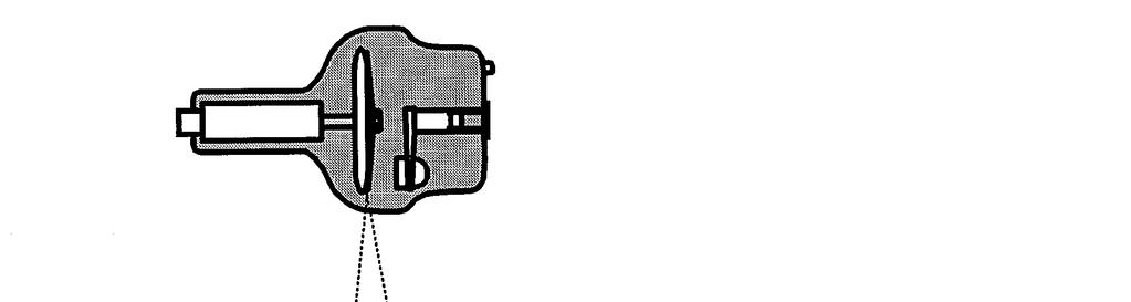
31 Anti-scatter Grid Grid cleans up scatter Increases contrast Increases dose Grid ratio = height / interspace width Typically: 5:1 (mammo) 8:1-12:1 (radiography) Carlos Vinhais 31
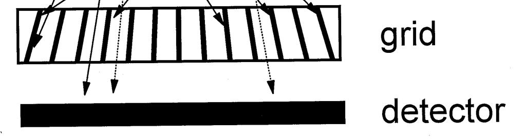
32 Anti-scatter Grid Carlos Vinhais 32


33 Anti-scatter Grid GRID (12:1) NO GRID Carlos Vinhais 33


34 Grid Artifacts Carlos Vinhais 34
35 Air Gap Air gaps used instead of grid to clean up scatter Magnification increases, FOV decreases Used in mammography Carlos Vinhais 35


36 Air Gap AIR GAP (12.5 cm) NO AIR GAP Carlos Vinhais 36
37 Question D12. L-4 is radiographed at a source-to-image distance (SID) of 100 cm, and an object-to-image distance (OID) of 20 cm. The width of L-4 measured on the radiograph is 35 mm. The true width is: A. 25 mm B. 28 mm C. 30 mm D. 35 mm E. 44 mm The magnification is M = I/O = SID/SOD = SID / (SID - OID), or 100 / (100-20) = M = I/O so the true size is 35 / 1.25 = 28. Carlos Vinhais 37
38 Question Object on fluoro table is 4 inches and projects as 7 inches on the image receptor which is 12 inches above the fluoro table. What is the distance from the x-ray tube (source) to the table? A. 8 in B. 12 in C. 16 in D. 20 in Using similar triangles, (SOD+12)/SOD = I/O, SOD= 16 Carlos Vinhais 38
39 Question D15. If the absorption efficiency of each intensifying screen in a dual screen system is 30%, what percentage of x-rays is stopped by the screens together? A. 9% B. 30% C. 51% D. 60% E. 70% 30% is absorbed in the first screen, 70% passes through. The second screen absorbs 30% of that 70% (or 21%). Total stopped is 30% + 21%. Carlos Vinhais 39
40 Question D19. Changing to a higher speed film will: A. Decrease patient exposure and increase noise. B. Decrease patient exposure and decrease noise. C. Not change exposure or noise, but decrease contrast. D. Increase patient exposure and increase noise. E. Increase patient exposure and decrease noise. Carlos Vinhais 40
41 Question G70. A radiograph has little contrast in density from one region to the next. Which of the following would improve contrast in a retake film? 1. Change to higher ratio grid. 2. Move the film closer to the patient. 3. Collimate the beam to as small a field as possible. 4. Raise the kvp to lower the exposure time. A. 1, 3 B. 1, 4 C. 2, 3 D. 1, 2 and 4 E. 1, 2, 3, and 4 Carlos Vinhais 41
42 Question G77. The purpose of a screen is to: 1. Convert x-rays to light photons. 2. Reduce scatter reaching the film. 3. Reduce patient's exposure. 4. Increase radiographic resolution. A. 1, 2, 3 and 4 B. 2 only C. 2, 4 D. 1, 3 E. 4 only Carlos Vinhais 42
43 Question G72. Which of the following does not reduce patient dose (for the same optical density on the film)? A. Use of screens B. Using a high kvp C. Using a high ratio grid D. Collimation Carlos Vinhais 43
44 Question G72. A radiograph transmits 10% of the light from a viewbox with an illumination level of 400 lux. The optical density of the radiograph is: A. 10 B. 2 C. 1 D. 0.1 E OD = -log10(t) = log10(1/t) = log10(1/0.1) = log10(10) = 1 Carlos Vinhais 44
45 Question D20. Optical density (OD) regions on film of 1.0, 1.3, and 2.0 will transmit of the light from a viewbox: A. 10%, 5%, 1% B. 10%, 13%, 20% C. 1%, 5%, 10% D. 90%, 87%, 20% E. 50%, 33%, 25% T = 10-OD Carlos Vinhais 45
46 Question D24. In some situations, e.g., a chest exam, it is important to see radiographic anatomy in both highand low-density regions. To aid in this, one could choose a film with a. A. High gradient B. High gamma C. Slow speed D. Long latitude E. Low fog Carlos Vinhais 46
47 Question D19. In order to decrease the optical density of an overexposed radiograph from 2.0 to 1.2, the mas should be reduced by approximately %. (Assume a slope of the characteristic curve of 3.0): A B C D E. Greater than 95 OD2 - OD1 = Average Gradient x log10 (E2/E1) where E is exposure, proportional to mas = 3.0 log10 (E2/E1), E2/E1 = 0.54 or 46% reduction. Carlos Vinhais 47
48 End of Lecture!
LECTURE 1 The Radiographic Image
 LECTURE 1 The Radiographic Image Prepared by:- KAMARUL AMIN ABDULLAH @ ABU BAKAR UiTM Faculty of Health Sciences Medical Imaging Department 11/23/2011 KAMARUL AMIN (C) 1 Lesson Objectives At the end of
Multiple Choice Identify the letter of the choice that best completes the statement or answers the question.
 RA110 test 3 Multiple Choice Identify the letter of the choice that best completes the statement or answers the question. 1. An object 35 cm in width is radiographed at 100 cm SID and at a 50 cm SOD. What
Introduction. Chapter 16 Diagnostic Radiology. Primary radiological image. Primary radiological image
 Introduction Chapter 16 Diagnostic Radiology Radiation Dosimetry I Text: H.E Johns and J.R. Cunningham, The physics of radiology, 4 th ed. http://www.utoledo.edu/med/depts/radther In diagnostic radiology
Mammography is a radiographic procedure specially designed for detecting breast pathology Approximately 1 woman in 8 will develop breast cancer over
 Mammography is a radiographic procedure specially designed for detecting breast pathology Approximately 1 woman in 8 will develop breast cancer over a lifetime Breast cancer screening programs rely on
Radiology. Radiograph: Is the image of an object made with use of X- ray instead of light.
 Radiology د. اريج Lec. 3 X Ray Films Radiograph: Is the image of an object made with use of X- ray instead of light. Dental x- ray film: Is a recording media on which image of the object was made by exposing
Contrast. Contrast: the difference in density on adjacent areas of a radiograph or other image receptor. Subjective. Long Scale (Low Contrast)
") Contrast Contrast: the difference in density on adjacent areas of a radiograph or other image receptor. Subject Subjective Radiographic Long Scale (Low Contrast) Short Scale (High Contrast) Factors affecting
Contrast Contrast: the difference in density on adjacent areas of a radiograph or other image receptor. Subject Subjective Radiographic Long Scale (Low Contrast) Short Scale (High Contrast) Factors affecting
Beam-Restricting Devices
 Beam-Restricting Devices Three factors contribute to an increase in scatter radiation: Increased kvp Increased Field Size Increased Patient or Body Part Size. X-ray Interactions a some interact with the
Overview. Professor Roentgen was a Physicist!!! The Physics of Radiation Oncology X-ray Imaging
 The Physics of Radiation Oncology X-ray Imaging Charles E. Willis, Ph.D. DABR Associate Professor Department of Imaging Physics The University of Texas M.D. Anderson Cancer Center Houston, Texas Overview
COMPUTED RADIOGRAPHY CHAPTER 4 EFFECTIVE USE OF CR
 This presentation is a professional collaboration of development time prepared by: Rex Christensen Terri Jurkiewicz and Diane Kawamura New Technology https://www.youtube.com/watch?v=ptkzznazb 7U COMPUTED
SPRINGFIELD TECHNICAL COMMUNITY COLLEGE ACADEMIC AFFAIRS
 SPRINGFIELD TECHNICAL COMMUNITY COLLEGE ACADEMIC AFFAIRS Course Number: RADG 112 Department: Radiography Course Title: Image Production & Eval. Semester: Spring Year: 1997 Objectives/ Unit One: Introduction
3/31/2011. Objectives. Emory University. Historical Development. Historical Development. Historical Development
 Teaching Radiographic Technique in a Digital Imaging Paradigm Objectives 1. Discuss the historical development of digital imaging. Dawn Couch Moore, M.M.Sc., RT(R) Assistant Professor and Director Emory
10/3/2012. Study Harder
 This presentation is a professional collaboration of development time prepared by: Rex Christensen Terri Jurkiewicz and Diane Kawamura Study Harder CR detection is inefficient, inferior to film screen
RAD 150 RADIOLOGIC EXPOSURE TECHNIQUE II
 RAD 150 RADIOLOGIC EXPOSURE TECHNIQUE II APPROVED 12/O2/2011 EFFECTIVE SPRING 2013-14 Prefix & Number RAD 150 Course Title: Radiologic Exposure Technique II & Lab Purpose of this submission: New Change/Updated
CR Basics and FAQ. Overview. Historical Perspective
 Page: 1 of 6 CR Basics and FAQ Overview Computed Radiography is a term used to describe a system that electronically records a radiographic image. Computed Radiographic systems use unique image receptors
10/26/2015. Study Harder
 This presentation is a professional collaboration of development time prepared by: Rex Christensen Terri Jurkiewicz and Diane Kawamura Study Harder CR detection is inefficient, inferior to film screen
STUDENT REVIEW QUESTION SET K CR/DR CONTENT AREA
 STUDENT REVIEW QUESTION SET K CR/DR CONTENT AREA RADT 2913 COMPREHENSIVE REVIEW 1 The CR cassette is backed by aluminum that: A. reflects x-rays B. absorbs x-rays C. captures the image D. transmits x-rays
SECTION I - CHAPTER 1 DIGITAL RADIOGRAPHY: AN OVERVIEW OF THE TEXT. Exam Content Specifications 8/22/2012 RADT 3463 COMPUTERIZED IMAGING
 RADT 3463 - COMPUTERIZED IMAGING Section I: Chapter 1 RADT 3463 Computerized Imaging 1 SECTION I - CHAPTER 1 DIGITAL RADIOGRAPHY: AN OVERVIEW OF THE TEXT RADT 3463 COMPUTERIZED IMAGING Section I: Chapter
Seminar 8. Radiology S8 1
 Seminar 8 Radiology Medical imaging. X-ray image formation. Energizing and controlling the X-ray tube. Image detectors. The acquisition of analog and digital images. Digital image processing. Selected
Do you have any other questions? Please call us at (Toll Free) or , or
 or , or") INSTRUCTIONS Read the appropriate course/ textbook. This is an open book test. A score of 75% or higher is needed to receive CE credit. You will have a maximum of three attempts to pass this course. Please
INSTRUCTIONS Read the appropriate course/ textbook. This is an open book test. A score of 75% or higher is needed to receive CE credit. You will have a maximum of three attempts to pass this course. Please
1. Carlton, Richard R., and Arlene M. Adler. Principles of Radiographic Imaging: An Art and a Science, 5th edition (2013).
.") CODE: RADT 151 INSTITUTE: Health Science TITLE: Radiographic Exposure DEPARTMENT: Radiologic Technology COURSE DESCRIPTION: This course covers the principles of radiographic exposure selection and manipulation
CODE: RADT 151 INSTITUTE: Health Science TITLE: Radiographic Exposure DEPARTMENT: Radiologic Technology COURSE DESCRIPTION: This course covers the principles of radiographic exposure selection and manipulation
Visibility of Detail
 Visibility of Detail Radiographic Quality Quality radiographic images represents the, and information is for diagnosis. The of the anatomic structures and the accuracy of their ( ) determine the overall
- KiloVoltage. Technique 101: Getting Back to Basics
 Why do I need to know technique? Technique 101: Getting Back to Basics Presented by: Thomas G. Sandridge, M.S., M.Ed., R.T.(R) Program Director Northwestern Memorial Hospital School of Radiography Chicago,
X-RAY IMAGING EE 472 F2017. Prof. Yasser Mostafa Kadah
 X-RAY IMAGING EE 472 F2017 Prof. Yasser Mostafa Kadah www.k-space.org Recommended Textbook Stewart C. Bushong, Radiologic Science for Technologists: Physics, Biology, and Protection, 10 th ed., Mosby,
Acquisition, Processing and Display
 Acquisition, Processing and Display Terri L. Fauber, R.T. (R)(M) Department of Radiation Sciences School of Allied Health Professions Virginia Commonwealth University Topics Image Characteristics Image
NPTEL NPTEL ONLINE COURSE. NPTEL Online Certification Course (NOC) NPTEL. Theory and Practice of Non Destructive Testing
 NPTEL. Theory and Practice of Non Destructive Testing") NPTEL NPTEL ONLINE COURSE NPTEL Online Certification Course (NOC) NPTEL Theory and Practice of Non Destructive Testing Dr. Ranjit Bauri Dept. of Metallurgical & Materials Engineering IIT Madras, Chennai
NPTEL NPTEL ONLINE COURSE NPTEL Online Certification Course (NOC) NPTEL Theory and Practice of Non Destructive Testing Dr. Ranjit Bauri Dept. of Metallurgical & Materials Engineering IIT Madras, Chennai
Quality Control for Stereotactic Breast Biopsy. Robert J. Pizzutiello, Jr., F.A.C.M.P. Upstate Medical Physics, Inc
 Quality Control for Stereotactic Breast Biopsy Robert J. Pizzutiello, Jr., F.A.C.M.P. Upstate Medical Physics, Inc. 716-924-0350 Methods of Imaging Guided Breast Biopsy Ultrasound guided, hand-held needle
4. Contrast is the. There must The function of contrast is to:. The types of contrast are.
 RADIOGRAPHIC VISIBILITY OF DETAIL STUDY QUESTIONS 1. What is visibility of detail? It is controlled by properties. What are the two factors that affect it? 2. What is sharpness of detail? It is controlled
Exposure Indices and Target Values in Radiography: What Are They and How Can You Use Them?
 Exposure Indices and Target Values in Radiography: What Are They and How Can You Use Them? Definition and Validation of Exposure Indices Ingrid Reiser, PhD DABR Department of Radiology University of Chicago
Mammography: Physics of Imaging
 Mammography: Physics of Imaging Robert G. Gould, Sc.D. Professor and Vice Chair Department of Radiology and Biomedical Imaging University of California San Francisco, California Mammographic Imaging: Uniqueness
X-RAY. Lecture No.4. Image Characteristics:
 Lecture No.4 X-RAY أ.م.د. اسامة مراد ابراهيم Image Characteristics: *Radiographic density: It s the degree of blackness of the film. when a film is exposed by an x-ray beam (or by light in case of screenfilm
Essentials of Digital Imaging
 Essentials of Digital Imaging Module 1 Transcript 2016 ASRT. All rights reserved. Essentials of Digital Imaging Module 1 Fundamentals 1. ASRT Animation 2. Welcome Welcome to Essentials of Digital Imaging
X-ray Tube and Generator Basic principles and construction
 X-ray Tube and Generator Basic principles and construction Dr Slavik Tabakov - Production of X-rays OBJECTIVES - X-ray tube construction - Anode - types, efficiency - X-ray tube working characteristics
Photomultiplier Tube
 Nuclear Medicine Uses a device known as a Gamma Camera. Also known as a Scintillation or Anger Camera. Detects the release of gamma rays from Radionuclide. The radionuclide can be injected, inhaled or
JEFFERSON COLLEGE. Radiographic Exposures
 JEFFERSON COLLEGE COURSE SYLLABUS RAD140 Radiographic Exposures 3 Credit Hours Revised by: Janet E. Akers BS RT (R)(M) Date: September 30, 2013 Kenny Wilson, Director, Health Occupation Programs Dena McCaffrey,
Veterinary Science Preparatory Training for the Veterinary Assistant. Floron C. Faries, Jr., DVM, MS
 Veterinary Science Preparatory Training for the Veterinary Assistant Floron C. Faries, Jr., DVM, MS Radiology Floron C. Faries, Jr., DVM, MS Objectives Determine the appropriate machine settings for making
X-ray Tube and Generator Basic principles and construction
 X-ray Tube and Generator Basic principles and construction Dr Slavik Tabakov - Production of X-rays and Patient Dose OBJECTIVES - X-ray tube construction - Anode - types, efficiency - Classical X-ray generator
RADIOGRAPHIC EXPOSURE
 RADIOGRAPHIC EXPOSURE Receptor Exposure Receptor Exposure the that interacts with the receptor. Computed Radiography ( ) requires a. Direct Digital Radiography (DR) requires a. Exposure Indicators Exposure
The Evaluation of Collimator Alignment of Diagnostic X-ray Tube Using Computed Radiography System
 The Evaluation of Collimator Alignment of Diagnostic X-ray Tube Using Computed Radiography System The Evaluation of Collimator Alignment of Diagnostic X-ray Tube Using Computed Radiography System Manus
PD233: Design of Biomedical Devices and Systems
 PD233: Design of Biomedical Devices and Systems (Lecture-8 Medical Imaging Systems) (Imaging Systems Basics, X-ray and CT) Dr. Manish Arora CPDM, IISc Course Website: http://cpdm.iisc.ac.in/utsaah/courses/
A Comprehensive Review of Image Production
 A Comprehensive Review of Image Production Presented by: John Fleming, M.Ed., RT(R)(MR)(CT) St. Petersburg College Office: (727) 341-3758 E-mail: flemingj@spcollege.edu Lesson Objectives: ARRT Content
Current technology in digital image production (CR/DR and other modalities) Jaroonroj Wongnil 25 Mar 2016
 Jaroonroj Wongnil 25 Mar 2016") Current technology in digital image production (CR/DR and other modalities) Jaroonroj Wongnil 25 Mar 2016 Current technology in digital image production (CR/DR and other modalities) 2/ Overview Digital
Current technology in digital image production (CR/DR and other modalities) Jaroonroj Wongnil 25 Mar 2016 Current technology in digital image production (CR/DR and other modalities) 2/ Overview Digital
The importance of radiation quality for optimisation in radiology
 Available online at http://www.biij.org/2007/2/e38 doi: 10.2349/biij.3.2.e38 biij Biomedical Imaging and Intervention Journal COMMENTARY The importance of radiation quality for optimisation in radiology
SYLLABUS. TITLE: Equipment Operation I. DEPARTMENT: Radiologic Technology
 CODE: RADT 156 INSTITUTE: Health Science TITLE: Equipment Operation I DEPARTMENT: Radiologic Technology COURSE DESCRIPTION: This course covers the principles of equipment operation and maintenance of radiographic
INTRODUCTION TO FLEXIBLE BRONCHOSCOPY. Fluoroscopy Synopsis HENRI G COLT MD SECOND EDITION THE BRONCHOSCOPY EDUCATION PROJECT SERIES
 SECOND EDITION INTRODUCTION TO FLEXIBLE BRONCHOSCOPY Fluoroscopy Synopsis HENRI G COLT MD With contributions from Dr. S. Murgu THE BRONCHOSCOPY EDUCATION PROJECT SERIES FLUOROSCOPY SYNOPSIS The purpose
Radiology Physics Lectures: Digital Radiography. Digital Radiography. D. J. Hall, Ph.D. x20893
 Digital Radiography D. J. Hall, Ph.D. x20893 djhall@ucsd.edu Background Common Digital Modalities Digital Chest Radiograph - 4096 x 4096 x 12 bit CT - 512 x 512 x 12 bit SPECT - 128 x 128 x 8 bit MRI -
Film Replacement in Radiographic Weld Inspection The New ISO Standard
 BAM Berlin Film Replacement in Radiographic Weld Inspection The New ISO Standard 17636-2 Uwe Ewert, Uwe Zscherpel, Mirko Jechow Requests and information to: uwez@bam.de 1 Outline - The 3 essential parameters
2017 West Coast Educators Conference Orlando. Projection Geometry. 1. Review hierarchy of image qualities (amplified version):
:") Spatial Resolution in the Digital Age: NOTES Quinn B. Carroll, MEd, RT 2017 West Coast Educators Conference Orlando Projection Geometry 1. Review hierarchy of image qualities (amplified version): a. Maximum
Spatial Resolution in the Digital Age: NOTES Quinn B. Carroll, MEd, RT 2017 West Coast Educators Conference Orlando Projection Geometry 1. Review hierarchy of image qualities (amplified version): a. Maximum
Unit thickness. Unit area. σ = NΔX = ΔI / I 0
 Unit thickness I 0 ΔI I σ = ΔI I 0 NΔX = ΔI / I 0 NΔX Unit area Δx Average probability of reaction with atom for the incident photons at unit area with the thickness of Delta-X Atom number at unit area
Nuclear Associates
 Nuclear Associates 07-644 Grid Alignment Test Tool Users Manual March 2005 Manual No. 07-644-1 Rev. 2 2004, 2005 Fluke Corporation, All rights reserved. Printed in U.S.A. All product names are trademarks
Digital Imaging Considerations Computed Radiography
 Digital Imaging Considerations Digital Radiography Computed Radiography o Cassette based Direct or Indirect Digital Radiography o Cassetteless Computed Radiography 1 CR Image Acquisition Most like conventional
Amorphous Selenium Direct Radiography for Industrial Imaging
 DGZfP Proceedings BB 67-CD Paper 22 Computerized Tomography for Industrial Applications and Image Processing in Radiology March 15-17, 1999, Berlin, Germany Amorphous Selenium Direct Radiography for Industrial
Radiographic sensitivity improved by optimized high resolution X -ray detector design.
 DIR 2007 - International Symposium on Digital industrial Radiology and Computed Tomography, June 25-27, 2007, Lyon, France Radiographic sensitivity improved by optimized high resolution X -ray detector
I. PERFORMANCE OF X-RAY PRODUCTION COMPONENTS FLUOROSCOPIC ACCEPTANCE TESTING: TEST PROCEDURES & PERFORMANCE CRITERIA
 FLUOROSCOPIC ACCEPTANCE TESTING: TEST PROCEDURES & PERFORMANCE CRITERIA EDWARD L. NICKOLOFF DEPARTMENT OF RADIOLOGY COLUMBIA UNIVERSITY NEW YORK, NY ACCEPTANCE TESTING GOALS PRIOR TO 1st CLINICAL USAGE
Moving from film to digital: A study of digital x-ray benefits, challenges and best practices
 Moving from film to digital: A study of digital x-ray benefits, challenges and best practices H.U. Pöhler 1 and N. D Ademo 2 DÜRR NDT GmbH & Co. KG, Höpfigheimer Straße 22, Bietigheim-Bissingen, 74321,
Course Instructions: Check your for your CE certification of completion (please check your junk/spam folder as well). About SMS CE courses:
. About SMS CE courses:") 2017 Course #4 Self-Study Course Contact Us: Phone 614-292-6737 Toll Free 1-888-476-7678 Fax 614-292-8752 E-mail smsosu@osu.edu Web dentistry.osu.edu/sms The Ohio State University College of Dentistry
2017 Course #4 Self-Study Course Contact Us: Phone 614-292-6737 Toll Free 1-888-476-7678 Fax 614-292-8752 E-mail smsosu@osu.edu Web dentistry.osu.edu/sms The Ohio State University College of Dentistry
SYLLABUS. 1. Identification of Subject:
 SYLLABUS Date/ Revision : 30 January 2017/1 Faculty : Life Sciences Approval : Dean, Faculty of Life Sciences SUBJECT : Biophysics 1. Identification of Subject: Name of Subject : Biophysics Code of Subject
Multiple Choice Identify the letter of the choice that best completes the statement or answers the question.
 RA202 image production class two Multiple Choice Identify the letter of the choice that best completes the statement or answers the question. 1. What removes excess chemistry from the film prior to it
The effect of compensating filter on image quality in lateral projection of thoraco lumbar radiography
 Journal of Physics: Conference Series OPEN ACCESS The effect of compensating filter on image quality in lateral projection of thoraco lumbar radiography To cite this article: N A A Daud et al 2014 J. Phys.:
Lecture 9. Lecture 9. t (min)
") Sensitivity of the Eye Lecture 9 The eye is capable of dark adaptation. This comes about by opening of the iris, as well as a change in rod cell photochemistry fovea only least perceptible brightness 10
Sensitivity of the Eye Lecture 9 The eye is capable of dark adaptation. This comes about by opening of the iris, as well as a change in rod cell photochemistry fovea only least perceptible brightness 10
ABSORBED DOSE DISTRIBUTIONS USING THE ISODENSITOMETRIC METHOD FOR EXPOSURES WITH FILTER EMPLOYED FOR MAMMOGRAPHIES
 Romanian Reports in Physics, Vol. 65, No. 1, P. 168 177, 213 ABSORBED DOSE DISTRIBUTIONS USING THE ISODENSITOMETRIC METHOD FOR EXPOSURES WITH FILTER EMPLOYED FOR MAMMOGRAPHIES F. SCARLAT 1, A. SCARISOREANU
BASICS OF FLUOROSCOPY
 Medical Physics Residents Training Program BASICS OF FLUOROSCOPY Dr. Khalid Alyousef, PhD Department of Medical Imaging King Abdulaziz Medical City- Riyadh Edison examining the hand of Clarence Dally with
Investigation of the line-pair pattern method for evaluating mammographic focal spot performance
 Investigation of the line-pair pattern method for evaluating mammographic focal spot performance Mitchell M. Goodsitt, a) Heang-Ping Chan, and Bob Liu Department of Radiology, University of Michigan, Ann
X-RAYS - NO UNAUTHORISED ENTRY
 Licencing of premises Premises Refer Guidelines A radiation warning sign and warning notice, X-RAYS - NO UNAUTHORISED ENTRY must be displayed at all entrances leading to the rooms where x-ray units are
Safelight Fog does what to contrast and density on film?
 Terri Jurkiewicz Safelight Fog does what to contrast and density on film? ANSWER INCREASES DENSITY DECREASES CONTRAST Explain how you determine if the focal spot size is within appropriate limits.
Studies on reduction of exposure dose using digital scattered X-ray removal processing
 Studies on reduction of exposure dose using digital scattered X-ray removal processing Poster No.: C-1834 Congress: ECR 2015 Type: Scientific Exhibit Authors: K. Kashiyama, M. Funahashi, T. Nakaoka, T.
Studies on reduction of exposure dose using digital scattered X-ray removal processing
 Studies on reduction of exposure dose using digital scattered X-ray removal processing Poster No.: C-1834 Congress: ECR 2015 Type: Scientific Exhibit Authors: K. Kashiyama, M. Funahashi, T. Nakaoka, T.
Effect of Backscattered Radiation on X-Ray Image Contrast
 Applied Physics Research; Vol. 9, No. 1; 2017 ISSN 1916-9639 E-ISSN 1916-9647 Published by Canadian Center of Science and Education Effect of Backscattered Radiation on X-Ray Image Contrast A. T. Naji
Digital Imaging started in the 1972 with Digital subtraction angiography Clinical digital imaging was employed from the 1980 ~ 37 years ago Amount of
 Digital Imaging started in the 1972 with Digital subtraction angiography Clinical digital imaging was employed from the 1980 ~ 37 years ago Amount of radiation to the population due to Medical Imaging
Nuclear Associates
 Nuclear Associates 07-647 R/F QC Phantom Operators Manual March 2005 Manual No. 07-647-1 Rev. 2 2004, 2005 Fluke Corporation, All rights reserved. All product names are trademarks of their respective companies
Open. the Digitized world. Fuji Computed Radiography
 Open the Digitized world Fuji Computed Radiography If just one of these applies to you... Managing developing fluid is hard and darkroom work is a hassle... Images are not stable... Isn t digitalization
Appropriate Inspection Distance of Digital X-Ray Imaging Equipment for Diagnosis
 Indian Journal of Science and Technology Vol 8(S8), 380-386, April 2015 ISSN (Print) : 0974-6846 ISSN (Online) : 0974-5645 DOI: 10.17485/ijst/2015/v8iS8/70528 Appropriate Inspection Distance of Digital
X-rays. X-rays are produced when electrons are accelerated and collide with a target. X-rays are sometimes characterized by the generating voltage
 X-rays Ouch! 1 X-rays X-rays are produced when electrons are accelerated and collide with a target Bremsstrahlung x-rays Characteristic x-rays X-rays are sometimes characterized by the generating voltage
Dose Reduction and Image Preservation After the Introduction of a 0.1 mm Cu Filter into the LODOX Statscan unit above 110 kvp
 Dose Reduction and Image Preservation After the Introduction of a into the LODOX Statscan unit above 110 kvp Abstract: CJ Trauernicht 1, C Rall 1, T Perks 2, G Maree 1, E Hering 1, S Steiner 3 1) Division
Version 1.0. TechnicVR. Student Guide
 Version 1.0 TechnicVR s h a d e r w a r e. c o m Student Guide TechnicVR s h a d e r w a r e. c o m Student Guide shaderware 2008 PO Box 103 Saltburn Cleveland TS12 1WP w w w. s h a d e r w a r e. c o
Photons interaction with matter
 ب س م هللا الر ح من الر حیم Photons interaction with matter Ionization Ionization is the process of removing an electron from an electrically neutral atom to produce an ion pair. An ion is an atom or subatomic
Comparison of computed radiography and filmõscreen combination using a contrast-detail phantom
 JOURNAL OF APPLIED CLINICAL MEDICAL PHYSICS, VOLUME 4, NUMBER 1, WINTER 2003 Comparison of computed radiography and filmõscreen combination using a contrast-detail phantom Z. F. Lu,* E. L. Nickoloff, J.
Collimation Assessment Using GAFCHROMIC XR-M2
 Collimation Assessment Using GAFCHROMIC XR-M2 I. Introduction A method of collimation assessment for GE Senographe full-field digital mammography (FFDM) systems is described that uses a self-developing
X-Ray-Based Medical Imaging and Resolution
 Residents Section Physics Minimodule Huda and Abrahams Resolution on Radiographs Residents Section Physics Minimodule Residents inradiology Walter Huda 1 R. Brad Abrahams 2 Huda W, Abrahams RB Keywords:
Medical Imaging. X-rays, CT/CAT scans, Ultrasound, Magnetic Resonance Imaging
 Medical Imaging X-rays, CT/CAT scans, Ultrasound, Magnetic Resonance Imaging From: Physics for the IB Diploma Coursebook 6th Edition by Tsokos, Hoeben and Headlee And Higher Level Physics 2 nd Edition
TESTING FLAT-PANEL IMAGING SYSTEMS: What the Medical Physicist Needs to Know. JAMES A. TOMLINSON, M.S., D.A.B.R. Diagnostic Radiological Physicist
 TESTING FLAT-PANEL IMAGING SYSTEMS: What the Medical Physicist Needs to Know JAMES A. TOMLINSON, M.S., D.A.B.R. Diagnostic Radiological Physicist Topics Image Uniformity and Artifacts Image Quality - Detail
NDE SOLUTIONS RADIOGRAPHY COURSE OUTLINE
 NDE SOLUTIONS RADIOGRAPHY COURSE OUTLINE 80 Hour Course Length 1.0 NDT Qualification and Introduction (3 Hours) 1.1 NDT Introduction 1.2 NDT Qualification and Certification 1.2.1 NAS 410 1.2.2 SNT-TC-1A
PERFORMANCE CHARACTERIZATION OF AMORPHOUS SILICON DIGITAL DETECTOR ARRAYS FOR GAMMA RADIOGRAPHY
 12 th A-PCNDT 2006 Asia-Pacific Conference on NDT, 5 th 10 th Nov 2006, Auckland, New Zealand PERFORMANCE CHARACTERIZATION OF AMORPHOUS SILICON DIGITAL DETECTOR ARRAYS FOR GAMMA RADIOGRAPHY Rajashekar
Breast Tomosynthesis. Bob Liu, Ph.D. Department of Radiology Massachusetts General Hospital And Harvard Medical School
 Breast Tomosynthesis Bob Liu, Ph.D. Department of Radiology Massachusetts General Hospital And Harvard Medical School Outline Physics aspects of breast tomosynthesis Quality control of breast tomosynthesis
Principle of X-Ray Systems
 Principle of X-Ray Systems Hossein Ebrahimi Nasab PHYSICS OF X-RAYS Nature of X-rays Energy unit Interaction with matter INTERACTION WITH THE MATTER In vacuum: photon move along a straight line In materials,
Ansur TNT Users Manual. Plug-In
 Ansur TNT 12000 Plug-In Users Manual August 2009, Rev. 2, 12/09 2009 Fluke Corporation. All rights reserved. Specifications are subject to change without notice. All product names are trademarks of their
HISTORY. CT Physics with an Emphasis on Application in Thoracic and Cardiac Imaging SUNDAY. Shawn D. Teague, MD
 CT Physics with an Emphasis on Application in Thoracic and Cardiac Imaging Shawn D. Teague, MD DISCLOSURES 3DR- advisory committee CT PHYSICS WITH AN EMPHASIS ON APPLICATION IN THORACIC AND CARDIAC IMAGING
biij Optimisation in general radiography CJ Martin, PhD, FIPEM, FioP Biomedical Imaging and Intervention Journal REVIEW PAPER
 Available online at http://www.biij.org/2007/2/e18 doi: 10.2349/biij.3.2.e18 biij Biomedical Imaging and Intervention Journal REVIEW PAPER Optimisation in general radiography CJ Martin, PhD, FIPEM, FioP
Setting up digital imaging department!
 Outline Setting up digital imaging department! From screen/film to digital radiography PACS/Tele radiology Setting up digital department Digital Imaging Napapong Pongnapang, Ph.D. Department of Radiological
DIGITAL RADIOGRAPHY ARTIFACTS
 IMAGING LAB MPHY 487 DIGITAL RADIOGRAPHY ARTIFACTS Mohammad Esmael Alsulimane B.Sc, M.Sc Medical Physics Lecturer - Physics Department All Rights Reserved: Some information and figures in this presentation
Overview of Safety Code 35
 Common Quality Control Procedures for All s Quality Control Procedures Film All s Daily Quality Control Tests Equipment Warm-up (D1) According to manufacturers instructions Can include auto calibration(d1)
X-rays in medical diagnostics
 X-rays in medical diagnostics S.Dolanski Babić 2017/18. History W.C.Röntgen (1845-1923) discovered a new type of radiation Nature, Jan. 23. 1896.; Science, Feb.14. 1896. X- rays: Induced the ionization
Film and processing quality assurance
 Film and processing quality assurance Image Receptors Direct action non screen film Indirect action screen film Digital sensor Direct Action Non Screen Film Usually intra-oral film Non screen film reacts
Image Quality. HTC Grid High Transmission Cellular Grid provides higher contrast images
 B R E A S T I M A G I N G S O L U T I O N S Setting the benchmark for mammography M-IV Series Innovations in breast imaging The Lorad M-IV Series exemplifies Hologic's commitment to developing advanced
Quality Control of Full Field Digital Mammography Units
 Quality Control of Full Field Digital Mammography Units Melissa C. Martin, M.S., FACMP, FACR, FAAPM Melissa@TherapyPhysics.com 310-612-8127 ACMP Annual Meeting Virginia Beach, VA May 2, 2009 History of
Introduction. Sam R. Kottamasu Lawrence R. Kuhns
 Pediatr Radiol (1997) 27: 119 123 Springer-Verlag 1997 Sam R. Kottamasu Lawrence R. Kuhns Musculoskeletal computed radiography in children: scatter reduction and improvement in bony trabecular sharpness
Maximizing clinical outcomes
 Maximizing clinical outcomes Digital Tomosynthesis Dual Energy Subtraction Automated Long Length Imaging Improved image quality at a low dose Xray Xray Patented ISS capture technology promotes high sensitivity
Fluoroscopy - Chapter 9
 Fluoroscopy - Chapter 9 Kalpana Kanal, Ph.D., DABR Lecturer, Diagnostic Physics Dept. of Radiology UW Medicine a copy of this lecture may be found at: http://courses.washington.edu/radxphys/physicscourse04-05.html
DIGITAL IMAGE PROCESSING IN X-RAY IMAGING
 DIGITAL IMAGE PROCESSING IN X-RAY IMAGING Shalini Kumari 1, Bachan Prasad 2,Aliya Nasim 3 Department of Electronics And Communication Engineering R.V.S College of Engineering & Technology, Jamshedpur,
Teaching Digital Radiography and Fluoroscopic Radiation Protection
 Teaching Digital Radiography and Fluoroscopic Radiation Protection WCEC 20 th Student Educator Radiographer Conference Dennis Bowman, RT(R), CRT (R)(F) Community Hospital of the Monterey Peninsula (CHOMP)
10/15/2012 SECTION III - CHAPTER 6 DIGITAL FLUOROSCOPY RADT 3463 COMPUTERIZED IMAGING
 RADT 3463 - COMPUTERIZED IMAGING Section III: Chapter 6 RADT 3463 Computerized Imaging 1 SECTION III - CHAPTER 6 DIGITAL FLUOROSCOPY RADT 3463 COMPUTERIZED IMAGING Section III: Chapter 6 RADT 3463 Computerized