Unit thickness. Unit area. σ = NΔX = ΔI / I 0
|
|
|
- Magnus Green
- 6 years ago
- Views:
Transcription

1
2 Unit thickness I 0 ΔI I σ = ΔI I 0 NΔX = ΔI / I 0 NΔX Unit area Δx Average probability of reaction with atom for the incident photons at unit area with the thickness of Delta-X Atom number at unit area with the thickness of Delta-X ΔI / I = Nσ = ΔX Σ 0 Average probability of reaction with atom for the incident photons at unit area with the thickness of Delta-X Thickness of Delta-X
3 ! Linear attenuation coefficient Definition: Ratio of reaction photons to incident photons after traverse material with unit thickness, denoted as µ. Unit thickness µ = di Idx ln I I 0 = µ d HVL = ln2 µ I 0 di µ m µ ρ = Unit area Δx ΔI / I0 Σ = Nσ = ΔX
4 <30 kev 30 kev-30 MeV >30 MeV Low hv X-ray photon hv>e b Binding Energy High hv X-ray photon λ 0 hv>>w Ultra-high hv X-ray 10, 000 times photon hv>1.022 MeV 1, 000 kvp hoelectron absorbed Charact. X-ray <0.5 kev, absorbed Recoil(Campton) Photoelectric effect All photon energy is absorbed Electron ϕ θ Compton effect λ Scattered photon Positive electron Mev photon Electric pair effect - Negative electron

5 Low energy High energy
6 4. Quality evaluation parameter Reference: P39-58
7 !! ) )
8
9 !
10 !!

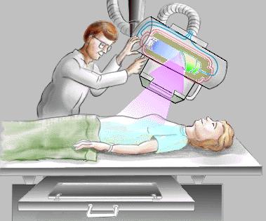
11 X-ray fluoroscopy Fluorescent screen Dynamic/ real time Low resolution image Image can not be kept Patient exposed to X-ray for a few seconds or minutes ED 0.05mSv ED 0.94mSv

12 Digital X-ray Imaging CR - DR
13 Part II Computed Radiography CR! 1982! (imaging plate, IP) IP! IP IP


14 Computed Radiography physics X-ray First excitation laser Second excitation Photon/electric Imaging reader A/D Digital image
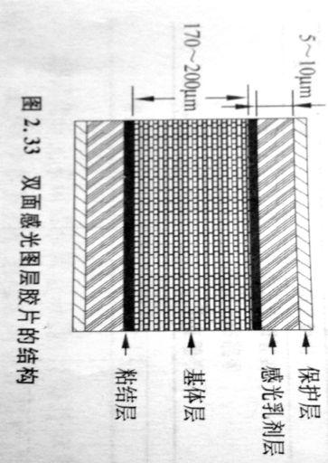
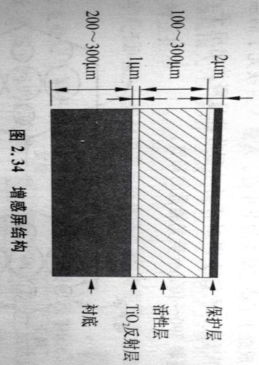
15 Imaging plate X Photo-stimulated luminescence, PSL BaFX: Eu 2+ Protective layer Fluorescence layer(psl) Support layer Protective layer Structure of IP 20cm*25cm X-ray -> IP -> latent image () -> image reader h Fading of PSL Deposit time should less than 8h

16 Imaging reader CCD " Six components: Laser optic system collector A/D IP transport erase
17 Imaging processing techniques! Standard & gray scale adjustment! Dynamic compression! Frequency adjustment - = Original smooth High freq. + Boundary sharpen An example of boundary enhancement technique
18 CR s Features Merits " Low dose " IP is repetitive " Good compatibility " No black room Drawbacks " Complicated process " Low time resolution (6 mins) " Spatial resolution 2.8lp/mm < 6-7lp/mm(film) " With image postprocessing " Digital storage Example: IP size is 35.56cm*43.18cm, and the digitized image is 2000*2510. Then, what is the pixel size? Rx=35.56/2000=0.18mm, Ry=43.18/2510=0.18mm. Line pairs: 1/(2*0.18)=2.8lp/mm
19 Part III Digital radiography -DR 1. DR introduction 2. DR classification 3. a-si, a-se plate detector 4. Line scanner X-ray imaging 5. Evaluation of DR
 improves workflow efficiency. \" Large-area digital image receptor, 35cm*43cm, 43cm*43 cm, 1:1 with low distortion.")
20 DR introduction Digital radiography is a form of x-ray imaging, where digital X-ray sensors are used instead of traditional photographic film. " Fast imaging process, <1.0 s " Digital display (Computer monitor) improves workflow efficiency. " Large-area digital image receptor, 35cm*43cm, 43cm*43 cm, 1:1 with low distortion. " As high as 65% detective quantum efficiency (DQE), low dose & early diagnose. " Wide dynamic imaging range, above 14 bit gray scale. " Good reliability, suit for the large turnover. " 5f/s, be able to realize DSA X-ray radiography: Preparation, Exposure, Developing, Quality control, Finish; 6 min CR: Preparation, Exposure, Scan, Finish; 7 min DR: Preparation, Exposure, Preview; Finish; 1.3 min
21 DR classification # Plane and line scan # Indirect digital radiography(idr) and direct digital radiography(ddr)
22 Cesium iodide CsI/ Gd2O2S:Tb/ DR classification INDIRECT CONVERSION X-ray Scintillator/ Visible light Photodiode/or CCD Thin film transistor(tft) / Electrical signal A/D DIRECT CONVERSION X-ray Photoconductor/ Electrical charge Thin film transistor(tft) / Electrical signal A/D Digital signal Digital signal a-si) CCD a-se) CMOS From the viewpoint of exposure, DR system can be divided into! Plane exposure, Flat Plane Detector! Line scan, the detectors own three types: a-se) PbI 2 ) CMOS

 Perfect match \" Photo transfer function Needle-shaped CsI(TI)crystal, 6-10 micro")

23 a-si plate detector (IDR) " Scintillator Cesium iodide CsI/ " Photoelectric effect Visible light s peakvalue is 540 nm, very close to the optimized response wavelength of a-si photodiode, nm. ( ) Perfect match " Photo transfer function Needle-shaped CsI(TI)crystal, 6-10 micro dia., height " a-si photodiode( Every photodiode is a pixel, 70~200 micro. GE TM a-si plate detector Field: 41cm*41cm Pixel: 2050*2050 Pixel size: 200 micro DQE: 77% Imaging process: 3s Space resolution: 2.5 linepair/mm Pixel (A/D): 14 bit
 + - + - X-ray")
24 a-se plate detector (DDR) X-ray a-se

25 a-se plate detector (DDR)! Reflective mode: Reflective mirror! Direct mode: Lens! Fiber mode: Fiber! Translation mode

 /CMOS")
26 Line scanner X-ray imaging 0.16mm*8 column 2048 detector form a row with the width of 41 cm Three types MWPC (Multi wires proportional chamber) /CMOS (CdZnTe, CZT)

27 Line scanner X-ray imaging
28 Linear scanner X-ray imaging features Merit 1. Direct conversion avoid energy and information loss during the conversion. 2. Reduce the disturbance of scattered ray. 3. High sensitivity reduces the dose. The dose of 0.01mGy for chest scan is much lower than the personal protection standard of 0.3 mgy. Drawback 1. Cannot acquire real-time images. 2. Relative low spatial resolution 1.6lp/mm.
29
30 Part IV-5 Detector Quality Evaluation Parameters 1. Pixel & spatial resolution 2. Dynamic range 3. Modulation transfer function 4. Detective quantum efficiency

31 Pixel & Spatial resolution Pixel- In digital image, a pixel (or picture element) is the smallest item of information in an image. Pixels are normally arranged in a 2-dimensional grid, and are often represented using dots, squares, or rectangles. Example 1 Calculate the pixel size & spatial resolution of DR system where one Flat Plane Detector (14*17 inch) is digitized into a matrix size of 2000*2510. Spatial resolution Example 2 Calculate the total number of bytes stored for this DR system with a 2048*2048 matrix if an 8-bit ( analog-to digital converter is used.
32 Pixel & Image quality Store & display cost Mammo radiography General Noise radiography limit Resolution limit High 70~ ~200 Quality Low Pixel (Micron)

33 Detector dynamic range! DR X Example Low limit: 1micro-Gy High limit: 10 mili-gy e ~14 6bit8bit 10bit
![Modulation transfer function, MTF ( I I )/( I I ) M = + max min here I max indicates the maximum signal, I min is the minimum.! MTF is located in the region of [0, 1];!](/docs-images/76/73754972/images/34-0.jpg "Higher MTF, higher contrast, and lower information transfer lose;! If MTF=0 the image can t be obtained;! Human can t tell the difference with MTF less than 0.025;!")
34 Modulation transfer function, MTF ( I I )/( I I ) M = + max min here I max indicates the maximum signal, I min is the minimum.! MTF is located in the region of [0, 1];! Higher MTF, higher contrast, and lower information transfer lose;! If MTF=0 the image can t be obtained;! Human can t tell the difference with MTF less than 0.025;! MTF is a function of the spatial resolution. So we must refer MTF@ known spatial resolution. max min Modulation Transfer

35 Detective Quantum Efficiency, DQE!! DQE DQE! X DQE! X DQE

36 DQE importance! DR-65%; CR-35%; Radiography (Screen-film) 25%.! Because of the higher DQE, flat-panel detectors are expected to use radiation dose more efficiently, and they could theoretically provide lower noise image over the ranger of spatial frequencies encountered in medical imaging.
37 Conclusion! Classification of X-ray radiography, CR, and DR! CR workflow and main components! DR classification( DDR & IDR, Plane & Line scan)! Quality evaluation parameter: DQE, MTF, dynamic range, Pixel & spatial parameters
38 Questions?
10/26/2015. Study Harder
 This presentation is a professional collaboration of development time prepared by: Rex Christensen Terri Jurkiewicz and Diane Kawamura Study Harder CR detection is inefficient, inferior to film screen
10/3/2012. Study Harder
 This presentation is a professional collaboration of development time prepared by: Rex Christensen Terri Jurkiewicz and Diane Kawamura Study Harder CR detection is inefficient, inferior to film screen
Seminar 8. Radiology S8 1
 Seminar 8 Radiology Medical imaging. X-ray image formation. Energizing and controlling the X-ray tube. Image detectors. The acquisition of analog and digital images. Digital image processing. Selected
Current technology in digital image production (CR/DR and other modalities) Jaroonroj Wongnil 25 Mar 2016
 Jaroonroj Wongnil 25 Mar 2016") Current technology in digital image production (CR/DR and other modalities) Jaroonroj Wongnil 25 Mar 2016 Current technology in digital image production (CR/DR and other modalities) 2/ Overview Digital
Current technology in digital image production (CR/DR and other modalities) Jaroonroj Wongnil 25 Mar 2016 Current technology in digital image production (CR/DR and other modalities) 2/ Overview Digital
Setting up digital imaging department!
 Outline Setting up digital imaging department! From screen/film to digital radiography PACS/Tele radiology Setting up digital department Digital Imaging Napapong Pongnapang, Ph.D. Department of Radiological
Introduction. Chapter 16 Diagnostic Radiology. Primary radiological image. Primary radiological image
 Introduction Chapter 16 Diagnostic Radiology Radiation Dosimetry I Text: H.E Johns and J.R. Cunningham, The physics of radiology, 4 th ed. http://www.utoledo.edu/med/depts/radther In diagnostic radiology
Basis of Computed Radiography & PACS
 Basis of Computed Radiography & PACS Slavik Tabakov Computed Radiography (CR) refers to new types of X-ray detectors (i.e. replaces the X-ray Film) The CR output media is a digital image, which can be
CR Basics and FAQ. Overview. Historical Perspective
 Page: 1 of 6 CR Basics and FAQ Overview Computed Radiography is a term used to describe a system that electronically records a radiographic image. Computed Radiographic systems use unique image receptors
STUDENT REVIEW QUESTION SET K CR/DR CONTENT AREA
 STUDENT REVIEW QUESTION SET K CR/DR CONTENT AREA RADT 2913 COMPREHENSIVE REVIEW 1 The CR cassette is backed by aluminum that: A. reflects x-rays B. absorbs x-rays C. captures the image D. transmits x-rays
Amorphous Selenium Direct Radiography for Industrial Imaging
 DGZfP Proceedings BB 67-CD Paper 22 Computerized Tomography for Industrial Applications and Image Processing in Radiology March 15-17, 1999, Berlin, Germany Amorphous Selenium Direct Radiography for Industrial
Acquisition, Processing and Display
 Acquisition, Processing and Display Terri L. Fauber, R.T. (R)(M) Department of Radiation Sciences School of Allied Health Professions Virginia Commonwealth University Topics Image Characteristics Image
Radiology Physics Lectures: Digital Radiography. Digital Radiography. D. J. Hall, Ph.D. x20893
 Digital Radiography D. J. Hall, Ph.D. x20893 djhall@ucsd.edu Background Common Digital Modalities Digital Chest Radiograph - 4096 x 4096 x 12 bit CT - 512 x 512 x 12 bit SPECT - 128 x 128 x 8 bit MRI -
Basis of Computed Radiography & PACS
 Basis of Computed Radiography & PACS Slavik Tabakov slavik.tabakov@emerald2.co.uk Digital Film-screen Image comparison and image transfer through various systems 1 Source: A. Pascoal CR system using laser
ISO INTERNATIONAL STANDARD
 INTERNATIONAL STANDARD ISO 16371-1 First edition 2011-10-01 Non-destructive testing Industrial computed radiography with storage phosphor imaging plates Part 1: Classification of systems Essais non destructifs
PERFORMANCE CHARACTERIZATION OF AMORPHOUS SILICON DIGITAL DETECTOR ARRAYS FOR GAMMA RADIOGRAPHY
 12 th A-PCNDT 2006 Asia-Pacific Conference on NDT, 5 th 10 th Nov 2006, Auckland, New Zealand PERFORMANCE CHARACTERIZATION OF AMORPHOUS SILICON DIGITAL DETECTOR ARRAYS FOR GAMMA RADIOGRAPHY Rajashekar
Mammography: Physics of Imaging
 Mammography: Physics of Imaging Robert G. Gould, Sc.D. Professor and Vice Chair Department of Radiology and Biomedical Imaging University of California San Francisco, California Mammographic Imaging: Uniqueness
Radiographic sensitivity improved by optimized high resolution X -ray detector design.
 DIR 2007 - International Symposium on Digital industrial Radiology and Computed Tomography, June 25-27, 2007, Lyon, France Radiographic sensitivity improved by optimized high resolution X -ray detector
BASICS OF FLUOROSCOPY
 Medical Physics Residents Training Program BASICS OF FLUOROSCOPY Dr. Khalid Alyousef, PhD Department of Medical Imaging King Abdulaziz Medical City- Riyadh Edison examining the hand of Clarence Dally with
DALLA LUCE VISIBILE AI RAGGI X: NUOVI RIVELATORI DI IMMAGINI PER RAGGI X A DISCRIMINAZIONE IN ENERGIA ED APPLICAZIONI
 DALLA LUCE VISIBILE AI RAGGI X: NUOVI RIVELATORI DI IMMAGINI PER RAGGI X A DISCRIMINAZIONE IN ENERGIA ED APPLICAZIONI D. Pacella ENEA - Frascati LIMS, Frascati 14-15 ottobre 2015 Come per la fotografia:
DELWORKS DR MEDICAL. take the next step
 DELWORKS DR MEDICAL take the next step DELWORKS MEDICAL DR If you are thinking of taking the next step to digital radiography, consider a DelWorks Medical DR Retrofit Package, the easy and affordable way
X-ray Imaging. PHYS Lecture. Carlos Vinhais. Departamento de Física Instituto Superior de Engenharia do Porto
 X-ray Imaging PHYS Lecture Carlos Vinhais Departamento de Física Instituto Superior de Engenharia do Porto cav@isep.ipp.pt Overview Projection Radiography Anode Angle Focal Spot Magnification Blurring
Digital Imaging Considerations Computed Radiography
 Digital Imaging Considerations Digital Radiography Computed Radiography o Cassette based Direct or Indirect Digital Radiography o Cassetteless Computed Radiography 1 CR Image Acquisition Most like conventional
Medical Imaging: A Look inside. Medical Imaging. Medical Imaging. Visible Human Project
 Medical Imaging: A Look inside Medical Imaging Allows physicians to see what had previously been unseeable: internal organs and tissues, bones, a beating heart, etc. Allows physicians to: detect brain
COMPUTED RADIOGRAPHY CHAPTER 4 EFFECTIVE USE OF CR
 This presentation is a professional collaboration of development time prepared by: Rex Christensen Terri Jurkiewicz and Diane Kawamura New Technology https://www.youtube.com/watch?v=ptkzznazb 7U COMPUTED
Do you have any other questions? Please call us at (Toll Free) or , or
 or , or") INSTRUCTIONS Read the appropriate course/ textbook. This is an open book test. A score of 75% or higher is needed to receive CE credit. You will have a maximum of three attempts to pass this course. Please
INSTRUCTIONS Read the appropriate course/ textbook. This is an open book test. A score of 75% or higher is needed to receive CE credit. You will have a maximum of three attempts to pass this course. Please
Moving from film to digital: A study of digital x-ray benefits, challenges and best practices
 Moving from film to digital: A study of digital x-ray benefits, challenges and best practices H.U. Pöhler 1 and N. D Ademo 2 DÜRR NDT GmbH & Co. KG, Höpfigheimer Straße 22, Bietigheim-Bissingen, 74321,
STATUS AND PROSPECTS OF DIGITAL DETECTOR TECHNOLOGY FOR CR AND DR Ulrich Neitzel Philips Medical Systems, Röntgenstrasse 24, D Hamburg, Germany
 Radiation Protection Dosimetry (2005), Vol. 114, Nos 1-3, pp. 32 38 doi:10.1093/rpd/nch532 INVITED PAPER STATUS AND PROSPECTS OF DIGITAL DETECTOR TECHNOLOGY FOR CR AND DR Ulrich Neitzel Philips Medical
Examination of Pipe Welds by Image Plate Based Computed Radiography System
 Examination of Pipe Welds by Image Plate Based Computed Radiography System Sanjoy Das, M.S.Rana, Benny Sebastian, D. Mukherjee and K.K. Abdulla Atomic Fuels Division Bhabha Atomic Research Centre Mumbai
Digital radiography: Practical advantages of Digital Radiography. Practical Advantages in image quality
 Digital radiography: Digital radiography is set to become the most common form of processing radiographic images in the next 10 years. This is due to a number of practical and image quality issues. Practical
DIGITAL IMAGE PROCESSING IN X-RAY IMAGING
 DIGITAL IMAGE PROCESSING IN X-RAY IMAGING Shalini Kumari 1, Bachan Prasad 2,Aliya Nasim 3 Department of Electronics And Communication Engineering R.V.S College of Engineering & Technology, Jamshedpur,
I. PERFORMANCE OF X-RAY PRODUCTION COMPONENTS FLUOROSCOPIC ACCEPTANCE TESTING: TEST PROCEDURES & PERFORMANCE CRITERIA
 FLUOROSCOPIC ACCEPTANCE TESTING: TEST PROCEDURES & PERFORMANCE CRITERIA EDWARD L. NICKOLOFF DEPARTMENT OF RADIOLOGY COLUMBIA UNIVERSITY NEW YORK, NY ACCEPTANCE TESTING GOALS PRIOR TO 1st CLINICAL USAGE
3/31/2011. Objectives. Emory University. Historical Development. Historical Development. Historical Development
 Teaching Radiographic Technique in a Digital Imaging Paradigm Objectives 1. Discuss the historical development of digital imaging. Dawn Couch Moore, M.M.Sc., RT(R) Assistant Professor and Director Emory
SECTION I - CHAPTER 1 DIGITAL RADIOGRAPHY: AN OVERVIEW OF THE TEXT. Exam Content Specifications 8/22/2012 RADT 3463 COMPUTERIZED IMAGING
 RADT 3463 - COMPUTERIZED IMAGING Section I: Chapter 1 RADT 3463 Computerized Imaging 1 SECTION I - CHAPTER 1 DIGITAL RADIOGRAPHY: AN OVERVIEW OF THE TEXT RADT 3463 COMPUTERIZED IMAGING Section I: Chapter
X-RAY IMAGING EE 472 F2017. Prof. Yasser Mostafa Kadah
 X-RAY IMAGING EE 472 F2017 Prof. Yasser Mostafa Kadah www.k-space.org Recommended Textbook Stewart C. Bushong, Radiologic Science for Technologists: Physics, Biology, and Protection, 10 th ed., Mosby,
Computed Radiography
 BAM Berlin Computed Radiography --INDE 2007, Kalpakkam, India -- Uwe Zscherpel, Uwe Ewert BAM Berlin, Division VIII.3 Requests Requests and and information information to: to: Dr. Dr. U. U. Zscherpel Zscherpel
9/10/2012. Computed Radiography Chapter 3 Physics and Technology. What is Computed Radiography?
 Computed Radiography Chapter 3 Physics and Technology This presentation is a professional collaboration of development time prepared by: Rex Christensen Terri Jurkiewicz and Diane Kawamura Today s Humor:
SYLLABUS. TITLE: Equipment Operation I. DEPARTMENT: Radiologic Technology
 CODE: RADT 156 INSTITUTE: Health Science TITLE: Equipment Operation I DEPARTMENT: Radiologic Technology COURSE DESCRIPTION: This course covers the principles of equipment operation and maintenance of radiographic
Beam-Restricting Devices
 Beam-Restricting Devices Three factors contribute to an increase in scatter radiation: Increased kvp Increased Field Size Increased Patient or Body Part Size. X-ray Interactions a some interact with the
Breast Tomosynthesis. Bob Liu, Ph.D. Department of Radiology Massachusetts General Hospital And Harvard Medical School
 Breast Tomosynthesis Bob Liu, Ph.D. Department of Radiology Massachusetts General Hospital And Harvard Medical School Outline Physics aspects of breast tomosynthesis Quality control of breast tomosynthesis
STEREOTACTIC BREAST BIOPSY EQUIPMENT SURVEYS
 STEREOTACTIC BREAST BIOPSY EQUIPMENT SURVEYS JAMES A. TOMLINSON, M.S. Diagnostic Radiological Physicist American Board of Radiology Certified Medical Physics Consultants, Inc. Bio 28 yrs experience 100%
Features and Weaknesses of Phantoms for CR/DR System Testing
 Physics testing of image detectors Parameters to test Features and Weaknesses of Phantoms for CR/DR System Testing Spatial resolution Contrast resolution Uniformity/geometric distortion Dose response/signal
Overview. Professor Roentgen was a Physicist!!! The Physics of Radiation Oncology X-ray Imaging
 The Physics of Radiation Oncology X-ray Imaging Charles E. Willis, Ph.D. DABR Associate Professor Department of Imaging Physics The University of Texas M.D. Anderson Cancer Center Houston, Texas Overview
Hardware for High Energy Applications 30 October 2009
 Paper No. 003 09 Hardware for High Energy Applications 30 October 2009 This document was created by the Federal Working Group on Industrial Digital Radiography. Reproduction is authorized. Federal Working
X-ray light valve (XLV): a novel detectors technology for digital mammography*
: a novel detectors technology for digital mammography*") X-ray light valve (XLV): a novel detectors technology for digital mammography* Sorin Marcovici, Vlad Sukhovatkin, Peter Oakham XLV Diagnostics Inc., Thunder Bay, ON P7A 7T1, Canada ABSTRACT A novel method,
X-ray light valve (XLV): a novel detectors technology for digital mammography* Sorin Marcovici, Vlad Sukhovatkin, Peter Oakham XLV Diagnostics Inc., Thunder Bay, ON P7A 7T1, Canada ABSTRACT A novel method,
SmartRAD. Advanced Digital Radiography System
 SmartRAD Advanced Digital Radiography System SmartRAD Expanding The Horizons Of Digital Radiography CMT introduces the SmartRAD Digital Radiography system, featuring an integrated flat panel digital detector
LARGE FORMATTED AND HIGH RESOLUTION CMOS FLAT PANEL SENSORS FOR X-RAY
 The 8 th International Conference of the Slovenian Society for Non-Destructive Testing»Application of Contemporary Non-Destructive Testing in Engineering«September 1-3, 2005, Portorož, Slovenia, pp. 165-172
PD233: Design of Biomedical Devices and Systems
 PD233: Design of Biomedical Devices and Systems (Lecture-8 Medical Imaging Systems) (Imaging Systems Basics, X-ray and CT) Dr. Manish Arora CPDM, IISc Course Website: http://cpdm.iisc.ac.in/utsaah/courses/
The importance of radiation quality for optimisation in radiology
 Available online at http://www.biij.org/2007/2/e38 doi: 10.2349/biij.3.2.e38 biij Biomedical Imaging and Intervention Journal COMMENTARY The importance of radiation quality for optimisation in radiology
Digital Detector Array Image Quality for Various GOS Scintillators
 Digital Detector Array Image Quality for Various GOS Scintillators More info about this article: http://www.ndt.net/?id=22768 Brian S. White 1, Mark E. Shafer 2, William H. Russel 3, Eric Fallet 4, Jacques
DIGITAL RADIOGRAPHY. Digital radiography is a film-less technology used to record radiographic images.
 DIGITAL RADIOGRAPHY Digital radiography is a film-less technology used to record radiographic images. 1 The purpose of digital imaging is to generate images that can be used in the diagnosis and assessment
Conversion to Digital Radiography from Film Radiography
 Conversion to Digital Radiography from Film Radiography Steve Mango Worldwide Technical Manager Carestream NDT Rochester, NY Overview: Overview of digital Basic computed radiography (CR) Basic digital
Shad-o-Box HS Product Family
 Shad-o-Box HS Product Family DATASHEET Overview Key Features Large active area up to 10x15 cm Up to 10 lp/mm resolution Gigabit Ethernet interface (Camera Link optional) 14-bit digital video output Energy
Gamex CR 2.0 Program description and operating manual
 Gamex CR 2.0 Program description and operating manual Issue No. : 2.0 Date of Issue : Jan. 2013 Z.U.T. NDT SOFT http://www.ndtsoft.eu Copyright (c) 2013 by Z.U.T. NDT SOFT All Rights Reserved Disclaimer
Essential Parameters for the Visibility of IQIs and Small Indications in Digital Radiography
 7 th European-American Workshop on Reliability of NDE Essential Parameters for the Visibility of IQIs and Small Indications in Digital Radiography Uwe EWERT, Uwe ZSCHERPEL, Justus VOGEL, Fangzhou ZHANG
Medical Imaging. X-rays, CT/CAT scans, Ultrasound, Magnetic Resonance Imaging
 Medical Imaging X-rays, CT/CAT scans, Ultrasound, Magnetic Resonance Imaging From: Physics for the IB Diploma Coursebook 6th Edition by Tsokos, Hoeben and Headlee And Higher Level Physics 2 nd Edition
Advances in X-Ray Scintillator Technology Roger D. Durst Bruker AXS Inc.
 Advances in X-Ray Scintillator Technology Roger D. Durst Inc. Acknowledgements T. Thorson, Y. Diawara, E. Westbrook, MBC J. Morse, ESRF C. Summers, Georgia Tech/PTCE B. Wagner, Georgia Tech/PTCE V. Valdna,
AeroDR HD. Wireless Digital Radiography System
 AeroDR HD Wireless Digital Radiography System AeroDR HD 2 Konica Minolta has developed the AeroDR HD: our most sophisticated detector with the highest resolution and sensitivity enabling the highest image
SAMSUNG SMART DIGITAL RADIOGRAPHY. XGEO GU60A series REDEFINING ERGONOMICS
 SAMSUNG SMART DIGITAL RADIOGRAPHY XGEO GU60A series REDEFINING ERGONOMICS U-arm Positioner (Fully Automated) The XGEO GU60A/60A-65 is a universal, fully motorized system. Its unique U-arm rotates +120
COST EFFECTIVE FLAT PANEL DIGITAL RADIOGRAPHY UPGRADE SOLUTIONS
 COST EFFECTIVE FLAT PANEL DIGITAL RADIOGRAPHY UPGRADE SOLUTIONS DRive is a digital imaging DR hardware & Software solution designed for General Radiography of anatomy. It intended to replace film/screen
A Comprehensive Review of Image Production
 A Comprehensive Review of Image Production Presented by: John Fleming, M.Ed., RT(R)(MR)(CT) St. Petersburg College Office: (727) 341-3758 E-mail: flemingj@spcollege.edu Lesson Objectives: ARRT Content
Exposure Indices and Target Values in Radiography: What Are They and How Can You Use Them?
 Exposure Indices and Target Values in Radiography: What Are They and How Can You Use Them? Definition and Validation of Exposure Indices Ingrid Reiser, PhD DABR Department of Radiology University of Chicago
AN ABSTRACT OF THE THESIS OF. W. Scott Helms for the degree of Master of Science in Radiation Health Physics
 AN ABSTRACT OF THE THESIS OF W. Scott Helms for the degree of Master of Science in Radiation Health Physics presented on November 24, 2014 Title: A Quantitative Comparison of Cardiovascular Imaging Systems
Essentials of Digital Imaging
 Essentials of Digital Imaging Module 2 Transcript 2016 ASRT. All rights reserved. Essentials of Digital Imaging Module 2 Processing 1. ASRT Animation 2. Welcome Welcome to Essentials of Digital Imaging
COMPUTED TOMOGRAPHY 1
 COMPUTED TOMOGRAPHY 1 Why CT? Conventional X ray picture of a chest 2 Introduction Why CT? In a normal X-ray picture, most soft tissue doesn't show up clearly. To focus in on organs, or to examine the
Essentials of Digital Imaging
 Essentials of Digital Imaging Module 1 Transcript 2016 ASRT. All rights reserved. Essentials of Digital Imaging Module 1 Fundamentals 1. ASRT Animation 2. Welcome Welcome to Essentials of Digital Imaging
X-ray detectors in healthcare and their applications
 X-ray detectors in healthcare and their applications Pixel 2012, Inawashiro September 4th, 2012 Martin Spahn, PhD Clinical applications of X-ray imaging Current X-ray detector technology (case study radiography
In-Vivo Imaging: IVIS Lumina XR. William R. Anderson IVIS Product Specialist
 In-Vivo Imaging: IVIS Lumina XR William R. Anderson IVIS Product Specialist 1 What will be covered? Introduction Principles of optical In Vivo Imaging Key IVIS Hardware components Overview of Living Image
We are IntechOpen, the first native scientific publisher of Open Access books. International authors and editors. Our authors are among the TOP 1%
 We are IntechOpen, the first native scientific publisher of Open Access books 3,350 108,000 1.7 M Open access books available International authors and editors Downloads Our authors are among the 151 Countries
CXDI-70C WIRELESS SPECIFICATIONS
 CXDI-70C WIRELESS SPECIFICATIONS Purpose Method Sensor Scintillator Pixel Pitch Pixels Image Size A/D Grayscale Wireless Standard Preview Image Access Time High Resolution Image Display Time Cycle Time
Film Replacement in Radiographic Weld Inspection The New ISO Standard
 BAM Berlin Film Replacement in Radiographic Weld Inspection The New ISO Standard 17636-2 Uwe Ewert, Uwe Zscherpel, Mirko Jechow Requests and information to: uwez@bam.de 1 Outline - The 3 essential parameters
Digital Radiography X-Ray System. X Twin with X Mobil Roesys GmbH [rshsmi] 1/11
![Digital Radiography X-Ray System. X Twin with X Mobil Roesys GmbH [rshsmi] 1/11](/thumbs/95/124313736.jpg "Digital Radiography X-Ray System. X Twin with X Mobil Roesys GmbH [rshsmi] 1/11") Digital Radiography X-Ray System 2017 Roesys GmbH [rshsmi] 1/11 1 General specifications The system is intended to be installed with floor mounted components only. It enables spacesaving installation without
Digital Radiography X-Ray System 2017 Roesys GmbH [rshsmi] 1/11 1 General specifications The system is intended to be installed with floor mounted components only. It enables spacesaving installation without
Exposure in Dental Radiology: A Comparison Between Intra-oral, Panoramic and Tomographic Examinations
 Exposure in Dental Radiology: A Comparison Between Intra-oral, Panoramic and Tomographic Examinations S. Baechler 1, P. Monnin 1, A. Aroua 1, J.F. Valley 1, M. Perrier, P. Trueb 3, F.R. Verdun 1 1 University
RADIOGRAPHY TERMS TO KNOW SELF STUDY DENTALELLE TUTORING
 RADIOGRAPHY TERMS TO KNOW SELF STUDY DENTALELLE TUTORING PLEASE NOTE You DO NOT need to study these for the board exam if this is why you bought our Radiography course, however if you come across any terms
Phase Contrast Imaging with X-ray tube
 Phase Contrast Imaging with X-ray tube Institute for Roentgen Optics /IRO/, Moscow Vladimir Shovkun and Muradin Kumakhov Proc. SPIE v.5943, 2005 Institute for Roentgen Optics. Vladimir Ya. Shovkun. E-mail:
HPX1-Plus. For Non-Destructive Testing. THE BENCHMARK IN COMPUTED RADIOGRAPHY.
 HPX1-Plus For Non-Destructive Testing THE BENCHMARK IN COMPUTED RADIOGRAPHY. Introducing the The HPX family of products has earned many of the NDT industry s most prestigious awards. It was no surprise
Real Time Linear Array Imaging. Brian Caccamise
 Real Time Linear Array Imaging Brian Caccamise 1 Real Time Linear Array Imaging What is Real Time Linear Array Imaging? Or Real Time Radiography (RTR)? 2 Real Time Linear Array Imaging It s Not This! Shoe
Veterinary Science Preparatory Training for the Veterinary Assistant. Floron C. Faries, Jr., DVM, MS
 Veterinary Science Preparatory Training for the Veterinary Assistant Floron C. Faries, Jr., DVM, MS Radiology Floron C. Faries, Jr., DVM, MS Objectives Determine the appropriate machine settings for making
Flat Panel Detectors in Industrial Radiography
 1. INTRODUCTION Flat Panel Detectors in Industrial Radiography P.R. Vaidya, Ph.D. Head, Quality Control Section Quality Assurance Division Bhabha Atomic Research Centre Bombay 400 085, India. pr_vaidya@yahoo.com
Gas scintillation Glass GEM detector for high-resolution X-ray imaging and CT
 Gas scintillation Glass GEM detector for high-resolution X-ray imaging and CT Takeshi Fujiwara 1, Yuki Mitsuya 2, Hiroyuki Takahashi 2, and Hiroyuki Toyokawa 2 1 National Institute of Advanced Industrial
Lecture Notes 10 Image Sensor Optics. Imaging optics. Pixel optics. Microlens
 Lecture Notes 10 Image Sensor Optics Imaging optics Space-invariant model Space-varying model Pixel optics Transmission Vignetting Microlens EE 392B: Image Sensor Optics 10-1 Image Sensor Optics Microlens
NM Module Section 2 6 th Edition Christian, Ch. 3
 NM 4303 Module Section 2 6 th Edition Christian, Ch. 3 Gas Filled Chamber Voltage Gas filled chamber uses Hand held detectors cutie pie Geiger counter Dose calibrators Cutie pie Chamber voltage in Ionization
10/15/2012 SECTION III - CHAPTER 6 DIGITAL FLUOROSCOPY RADT 3463 COMPUTERIZED IMAGING
 RADT 3463 - COMPUTERIZED IMAGING Section III: Chapter 6 RADT 3463 Computerized Imaging 1 SECTION III - CHAPTER 6 DIGITAL FLUOROSCOPY RADT 3463 COMPUTERIZED IMAGING Section III: Chapter 6 RADT 3463 Computerized
Small Animal Radiographic Techniques and Positioning COPYRIGHTED MATERIAL
 Small Animal Radiographic Techniques and Positioning COPYRIGHTED MATERIAL Section 1 Theory and Equipment 1 Introduction to Digital Imaging Small animal radiography has changed dramatically in the past
Overview of Safety Code 35
 Common Quality Control Procedures for All s Quality Control Procedures Film All s Daily Quality Control Tests Equipment Warm-up (D1) According to manufacturers instructions Can include auto calibration(d1)
Chapters 1-3. Chapter 1: Introduction and applications of photogrammetry Chapter 2: Electro-magnetic radiation. Chapter 3: Basic optics
 Chapters 1-3 Chapter 1: Introduction and applications of photogrammetry Chapter 2: Electro-magnetic radiation Radiation sources Classification of remote sensing systems (passive & active) Electromagnetic
AeroDR X60. Motorized X-ray System
 AeroDR X60 Motorized X-ray System AeroDR X60 2 MOTORIZED X-RAY SYSTEM The AeroDR X60 is a high quality, cost effective, compact motorized floor mounted X-ray system, built around Konica Minolta s renowned
Open. the Digitized world. Fuji Computed Radiography
 Open the Digitized world Fuji Computed Radiography If just one of these applies to you... Managing developing fluid is hard and darkroom work is a hassle... Images are not stable... Isn t digitalization
radiography detector
 Clinical evaluation of a full field digital projection radiography detector Gary S. Shaber'1, Denny L. Leeb, Jeffrey Belib, Gregory Poweii1', Andrew D.A. Maidment'1 a Thomas Jefferson University Hospital,
Experiences of users in Digital Radiography
 Computed Radiography Products & Applications Experiences of users in Digital Radiography Jimmy Opdekamp May Jimmy 2006Opdekamp Global Product Manager CR Int l Workshop Imaging NDT Chennai, 25-28 April
Fluoroscopy - Chapter 9
 Fluoroscopy - Chapter 9 Kalpana Kanal, Ph.D., DABR Lecturer, Diagnostic Physics Dept. of Radiology UW Medicine a copy of this lecture may be found at: http://courses.washington.edu/radxphys/physicscourse04-05.html
2017 West Coast Educators Conference Orlando. Projection Geometry. 1. Review hierarchy of image qualities (amplified version):
:") Spatial Resolution in the Digital Age: NOTES Quinn B. Carroll, MEd, RT 2017 West Coast Educators Conference Orlando Projection Geometry 1. Review hierarchy of image qualities (amplified version): a. Maximum
Spatial Resolution in the Digital Age: NOTES Quinn B. Carroll, MEd, RT 2017 West Coast Educators Conference Orlando Projection Geometry 1. Review hierarchy of image qualities (amplified version): a. Maximum
ACQUISITION HARDWARE FOR DIGITAL IMAGING
 ACQUISITION HARDWARE FOR DIGITAL IMAGING WILLIAM R. WIDMER Use of digital radiography is growing rapidly in veterinary medicine. Two basic digital imaging systems are available, computed radiography (CR)
Charged-Coupled Devices
 Charged-Coupled Devices Charged-Coupled Devices Useful texts: Handbook of CCD Astronomy Steve Howell- Chapters 2, 3, 4.4 Measuring the Universe George Rieke - 3.1-3.3, 3.6 CCDs CCDs were invented in 1969
Chromatic X-Ray imaging with a fine pitch CdTe sensor coupled to a large area photon counting pixel ASIC
 Chromatic X-Ray imaging with a fine pitch CdTe sensor coupled to a large area photon counting pixel ASIC R. Bellazzini a,b, G. Spandre a*, A. Brez a, M. Minuti a, M. Pinchera a and P. Mozzo b a INFN Pisa
AeroDR 2S. Wireless Digital Radiography System
 AeroDR 2S Wireless Digital Radiography System AERODR 2S 2 DIGITIZE YOUR WORKFLOW Upgrade your existing analog X-ray room in just a few simple steps with Konica Minolta s lightest 14x17 CsI Digital Detector
Unfors EDD-30 Radiation Protection in Fluoroscopy
 Unfors EDD-30 Radiation Protection in Fluoroscopy Immediate Warning Decrease Your Dose Interventional radiology procedures are considered to be essential to medical diagnosis and treatment. It is recognized,
inspexio SMX-225CT FPD HR
 Microfocus X-Ray CT System C251-E029A Advanced Operability and Excellent Image Quality That Overturns Conventional Assumptions Microfocus X-Ray CT System The is a high-performance microfocus X-ray CT system
An Activity in Computed Tomography
 Pre-lab Discussion An Activity in Computed Tomography X-rays X-rays are high energy electromagnetic radiation with wavelengths smaller than those in the visible spectrum (0.01-10nm and 4000-800nm respectively).
X-Ray Medical Imaging and Pixel detectors
 X-Ray Medical Imaging and Pixel detectors PIXEL 2000 Genova, June 5-8 th 2000 J.P.Moy, TRI XELL, Moirans, France 1 OUTLINE - X-ray medical imaging. The requirements, some particular features - Present
DR _ solutions. We understand that customers don t need just products, they want. solutions
 DR _ solutions We understand that customers don t need just products, they want solutions index company profile 1974-2005 2006-2007 - 2008 ITALRAY Srl was founded in 1974 as the production branch of Marzocchi