Veterinary Science Preparatory Training for the Veterinary Assistant. Floron C. Faries, Jr., DVM, MS
|
|
|
- Allan Boone
- 5 years ago
- Views:
Transcription


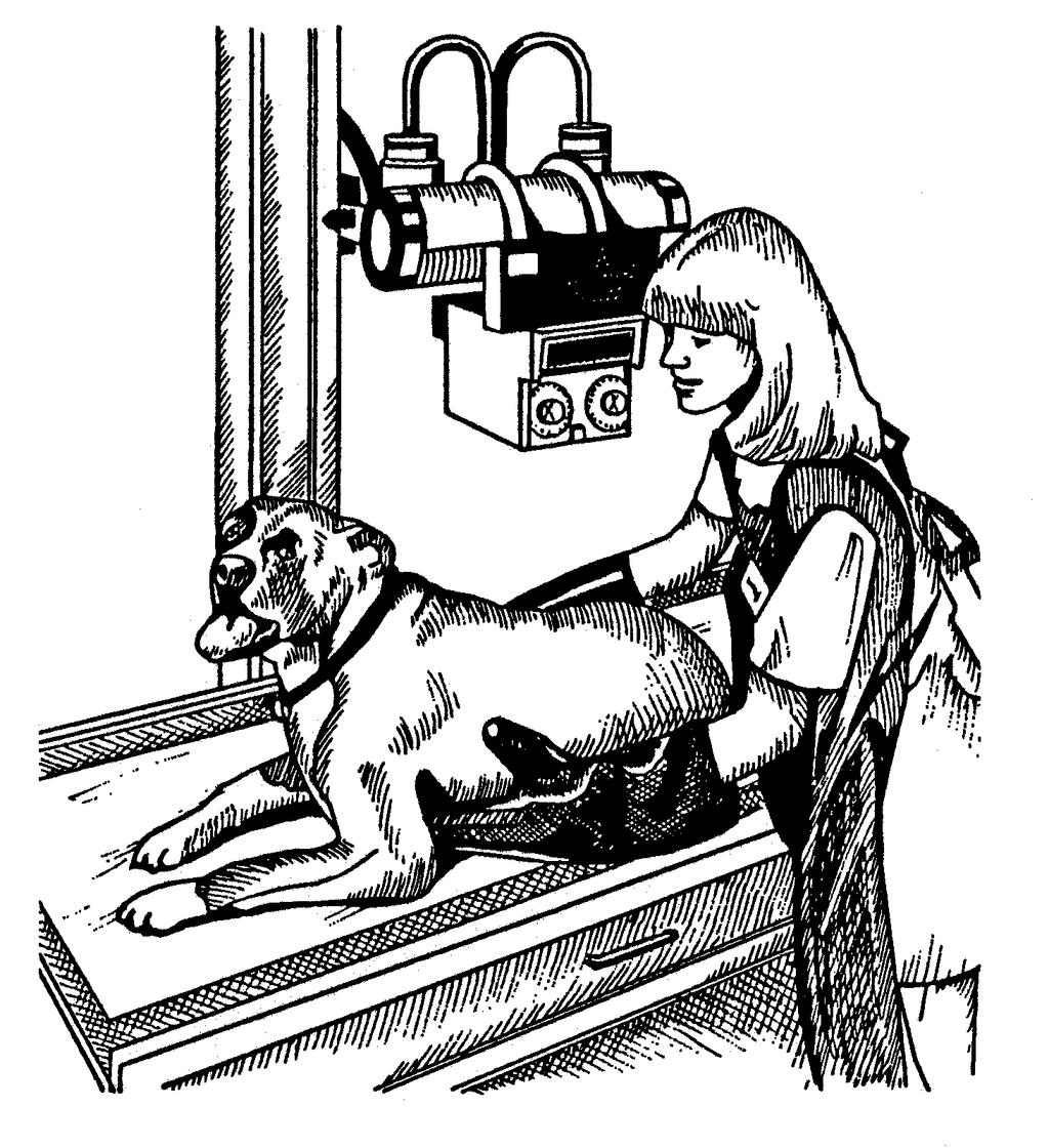
1 Veterinary Science Preparatory Training for the Veterinary Assistant Floron C. Faries, Jr., DVM, MS

2 Radiology Floron C. Faries, Jr., DVM, MS
3 Objectives Determine the appropriate machine settings for making a radiograph Describe essential radiograph accessories Describe the positions used to perform radiographs from different views Describe safety precautions and discuss their importance
 emitted Patient s tissues absorb Strike patient and redirect (scatter radiation) High voltage power source 30-150")
4 Terminology X-ray tube of radiograph machine Vacuum tube Cathode emits electrons Anode collects electrons Metal target on anode Electron beam (electrical current) perpendicularly directed X-rays (photons, radiation) emitted Patient s tissues absorb Strike patient and redirect (scatter radiation) High voltage power source kilovolts (kv)
 Too low kvp = white image (under-exposed) Low mas and high kvp = gray")
5 Milliamperage (ma) Unit of measure Controls amount of emitted x-rays ma x exposure time (S) = mas Kilovoltage peak (kvp) Value of voltage Controls radiographic contrast of image High mas and low kvp = black and white image Too high mas = black image (over-exposed) Too low kvp = white image (under-exposed) Low mas and high kvp = gray image
6 Source to image distance (SID) Distance from anode to receptor (film, plate) Increase distance = decreases beam intensity Lightens images Requires increasing mas settings Decrease distance = increases beam intensity Darkens images Requires reducing mas settings Reduces radiation exposure to patient
7 X-ray scatter control (reduces x-ray dose exposure to patient and x-ray scatter) Filtration device on x-ray tube Filtration device of lead strips (x-ray grid) Between anode and receptor (film, plate) Requires increasing mas settings Requires increasing kvp setting for large animal thorax
8 Determination of Machine Settings Measure thickness of patient s part Caliper instrument in centimeters Use technique chart for thickness measurement Position machine settings kvp ma Exposure time Set distance Set needle of voltage line meter 1/16 inch above center line Insures constant voltage coming into machine
9 Radiographic Positions Head, neck, body, tail Dorsal top view Ventral bottom view Lateral side view Leg Anterior front view Posterior back view Lateral outer side view Medial inner side view Two views One plane 90º angle to first view
10 Imaging Modalities Film/Screen Combinations Cassette holds film inside Image produced on film x-ray image (not visible) Film development image visible Chemical, water, and drying times Computer Radiology (CR) or Digital Radiography (DR) Cassette holds phosphor plate Image produced on plate x-ray image (not visible) Plate development image visible Plate in reader laser reads and digitizes into digital image Digital image sent to computer for viewing and storing Plate erased and refreshed for reuse by exposure to light
11 Computerized Axial Tomography (CAT) or Computerized Tomography (CT) Scan Utilizes x-rays Produces cross-sectional images of soft tissue and bone Ultrasonography (Ultrasound) Utilizes sound waves Produces images (sonograms) of soft tissues Motion images Still images Magnetic Resonance Imaging (MRI) Scan Utilizes radio waves Produces images in any plane of body
12 Images and Tissue Density Black image air Gray image soft tissue White image bone, minerals, sand, metal, dental enamel
13 Patient Motion Voluntary motion (controlled motion) restraint necessary Looking around Moving away Moving extremeties Paddling Pushing away Involuntary motion (uncontrolled motion) reduce exposure time, increase ma Heart beating Breathing Panting Discomfort moving
14 Heart, lungs, and thorax images Animal inhales make exposure Abdomen, pelvis, and spine images Animal exhales make exposure
15 Safety Devices and Precautions Remove anyone from the x-ray room that is not needed. Every person should wear a lead apron and lead gloves when holding the x-rayed animal. Check the condition of gloves and aprons periodically by using radiography to determine if they allow x-rays to pass through. Limit the beam to the size of the film with a cone or lead diaphragm. Do not direct the x-ray beam into another room or work area. Install an aluminum filter (1 to 2 mm thick) at the tube housing opening to eliminate radiation from useless wave lengths. Cover the bottom side of the x-ray table with lead to protect the feet. The hands should not be placed in the path of the direct beam. Do not use fluoroscopy when radiography will do the job. Fluoroscopy is more hazardous and requires additional safety precautions.
16
17 Lead Gloves and Aprons To help prevent deterioration of lead gloves and lead aprons: Roll or drape apron over curved surface rather than folding it. Store gloves by hanging or by placing cans with both ends open inside gloves to keep the gloves open and to allow moisture to evaporate.
RADIOGRAPHIC EXPOSURE
 RADIOGRAPHIC EXPOSURE Receptor Exposure Receptor Exposure the that interacts with the receptor. Computed Radiography ( ) requires a. Direct Digital Radiography (DR) requires a. Exposure Indicators Exposure
LECTURE 1 The Radiographic Image
 LECTURE 1 The Radiographic Image Prepared by:- KAMARUL AMIN ABDULLAH @ ABU BAKAR UiTM Faculty of Health Sciences Medical Imaging Department 11/23/2011 KAMARUL AMIN (C) 1 Lesson Objectives At the end of
Beam-Restricting Devices
 Beam-Restricting Devices Three factors contribute to an increase in scatter radiation: Increased kvp Increased Field Size Increased Patient or Body Part Size. X-ray Interactions a some interact with the
X-RAYS - NO UNAUTHORISED ENTRY
 Licencing of premises Premises Refer Guidelines A radiation warning sign and warning notice, X-RAYS - NO UNAUTHORISED ENTRY must be displayed at all entrances leading to the rooms where x-ray units are
1-1. GENERAL 1-2. DISCOVERY OF X-RAYS
 1-1. GENERAL Radiography is a highly technical field, indispensable to the modern dental practice, but presenting many potential hazards. The dental radiographic specialist must be thoroughly familiar
STUDENT REVIEW QUESTION SET K CR/DR CONTENT AREA
 STUDENT REVIEW QUESTION SET K CR/DR CONTENT AREA RADT 2913 COMPREHENSIVE REVIEW 1 The CR cassette is backed by aluminum that: A. reflects x-rays B. absorbs x-rays C. captures the image D. transmits x-rays
Introduction. Chapter 16 Diagnostic Radiology. Primary radiological image. Primary radiological image
 Introduction Chapter 16 Diagnostic Radiology Radiation Dosimetry I Text: H.E Johns and J.R. Cunningham, The physics of radiology, 4 th ed. http://www.utoledo.edu/med/depts/radther In diagnostic radiology
Contrast. Contrast: the difference in density on adjacent areas of a radiograph or other image receptor. Subjective. Long Scale (Low Contrast)
") Contrast Contrast: the difference in density on adjacent areas of a radiograph or other image receptor. Subject Subjective Radiographic Long Scale (Low Contrast) Short Scale (High Contrast) Factors affecting
Contrast Contrast: the difference in density on adjacent areas of a radiograph or other image receptor. Subject Subjective Radiographic Long Scale (Low Contrast) Short Scale (High Contrast) Factors affecting
Multiple Choice Identify the letter of the choice that best completes the statement or answers the question.
 RA110 test 3 Multiple Choice Identify the letter of the choice that best completes the statement or answers the question. 1. An object 35 cm in width is radiographed at 100 cm SID and at a 50 cm SOD. What
Exposure System Selection
 Principles of Imaging Science II (RAD120) Exposure Systems Exposure System Selection Radiographic exposure is a very complex process Best technique systems manipulate one variable while holding others
Visibility of Detail
 Visibility of Detail Radiographic Quality Quality radiographic images represents the, and information is for diagnosis. The of the anatomic structures and the accuracy of their ( ) determine the overall
X-RAY IMAGING EE 472 F2017. Prof. Yasser Mostafa Kadah
 X-RAY IMAGING EE 472 F2017 Prof. Yasser Mostafa Kadah www.k-space.org Recommended Textbook Stewart C. Bushong, Radiologic Science for Technologists: Physics, Biology, and Protection, 10 th ed., Mosby,
CR Basics and FAQ. Overview. Historical Perspective
 Page: 1 of 6 CR Basics and FAQ Overview Computed Radiography is a term used to describe a system that electronically records a radiographic image. Computed Radiographic systems use unique image receptors
DENTAL RADIOGRAPHY KAMARUL AMIN BIN ABU BAKAR
 DENTAL RADIOGRAPHY KAMARUL AMIN BIN ABDULLAH @ ABU BAKAR Components of the Dental X-Ray Machine Dental x-ray machines may vary somewhat in size and appearance, but all machines will have three primary
PD233: Design of Biomedical Devices and Systems
 PD233: Design of Biomedical Devices and Systems (Lecture-8 Medical Imaging Systems) (Imaging Systems Basics, X-ray and CT) Dr. Manish Arora CPDM, IISc Course Website: http://cpdm.iisc.ac.in/utsaah/courses/
COMPUTED RADIOGRAPHY CHAPTER 4 EFFECTIVE USE OF CR
 This presentation is a professional collaboration of development time prepared by: Rex Christensen Terri Jurkiewicz and Diane Kawamura New Technology https://www.youtube.com/watch?v=ptkzznazb 7U COMPUTED
SECTION I - CHAPTER 1 DIGITAL RADIOGRAPHY: AN OVERVIEW OF THE TEXT. Exam Content Specifications 8/22/2012 RADT 3463 COMPUTERIZED IMAGING
 RADT 3463 - COMPUTERIZED IMAGING Section I: Chapter 1 RADT 3463 Computerized Imaging 1 SECTION I - CHAPTER 1 DIGITAL RADIOGRAPHY: AN OVERVIEW OF THE TEXT RADT 3463 COMPUTERIZED IMAGING Section I: Chapter
SPRINGFIELD TECHNICAL COMMUNITY COLLEGE ACADEMIC AFFAIRS
 SPRINGFIELD TECHNICAL COMMUNITY COLLEGE ACADEMIC AFFAIRS Course Number: RADG 112 Department: Radiography Course Title: Image Production & Eval. Semester: Spring Year: 1997 Objectives/ Unit One: Introduction
SPRINGFIELD TECHNICAL COMMUNITY COLLEGE ACADEMIC AFFAIRS
 SPRINGFIELD TECHNICAL COMMUNITY COLLEGE ACADEMIC AFFAIRS Course Number: RADG 212 Department: Radiography Course Title: Equip. Operation & Maint. Semester: Spring Year: 1997 Objectives/ Unit One: The X-ray
- KiloVoltage. Technique 101: Getting Back to Basics
 Why do I need to know technique? Technique 101: Getting Back to Basics Presented by: Thomas G. Sandridge, M.S., M.Ed., R.T.(R) Program Director Northwestern Memorial Hospital School of Radiography Chicago,
X-ray Imaging. PHYS Lecture. Carlos Vinhais. Departamento de Física Instituto Superior de Engenharia do Porto
 X-ray Imaging PHYS Lecture Carlos Vinhais Departamento de Física Instituto Superior de Engenharia do Porto cav@isep.ipp.pt Overview Projection Radiography Anode Angle Focal Spot Magnification Blurring
Radiology Physics Lectures: Digital Radiography. Digital Radiography. D. J. Hall, Ph.D. x20893
 Digital Radiography D. J. Hall, Ph.D. x20893 djhall@ucsd.edu Background Common Digital Modalities Digital Chest Radiograph - 4096 x 4096 x 12 bit CT - 512 x 512 x 12 bit SPECT - 128 x 128 x 8 bit MRI -
Overview of Safety Code 35
 Common Quality Control Procedures for All s Quality Control Procedures Film All s Daily Quality Control Tests Equipment Warm-up (D1) According to manufacturers instructions Can include auto calibration(d1)
INNOVATION BY DESIGN. Toshiba A History of Leadership REMOTE CONTROL R/F SYSTEM
 INNOVATION BY DESIGN For over 130 years, Toshiba has led the world in developing technology to improve the quality of life. This Made for Life TM commitment is reflected in our family of leading-edge imaging
10/3/2012. Study Harder
 This presentation is a professional collaboration of development time prepared by: Rex Christensen Terri Jurkiewicz and Diane Kawamura Study Harder CR detection is inefficient, inferior to film screen
10/26/2015. Study Harder
 This presentation is a professional collaboration of development time prepared by: Rex Christensen Terri Jurkiewicz and Diane Kawamura Study Harder CR detection is inefficient, inferior to film screen
Nuclear Associates , , , , , ,
 Nuclear Associates 57-411, 57-412, 57-413 57-426, 57-431, 57-432 57-433, 57-435, 57-436 CLEAR-Pb Transparent X-Ray Compensation Filters Users Manual March 2005 Manual No. 57-XXX-1 Rev. 2 2003, 2005 Fluke
SYLLABUS. TITLE: Equipment Operation I. DEPARTMENT: Radiologic Technology
 CODE: RADT 156 INSTITUTE: Health Science TITLE: Equipment Operation I DEPARTMENT: Radiologic Technology COURSE DESCRIPTION: This course covers the principles of equipment operation and maintenance of radiographic
Fabrício Sampaio Péres Kury Federal University of Rio de Janeiro Medical School
 Fabrício Sampaio Péres Kury Federal University of Rio de Janeiro Medical School Harvard Medical School Exchange Clerkship Program Primary Care Radiology Clerkship Gillian Lieberman, M. D. Monday, September
Essentials of Digital Imaging
 Essentials of Digital Imaging Module 6 Transcript 2016 ASRT. All rights reserved. Essentials of Digital Imaging Module 6 Dose Reduction and Patient Safety 1. ASRT Animation 2. Welcome Welcome to Essentials
The effect of compensating filter on image quality in lateral projection of thoraco lumbar radiography
 Journal of Physics: Conference Series OPEN ACCESS The effect of compensating filter on image quality in lateral projection of thoraco lumbar radiography To cite this article: N A A Daud et al 2014 J. Phys.:
Artefacts found in computed radiography
 The British Journal of Radiology, 74 (2001), 195 202 E 2001 The British Institute of Radiology Pictorial review Artefacts found in computed radiography L J CESAR, RT(R)(QM), B A SCHUELER, PhD, F E ZINK,
MaxRay Handheld X-ray Systems Operator Training Exam
 MaxRay Handheld X-ray Systems Operator Training Exam Employee: Instructor: ate: Score: Instructions Read each question carefully and choose the best answer. 1) LR is 2) 3) 4) a. a safety principle meant
Mammography is a radiographic procedure specially designed for detecting breast pathology Approximately 1 woman in 8 will develop breast cancer over
 Mammography is a radiographic procedure specially designed for detecting breast pathology Approximately 1 woman in 8 will develop breast cancer over a lifetime Breast cancer screening programs rely on
RULES OF TENNESSEE DEPARTMENT OF ENVIRONMENT AND CONSERVATION DIVISION OF RADIOLOGICAL HEALTH CHAPTER USE OF X-RAY APPARATUS
 RULES OF TENNESSEE DEPARTMENT OF ENVIRONMENT AND CONSERVATION DIVISION OF RADIOLOGICAL HEALTH CHAPTER 0400-20-06 USE OF X-RAY APPARATUS TABLE OF CONTENTS 0400-20-06-.01 Purpose 0400-20-06-.06 Veterinary
Teaching Digital Radiography and Fluoroscopic Radiation Protection
 Teaching Digital Radiography and Fluoroscopic Radiation Protection WCEC 20 th Student Educator Radiographer Conference Dennis Bowman, RT(R), CRT (R)(F) Community Hospital of the Monterey Peninsula (CHOMP)
Radiology. Radiograph: Is the image of an object made with use of X- ray instead of light.
 Radiology د. اريج Lec. 3 X Ray Films Radiograph: Is the image of an object made with use of X- ray instead of light. Dental x- ray film: Is a recording media on which image of the object was made by exposing
The importance of radiation quality for optimisation in radiology
 Available online at http://www.biij.org/2007/2/e38 doi: 10.2349/biij.3.2.e38 biij Biomedical Imaging and Intervention Journal COMMENTARY The importance of radiation quality for optimisation in radiology
4. Contrast is the. There must The function of contrast is to:. The types of contrast are.
 RADIOGRAPHIC VISIBILITY OF DETAIL STUDY QUESTIONS 1. What is visibility of detail? It is controlled by properties. What are the two factors that affect it? 2. What is sharpness of detail? It is controlled
Digital Imaging Considerations Computed Radiography
 Digital Imaging Considerations Digital Radiography Computed Radiography o Cassette based Direct or Indirect Digital Radiography o Cassetteless Computed Radiography 1 CR Image Acquisition Most like conventional
Radiographic Testing (RT) [10]
![Radiographic Testing (RT) [10]](/thumbs/74/70215439.jpg "Radiographic Testing (RT) [10]") Radiographic Testing (RT) [10] Definition: An NDT method that utilizes x-rays or gamma radiation to detect discontinuities in materials, and to present their images on recording medium. 1> Electromagnetic
Radiographic Testing (RT) [10] Definition: An NDT method that utilizes x-rays or gamma radiation to detect discontinuities in materials, and to present their images on recording medium. 1> Electromagnetic
1. Carlton, Richard R., and Arlene M. Adler. Principles of Radiographic Imaging: An Art and a Science, 5th edition (2013).
.") CODE: RADT 151 INSTITUTE: Health Science TITLE: Radiographic Exposure DEPARTMENT: Radiologic Technology COURSE DESCRIPTION: This course covers the principles of radiographic exposure selection and manipulation
CODE: RADT 151 INSTITUTE: Health Science TITLE: Radiographic Exposure DEPARTMENT: Radiologic Technology COURSE DESCRIPTION: This course covers the principles of radiographic exposure selection and manipulation
X-rays. X-rays are produced when electrons are accelerated and collide with a target. X-rays are sometimes characterized by the generating voltage
 X-rays Ouch! 1 X-rays X-rays are produced when electrons are accelerated and collide with a target Bremsstrahlung x-rays Characteristic x-rays X-rays are sometimes characterized by the generating voltage
Nuclear Associates
 Nuclear Associates 07-649 CDRH Fluoroscopic Phantom Users Manual March 2005 Manual No. 07-649-1 Rev. 2 2004, 2005 Fluke Corporation, All rights reserved. Printed in U.S.A. All product names are trademarks
X-ray Tube and Generator Basic principles and construction
 X-ray Tube and Generator Basic principles and construction Dr Slavik Tabakov - Production of X-rays and Patient Dose OBJECTIVES - X-ray tube construction - Anode - types, efficiency - Classical X-ray generator
Acquisition, Processing and Display
 Acquisition, Processing and Display Terri L. Fauber, R.T. (R)(M) Department of Radiation Sciences School of Allied Health Professions Virginia Commonwealth University Topics Image Characteristics Image
Photons interaction with matter
 ب س م هللا الر ح من الر حیم Photons interaction with matter Ionization Ionization is the process of removing an electron from an electrically neutral atom to produce an ion pair. An ion is an atom or subatomic
INTRODUCTION TO FLEXIBLE BRONCHOSCOPY. Fluoroscopy Synopsis HENRI G COLT MD SECOND EDITION THE BRONCHOSCOPY EDUCATION PROJECT SERIES
 SECOND EDITION INTRODUCTION TO FLEXIBLE BRONCHOSCOPY Fluoroscopy Synopsis HENRI G COLT MD With contributions from Dr. S. Murgu THE BRONCHOSCOPY EDUCATION PROJECT SERIES FLUOROSCOPY SYNOPSIS The purpose
Medical Imaging. X-rays, CT/CAT scans, Ultrasound, Magnetic Resonance Imaging
 Medical Imaging X-rays, CT/CAT scans, Ultrasound, Magnetic Resonance Imaging From: Physics for the IB Diploma Coursebook 6th Edition by Tsokos, Hoeben and Headlee And Higher Level Physics 2 nd Edition
X-RAY. Lecture No.4. Image Characteristics:
 Lecture No.4 X-RAY أ.م.د. اسامة مراد ابراهيم Image Characteristics: *Radiographic density: It s the degree of blackness of the film. when a film is exposed by an x-ray beam (or by light in case of screenfilm
3/31/2011. Objectives. Emory University. Historical Development. Historical Development. Historical Development
 Teaching Radiographic Technique in a Digital Imaging Paradigm Objectives 1. Discuss the historical development of digital imaging. Dawn Couch Moore, M.M.Sc., RT(R) Assistant Professor and Director Emory
COMPUTED RADIOGRAPHY (CR)
") COMPUTED RADIOGRAPHY (CR) Moving with the time Avi Avner BVSc BSc CVR DVDI MRCVS CR-Basics A five step process: 1. X-ray image received on phosphor plate 2. Image extracted from phosphor plate by Laser
COMPUTED RADIOGRAPHY (CR) Moving with the time Avi Avner BVSc BSc CVR DVDI MRCVS CR-Basics A five step process: 1. X-ray image received on phosphor plate 2. Image extracted from phosphor plate by Laser
DIGITAL RADIOGRAPHY. Digital radiography is a film-less technology used to record radiographic images.
 DIGITAL RADIOGRAPHY Digital radiography is a film-less technology used to record radiographic images. 1 The purpose of digital imaging is to generate images that can be used in the diagnosis and assessment
Ludlum Medical Physics
 Ludlum Medical Physics Medical Imaging Radiology QA Test Tools NEW LUDLUM PRODUCT LINE Medical Physics Products Medical Physics Products What are they? Products used to measure radiation output and to
Examination of Pipe Welds by Image Plate Based Computed Radiography System
 Examination of Pipe Welds by Image Plate Based Computed Radiography System Sanjoy Das, M.S.Rana, Benny Sebastian, D. Mukherjee and K.K. Abdulla Atomic Fuels Division Bhabha Atomic Research Centre Mumbai
China Resources Wandong Medical Equipment Co., Ltd. High Frequency 50kW, 150kV Radiography System - HF50-R
 China Resources Wandong Medical Equipment Co., Ltd. High Frequency 50kW, 150kV Radiography System - HF50-R Building 3, No.9, Jiuxianqiaodong Road, Chaoyang District, Beijing 100015, P.R. China E-mail:
Explain what is meant by a photon and state one of its main properties [2]
![Explain what is meant by a photon and state one of its main properties [2]](/thumbs/80/82516318.jpg "Explain what is meant by a photon and state one of its main properties [2]") 1 (a) A patient has an X-ray scan taken in hospital. The high-energy X-ray photons interact with the atoms inside the body of the patient. Explain what is meant by a photon and state one of its main properties....
1 (a) A patient has an X-ray scan taken in hospital. The high-energy X-ray photons interact with the atoms inside the body of the patient. Explain what is meant by a photon and state one of its main properties....
Course Instructions: Check your for your CE certification of completion (please check your junk/spam folder as well). About SMS CE courses:
. About SMS CE courses:") 2017 Course #4 Self-Study Course Contact Us: Phone 614-292-6737 Toll Free 1-888-476-7678 Fax 614-292-8752 E-mail smsosu@osu.edu Web dentistry.osu.edu/sms The Ohio State University College of Dentistry
2017 Course #4 Self-Study Course Contact Us: Phone 614-292-6737 Toll Free 1-888-476-7678 Fax 614-292-8752 E-mail smsosu@osu.edu Web dentistry.osu.edu/sms The Ohio State University College of Dentistry
Nuclear Associates
 Nuclear Associates 07-644 Grid Alignment Test Tool Users Manual March 2005 Manual No. 07-644-1 Rev. 2 2004, 2005 Fluke Corporation, All rights reserved. Printed in U.S.A. All product names are trademarks
RAD 150 RADIOLOGIC EXPOSURE TECHNIQUE II
 RAD 150 RADIOLOGIC EXPOSURE TECHNIQUE II APPROVED 12/O2/2011 EFFECTIVE SPRING 2013-14 Prefix & Number RAD 150 Course Title: Radiologic Exposure Technique II & Lab Purpose of this submission: New Change/Updated
Ch. 223 VETERINARY MEDICINE CHAPTER 223. VETERINARY MEDICINE GENERAL PROVISIONS X-RAYS RADIOACTIVE MATERIAL. Authority
 Ch. 223 VETERINARY MEDICINE 25 223.1 CHAPTER 223. VETERINARY MEDICINE Sec. 223.1. Purpose and scope. 223.2. [Reserved]. 223.2a. Definitions. 223.3 223.6. [Reserved]. 223.7. Structural shielding. 223.8.
Seminar 8. Radiology S8 1
 Seminar 8 Radiology Medical imaging. X-ray image formation. Energizing and controlling the X-ray tube. Image detectors. The acquisition of analog and digital images. Digital image processing. Selected
Small Animal Radiographic Techniques and Positioning COPYRIGHTED MATERIAL
 Small Animal Radiographic Techniques and Positioning COPYRIGHTED MATERIAL Section 1 Theory and Equipment 1 Introduction to Digital Imaging Small animal radiography has changed dramatically in the past
BASICS OF FLUOROSCOPY
 Medical Physics Residents Training Program BASICS OF FLUOROSCOPY Dr. Khalid Alyousef, PhD Department of Medical Imaging King Abdulaziz Medical City- Riyadh Edison examining the hand of Clarence Dally with
CARESTREAM INDUSTREX Digital Imaging Plates
 2016-09-29 TI-2632 CARESTREAM INDUSTREX Digital Imaging Plates CARESTREAM INDUSTREX Flex Digital Imaging Plates (IPs) are designed for computed radiography in nondestructive testing applications. These
Digital Imaging started in the 1972 with Digital subtraction angiography Clinical digital imaging was employed from the 1980 ~ 37 years ago Amount of
 Digital Imaging started in the 1972 with Digital subtraction angiography Clinical digital imaging was employed from the 1980 ~ 37 years ago Amount of radiation to the population due to Medical Imaging
COMPUTED TOMOGRAPHY 1
 COMPUTED TOMOGRAPHY 1 Why CT? Conventional X ray picture of a chest 2 Introduction Why CT? In a normal X-ray picture, most soft tissue doesn't show up clearly. To focus in on organs, or to examine the
10/15/2012 SECTION III - CHAPTER 6 DIGITAL FLUOROSCOPY RADT 3463 COMPUTERIZED IMAGING
 RADT 3463 - COMPUTERIZED IMAGING Section III: Chapter 6 RADT 3463 Computerized Imaging 1 SECTION III - CHAPTER 6 DIGITAL FLUOROSCOPY RADT 3463 COMPUTERIZED IMAGING Section III: Chapter 6 RADT 3463 Computerized
Dose Reduction and Image Preservation After the Introduction of a 0.1 mm Cu Filter into the LODOX Statscan unit above 110 kvp
 Dose Reduction and Image Preservation After the Introduction of a into the LODOX Statscan unit above 110 kvp Abstract: CJ Trauernicht 1, C Rall 1, T Perks 2, G Maree 1, E Hering 1, S Steiner 3 1) Division
LECTURE 20 ELECTROMAGNETIC WAVES. Instructor: Kazumi Tolich
 LECTURE 20 ELECTROMAGNETIC WAVES Instructor: Kazumi Tolich Lecture 20 2 25.6 The photon model of electromagnetic waves 25.7 The electromagnetic spectrum Radio waves and microwaves Infrared, visible light,
Overview. Professor Roentgen was a Physicist!!! The Physics of Radiation Oncology X-ray Imaging
 The Physics of Radiation Oncology X-ray Imaging Charles E. Willis, Ph.D. DABR Associate Professor Department of Imaging Physics The University of Texas M.D. Anderson Cancer Center Houston, Texas Overview
Principle of X-Ray Systems
 Principle of X-Ray Systems Hossein Ebrahimi Nasab PHYSICS OF X-RAYS Nature of X-rays Energy unit Interaction with matter INTERACTION WITH THE MATTER In vacuum: photon move along a straight line In materials,
Enhanced Functionality of High-Speed Image Processing Engine SUREengine PRO. Sharpness (spatial resolution) Graininess (noise intensity)
 Graininess (noise intensity)") Vascular Enhanced Functionality of High-Speed Image Processing Engine SUREengine PRO Medical Systems Division, Shimadzu Corporation Yoshiaki Miura 1. Introduction In recent years, digital cardiovascular
Vascular Enhanced Functionality of High-Speed Image Processing Engine SUREengine PRO Medical Systems Division, Shimadzu Corporation Yoshiaki Miura 1. Introduction In recent years, digital cardiovascular
X-ray Tube and Generator Basic principles and construction
 X-ray Tube and Generator Basic principles and construction Dr Slavik Tabakov - Production of X-rays OBJECTIVES - X-ray tube construction - Anode - types, efficiency - X-ray tube working characteristics
CARESTREAM INDUSTREX Digital Imaging Plates
 TECHNICAL DATA / Non-Destructive Testing February 2011 TI-2632 CARESTREAM INDUSTREX Digital Imaging Plates CARESTREAM INDUSTREX Flex XL, GP and HR Digital Imaging Plates (IPs) are designed for computed
Digital Imaging CT & MR
 Digital Imaging CT & MR January 22, 2008 Digital Radiography, CT and MRI generate images in a digital format What is a Digital Image? A digital image is made up of picture elements, pixels row by column
QC Testing for Computed Tomography (CT) Scanner
 Scanner") QC Testing for Computed Tomography (CT) Scanner QA - Quality Assurance All planned and systematic actions needed to provide confidence on a structure, system or component. all-encompassing program, including
QC Testing for Computed Tomography (CT) Scanner QA - Quality Assurance All planned and systematic actions needed to provide confidence on a structure, system or component. all-encompassing program, including
Nuclear Associates
 Nuclear Associates 76-700 Digital Subtraction Angiography Phantom Users Manual March 2005 Manual No. 76-700-1 Rev. 2 2004, 2005 Fluke Corporation, All rights reserved. Printed in U.S.A. All product names
Digital radiography. bucky table and wall stand as a dual detector or wireless system. Amadeo DR Systems
 Amadeo DR Systems with dicom PACS DX-R Software X-ray Systems for the Future Digital radiography with Amadeo R-DR including bucky table and wall stand as a dual detector or wireless system High contrast
Comparison of computed radiography and filmõscreen combination using a contrast-detail phantom
 JOURNAL OF APPLIED CLINICAL MEDICAL PHYSICS, VOLUME 4, NUMBER 1, WINTER 2003 Comparison of computed radiography and filmõscreen combination using a contrast-detail phantom Z. F. Lu,* E. L. Nickoloff, J.
SYLLABUS. 1. Identification of Subject:
 SYLLABUS Date/ Revision : 30 January 2017/1 Faculty : Life Sciences Approval : Dean, Faculty of Life Sciences SUBJECT : Biophysics 1. Identification of Subject: Name of Subject : Biophysics Code of Subject
Essentials of Digital Imaging
 Essentials of Digital Imaging Module 1 Transcript 2016 ASRT. All rights reserved. Essentials of Digital Imaging Module 1 Fundamentals 1. ASRT Animation 2. Welcome Welcome to Essentials of Digital Imaging
Current technology in digital image production (CR/DR and other modalities) Jaroonroj Wongnil 25 Mar 2016
 Jaroonroj Wongnil 25 Mar 2016") Current technology in digital image production (CR/DR and other modalities) Jaroonroj Wongnil 25 Mar 2016 Current technology in digital image production (CR/DR and other modalities) 2/ Overview Digital
Current technology in digital image production (CR/DR and other modalities) Jaroonroj Wongnil 25 Mar 2016 Current technology in digital image production (CR/DR and other modalities) 2/ Overview Digital
MXHF-1500RF is controlled by Digital key panel console that displays KV, ma and mas with APR menu programmed.
 R/F TV X-RAY SYSTEM DIAGNOSTIC RADIOGRAPHIC FLUOROSCOPIC TV SYSTEM MXHF-1500RF SYSTEM OUTLINE Product Data No. 041021-01 MXHF-1500RF is controlled by Digital key panel console that displays KV, ma and
DIGITAL RADIOGRAPHY ARTIFACTS
 IMAGING LAB MPHY 487 DIGITAL RADIOGRAPHY ARTIFACTS Mohammad Esmael Alsulimane B.Sc, M.Sc Medical Physics Lecturer - Physics Department All Rights Reserved: Some information and figures in this presentation
A Comprehensive Review of Image Production
 A Comprehensive Review of Image Production Presented by: John Fleming, M.Ed., RT(R)(MR)(CT) St. Petersburg College Office: (727) 341-3758 E-mail: flemingj@spcollege.edu Lesson Objectives: ARRT Content
U.S. ARMY MEDICAL DEPARTMENT CENTER AND SCHOOL FORT SAM HOUSTON, TEXAS PRINCIPLES OF RADIOGRAPHIC EXPOSURE SUBCOURSE MD0952 EDITION 200
 U.S. ARMY MEDICAL DEPARTMENT CENTER AND SCHOOL FORT SAM HOUSTON, TEXAS 78234-6100 PRINCIPLES OF RADIOGRAPHIC EXPOSURE SUBCOURSE MD0952 EDITION 200 DEVELOPMENT This subcourse is approved for resident and
Digital Image Processing
 What is an image? Digital Image Processing Picture, Photograph Visual data Usually two- or three-dimensional What is a digital image? An image which is discretized, i.e., defined on a discrete grid (ex.
STUDENT REVIEW QUESTION SET I RANDOM CONTENT AREA RADT 2913 COMPREHENSIVE REVIEW
 STUDENT REVIEW QUESTION SET I RANDOM CONTENT AREA RADT 2913 COMPREHENSIVE REVIEW 1 What is the most probable source for the radiographic artifact which appears as high-density artifacts resembling lightning
DIAGNOSTIC ACCREDITATION PROGRAM. Radiology and CT Quality Control Procedures Workbook
 DIAGNOSTIC ACCREDITATION PROGRAM Radiology and CT Quality Control Procedures Workbook Quality Control Procedures Radiography/CR/DR Safety Code 35 Summary For more detail about each quality control (QC)
Half value layer and AEC receptor dose compliance survey in Estonia
 Half value layer and AEC receptor dose compliance survey in Estonia K. Kepler, A. Vladimirov Training Centre of Medical Physics, University of Tartu Testing Centre of the University of Tartu, Estonia E-mail:
Dental Radiography. One of the problems of dental radiography is having different dimensions than normal.
 The prototype receptor (the recording medium) most commonly used in dental radiography is the radiographic film. However, there are many other new more efficient receptors than the formed one that can
Diffraction-enhanced X-ray Imaging (DEXI) Medical Solutions. More information using less radiation
 Medical Solutions. More information using less radiation") Diffraction-enhanced X-ray Imaging (DEXI) Medical Solutions More information using less radiation Medical Small Animal Security NDE/NDT Diffraction-Enhanced X-ray Imaging Medical Solutions Safe non-invasive
Diffraction-enhanced X-ray Imaging (DEXI) Medical Solutions More information using less radiation Medical Small Animal Security NDE/NDT Diffraction-Enhanced X-ray Imaging Medical Solutions Safe non-invasive
Photomultiplier Tube
 Nuclear Medicine Uses a device known as a Gamma Camera. Also known as a Scintillation or Anger Camera. Detects the release of gamma rays from Radionuclide. The radionuclide can be injected, inhaled or
Exposure Indices and Target Values in Radiography: What Are They and How Can You Use Them?
 Exposure Indices and Target Values in Radiography: What Are They and How Can You Use Them? Definition and Validation of Exposure Indices Ingrid Reiser, PhD DABR Department of Radiology University of Chicago
JEFFERSON COLLEGE COURSE SYLLABUS BET220 DIAGNOSTIC IMAGING. 3 Credit Hours. Prepared by: Scott Sebaugh Date: 2/20/2012
 JEFFERSON COLLEGE COURSE SYLLABUS BET220 DIAGNOSTIC IMAGING 3 Credit Hours Prepared by: Scott Sebaugh Date: 2/20/2012 Mary Beth Ottinger, Division Chair Elizabeth Check, Dean, Career & Technical Education
Sarah Hughes, MS, DABR Radiation Safety Officer
 Sarah Hughes, MS, DABR Radiation Safety Officer 502-852-6146 sarah.hughes@louisville.edu Mo my back is burnin!!! I got it MAG the cine! Sumthin s not right. Where s his heart? Fluoroscopy http://dccwww.bumc.bu.edu/fluoroscopy/def
Digital radiography: Practical advantages of Digital Radiography. Practical Advantages in image quality
 Digital radiography: Digital radiography is set to become the most common form of processing radiographic images in the next 10 years. This is due to a number of practical and image quality issues. Practical
P R E S E N T E D B Y. K A M A R U L A M I N A B D U L L A H Dip. MED. IMG., BSc. MED. IMG. (UiTM)
") + - P R E S E N T E D B Y K A M A R U L A M I N A B D U L L A H Dip. MED. IMG., BSc. MED. IMG. (UiTM) 1 I N T R O D U C T I O N : An x-ray generator is a device that Supplies electrical power to x-ray
+ - P R E S E N T E D B Y K A M A R U L A M I N A B D U L L A H Dip. MED. IMG., BSc. MED. IMG. (UiTM) 1 I N T R O D U C T I O N : An x-ray generator is a device that Supplies electrical power to x-ray
ProX Intraoral X-ray. PLANMECA is proud to introduce a new intraoral X-ray unit to its comprehensive collection of imaging products- the ProX.
 The premium intraoral X-ray unit... ProX Intraoral X-ray PLANMECA is proud to introduce a new intraoral X-ray unit to its comprehensive collection of imaging products- the ProX. This advanced unit provides