COMPUTED TOMOGRAPHY 1
|
|
|
- Francine Wilson
- 5 years ago
- Views:
Transcription

1 COMPUTED TOMOGRAPHY 1

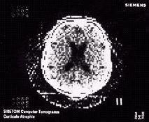
2 Why CT? Conventional X ray picture of a chest 2
3 Introduction Why CT? In a normal X-ray picture, most soft tissue doesn't show up clearly. To focus in on organs, or to examine the blood vessels, doctors must introduce contrast media into the body. Contrast media are liquids that absorb X-rays more effectively than the surrounding tissue. 3
4 Introduction CT scans are special x-ray tests that produce cross-sectional images of the body and the images allow to look at the inside of the body using x-rays and a computer. 4
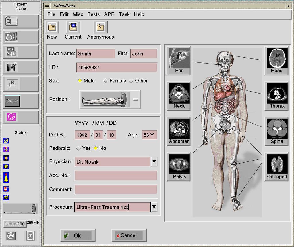
5 System Components CT Gantry. Computer. Operating console. The patient support table 5
6 System Components 6
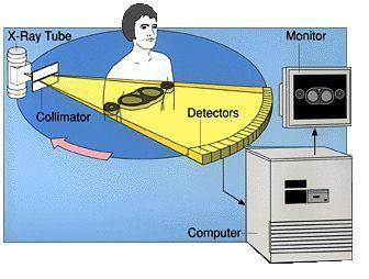
7 CT Gantry The CT gantry contains: X-ray tube. Detection system. An other associated mechanisms. Gantry Design:An x-ray tube is mounted within the scanner, opposite a set of detectors. 7
8 8
9 9
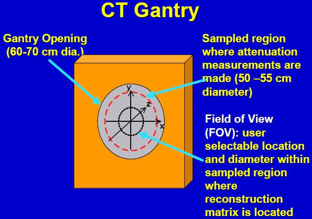
10 The patient support table The patient support table allows for insertion of the patient into the hole of gantry, which is approximately cm in diameter. Most gantries can be tilted +/- 30 degree in order to obtain oblique slices. 10
11 Computer The computer system plays a central role in CT scanning because without it, there would be no image computation and formation. The computer controls of: x-ray generation gantry and table motion data acquisition image formation display storage. 11
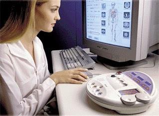
12 Control console Set scan parameters :kvp, ma, scan time, reconstruction filter, etc. Set scan mode:- Digital radiograph, axial or volume Reconstructor Review and archive images Post-processing 12
13 13
14 14
15 Detectors Scintillation Detectors: Types of material will scintillate when irradiated; that is, they emit a flash of light immediately in response to absorption of ionizing radiation. The amount of light emitted is proportional to the amount of energy absorbed by the crystal. 15
16 Detectors Early scintillation detector arrays contained scintillation crystal (Sodium iodide NaI) photomultiplier tube. Scintillation crystal coupled to the window of the crystal is a photomultiplier (PM) tube that converts the light flashes from the scintillator into electrical pulses. These detectors could not be packed together and they required a power supply for each photomultiplier. 16
17 They have been replaced with scintillation crystal (cesium iodide CsI). photodiode Photodiodes convert light into electronic signal, are smaller, cheaper and don t require a power supply for each. Approximately 90% of the x-rays incident on the detector are absorbed and contribute to the output signal 17
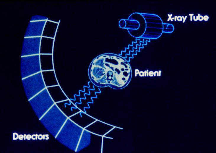
18 CT Principles CT uses X-rays to generate cross-sectional, images of the body. An x-ray tube is mounted within the scanner, opposite a set of detectors. The detectors receive the x-rays after they pass through the patients' body. 18
19 CT Principles Both the x-ray tube and detectors rotate around the patient, resulting in x-rays from 360 degrees. This scanned data is used by the CT computer to produce two or three-dimensional cross- sectional images. 19
20 History
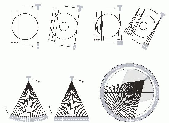
21 Operational Modes First generation: rotate/translate. Second generation: rotate/translate. Third generation: rotate/rotate. Fourth generation: rotate/stationary. Fifth generation: fixed tubes, fixed detectors
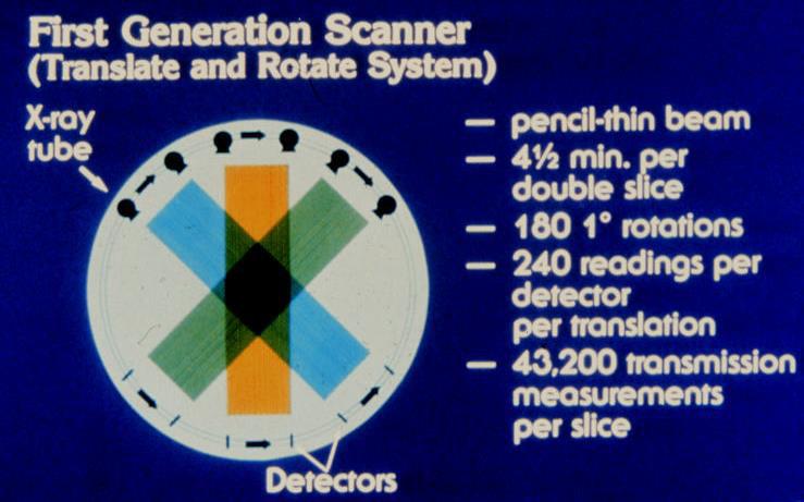
22 First generation: rotate/translate, pencil beam This system used parallel ray geometry - starting at a particular angle. The X-ray tube and detector system translated linearly across the field of view (FOV), acquiring 160 rays across a 24 - cm FOV, for the projection at that particular angle. When the X - ray tube/detector system was finished with its translation, the whole system was rotated and then another translation was used to acquire the next projection set.
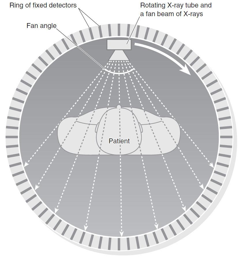
23 First generation 23
24 24
25 First generation The image was not perfect. One advantage of the first - generation CT scanner was that it accepted only a very small pencil ray of X - rays. 25
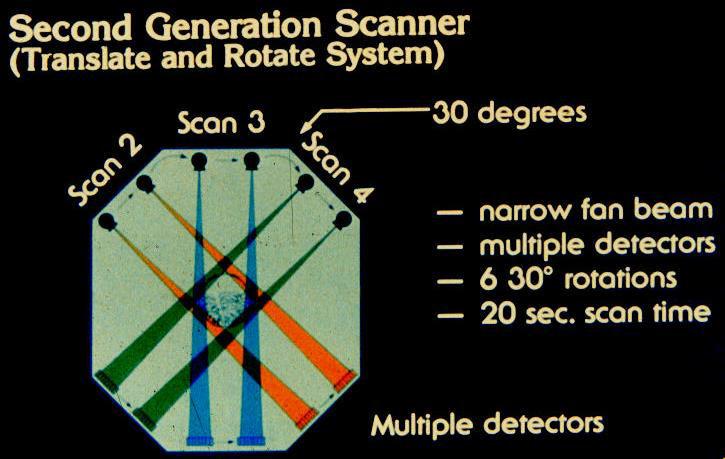
26 Second generation: rotate/translate, narrow fan beam The next incremental improvement to the CT scanner was the incorporation of linear array of (3-52) detectors. The use of 30 detectors increased the utilization of the X ray beam by 30 times over the single detector used per slice in first - generation systems. A relatively narrow fan angle of 10 degree was used.
27 Second generation 27
28 28
29 Second generation The disadvantages of the fan beam was the increased scatter radiation. This affected the final image. The translation motion of first and second generation CT scanners was a major limitation because at the end of each translation, the X - ray tube/detector system had to be stopped, the whole system rotated and then the translational motion had to be restarted. 29
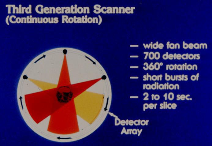
30 Third generation: rotate/rotate, wide fan beam The motion of third - generation CT is rotate/rotate, referring to the rotation of the X - ray tube and the rotation of the detector array. It was an evolution, because third - generation scanners could deliver scan times shorter than 5 seconds. The number of detectors used in third - generation scanners was increased (up to about 750 detectors).

31 . Third generation 31
32 32
33 Third generation The rotate/rotate geometry solved the mechanical problems by removing the translational motion, practical problems remained 33
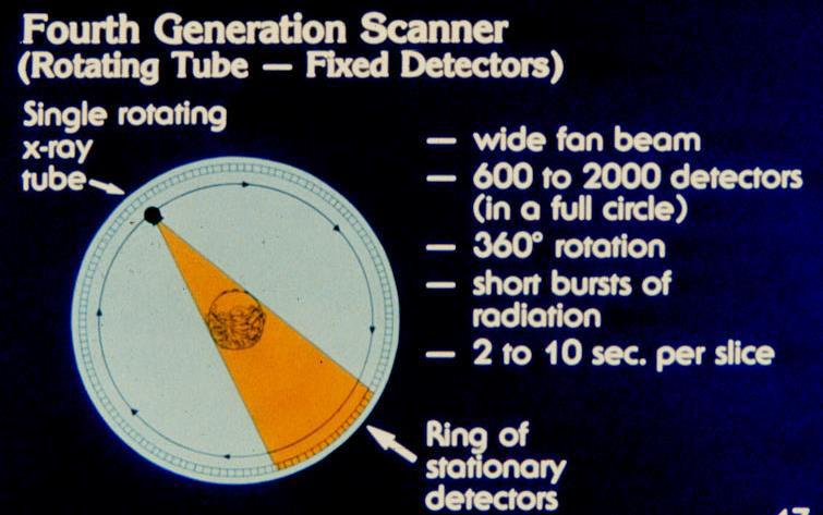
34 Fourth generation: rotate/stationary: Fourth - generation CT scanners were designed to overcome the problem of electronic drift between the many detectors used in the system. Fourth - generation uses a stationary ring of detectors positioned around the patient. Only the x-ray source rotates with a wide fan beam geometry, while the detectors are stationary.
35 Fourth generation With fourth - generation design, the detectors are removed from the rotating gantry and are placed on a stationary annulus around the patients. Modern fourth - generation CT systems use from 1200 to 4800 individual detectors. 35

36 Diagram of the fourth-generation CT scanner 36
37 37
38 38
39 Fourth generation The principal disadvantage of fourth generation CT scanners is patient dose, which is higher than that with other types of scanners. The cost of these units may also be higher because of the large number of detectors and their associated electronics. 39
40 Fifth generation: fixed tubes, fixed detectors. Real time imaging with no moving parts, heart imaging, expensive. Newer versions use 28 image intensifiers (halfcircle) with gantry rotation at 15 RPM. 3-D real time imaging. 40
41 41
42 42
43 Fifth generation: fixed tubes, fixed detectors. Real time imaging with no moving parts, heart imaging, expensive. Newer versions use 28 image intensifiers (half- circle) with gantry rotation at 15 RPM. 3-D real time imaging. 43
44 44
45 Benefits 1\ CT scanning is painless, noninvasive and accurate 2\ A major advantage of CT is that it is able to image bone, soft tissue and blood vessels all at the same time 3\ Unlike conventional x-rays, CT scanning provides very detailed images of many types of tissue well as the lungs, bones, and blood vessels 45
46 4 \ CT examinations are fast and simple; in emergency cases, they can reveal internal quickly enough to help save lives 6\ CT is less sensitive to patient movement than MRI 7\ CT can be performed if you have an implanted medical device of any kind, unlike MRI 46
47 8\ No radiation remains in a patient's body after a CT examination 9\ X-rays used in CT scans usually have no side effects 47
48 Risks 1\ The effective radiation dose from this procedure ranges from approximately two to 10 msv, which is about the same as the average person receives from background radiation in three to five years. 48
49 Risks 3\ CT scanning is, in general, not recommended for pregnant women unless medically necessary because of potential risk to the baby. 4\ Children should have a CT study only if it is essential for making a diagnosis and should not have repeated CT studies unless absolutely necessary 49
50 The limitations of CT Scanning of the Body. A person who is very large may not fit into the opening of a conventional CT scanner or may.be over the weight limit for the moving table 50
HISTORY. CT Physics with an Emphasis on Application in Thoracic and Cardiac Imaging SUNDAY. Shawn D. Teague, MD
 CT Physics with an Emphasis on Application in Thoracic and Cardiac Imaging Shawn D. Teague, MD DISCLOSURES 3DR- advisory committee CT PHYSICS WITH AN EMPHASIS ON APPLICATION IN THORACIC AND CARDIAC IMAGING
Introduction. Chapter 16 Diagnostic Radiology. Primary radiological image. Primary radiological image
 Introduction Chapter 16 Diagnostic Radiology Radiation Dosimetry I Text: H.E Johns and J.R. Cunningham, The physics of radiology, 4 th ed. http://www.utoledo.edu/med/depts/radther In diagnostic radiology
Medical Imaging. X-rays, CT/CAT scans, Ultrasound, Magnetic Resonance Imaging
 Medical Imaging X-rays, CT/CAT scans, Ultrasound, Magnetic Resonance Imaging From: Physics for the IB Diploma Coursebook 6th Edition by Tsokos, Hoeben and Headlee And Higher Level Physics 2 nd Edition
X-rays in medical diagnostics
 X-rays in medical diagnostics S.Dolanski Babić 2017/18. History W.C.Röntgen (1845-1923) discovered a new type of radiation Nature, Jan. 23. 1896.; Science, Feb.14. 1896. X- rays: Induced the ionization
Research Support. Dual-Source CT: What is it and How Do I Test it? Cynthia H. McCollough, Ph.D.
 Dual-Source CT: What is it and How Do I Test it? Cynthia H. McCollough, Ph.D. CT Clinical Innovation Center Department of Radiology Mayo Clinic College of Medicine Rochester, MN Research Support National
PD233: Design of Biomedical Devices and Systems
 PD233: Design of Biomedical Devices and Systems (Lecture-8 Medical Imaging Systems) (Imaging Systems Basics, X-ray and CT) Dr. Manish Arora CPDM, IISc Course Website: http://cpdm.iisc.ac.in/utsaah/courses/
Radiology Physics Lectures: Digital Radiography. Digital Radiography. D. J. Hall, Ph.D. x20893
 Digital Radiography D. J. Hall, Ph.D. x20893 djhall@ucsd.edu Background Common Digital Modalities Digital Chest Radiograph - 4096 x 4096 x 12 bit CT - 512 x 512 x 12 bit SPECT - 128 x 128 x 8 bit MRI -
Dose Reduction and Image Preservation After the Introduction of a 0.1 mm Cu Filter into the LODOX Statscan unit above 110 kvp
 Dose Reduction and Image Preservation After the Introduction of a into the LODOX Statscan unit above 110 kvp Abstract: CJ Trauernicht 1, C Rall 1, T Perks 2, G Maree 1, E Hering 1, S Steiner 3 1) Division
SYLLABUS. 1. Identification of Subject:
 SYLLABUS Date/ Revision : 30 January 2017/1 Faculty : Life Sciences Approval : Dean, Faculty of Life Sciences SUBJECT : Biophysics 1. Identification of Subject: Name of Subject : Biophysics Code of Subject
Explain what is meant by a photon and state one of its main properties [2]
![Explain what is meant by a photon and state one of its main properties [2]](/thumbs/80/82516318.jpg "Explain what is meant by a photon and state one of its main properties [2]") 1 (a) A patient has an X-ray scan taken in hospital. The high-energy X-ray photons interact with the atoms inside the body of the patient. Explain what is meant by a photon and state one of its main properties....
1 (a) A patient has an X-ray scan taken in hospital. The high-energy X-ray photons interact with the atoms inside the body of the patient. Explain what is meant by a photon and state one of its main properties....
Photomultiplier Tube
 Nuclear Medicine Uses a device known as a Gamma Camera. Also known as a Scintillation or Anger Camera. Detects the release of gamma rays from Radionuclide. The radionuclide can be injected, inhaled or
CHAPTER 2 COMMISSIONING OF KILO-VOLTAGE CONE BEAM COMPUTED TOMOGRAPHY FOR IMAGE-GUIDED RADIOTHERAPY
 14 CHAPTER 2 COMMISSIONING OF KILO-VOLTAGE CONE BEAM COMPUTED TOMOGRAPHY FOR IMAGE-GUIDED RADIOTHERAPY 2.1 INTRODUCTION kv-cbct integrated with linear accelerators as a tool for IGRT, was developed to
12/21/2016. Siemens Medical Systems Research Agreement Philips Healthcare Research Agreement AAN and ASN Committees
 Joseph V. Fritz, PhD Nandor Pintor, MD Dent Neurologic Institute ASN 2017 Friday, January 20, 2017 Siemens Medical Systems Research Agreement Philips Healthcare Research Agreement AAN and ASN Committees
QC Testing for Computed Tomography (CT) Scanner
 Scanner") QC Testing for Computed Tomography (CT) Scanner QA - Quality Assurance All planned and systematic actions needed to provide confidence on a structure, system or component. all-encompassing program, including
QC Testing for Computed Tomography (CT) Scanner QA - Quality Assurance All planned and systematic actions needed to provide confidence on a structure, system or component. all-encompassing program, including
Current technology in digital image production (CR/DR and other modalities) Jaroonroj Wongnil 25 Mar 2016
 Jaroonroj Wongnil 25 Mar 2016") Current technology in digital image production (CR/DR and other modalities) Jaroonroj Wongnil 25 Mar 2016 Current technology in digital image production (CR/DR and other modalities) 2/ Overview Digital
Current technology in digital image production (CR/DR and other modalities) Jaroonroj Wongnil 25 Mar 2016 Current technology in digital image production (CR/DR and other modalities) 2/ Overview Digital
Beam-Restricting Devices
 Beam-Restricting Devices Three factors contribute to an increase in scatter radiation: Increased kvp Increased Field Size Increased Patient or Body Part Size. X-ray Interactions a some interact with the
10/26/2015. Study Harder
 This presentation is a professional collaboration of development time prepared by: Rex Christensen Terri Jurkiewicz and Diane Kawamura Study Harder CR detection is inefficient, inferior to film screen
Studies on reduction of exposure dose using digital scattered X-ray removal processing
 Studies on reduction of exposure dose using digital scattered X-ray removal processing Poster No.: C-1834 Congress: ECR 2015 Type: Scientific Exhibit Authors: K. Kashiyama, M. Funahashi, T. Nakaoka, T.
Studies on reduction of exposure dose using digital scattered X-ray removal processing
 Studies on reduction of exposure dose using digital scattered X-ray removal processing Poster No.: C-1834 Congress: ECR 2015 Type: Scientific Exhibit Authors: K. Kashiyama, M. Funahashi, T. Nakaoka, T.
NM Module Section 2 6 th Edition Christian, Ch. 3
 NM 4303 Module Section 2 6 th Edition Christian, Ch. 3 Gas Filled Chamber Voltage Gas filled chamber uses Hand held detectors cutie pie Geiger counter Dose calibrators Cutie pie Chamber voltage in Ionization
Maximum Performance, Minimum Space
 TECHNOLOGY HISTORY For over 130 years, Toshiba has been a world leader in developing technology to improve the quality of life. Our 50,000 global patents demonstrate a long, rich history of leading innovation.
Computerized transverse axial scanning (tomography): Part I. Description of system
: Part I. Description of system") 1973, British Journal of Radiology, 46, 1016-1022 Computerized transverse axial scanning (tomography): Part I. Description of system Central Research Laboratories of EMI Limited, Hayes, Middlesex (Received
1973, British Journal of Radiology, 46, 1016-1022 Computerized transverse axial scanning (tomography): Part I. Description of system Central Research Laboratories of EMI Limited, Hayes, Middlesex (Received
CR Basics and FAQ. Overview. Historical Perspective
 Page: 1 of 6 CR Basics and FAQ Overview Computed Radiography is a term used to describe a system that electronically records a radiographic image. Computed Radiographic systems use unique image receptors
CT Basics: Data Acquisition Module 3
 Module 3 Transcript For educational and institutional use. This transcript is licensed for noncommercial, educational inhouse or online educational course use only in educational and corporate institutions.
Radionuclide Imaging MII Single Photon Emission Computed Tomography (SPECT)
") Radionuclide Imaging MII 3073 Single Photon Emission Computed Tomography (SPECT) Single Photon Emission Computed Tomography (SPECT) The successful application of computer algorithms to x-ray imaging in
Radionuclide Imaging MII 3073 Single Photon Emission Computed Tomography (SPECT) Single Photon Emission Computed Tomography (SPECT) The successful application of computer algorithms to x-ray imaging in
Data. microcat +SPECT
 Data microcat +SPECT microcat at a Glance Designed to meet the throughput, resolution and image quality requirements of academic and pharmaceutical research, the Siemens microcat sets the standard for
10/3/2012. Study Harder
 This presentation is a professional collaboration of development time prepared by: Rex Christensen Terri Jurkiewicz and Diane Kawamura Study Harder CR detection is inefficient, inferior to film screen
Pitfalls and Remedies of MDCT Scanners as Quantitative Instruments
 intensity m(e) m (/cm) 000 00 0 0. 0 50 0 50 Pitfalls and Remedies of MDCT Scanners as Jiang Hsieh, PhD GE Healthcare Technology University of Wisconsin-Madison Root-Causes of CT Number Inaccuracies Nature
TOPICS: CT Protocol Optimization over the Range of Patient Age & Size and for Different CT Scanner Types: Recommendations & Misconceptions
 CT Protocol Optimization over the Range of Patient Age & Size and for Different CT Scanner Types: Recommendations & Misconceptions TOPICS: Computed Tomography Quick Overview CT Dosimetry Effects of CT
Diffraction-enhanced X-ray Imaging (DEXI) Medical Solutions. More information using less radiation
 Medical Solutions. More information using less radiation") Diffraction-enhanced X-ray Imaging (DEXI) Medical Solutions More information using less radiation Medical Small Animal Security NDE/NDT Diffraction-Enhanced X-ray Imaging Medical Solutions Safe non-invasive
Diffraction-enhanced X-ray Imaging (DEXI) Medical Solutions More information using less radiation Medical Small Animal Security NDE/NDT Diffraction-Enhanced X-ray Imaging Medical Solutions Safe non-invasive
An Activity in Computed Tomography
 Pre-lab Discussion An Activity in Computed Tomography X-rays X-rays are high energy electromagnetic radiation with wavelengths smaller than those in the visible spectrum (0.01-10nm and 4000-800nm respectively).
High Energy Digital Radiography & 3D-CT for Industrial Systems
 DIR 2007 - International Symposium on Digital industrial Radiology and Computed Tomography, June 25-27, 2007, Lyon, France High Energy Digital Radiography & 3D-CT for Industrial Systems Non-Destructive
An Activity in Computed Tomography
 Pre-lab Discussion An Activity in Computed Tomography X-rays X-rays are high energy electromagnetic radiation with wavelengths smaller than those in the visible spectrum (0.01-10nm and 4000-800nm respectively).
Computed Tomography. The Fundamentals of... THE FUNDAMENTALS OF... Jason H. Launders, MSc. Current Technology
 The Fundamentals of... Computed Tomography Computed Tomography (CT) systems use x-rays to produce images of slices through a patient s anatomy. Despite having lower spatial resolution than other x-ray
CT Scanner Dose Survey
 CT Scanner Dose Survey Measurement Protocol Version 5.0 July 1997 Co-ordinated by ImPACT and The Medical Physics Department,, London SW17, UK. 0181-725-3366 CT Scanner Dose Survey: Measurement Protocol
Fig. 1
 PhysicsAndMathsTutor.com 1 1. Fig. 1 shows data for the intensity of a parallel beam of X-rays after penetration through varying thicknesses of a material. intensity / MW m 2 thickness / mm 0.91 0.40 0.69
Four-dimensional Computed Tomography (4D CT) Concepts and Preliminary Development
 Concepts and Preliminary Development") ORIGINAL ARTICLE ORIGINAL ARTICLE Radiation Medicine: Vol. 21 No. 1, 17 22 p.p., 2003 Four-dimensional Computed Tomography (4D CT) Concepts and Preliminary Development Masahiro Endo,* Takanori Tsunoo,*
ORIGINAL ARTICLE ORIGINAL ARTICLE Radiation Medicine: Vol. 21 No. 1, 17 22 p.p., 2003 Four-dimensional Computed Tomography (4D CT) Concepts and Preliminary Development Masahiro Endo,* Takanori Tsunoo,*
Hardware for High Energy Applications 30 October 2009
 Paper No. 003 09 Hardware for High Energy Applications 30 October 2009 This document was created by the Federal Working Group on Industrial Digital Radiography. Reproduction is authorized. Federal Working
160-slice CT SCANNER / New Standard for the Future
 TECHNOLOGY HISTORY For over 130 years, Toshiba has been a world leader in developing technology to improve the quality of life. Our 50,000 global patents demonstrate a long, rich history of leading innovation.
Lunar Technology Advantages
 Lunar Technology Advantages DXA stands for Dual-Energy X-ray Absorptiometry. It is a measurement method that uses the differences in the absorption of high energy and low energy X-ray photons by different
Detector technology in simultaneous spectral imaging
 Computed tomography Detector technology in simultaneous spectral imaging Philips IQon Spectral CT Z. Romman, I. Uman, Y. Yagil, D. Finzi, N. Wainer, D. Milstein; Philips Healthcare While CT has become
Radionuclide Imaging MII 3073 RADIONUCLIDE IMAGING SYSTEM
 Radionuclide Imaging MII 3073 RADIONUCLIDE IMAGING SYSTEM Preamplifiers and amplifiers The current from PMT must be further amplified before it can be processed and counted (the number of electrons yielded
IMPROVEMENTS IN X-RAY CT
 The First International Proficiency Testing Conference Sinaia, România 11 th 13 th October, 2007 IMPROVEMENTS IN X-RAY CT Emilia Dana Seleţchi Faculty of Physics, University of Bucharest, Măgurele, CP
diagnostic examination
 RADIOLOGICAL PHYSICS 2011 Raphex diagnostic examination Adel A. Mustafa, Ph.D., Editor PUBLISHED FOR: RAMPS (Radiological and Medical Physics Society of New York) preface The RAPHEX Diagnostic exam 2011
Wide-Detector CT for TAVR Planning:
 Wide-Detector CT for TAVR Planning: Impact on Iodine Dose, Radiation Dose, and Image Quality SCBTMR 2015 Annual Course Thursday, October 8 William P. Shuman MD FSCBTMR Department of Radiology University
Veterinary Science Preparatory Training for the Veterinary Assistant. Floron C. Faries, Jr., DVM, MS
 Veterinary Science Preparatory Training for the Veterinary Assistant Floron C. Faries, Jr., DVM, MS Radiology Floron C. Faries, Jr., DVM, MS Objectives Determine the appropriate machine settings for making
LECTURE 1 The Radiographic Image
 LECTURE 1 The Radiographic Image Prepared by:- KAMARUL AMIN ABDULLAH @ ABU BAKAR UiTM Faculty of Health Sciences Medical Imaging Department 11/23/2011 KAMARUL AMIN (C) 1 Lesson Objectives At the end of
12/26/2017. Alberto Ardon M.D.
 Alberto Ardon M.D. 1 Preparatory Work Ultrasound Physics http://www.nysora.com/mobile/regionalanesthesia/foundations-of-us-guided-nerve-blockstechniques/index.1.html Basic Ultrasound Handling https://www.youtube.com/watch?v=q2otukhrruc
Digital radiography: Practical advantages of Digital Radiography. Practical Advantages in image quality
 Digital radiography: Digital radiography is set to become the most common form of processing radiographic images in the next 10 years. This is due to a number of practical and image quality issues. Practical
BASICS OF FLUOROSCOPY
 Medical Physics Residents Training Program BASICS OF FLUOROSCOPY Dr. Khalid Alyousef, PhD Department of Medical Imaging King Abdulaziz Medical City- Riyadh Edison examining the hand of Clarence Dally with
of sufficient quality and quantity
 of sufficient quality and quantity The patient s body attenuates the beam as it passes though the body More energy is deposited in organs located near the entry of the beam than near the exit of the beam
Software and Hardware in CCTA. Elly Castellano PhD
 Software and Hardware in CCTA Elly Castellano PhD Outline technical requirements for coronary CTA the modern cardiac CT scanner ECG-gating technology image reconstruction algorithms 2 Technical requirements
PET/CT Instrumentation Basics
 / Instrumentation Basics 1. Motivations for / imaging 2. What is a / Scanner 3. Typical Protocols 4. Attenuation Correction 5. Problems and Challenges with / 6. Examples Motivations for / Imaging Desire
PRODUCT 4.06 IMAGE MANAGEMENT
 IT EDUCTRA TELEMATICS APPLICATIONS PROGRAMME Sector: Healthcare PRODUCT 4.06 IMAGE MANAGEMENT Arie HASMAN This Product Section outlines the different methods of generating and analysing images, where each
CT Basics: Equipment and Instrumentation Module 2
 Module 2 Transcript For educational and institutional use. This transcript is licensed for noncommercial, educational in-house or online educational course use only in educational and corporate institutions.
TORNIER BLUEPRINT. 3D Planning + PSI SCAN PROTOCOL
 TORNIER BLUEPRINT 3D Planning + PSI SCAN PROTOCOL Contents 3 Introduction 3 Patient preparation 3 Scanning instructions 4 Image instructions 5 Scanning parameters 6 Technical instructions 2 BLUEPRINT 3D
Image Quality and Dose. Image Quality and Dose. Image Quality and Dose Issues in MSCT. Scanner parameters affecting IQ and Dose
 Image Quality and Dose Issues in MSCT Image Quality and Dose Image quality Image noise Spatial resolution Contrast Artefacts Speckle and sharpness S. Edyvean St. George s Hospital London SW17 0QT Radiation
RADspeed Pro. EDGEpackage C501-E041C
 RADspeed Pro EDGEpackage C501-E041C 2 Some of the FPDs may be not available in your country. Please contact us to check the availability in your country. 3 Tomosynthesis in the Standing Position Tomosynthesis
The physics of ultrasound. Dr Graeme Taylor Guy s & St Thomas NHS Trust
 The physics of ultrasound Dr Graeme Taylor Guy s & St Thomas NHS Trust Physics & Instrumentation Modern ultrasound equipment is continually evolving This talk will cover the basics What will be covered?
INNOVATION BY DESIGN. Toshiba A History of Leadership REMOTE CONTROL R/F SYSTEM
 INNOVATION BY DESIGN For over 130 years, Toshiba has led the world in developing technology to improve the quality of life. This Made for Life TM commitment is reflected in our family of leading-edge imaging
Acquisition, Processing and Display
 Acquisition, Processing and Display Terri L. Fauber, R.T. (R)(M) Department of Radiation Sciences School of Allied Health Professions Virginia Commonwealth University Topics Image Characteristics Image
I. Introduction.
 JOURNAL OF APPLIED CLINICAL MEDICAL PHYSICS, VOLUME 15, NUMBER 1, 2014 Accuracy of measuring half- and quarter-value layers and appropriate aperture width of a convenient method using a lead-covered case
Unit thickness. Unit area. σ = NΔX = ΔI / I 0
 Unit thickness I 0 ΔI I σ = ΔI I 0 NΔX = ΔI / I 0 NΔX Unit area Δx Average probability of reaction with atom for the incident photons at unit area with the thickness of Delta-X Atom number at unit area
CT Basics: Image Quality Module 6
 Module 6 For educational and institutional use. This transcript is licensed for noncommercial, educational inhouse or online educational course use only in educational and corporate institutions. Any broadcast,
1. Patient size AEC. Large Patient High ma. Small Patient Low ma
 Comparison of the function and performance of CT AEC systems CTUG meeting by Emily Field Trainee clinical scientist 14 th th Breakdown CT Automatic Exposure Control (AEC) Background Project Description
Gamma Spectrometer Initial Project Proposal
 Gamma Spectrometer Initial Project Proposal Group 9 Aman Kataria Johnny Klarenbeek Dean Sullivan David Valentine Introduction There are currently two main types of gamma radiation detectors used for gamma
A NOVEL CONCEPT FOR A POSITRON EMISSION TOMOGRAPHY SCANNER
 A NOVEL CONCEPT FOR A POSITRON EMISSION TOMOGRAPHY SCANNER An Undergraduate Research Scholars Thesis by BRIAN KELLY, MATTHEW LEE ELLIOT LEVIN and JEENA KHATRI Submitted to Honors and Undergraduate Research
Interaction of Sound and. logarithms. Logarithms continued. Decibels (db) Decibels (db) continued. Interaction of Sound and Media continued
 Decibels (db) continued. Interaction of Sound and Media continued") Interaction of Sound and Media continued Interaction of Sound and Media Chapter 6 As sound travels through a media and interacts with normal anatomical structures its intensity weakens through what is
Interaction of Sound and Media continued Interaction of Sound and Media Chapter 6 As sound travels through a media and interacts with normal anatomical structures its intensity weakens through what is
Projection Data and Scanograms in X-ray Computed Tomography
 DICTA2002: Digital Image Computing Techniques and Applications, 21 22 January 2002, Melbourne, Australia 1 Projection Data and Scanograms in X-ray Computed Tomography Russell Horney Imants Svalbe Peter
X-ray Imaging. PHYS Lecture. Carlos Vinhais. Departamento de Física Instituto Superior de Engenharia do Porto
 X-ray Imaging PHYS Lecture Carlos Vinhais Departamento de Física Instituto Superior de Engenharia do Porto cav@isep.ipp.pt Overview Projection Radiography Anode Angle Focal Spot Magnification Blurring
X-RAYS - NO UNAUTHORISED ENTRY
 Licencing of premises Premises Refer Guidelines A radiation warning sign and warning notice, X-RAYS - NO UNAUTHORISED ENTRY must be displayed at all entrances leading to the rooms where x-ray units are
Do you have any other questions? Please call us at (Toll Free) or , or
 or , or") INSTRUCTIONS Read the appropriate course/ textbook. This is an open book test. A score of 75% or higher is needed to receive CE credit. You will have a maximum of three attempts to pass this course. Please
INSTRUCTIONS Read the appropriate course/ textbook. This is an open book test. A score of 75% or higher is needed to receive CE credit. You will have a maximum of three attempts to pass this course. Please
X-ray detectors in healthcare and their applications
 X-ray detectors in healthcare and their applications Pixel 2012, Inawashiro September 4th, 2012 Martin Spahn, PhD Clinical applications of X-ray imaging Current X-ray detector technology (case study radiography
Wide beam CT dosimetry. Elly Castellano
 Wide beam CT dosimetry Elly Castellano Outline revision: CT dose indices wide-beam CT: the end of the road for CTDI? the IEC rescue plan for CTDI 100 the american way AAPM report 111 better estimates of
QC by the MPE in Belgium
 Acceptance testing of state-of-the-art CT scanners using a new national protocol: first experience on a large number of scanners of different make and model the working group Radiology of the Belgian Hospital
SECTION I - CHAPTER 2 DIGITAL IMAGING PROCESSING CONCEPTS
 RADT 3463 - COMPUTERIZED IMAGING Section I: Chapter 2 RADT 3463 Computerized Imaging 1 SECTION I - CHAPTER 2 DIGITAL IMAGING PROCESSING CONCEPTS RADT 3463 COMPUTERIZED IMAGING Section I: Chapter 2 RADT
Automated dose control in multi-slice CT. Nicholas Keat Formerly ImPACT, St George's Hospital, London
 Automated dose control in multi-slice CT Nicholas Keat Formerly ImPACT, St George's Hospital, London Introduction to presentation CT contributes ~50+ % of all medical radiation dose Ideally all patients
Seminar 8. Radiology S8 1
 Seminar 8 Radiology Medical imaging. X-ray image formation. Energizing and controlling the X-ray tube. Image detectors. The acquisition of analog and digital images. Digital image processing. Selected
Medical Imaging: A Look inside. Medical Imaging. Medical Imaging. Visible Human Project
 Medical Imaging: A Look inside Medical Imaging Allows physicians to see what had previously been unseeable: internal organs and tissues, bones, a beating heart, etc. Allows physicians to: detect brain
The Influence of Filtration, Tube Current and Number of Projections on CBCT Image quality
 Adeola Olubamiji The Influence of Filtration, Tube Current and Number of Projections on CBCT Image quality Master of Science Thesis Subject and Supervisors approved by the Department Council on June 6,
Amorphous Selenium Direct Radiography for Industrial Imaging
 DGZfP Proceedings BB 67-CD Paper 22 Computerized Tomography for Industrial Applications and Image Processing in Radiology March 15-17, 1999, Berlin, Germany Amorphous Selenium Direct Radiography for Industrial
V SALAI SELVAM, AP & HOD, ECE, Sriram Engg. College, Perumalpattu 1 MEDICAL ELECTRONICS UNIT IV
 V SALAI SELVAM, AP & HOD, ECE, Sriram Engg. College, Perumalpattu 1 MEDICAL ELECTRONICS UNIT IV Ionizing and non-ionizing radiations: The radiation that ionizes the gases through which it travels is known
SECTION I - CHAPTER 1 DIGITAL RADIOGRAPHY: AN OVERVIEW OF THE TEXT. Exam Content Specifications 8/22/2012 RADT 3463 COMPUTERIZED IMAGING
 RADT 3463 - COMPUTERIZED IMAGING Section I: Chapter 1 RADT 3463 Computerized Imaging 1 SECTION I - CHAPTER 1 DIGITAL RADIOGRAPHY: AN OVERVIEW OF THE TEXT RADT 3463 COMPUTERIZED IMAGING Section I: Chapter
Real Time Linear Array Imaging. Brian Caccamise
 Real Time Linear Array Imaging Brian Caccamise 1 Real Time Linear Array Imaging What is Real Time Linear Array Imaging? Or Real Time Radiography (RTR)? 2 Real Time Linear Array Imaging It s Not This! Shoe
Mammography is a radiographic procedure specially designed for detecting breast pathology Approximately 1 woman in 8 will develop breast cancer over
 Mammography is a radiographic procedure specially designed for detecting breast pathology Approximately 1 woman in 8 will develop breast cancer over a lifetime Breast cancer screening programs rely on
JEFFERSON COLLEGE COURSE SYLLABUS BET220 DIAGNOSTIC IMAGING. 3 Credit Hours. Prepared by: Scott Sebaugh Date: 2/20/2012
 JEFFERSON COLLEGE COURSE SYLLABUS BET220 DIAGNOSTIC IMAGING 3 Credit Hours Prepared by: Scott Sebaugh Date: 2/20/2012 Mary Beth Ottinger, Division Chair Elizabeth Check, Dean, Career & Technical Education
HIGH SPEED, HIGH RESOLUTION AND LOW COST DIGITAL RADIOGRAPHY
 HIGH SPEED, HIGH RESOLUTION AND LOW COST DIGITAL RADIOGRAPHY AND COMPUTED TOMOGRAPHY SYSTEM Kasiviswanathan Rangarajan1,2 and T. Jensen 1 Department of Computer Engineering 2 Center for Nondestructive
Exposure in Dental Radiology: A Comparison Between Intra-oral, Panoramic and Tomographic Examinations
 Exposure in Dental Radiology: A Comparison Between Intra-oral, Panoramic and Tomographic Examinations S. Baechler 1, P. Monnin 1, A. Aroua 1, J.F. Valley 1, M. Perrier, P. Trueb 3, F.R. Verdun 1 1 University
Chiara Secco. PET Performance measurements of the new LSO-Based Whole Body PET/CT. Scanner biograph 16 HI-REZ using the NEMA NU Standard.
 Chiara Secco PET Performance measurements of the new LSO-Based Whole Body PET/CT Scanner biograph 16 HI-REZ using the NEMA NU 2-2001 Standard. INTRODUCTION Since its introduction, CT has become a fundamental
Characterization of photon counting CZT detectors for medical x-ray imaging and spectroscopy
 Louisiana State University LSU Digital Commons LSU Doctoral Dissertations Graduate School 2011 Characterization of photon counting CZT detectors for medical x-ray imaging and spectroscopy Shannon Fritz
The Physics of Echo. The Physics of Echo. The Physics of Echo Is there pericardial calcification? 9/30/13
 Basic Ultrasound Physics Kirk Spencer MD Speaker has no disclosures to make Sound Audible range 20Khz Medical ultrasound Megahertz range Advantages of imaging with ultrasound Directed as a beam Tomographic
How are X-ray slides formed?
 P3 Revision. How are X-ray slides formed? X-rays can penetrate soft tissue but not bone. X-rays are absorbed more by some materials than others. Photographic film can be used to detect X-rays, but these
FOREWORD. Acknowledgements
 ΠΑΝΕΠΙΣΗΜΙΟ ΠΑΣΡΩΝ Διαημημαηικό Πρόγραμμα Μεηαπηστιακών ποσδών ζηην Ιαηρική Φσζική Διπλωμαηική εργαζία «ΔΟΙΜΕΣΡΙΑ ΑΘΕΝΩΝ Ε ΕΞΕΣΑΕΙ ΤΠΟΛΟΓΙΣΙΚΗ ΣΟΜΟΓΡΑΦΙΑ ΠΟΛΛΑΠΛΩΝ ΣΟΜΩΝ» ηέλλα Γ. Θαλαζζινού Α.Μ : 1575
HIGH RESOLUTION COMPUTERIZED TOMOGRAPHY SYSTEM USING AN IMAGING PLATE
 HIGH RESOLUTION COMPUTERIZED TOMOGRAPHY SYSTEM USING AN IMAGING PLATE Takeyuki Hashimoto 1), Morio Onoe 2), Hiroshi Nakamura 3), Tamon Inouye 4), Hiromichi Jumonji 5), Iwao Takahashi 6); 1)Yokohama Soei
Clinical Experience Using the Open Bore Multislice CT System Supria (16 slice CT) MEDIX VOL. 61 P.8 P.11
 MEDIX VOL. 61 P.8 P.11") Clinical Experience Using the Open Bore Multislice CT System Supria (16 slice CT) Hiroki Kadoya Yukiko Kitagawa MEDIX VOL. 61 P.8 P.11 Clinical Experience Using the Open Bore Multislice CT System Supria
Clinical Experience Using the Open Bore Multislice CT System Supria (16 slice CT) Hiroki Kadoya Yukiko Kitagawa MEDIX VOL. 61 P.8 P.11 Clinical Experience Using the Open Bore Multislice CT System Supria
Nuclear Associates
 Nuclear Associates 76-700 Digital Subtraction Angiography Phantom Users Manual March 2005 Manual No. 76-700-1 Rev. 2 2004, 2005 Fluke Corporation, All rights reserved. Printed in U.S.A. All product names
CT Basics: Computed Tomography Fundamentals Module 1
 Module 1 Transcript For educational and institutional use. This transcript is licensed for noncommercial, educational inhouse or online educational course use only in educational and corporate institutions.
Philip Sperling. Sales Science and New Materials, YXLON International GmbH, Essener Bogen 15, Hamburg, Germany.
 A new generation of x-ray computed tomography devices for quality inspection and metrology inspection in the field of additive manufacturing and other sciences Philip Sperling Sales Science and New Materials,
NeuViz 16 Computed Tomography. Elevating routine imaging for exceptional results
 NeuViz 16 Computed Tomography Elevating routine imaging for exceptional results Essence NeuViz 16 Raising the bar on clinical utility in routine imaging. Get more. More clinical information for patients.
2D, 3D CT Intervention, and CT Fluoroscopy
 2D, 3D CT Intervention, and CT Fluoroscopy SOMATOM Definition, Definition AS, Definition Flash Answers for life. Siemens CT Vision Siemens CT Vision The justification for the existence of the entire medical