Wide beam CT dosimetry. Elly Castellano
|
|
|
- Tabitha Richards
- 6 years ago
- Views:
Transcription


1 Wide beam CT dosimetry Elly Castellano
2 Outline revision: CT dose indices wide-beam CT: the end of the road for CTDI? the IEC rescue plan for CTDI 100 the american way AAPM report 111 better estimates of patient dose AAPM report 204 effective dose calculations options for wide beam CT 2
3 Revision: CT dose indices 3
4 Multiple Scan Average Dose axial scanning beam rotation + translation beam width T = increment NT for MSCT MSAD = average dose in centre of irradiated volume tends towards equilibrium value MSAD Shope et al
5 CT Dose Index MSAD can be measured instead on one axial scan CTDI = CT dose index CTDI 1 T D z d or NT for MSCT z CTDI = MSAD for equivalent scan range Shope et al

6 CTDI in practice measured parallel to axis of scanner using pencil ionisation chamber 100 mm integration length CTDI 100 free-in-air, or in dose phantoms in terms of air kerma 6
7 CTDI in practice measured parallel to axis of scanner using pencil ionisation chamber 100 mm integration length CTDI 100 free-in-air, or in dose phantoms in terms of air kerma dose (mgy) distance (mm) 7

8 CTDI 100 in dose phantoms cylindrical PMMA phantoms with holes for pencil chamber 32 cm body phantom 16 cm head phantom 14 cm depth CTDI 100 measured at centre and 1 cm below surface 8

9 CTDI 100 in dose phantoms cylindrical PMMA phantoms with holes for pencil chamber 32 cm body phantom 16 cm head phantom 14 cm depth CTDI 100 measured at centre and 1 cm below surface IAEA HHR #5 9
10 CT dose descriptors based on CTDI 100 combined with actual scan parameters to indicate dose to patient CTDI w weighted value of central & peripheral values in PMMA phantom 1 2 CTDI w CTDI 100, c CTDI 100, 3 3 linear increase in dose along radius assumed p 10
11 CT dose descriptors CTDI vol takes account of helical pitch or axial scan increment CTDI w CTDI vol p DLP takes into account scan range DLP CTDI vol R CTDI vol and DLP displayed on scanner console 11
12 CTDI mgy Limitations of CTDI 100 underestimates MSAD for scan ranges > 100 mm typical scan range mm overestimates MSAD for scan ranges < 100 mm defined for axial scanning only overestimates MSAD for stationary scans extension to helical scanning presumptuous scan range mm 12
13 Limitations of CT dose descriptors CTDI w defined empirically CTDI vol inaccurate under AEC DLP good indicator of integral dose Dixon and Boone 2013 they are not patient dose phantoms not representative of human body 100 mm unrepresentative of clinical scan ranges 13
14 Wide beam CT: the end of the road for CTDI? 14
 80")
")


15 Wide-beam CT MSCT 40 mm (Siemens) 80 mm (Philips, GE) 160 mm (Toshiba) CBCT interventional units linac on-board imaging NM localisation imaging 15

16 D(z) and increasing beam width free-in-air Mori et al

17 D(z) and increasing beam width centre of 900 mm long body phantom Mori et al

18 D(z) and increasing beam width periphery of 900 mm long body phantom Mori et al
19 D(z) and increasing beam width free-in-air dose profile widens with collimation heel effect becomes evident in phantom dose profile widens with collimation D(0) increases with collimation tends to equilibrium value analogous to train of contiguous narrow beam profiles 19
20 CTDI 100 and increasing beam width free-in-air well-defined <100 mm definition breaks down for NT 100 mm 20
21 CTDI 100 and increasing beam in long phantom stable 40 mm decreases for mm definition breaks down for 80 mm diverging primary beam wider than 100 mm width CTDI 100 efficiency = CTDI 100 CTDI Boone
22 The IEC rescue plan for CTDI
23 IEC Edition 3 two definitions of CTDI 100 : choice of denominator NT for NT < 100 mm 100 mm for NT > 100 mm 23
24 IEC Edition 3 for beams < 100 mm no change for beams > 100mm measure average dose over 100 mm CTDI 300 for 160 mm beam (Geleijns et al 2009) but different CTDI efficiency CTDI mgy scan range mm 24
25 IEC Edition 3 for NT 40 mm Amendment 1 for NT > 40 mm NT ref 20 mm integration length for CTDI free-in-air max{nt+40 mm, 100 mm} 25
26 IEC Edition 3 Amendment 1 no change for beams 40 mm for beams > 40mm same CTDI efficiency body phantom IAEA HHR #5 26
27 Why so much effort to hang on to CTDI 100? test equipment available 100 mm pencil chambers 140 mm head and body phantoms practicalities transporting 300 mm PMMA phantoms no consensus on new phantom length 27
28 IEC Edition 3 Amendment 1: in practice modality MSCT CBCT - DR CBCT - RT CBCT - NM availability of NT ref range of collimations available in axial mode collimation set manually Varian user choice Elekta 20 mm collimator narrow collimation available in service mode 28

29 IEC Edition 3 Amendment 1: in practice CTDI free-in-air CT chamber centred well beyond table to reduce scatter use 100 mm table feed to step chamber through beam 160 mm beam IAEA HHR #5 29
30 The american way: AAPM report
31 Rationale for report 111 CTDI has been outgrown defined for axial scanning only helical and CBCT geometries now ubiquitous CTDI 100 breaks down for wide beams 31
32 Rationale for report 111 new CT metrics for acceptance testing and QC axial, helical and stationary scanning all beam widths and scan lengths uniform phantoms of sufficient length new CT dose descriptors for patient dose estimates 32
33 Glossary f(z) single-scan dose profile nt nominal beam width a collimation width half f(0) b table increment per rotation b= helically L scan range along z-axis L=Nb axially L= t helically AAPM report
34 Cumulative dose in phantoms for scans with table translation 34
35 Cumulative dose cumulative dose profile D L (z) smoothed for general applicability Shope et al 1981 AAPM report
36 Cumulative dose: axial scanning oscillatory with period b smoothed by averaging over z ± b/2 at each value of z convolution with rectangular function Π (z/l) 1 L/2 L/2 z 36
37 Cumulative dose: helical scanning no smoothing required along central axis non-oscillatory function for right cylindrical phantoms smoothed by angular averaging over 2 at each value of z equivalent to smoothing along z axis 37
38 Cumulative dose: helical scanning 38
39 Cumulative dose at the scan general expression: midpoint at scan midpoint: at midpoint for L= 39
40 CT metric # 1: equilibrium dose D eq equilibrium reached at L eq ~ 400 mm for this example measurements practical D eq a/b dose distribution broadens for L > L eq no scatter reaches z=0 40
41 CT metric # 2: equilibrium dosepitch product where p = pitch independent of p, b can be measured at any convenient p =b/nt equal to CTDI 41
42 CT metric # 3: equilibrium dose constant independent of a, b equilibrium dose when increment = beam width complete specification of the midpoint dose and can be calculated for any other beam widths note nt/a is 1/geometric efficiency (tabulated) 42
43 Cumulative dose in phantoms for stationary scans 43
44 Cumulative dose for N rotations: at scan midpoint: analogous measurement to D L (0) unlikely to reach D eq beam widths too narrow 44
45 Dose free-in-air 45
46 Dose free-in-air expressed in terms of equilibrium dosepitch product: f air (z) dose profile free-in-air for single axial rotation equal to CTDI 46
47 CT dose descriptors 47
48 Integral dose total energy absorbed in phantom where f(r,z) axial dose profile at radius r from central axis R phantom radius phantom density 48
49 Planar average equilibrium dose denoted by related to E tot by valid for any scanning length L 49
50 Comments on CT dose descriptors E tot is not the energy deposited in the scanned volume is not the average dose over the scanned volume 50
51 AAPM report 111 in practice 51

52 Test equipment phantoms uniform sufficiently long > 450 mm shape, size and composition not yet specified 30 cm water phantom, 50 cm long 32 cm PMMA phantom, 45 cm long 52

53 Test equipment detectors thimble chamber mm active length volume > 0.6 cm 3 other point dosimeters TLDs solid state detector RadCal 0.6 cm 3 chamber 53
54 Measurement technique with translation chamber in midpoint along chosen axis centre, 1 cm below surface typical select reference scan protocol kvp, mas, focus beam-shaping filter beam width table increment / pitch < ½ chamber length average out oscillations 54
55 Measurement technique with L eq is unknown translation so measure approach-to-equilibrium function h(l) for selection of scan ranges L beware of helical overranging! 55
56 Measurement technique with fit function of form* translation evaluate D eq and L eq calculate equilibrium dose-pitch product calculate equilibrium dose constant if a is known * for 32 cm PPM phantom, Dixon and Ballard
57 Measurement technique with translation equilibrium dose-pitch product for other collimations can be calculated if a / nt is known or measured at L L eq repeat all measurements for other kvps, beam-shaping filters repeat along other axes repeat with other phantoms 57
 directly reference scan protocol other")
58 Measurement technique without translation chamber in midpoint along chosen axis centre, 1 cm below surface typical select reference scan protocol kvp, mas, focus beam-shaping filter beam width measure f(0) directly reference scan protocol other permutations 58
59 Measurement technique free-inair chamber centred free-in-air, clear of table select reference scan protocol measure dose integral translate chamber through beam calculate equilibrium dose-pitch product if a/nt is known, calculate for other collimations, otherwise measure measure for other permutations 59
60 Why are we not embracing AAPM report 111? equipment not available too heavy to carry fillable water phantoms pose an electrical hazard extensive measurements required more suited to type testing than QC? no closer to patient dose than CTDI
61 Better estimates of patient dose: AAPM report
62 Rationale for AAPM report 204 patient dose depends on scanner radiation output patient size CTDI vol provides information only on scanner output adopting CTDI vol as patient dose can result in large underestimates e.g. factor 2-3 for paediatric scans using 32 cm dose phantom 62
63 Objectives conversion factors to estimate patient dose applied to displayed CTDI vol for subjects of all sizes user-friendly radiologists, technologists, physicists 63
64 General approach estimate patient dose from patient size size-specific conversion factor radiation output metric normalisation of conversion factors by CTDI vol eliminates variations due to different beam qualities kvp, scanner filtration, geometry etc Turner et al
65 General approach patient size described by AP dimension LAT dimension AP+LAT effective diameter (AP x LAT) patient size determined using electronic calliper on SPR or CT scan physical calliper on patient 65
66 AAPM report 204: deriving conversion factors 66
67 Review of research studies data from 4 research groups combined different materials and methods 2 phantom-based studies 2 Monte Carlo-based studies 67
 at centre and periphery calculated area mean D L (0) (1/3 C + 2/3 P) normalised by displayed CTDI vol")
68 Group 1: Mc group anthropomorphic torso phantoms 11 sizes: 9 to 39 cm LAT dimension additional scattering material superior and inferior 4 CT scanner models abdominal protocol helical, axial, cine clinical scan ranges measured D L (0) at centre and periphery calculated area mean D L (0) (1/3 C + 2/3 P) normalised by displayed CTDI vol 68

69 Group 2: TS group uniform PMMA phantoms 3 sizes: 10, 16, 32 cm diameter, 15 cm long water-equivalent diameter (WED) calculated for each all CT vendors, 18 models, range of kvps measured CTDI vol and normalised as function of WED 100 mm scan range implicit established WEDs and LAT sizes of head, chest and body of children from SPRs and CT scans produced tables of CTDI vol factors v. LAT size a 69

70 Group 3: MG group mathematical voxel phantoms 8 sizes: newborn to large adult MCNPX code 4 CT scanner models abdominal protocol 150 to 330 mm scan range calculated organ doses within irradiated volume normalised by calculated CTDI vol and averaged over all scanners plotted against patient perimeter at central slice x z y 70
71 Group 4: ZB group mathematical uniform cylindrical phantoms many sizes: 1 to 50 cm, infinitely long water, PMMA SIERRA code 1 CT scanner model, range of kvps 10, 100 mm and scan range interpolate to estimate 200 to 300 mm scan range calculated dose to water at centre and periphery calculated mean (1/3 C + 2/3 P) x z y normalised by calculated CTDI vol 71

72 Comparison of data 120 kvp coefficients normalised to 32 cm phantom good agreement 10 cm spectral differences? good agreement with Huda 2000 thoracic study similar result for 16 cm phantom 72
73 Comparison of data coefficients for range of kvps normalised to 32 cm phantom data from 2 groups R 2 =0.973 for single best fit within 5.1% of 120 kvp data 73

74 Outcomes 120 kvp coefficients adopted considered most robust data look-up tables generated conversion factor v. AP, LAT, AP+LAT, effective diameter 32 cm dose phantom 74
75 AAPM report 204 in practice 75
76 User-friendly dose estimate estimate of mean dose at the central slice of a clinically realistic scan range f defined for patient size indicator X, 32 cm phantom X = A for AP, L for LAT, D for effective diameter equivalent expression for 16 cm phantom 76
77 User-friendly dose estimate obtain CTDI vol for scan series confirm reference dose phantom determine patient dimension LAT from SPR beware of miscentering AP and LAT from CT scan beware of FOV LAT from direct measurement effective diameter from age select and calculate SSDE from ICRU 74 77
78 Will SSDE catch on? IEC exploring mandatory implementation patient size measured automatically SSDE displayed with CTDI vol and DLP risk of drifting towards constant dose scan protocols lower doses required to image smaller patients weight kg SSDE for equal noise mgy 78
79 Effective dose calculations: options for wide beam CT 79

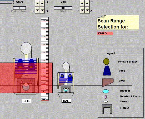
80 Available CT dose calculators Monte Carlo source data phantoms normalisation measurable quantities NRPB R-248 to R ImPACT dose calculator adult male MIRD, fixed weight CTDI ICRUmuscle free-in-air CTDI air free-inair CTDI air free-in-air GSF CT conversion factors + CT-Expo adult male and female MIRD, Child and Baby, fixed weight CTDI air free-inair CTDI w CTDI w 80


81 ImPACT dose calculator courtesy: ImPACT 81

82 CT-Expo courtesy: Stamm 82
83 Effective dose estimates for wide beam CT ImPACT dose calculator can be used for full rotation scans partial rotation scans with random start adopt IEC edition 3 amendment 1 experimental methods reference collimation to match scanner to available MC data set new integration limits to measure CTDI-in-air 83
84 Effective dose estimates for wide beam CT PCXMC20Rotation could be used for full rotation scans partial rotation scans with random or fixed start x-ray beam modelled by adding beams of varying sizes x-ray beam quality parameters required air kerma at isocentre, rather than CTDI, to calculate organ doses 84

85 PCXMC20Rotation courtesy: Stamm 85
86 Primary references Status of Computed Tomography Dosimetry for Wide Cone Beam Scanners, IAEA Human Health report 5, 2011 Comprehensive Methodology for the Evaluation of Radiation Dose in X-Ray Computed Tomography, AAPM report 111, 2010 Size-specific Dose Estimates (SSDE) in Pediatric and Adult Body CT Examinations, AAPM report 204,
87 Secondary references 87
2012 :15th SESSION of ESMP
 2012 :15th SESSION of ESMP Lecture presented in Archamps (Salève Building) by : Elly CASTELLANO (London) Patient dosimetry in x-ray imaging and CT Elly Castellano Objectives measurable dose quantities
Image Quality and Dose. Image Quality and Dose. Image Quality and Dose Issues in MSCT. Scanner parameters affecting IQ and Dose
 Image Quality and Dose Issues in MSCT Image Quality and Dose Image quality Image noise Spatial resolution Contrast Artefacts Speckle and sharpness S. Edyvean St. George s Hospital London SW17 0QT Radiation
Technical aspects on DAP calibration and CT calibration
 EURADOS Report 2015-03 European Radiation Dosimetry Group e. V. Braunschweig, August 2015 Technical aspects on DAP calibration and CT calibration H. Järvinen, O. Turak, M. Ginjaume, J. Daures, C. Hourdakis,
TOPICS: CT Protocol Optimization over the Range of Patient Age & Size and for Different CT Scanner Types: Recommendations & Misconceptions
 CT Protocol Optimization over the Range of Patient Age & Size and for Different CT Scanner Types: Recommendations & Misconceptions TOPICS: Computed Tomography Quick Overview CT Dosimetry Effects of CT
HISTORY. CT Physics with an Emphasis on Application in Thoracic and Cardiac Imaging SUNDAY. Shawn D. Teague, MD
 CT Physics with an Emphasis on Application in Thoracic and Cardiac Imaging Shawn D. Teague, MD DISCLOSURES 3DR- advisory committee CT PHYSICS WITH AN EMPHASIS ON APPLICATION IN THORACIC AND CARDIAC IMAGING
computed tomography, computed tomography dose index, dosimetry
 Received: 18 January 2018 Revised: 6 April 2018 Accepted: 1 May 2018 DOI: 10.1002/acm2.12363 MEDICAL IMAGING Applying three different methods of measuring CTDI free air to the extended CTDI formalism for
Diagnostic X-Ray Shielding
 Diagnostic X-Ray Shielding Multi-Slice CT Scanners Using NCRP 147 Methodology Melissa C. Martin, M.S., FAAPM, FACR Therapy Physics Inc., Bellflower, CA AAPM Annual Meeting, Orlando, FL FL Refresher Course
Clinical Experiences with a Patient Skin Dose Monitoring and Tracking Program
 Clinical Experiences with a Patient Skin Dose Monitoring and Tracking Program Allen R. Goode, MS, DABR Chief Diagnostic Medical Physicist Department of Radiology & Medical Imaging University of Virginia
Automated dose control in multi-slice CT. Nicholas Keat Formerly ImPACT, St George's Hospital, London
 Automated dose control in multi-slice CT Nicholas Keat Formerly ImPACT, St George's Hospital, London Introduction to presentation CT contributes ~50+ % of all medical radiation dose Ideally all patients
Test Equipment for Radiology and CT Quality Control Contents
 Test Equipment for Radiology and CT Quality Control Contents Quality Control Testing...2 Photometers for Digital Clinical Display QC...3 Primary Workstations...3 Secondary Workstations...3 Testing of workstations...3
CHAPTER 2 COMMISSIONING OF KILO-VOLTAGE CONE BEAM COMPUTED TOMOGRAPHY FOR IMAGE-GUIDED RADIOTHERAPY
 14 CHAPTER 2 COMMISSIONING OF KILO-VOLTAGE CONE BEAM COMPUTED TOMOGRAPHY FOR IMAGE-GUIDED RADIOTHERAPY 2.1 INTRODUCTION kv-cbct integrated with linear accelerators as a tool for IGRT, was developed to
diagnostic examination
 RADIOLOGICAL PHYSICS 2011 Raphex diagnostic examination Adel A. Mustafa, Ph.D., Editor PUBLISHED FOR: RAMPS (Radiological and Medical Physics Society of New York) preface The RAPHEX Diagnostic exam 2011
Dose Reduction and Image Preservation After the Introduction of a 0.1 mm Cu Filter into the LODOX Statscan unit above 110 kvp
 Dose Reduction and Image Preservation After the Introduction of a into the LODOX Statscan unit above 110 kvp Abstract: CJ Trauernicht 1, C Rall 1, T Perks 2, G Maree 1, E Hering 1, S Steiner 3 1) Division
Development of new dosimeter for measuring dose distribution in CT
 Development of new dosimeter for measuring dose distribution in CT Poster No.: C-2925 Congress: ECR 2010 Type: Scientific Exhibit Topic: Physics in Radiology - Without Subtopic Authors: Y. Muramatsu, K.
1. Patient size AEC. Large Patient High ma. Small Patient Low ma
 Comparison of the function and performance of CT AEC systems CTUG meeting by Emily Field Trainee clinical scientist 14 th th Breakdown CT Automatic Exposure Control (AEC) Background Project Description
FOREWORD. Acknowledgements
 ΠΑΝΕΠΙΣΗΜΙΟ ΠΑΣΡΩΝ Διαημημαηικό Πρόγραμμα Μεηαπηστιακών ποσδών ζηην Ιαηρική Φσζική Διπλωμαηική εργαζία «ΔΟΙΜΕΣΡΙΑ ΑΘΕΝΩΝ Ε ΕΞΕΣΑΕΙ ΤΠΟΛΟΓΙΣΙΚΗ ΣΟΜΟΓΡΑΦΙΑ ΠΟΛΛΑΠΛΩΝ ΣΟΜΩΝ» ηέλλα Γ. Θαλαζζινού Α.Μ : 1575
CT Scanner Dose Survey
 CT Scanner Dose Survey Measurement Protocol Version 5.0 July 1997 Co-ordinated by ImPACT and The Medical Physics Department,, London SW17, UK. 0181-725-3366 CT Scanner Dose Survey: Measurement Protocol
QC Testing for Computed Tomography (CT) Scanner
 Scanner") QC Testing for Computed Tomography (CT) Scanner QA - Quality Assurance All planned and systematic actions needed to provide confidence on a structure, system or component. all-encompassing program, including
QC Testing for Computed Tomography (CT) Scanner QA - Quality Assurance All planned and systematic actions needed to provide confidence on a structure, system or component. all-encompassing program, including
Calibration of KAP meters
 Calibration of KAP meters Alexandr Malusek! Division of Radiological Sciences Department of Medical and Health Sciences Linköping University! 2014-04-15 1 Outline 1. KAP meter construction 2. Air kerma-area
COMPUTED TOMOGRAPHY 1
 COMPUTED TOMOGRAPHY 1 Why CT? Conventional X ray picture of a chest 2 Introduction Why CT? In a normal X-ray picture, most soft tissue doesn't show up clearly. To focus in on organs, or to examine the
Exposure in Dental Radiology: A Comparison Between Intra-oral, Panoramic and Tomographic Examinations
 Exposure in Dental Radiology: A Comparison Between Intra-oral, Panoramic and Tomographic Examinations S. Baechler 1, P. Monnin 1, A. Aroua 1, J.F. Valley 1, M. Perrier, P. Trueb 3, F.R. Verdun 1 1 University
Y11-DR Digital Radiography (DR) Image Quality
 Image Quality") Y11-DR Digital Radiography (DR) Image Quality Image quality is stressed for all systems in Safety Code 35. In the relevant sections Health Canada s advice is the manufacturer s recommended test procedures
Y11-DR Digital Radiography (DR) Image Quality Image quality is stressed for all systems in Safety Code 35. In the relevant sections Health Canada s advice is the manufacturer s recommended test procedures
Nuclear Associates , , CT Head and Body Dose Phantom
 Nuclear Associates 76-414,76-414-4150,76-415 CT Head and Body Dose Phantom Users Manual March 2005 Manual No. 76-414-1 Rev. 2 2004, 2005 Fluke Corporation, All rights reserved. Printed in U.S.A. All product
Detector technology in simultaneous spectral imaging
 Computed tomography Detector technology in simultaneous spectral imaging Philips IQon Spectral CT Z. Romman, I. Uman, Y. Yagil, D. Finzi, N. Wainer, D. Milstein; Philips Healthcare While CT has become
Dose Reduction in Helical CT: Dynamically Adjustable z-axis X-Ray Beam Collimation
 Medical Physics and Informatics Original Research Christner et al. CT Dose Reduction Medical Physics and Informatics Original Research Downloaded from www.ajronline.org by 8.243.133.8 on 2/26/18 from IP
Introduction of a Single Chip TLD System for Patient Dosimetry
 Introduction of a Single Chip TLD System for Patient Dosimetry C. Hranitzky a, M. Halda a, G. Müller a, B. Obryk b, H. Stadtmann a* a Austrian Research Centers GmbH ARC, 2444 Seibersdorf, Austria. b Institute
Exposure Indices and Target Values in Radiography: What Are They and How Can You Use Them?
 Exposure Indices and Target Values in Radiography: What Are They and How Can You Use Them? Definition and Validation of Exposure Indices Ingrid Reiser, PhD DABR Department of Radiology University of Chicago
of sufficient quality and quantity
 of sufficient quality and quantity The patient s body attenuates the beam as it passes though the body More energy is deposited in organs located near the entry of the beam than near the exit of the beam
QC by the MPE in Belgium
 Acceptance testing of state-of-the-art CT scanners using a new national protocol: first experience on a large number of scanners of different make and model the working group Radiology of the Belgian Hospital
Overview of Safety Code 35
 Common Quality Control Procedures for All s Quality Control Procedures Film All s Daily Quality Control Tests Equipment Warm-up (D1) According to manufacturers instructions Can include auto calibration(d1)
I. PERFORMANCE OF X-RAY PRODUCTION COMPONENTS FLUOROSCOPIC ACCEPTANCE TESTING: TEST PROCEDURES & PERFORMANCE CRITERIA
 FLUOROSCOPIC ACCEPTANCE TESTING: TEST PROCEDURES & PERFORMANCE CRITERIA EDWARD L. NICKOLOFF DEPARTMENT OF RADIOLOGY COLUMBIA UNIVERSITY NEW YORK, NY ACCEPTANCE TESTING GOALS PRIOR TO 1st CLINICAL USAGE
Calculati tions - BIR meth thod and NCRP 147
 Calculations l - BIR method and NCRP 147 David Sutton PhD 1. Cath Lab Indicative of all C-arm applications Only secondary radiation Primary Beam is stopped by the image intensifier Geometry BIR 9.5 m Preparation
abc MHRA Philips Mx8000 IDT CT scanner technical evaluation September 2004 Best choice best practice nww.medical-devices.nhs.
 abc September 2004 MHRA 04099 Philips Mx8000 IDT CT scanner technical evaluation Best choice best practice www.mhra.gov.uk nww.medical-devices.nhs.uk About MHRA evaluation reports. What you can expect.
PHYSICS QUESTIONNAIRE FORM
 PHYSICS QUESTIONNAIRE FORM Institution Name: Date: Contact Information (name, address, phone, fax, email): Physicist: Radiation Oncologist: Dosimetrist (if applicable): Study Coordinator (if applicable):
12/21/2016. Siemens Medical Systems Research Agreement Philips Healthcare Research Agreement AAN and ASN Committees
 Joseph V. Fritz, PhD Nandor Pintor, MD Dent Neurologic Institute ASN 2017 Friday, January 20, 2017 Siemens Medical Systems Research Agreement Philips Healthcare Research Agreement AAN and ASN Committees
Practical Medical Physics Session: TG-151 Dose Monitoring. August 5, 2013 Katie Hulme, M.S.
 Practical Medical Physics Session: TG-151 Dose Monitoring August 5, 2013 Katie Hulme, M.S. Digital Imaging and Dose Creep Images courtesy of Agfa Healthcare Under-Exposed Over-Exposed Freedman et al.,
Pitfalls and Remedies of MDCT Scanners as Quantitative Instruments
 intensity m(e) m (/cm) 000 00 0 0. 0 50 0 50 Pitfalls and Remedies of MDCT Scanners as Jiang Hsieh, PhD GE Healthcare Technology University of Wisconsin-Madison Root-Causes of CT Number Inaccuracies Nature
DIAGNOSTIC ACCREDITATION PROGRAM. Radiology and CT Quality Control Procedures Workbook
 DIAGNOSTIC ACCREDITATION PROGRAM Radiology and CT Quality Control Procedures Workbook Quality Control Procedures Radiography/CR/DR Safety Code 35 Summary For more detail about each quality control (QC)
X-RAYS - NO UNAUTHORISED ENTRY
 Licencing of premises Premises Refer Guidelines A radiation warning sign and warning notice, X-RAYS - NO UNAUTHORISED ENTRY must be displayed at all entrances leading to the rooms where x-ray units are
Joint ICTP/IAEA Advanced School on Dosimetry in Diagnostic Radiology and its Clinical Implementation May 2009
 2033-6 Joint ICTP/IAEA Advanced School on Dosimetry in Diagnostic Radiology and its Clinical Implementation 11-15 May 2009 Dosimetry for Fluoroscopy Basics Renato Padovani EFOMP Joint ICTP-IAEA Advanced
Update on Fluoroscopy Physics AAPM MO-A-210A-1 Stephen Balter, Ph.D.
 Update on Fluoroscopy Physics Stephen Balter, PhD Columbia University Draft for MO-A-210A-1 2009 AAPM Educational objectives Understand dosimetric concepts relating to interventional fluoroscopy Characterize
Translating Protocols Between Scanner Manufacturer and Model
 Translating Protocols Between Scanner Manufacturer and Model Robert J. Pizzutiello, MS, FAAPM, FACMP Sr. Vice-President, Global Physics Solutions President, Upstate Medical Physics Objectives Understand
3D Diode Array Commissioning: Building Confidence in 3D QA Technology
 3D Diode Array Commissioning: Building Confidence in 3D QA Technology Caroline Yount, MS CANCER CENTER 3D QA The complex three-dimensional (3D) shapes of intensity modulated radiation therapy (IMRT) dose
When small things matter. Small Field Dosimetry Application Guide
 R A D I AT I O N T H E R A P Y When small things matter. Small Field Dosimetry Application Guide Contents 1 Introduction 1 Introduction 2 2 The Physics of Small Fields 3 3 Detector Types 10 4 Detector
Digital radiography (DR) post processing techniques for pediatric radiology
 post processing techniques for pediatric radiology") Digital radiography (DR) post processing techniques for pediatric radiology St Jude Children s Research Hospital Samuel Brady, MS PhD DABR samuel.brady@stjude.org Purpose Review common issues and solutions
Digital radiography (DR) post processing techniques for pediatric radiology St Jude Children s Research Hospital Samuel Brady, MS PhD DABR samuel.brady@stjude.org Purpose Review common issues and solutions
Initial Survey Results on the Accuracy of Dose Data in the DICOM Objects
 Initial Survey Results on the Accuracy of Dose Data in the DICOM Objects Renato Padovani, Annalisa Trianni University Hospital S. Maria della Misericordia Udine, Italy INTRODUCTION The EC MED has forced
T h e P h a n t o m L a b o r a t o r y
 T h e P h a n t o m L a b o r a t o r y 1 CCT228 ATCM Phantom Manual Copyright 2017 WARRANTY THE PHANTOM LABORATORY INCORPORATED ( Seller ) warrants that this product shall remain in good working order
Reducing Radiation Exposure from Survey CT Scans
 Reducing Survey CT Scan Exposure Pediatric Imaging Original Research Jennifer C. O Daniel 1 Donna M. Stevens 2 Dianna D. Cody 2 O Daniel JC, Stevens DM, Cody DD Received July 28, 2004; accepted after revision
PATIENT EFFECTIVE DOSES IN DIAGNOSTIC RADIOLOGY, NA
 Title of Paper: Patient effective doses in diagnostic radiology Authors: N.A. Gkanatsios, and W. Huda * Corresponding Author: Department of Radiology, University of Florida, P.O. Box 100374, Gainesville,
Wide-Detector CT for TAVR Planning:
 Wide-Detector CT for TAVR Planning: Impact on Iodine Dose, Radiation Dose, and Image Quality SCBTMR 2015 Annual Course Thursday, October 8 William P. Shuman MD FSCBTMR Department of Radiology University
Slide 1. Slide 2. Slide 3 ACR CT Accreditation. Multi-Slice CT Artifacts and Quality Control. What are the rules or recommendations for CT QC?
 Slide 1 Multi-Slice CT Artifacts and Quality Control Dianna Cody, Ph.D. Chief, Radiologic Physics UT MD Anderson Cancer Center Houston, TX Slide 2 What are the rules or recommendations for CT QC? AAPM
Computed Tomography. The Fundamentals of... THE FUNDAMENTALS OF... Jason H. Launders, MSc. Current Technology
 The Fundamentals of... Computed Tomography Computed Tomography (CT) systems use x-rays to produce images of slices through a patient s anatomy. Despite having lower spatial resolution than other x-ray
The influence of bowtie filtration on cone-beam CT image quality
 The influence of bowtie filtration on cone-beam CT image quality N. Mail Radiation Medicine Program, Princess Margaret Hospital, Toronto, Ontario M5G 2M9, Canada and Ontario Cancer Institute, University
Research Support. Dual-Source CT: What is it and How Do I Test it? Cynthia H. McCollough, Ph.D.
 Dual-Source CT: What is it and How Do I Test it? Cynthia H. McCollough, Ph.D. CT Clinical Innovation Center Department of Radiology Mayo Clinic College of Medicine Rochester, MN Research Support National
A comparison of two methods for the determination of freein-air geometric efficiency in MDCT
 A comparison of two methods for the determination of freein-air geometric efficiency in MDCT Theocharis Berris *1, Kostas Perisinakis 1,, Antonios E. Papadakis and John Damilakis 1, 1 Department of Medical
SPECIFICATION. Kilovoltage X-ray calibration system for protection and diagnostic level dosimetry. Prepared by
 SPECIFICATION Kilovoltage X-ray Prepared by Igor Gomola, Technical Officer, Project ECU6023, Date 2015-Oct-06 Revision Date Status Comments 0.1 2015-Oct-06 Draft Igor Gomola Page 1 of 12 1. Scope This
7/24/2014. Image Quality for the Radiation Oncology Physicist: Review of the Fundamentals and Implementation. Disclosures. Outline
 Image Quality for the Radiation Oncology Physicist: Review of the Fundamentals and Implementation Image Quality Review I: Basics and Image Quality TH-A-16A-1 Thursday 7:30AM - 9:30AM Room: 16A J. Anthony
Radiation Dose Index monitoring (RDIM) systems and establishment of local DRLs
 systems and establishment of local DRLs") IAEA RER/9/135 COURSE ON OPTIMIZATION IN COMPUTED TOMOGRAPHY Sofia, Bulgaria, 21017 Radiation Dose Index monitoring (RDIM) systems and establishment of local DRLs Dean Pekarovič UMC Ljubljana, Institute
IAEA RER/9/135 COURSE ON OPTIMIZATION IN COMPUTED TOMOGRAPHY Sofia, Bulgaria, 21017 Radiation Dose Index monitoring (RDIM) systems and establishment of local DRLs Dean Pekarovič UMC Ljubljana, Institute
Breast Tomosynthesis. Bob Liu, Ph.D. Department of Radiology Massachusetts General Hospital And Harvard Medical School
 Breast Tomosynthesis Bob Liu, Ph.D. Department of Radiology Massachusetts General Hospital And Harvard Medical School Outline Physics aspects of breast tomosynthesis Quality control of breast tomosynthesis
I. Introduction.
 JOURNAL OF APPLIED CLINICAL MEDICAL PHYSICS, VOLUME 15, NUMBER 1, 2014 Accuracy of measuring half- and quarter-value layers and appropriate aperture width of a convenient method using a lead-covered case
CHAPTER 6 QUALITY ASSURANCE OF VARIAN ON-BOARD IMAGER
 127 CHAPTER 6 QUALITY ASSURANCE OF VARIAN ON-BOARD IMAGER 6.1 INTRODUCTION Accurate and repeatable setup of patients is a requisite in radiotherapy. In the treatment of head-and-neck tumors, accurate setup
Installation und Kommissionierung des Viewray MRIdian Linac Hamburg, 28. Mai 2018 Sebastian Klüter
 Installation und Kommissionierung des Viewray MRIdian Linac Hamburg, 28. Mai 2018 Sebastian Klüter MR-guided RT in Heidelberg Funded by the German Research Foundation (DFG) Heidelberg consortium received
A Generalized Strategy for 3D Dose Verification of IMRT/VMAT Using EPID-measured Transit Images
 A Generalized Strategy for 3D Dose Verification of IMRT/VMAT Using EPID-measured Transit Images Aiping Ding, Bin Han, Lei Wang, Lei Xing Department of Radiation Oncology, Stanford University School of
An Introduction to TG-142 Imaging QA Using Standard Imaging Products. Mark Wiesmeyer, PhD, DABR Technical Product Manager Standard Imaging, Inc.
 An Introduction to TG-142 Imaging QA Using Standard Imaging Products Mark Wiesmeyer, PhD, DABR Technical Product Manager Standard Imaging, Inc. Goals Understand the nature and intent of TG 142 imaging
Key words: fluoroscopy, dose-area-product, kerma-area-product, calibration of KAP meters, patient exposure
 Accuracy and calibration of integrated radiation output indicators in diagnostic radiology: A report of the AAPM Imaging Physics Committee Task Group 190 Pei-Jan P. Lin a) Virginia Commonwealth University
Multi-Access Biplane Lab
 Multi-Access Biplane Lab Advanced technolo gies deliver optimized biplane imaging Designed in concert with leading physicians, the Infinix VF-i/BP provides advanced, versatile patient access to meet the
MC SIMULATION OF SCATTER INTENSITIES IN A CONE-BEAM CT SYSTEM EMPLOYING A 450 kv X-RAY TUBE
 MC SIMULATION OF SCATTER INTENSITIES IN A CONE-BEAM CT SYSTEM EMPLOYING A 450 kv X-RAY TUBE A. Miceli ab, R. Thierry a, A. Flisch a, U. Sennhauser a, F. Casali b a Empa - Swiss Federal Laboratories for
Quantitation of clinical feedback on image quality differences between two CT scanner models
 Received: 4 August 2016 Revised: 4 November 2016 Accepted: 12 December 2016 DOI: 10.1002/acm2.12050 MEDICAL IMAGING Quantitation of clinical feedback on image quality differences between two CT scanner
Authors: Cabral, Ricardo 1 ; Carvoeiras, Pedro 2 ; Fatana, João 2, ; Alves, Rita 1. 1 Centro Hospitalar Lisboa Norte - Hospital de Santa Maria; 2
 Authors: Cabral, Ricardo 1 ; Carvoeiras, Pedro 2 ; Fatana, João 2, ; Alves, Rita 1. 1 Centro Hospitalar Lisboa Norte - Hospital de Santa Maria; 2 Medical Consult, SA; Establish a method to correlate image
Quantitative performance characterization of image quality and radiation dose for dental CBCT machine (CS9300) Elham Abouei
 Elham Abouei") Quantitative performance characterization of image quality and radiation dose for dental CBCT machine (CS9300) by Elham Abouei B.Sc., Sharif University of Technology, 2011 A THESIS SUBMITTED IN PARTIAL
Quantitative performance characterization of image quality and radiation dose for dental CBCT machine (CS9300) by Elham Abouei B.Sc., Sharif University of Technology, 2011 A THESIS SUBMITTED IN PARTIAL
STEREOTACTIC BREAST BIOPSY EQUIPMENT SURVEYS
 STEREOTACTIC BREAST BIOPSY EQUIPMENT SURVEYS JAMES A. TOMLINSON, M.S. Diagnostic Radiological Physicist American Board of Radiology Certified Medical Physics Consultants, Inc. Bio 28 yrs experience 100%
Phantoms in Medical Physics (RT) U. Oelfke. Division of Radiotherapy & Imaging
 U. Oelfke. Division of Radiotherapy & Imaging") in partnership with Phantoms in Medical Physics (RT) U. Oelfke Division of Radiotherapy & Imaging uwe.oelfke@icr.ac.uk Making the discoveries that defeat cancer 1. Introduction What is a phantom? Wiki:
in partnership with Phantoms in Medical Physics (RT) U. Oelfke Division of Radiotherapy & Imaging uwe.oelfke@icr.ac.uk Making the discoveries that defeat cancer 1. Introduction What is a phantom? Wiki:
Comparison of peripheral dose measurements using Ionization chamber and MOSFET detector
 ORIGINAL ARTICLES Comparison of peripheral dose measurements using Ionization chamber and MOSFET detector Gopiraj ANNAMALAI 1, Ramasubramanian VELAYUDHAM 2 ABSTRACT Received: 7.07.2009 Accepted: 2.11.2009
CT Basics: Image Quality Module 6
 Module 6 For educational and institutional use. This transcript is licensed for noncommercial, educational inhouse or online educational course use only in educational and corporate institutions. Any broadcast,
RaySafe X2. Effortless measurements of X-ray
 RaySafe X2 Effortless measurements of X-ray At your fingertips We ve grown accustomed to intuitive interactions with our devices. After all, it s not the device that s most important, but what you can
DETECTORS UNCOMPROMISING QUALITY. The standard in dosimetry measurements for over 40 years. EXRADIN DETECTORS
 DETECTORS UNCOMPROMISING QUALITY The standard in dosimetry measurements for over 40 years. EXRADIN DETECTORS The Exradin Advantage Better Components Waterproof construction eliminates the need for sleeves
SCINTILLATING FIBER DOSIMETER ARRAY
 SCINTILLATING FIBER DOSIMETER ARRAY FIELD OF THE INVENTION [0001] This invention relates generally to the field of dosimetry and, more particularly, to rapid, high-resolution dosimeters for advanced treatment
Software and Hardware in CCTA. Elly Castellano PhD
 Software and Hardware in CCTA Elly Castellano PhD Outline technical requirements for coronary CTA the modern cardiac CT scanner ECG-gating technology image reconstruction algorithms 2 Technical requirements
Chiara Secco. PET Performance measurements of the new LSO-Based Whole Body PET/CT. Scanner biograph 16 HI-REZ using the NEMA NU Standard.
 Chiara Secco PET Performance measurements of the new LSO-Based Whole Body PET/CT Scanner biograph 16 HI-REZ using the NEMA NU 2-2001 Standard. INTRODUCTION Since its introduction, CT has become a fundamental
IQM Detector Characteristics: Signal reproducibility
 The Integral Quality Monitor (IQM) System is a real-time beam verification system that monitors the accuracy of radiation delivery throughout each patient treatment without any user interaction. IQM continuously
MEASURING CONDITIONS AND UNCERTAINTIES FOR THE COMPARISON AND CALIBRATION OF NATIONAL DOSIMETRIC STANDARDS AT THE BIPM *
 MEASURING CONDITIONS AND UNCERTAINTIES FOR THE COMPARISON AND CALIBRATION OF NATIONAL DOSIMETRIC STANDARDS AT THE BIPM * C. Kessler and D.T. Burns December 2018 BUREAU INTERNATIONAL DES POIDS ET MESURES
Effect of patient support pads on image quality and dose in fluoroscopy a)
") Effect of patient support pads on image quality and dose in fluoroscopy a) William R. Geiser, Walter Huda, b) and Nikolaos A. Gkanatsios Department of Radiology, University of Florida, Gainesville, Florida
Effect of patient support pads on image quality and dose in fluoroscopy a) William R. Geiser, Walter Huda, b) and Nikolaos A. Gkanatsios Department of Radiology, University of Florida, Gainesville, Florida
NOT FOR DISTRIBUTION JINST_083P_0914 v1
 Use of XR-QA2 radiochromic films for quantitative imaging of a synchrotron radiation beam F. Di Lillo a,b, D. Dreossi c, F. Emiro a,b, C. Fedon d,e, R. Longo d,e, G. Mettivier a,b,*, L. Rigon d,e, P. Russo
Clinical Experience Using the Open Bore Multislice CT System Supria (16 slice CT) MEDIX VOL. 61 P.8 P.11
 MEDIX VOL. 61 P.8 P.11") Clinical Experience Using the Open Bore Multislice CT System Supria (16 slice CT) Hiroki Kadoya Yukiko Kitagawa MEDIX VOL. 61 P.8 P.11 Clinical Experience Using the Open Bore Multislice CT System Supria
Clinical Experience Using the Open Bore Multislice CT System Supria (16 slice CT) Hiroki Kadoya Yukiko Kitagawa MEDIX VOL. 61 P.8 P.11 Clinical Experience Using the Open Bore Multislice CT System Supria
The disclaimer on page 1 is an integral part of this document. Copyright November 30, 2017 by AAPM. All rights reserved.
 DISCLAIMER: TO THE EXTENT ALLOWED BY LOCAL LAW, THIS INFORMATION IS PROVIDED TO YOU BY THE AMERICAN ASSOCIATION OF PHYSICISTS IN MEDICINE, A NON-PROFIT ORGANIZATION ORGANIZED TO PROMOTE THE APPLICATION
Acceptance Testing of a Digital Breast Tomosynthesis Unit
 Acceptance Testing of a Digital Breast Tomosynthesis Unit 2012 AAPM Spring Clinical Meeting Jessica Clements, M.S., DABR Objectives Review of technology and clinical advantages Acceptance Testing Procedures
CT AutoQA Lite Software Model
 CT AutoQA Lite Software Model 49-802 Fast automated CT analysis for routine QC or acceptance testing Generates easy to read results with hardcopy output Comprehensive trend analysis Can be configured with
An Activity in Computed Tomography
 Pre-lab Discussion An Activity in Computed Tomography X-rays X-rays are high energy electromagnetic radiation with wavelengths smaller than those in the visible spectrum (0.01-10nm and 4000-800nm respectively).
Radiation Dose Modulation. the Multidetector CT Era: From Basics to Practice 1
 Note: This copy is for your personal non-commercial use only. To order presentation-ready copies for distribution to your colleagues or clients, contact us at www.rsna.org/rsnarights. EDUCATION EXHIBIT
1. Queries are issued to the image archive for information about computed tomographic (CT)
") Appendix E1 Exposure Extraction Method examinations. 1. Queries are issued to the image archive for information about computed tomographic (CT) 2. Potential dose report screen captures (hereafter, dose
Appendix E1 Exposure Extraction Method examinations. 1. Queries are issued to the image archive for information about computed tomographic (CT) 2. Potential dose report screen captures (hereafter, dose
Cylindrical Ion Chambers
 Cylindrical Ion Chambers Radiation Oncology ON Victoreen Model 550T Series Cylindrical Ion Chambers for use with Model 35040 and Model 560 electrometers Wide range of applications in Diagnostic X-Ray and
160-slice CT SCANNER / New Standard for the Future
 TECHNOLOGY HISTORY For over 130 years, Toshiba has been a world leader in developing technology to improve the quality of life. Our 50,000 global patents demonstrate a long, rich history of leading innovation.
SRS MapCHECK. SRS Patient QA, No Film. Your Most Valuable QA and Dosimetry Tools
 SRS MapCHECK SRS Patient QA, No Film Your Most Valuable QA and Dosimetry Tools SRS Patient QA, No Film With improvements in targeting and localization, stereotactic treatments have become prevalent. To
Interventional Radiological Equipment selection and installation
 Interventional Radiological Equipment selection and installation Renato Padovani ICTP Learning objectives To understand the main components of an interventional radiology equipment To understand the relevance
Received 3 December 2016; revised 17 May 2017; editorial decision 21 June 2017; accepted 4 July 2017
 Radiation Protection Dosimetry (2018), Vol. 178, No. 2, pp. 235 241 Advance Access publication 15 July 2017 doi:10.1093/rpd/ncx091 NOTE COMPARISON OF PERSONAL DOSE EQUIVALENT Hp(10) IN 137 CS RADIATION
Nuclear Associates
 Nuclear Associates 07-706 Patient Phantom/Penetrometer System Users Manual March 2005 Manual No. 07-706-1 Rev. 2 2004, 2005 Fluke Corporation, All rights reserved. Printed in U.S.A. All product names are
熊本大学学術リポジトリ. Kumamoto University Repositor
 熊本大学学術リポジトリ Kumamoto University Repositor Title Monte Carlo calculations of the rep correction factor, Ρ_, for cy chamber cav Author(s) Araki, Fujio CitationRadiological Physics and Technology Issue
QA Considerations. QA for LGK Perfexion : : Follow NRC licensing guidelines (10( CFR ) Leksell Gamma Knife Perfexion
 Leksell Gamma Knife Perfexion") Leksell Gamma Knife Perfexion QA Considerations Paula L. Petti, Ph.D. Taylor McAdam Bell Neuroscience Institute Washington Hospital Healthcare System Fremont, CA 1 QA for LGK Perfexion : : Follow NRC licensing
Leksell Gamma Knife Perfexion QA Considerations Paula L. Petti, Ph.D. Taylor McAdam Bell Neuroscience Institute Washington Hospital Healthcare System Fremont, CA 1 QA for LGK Perfexion : : Follow NRC licensing
Characterization of an in vivo diode dosimetry system for clinical use
 JOURNAL OF APPLIED CLINICAL MEDICAL PHYSICS, VOLUME 4, NUMBER 2, SPRING 2003 Characterization of an in vivo diode dosimetry system for clinical use Kai Huang, 1, * William S. Bice, Jr., 2, and Oscar Hidalgo-Salvatierra
Predicted image quality of a CMOS APS X-ray detector across a range of mammographic beam qualities
 Journal of Physics: Conference Series PAPER OPEN ACCESS Predicted image quality of a CMOS APS X-ray detector across a range of mammographic beam qualities Recent citations - Resolution Properties of a