Joint ICTP/IAEA Advanced School on Dosimetry in Diagnostic Radiology and its Clinical Implementation May 2009
|
|
|
- Beryl Davis
- 5 years ago
- Views:
Transcription
1 Joint ICTP/IAEA Advanced School on Dosimetry in Diagnostic Radiology and its Clinical Implementation May 2009 Dosimetry for Fluoroscopy Basics Renato Padovani EFOMP


2 Joint ICTP-IAEA Advanced school on Dosimetry in Diagnostic Radiology: And its Clinical Implementation May 2009; Miramare, Trieste, Italy Dosimetry for fluoroscopy - basics Renato Padovani Medical Physics Department University Hospital, Udine, Italy IAEA International Atomic Energy Agency
3 Introduction Fluoroscopy equipment Air-kerma rates and KAP measurement Phantom and patient dosimetry IAEA 2

4 Fluoroscopy: a see-through operation with motion Used to visualize motion of internal fluid, structures Operator controls activation of tube and position over patient Early fluoroscopy gave dim image on fluorescent screen Physician seared in dark room Modern systems include image intensifier with television screen display and choice of recording devices IAEA 3
5 Direct Fluoroscopy: obsolete In older fluoroscopic examinations radiologist stands behind screen and view the picture Radiologist receives high exposure; despite protective glass, lead shielding in stand, apron and perhaps goggles Main source staff exposure is NOT the patient but direct beam IAEA 4


6 Older Fluoroscopic Equipment (still in use in some countries) Staff in DIRECT beam Even no protection IAEA 5
7 New Fluoroscopic Equipment IAEA 6
8 Direct fluoroscopy AVOID USE OF DIRECT FLUOROSCOPY Directive 97/43Euratom Art 8.4. In the case of fluoroscopy, examinations without an image intensification or equivalent techniques are not justified and shall therefore be prohibited. Direct fluoroscopy will not comply with BSS App.II.25 performance of diagnostic radiography and fluoroscopy equipment and of nuclear medicine equipment should be assessed on the basis of comparison with the guidance levels IAEA 7

9 Modern fluoroscopic system components Display control Automatic control display brightness radiation dose film exposure Timer IAEA 8
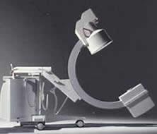
10 Different fluoroscopy systems Remote control systems Not requiring the presence of medical specialists inside the X Ray room Mobile C-arms Mostly used in surgical theatres. IAEA 9
11 Different fluoroscopy systems Interventional radiology systems Requiring specific safety considerations. In interventional radiology the surgeon can be near the patient during the procedure. Multipurpose fluoroscopy systems They can be used as a remote control system or as a system to perform simple interventional procedures IAEA 10
12 The image intensifier (I.I.) I.I. Input Screen Photocathode Electrode E 1 Electrons Path + Electrode E 2 Electrode E 3 I.I.Output Screen Input screen: conversion of incident X Rays into light photons (CsI) 1 X Ray photon creates 3,000 light photons Photocathode: conversion of light photons into electrons only 10 to 20% of light photons are converted into photoelectrons Electrodes : focalization of electrons onto the output screen electrodes provide the electronic magnification Output screen: conversion of accelerated electrons into light photons IAEA 11

13 Image intensifier systems IAEA 12
14 X Ray TUBE kv FILM PM VIDICON REFERENCE CONTROLLER kv GENERAL SCHEME OF FLUOROSCOPY IAEA 13
15 X Ray TUBE kv CINE MODE I 2 CONTROLLER I 3 I 1 FILM PM C 1 C 2 Ref. VIDICON IAEA 14
16 Type of TV camera VIDICON TV camera improvement of contrast improvement of signal to noise ratio high image lag PLUMBICON TV camera (suitable for cardiology) lower image lag (follow up of organ motions) higher quantum noise level CCD TV camera (digital fluoroscopy) digital fluoroscopy spot films are limited in resolution, since they depend on the TV camera (no better than about 2 lp/mm) for a 1000 line TV system IAEA 15
17 Digital radiography principle Image intensifier ANALOGUE SIGNAL I ADC Memory t Iris DIGITAL SIGNAL Clock t IAEA 16

18 Digital radiography principle Dynamic Digital Flat Panel ANALOGUE SIGNAL ADC Memory t DIGITAL SIGNAL Clock t IAEA 17


19 Flat panel technology: indirect conversion IAEA 18
20 Automatic Exposure Control in fluoroscopy kv, ma changes as a function of: Patient body absorption Image quality requested Field of view (FOV) mgy/min Entrance dose PMMA with BS (II 23 cm) R2_08/ cm PMMA IAEA 19 low med high
21 IAEA Code of Practice Dosimetry in fluoroscopy IAEA 20
22 Dosimetry in fluoroscopy Quality assurance Acceptance and constancy test air kerma rate for different acquisition modalities Patient dosimetry Comparison with reference levels Air kerma area product Dose analogues: fluoroscopy time and no. of acquired images Organ dose evaluation IAEA 21
23 CoP Entrance surface air kerma rate is the principal quantity to be measured in fluoroscopy using phantoms. For measurements on patients, the air kerma area product, a readily measured quantity closely related to the energy imparted to the patient and to the effective dose, is the recommended dosimetric quantity. IAEA 22
24 Incident air kerma & Entrance surface air kerma The incident air kerma, K i, is the kerma to air from an incident X ray beam measured on the central beam axis at the position of the patient or phantom surface. Only the radiation incident on the patient or phantom and not the backscattered radiation is included. The entrance surface air kerma, K e, is the kerma to air measured on the central beam axis at the position of the patient or phantom surface. The radiation incident on the patient or phantom and the backscattered radiation (B) are included. IAEA K 23 e = K i B
25 Measurements using phantoms The entrance surface air kerma rate is measured using a water phantom or a PMMA phantom. It is important that the detector responds to both direct as well as backscattered radiation. For detectors that do not respond to backscatter, the entrance surface air kerma rate is calculated from the incident air kerma rate and an appropriate backscatter factor. Semiconductor detector systems often possess this property IAEA 24
26 Equipment Diagnostic dosimeter calibrated for beam qualities used in fluoroscopy Water phantom of 20 cm thickness and crosssection of cm 2 ; additional water phantom (or PMMA) of 10 cm thickness for simulation of larger patients Or 185 cm thick PMMA phantom (correction factor for the different backscatter properties of PMMA) Ruler, Thermometer and barometer (for measurements with an ionization chamber) IAEA 25
27 Method The fluoroscopic unit should be operated under automatic brightness Control (ABC). ABC has to be stabilized before measurements Measurements for all image intensifier field sizes (FOV), dose rates and automatic brightness control options (image quality) reflecting normal clinical use. The focus to intensifier and focus to chamber distances, tube voltage, tube current and any filtration selected should be recorded for each measurement. The measurements are strongly dependent on the relative positions of the X ray tube, patient entrance surface and image intensifier. IAEA 26
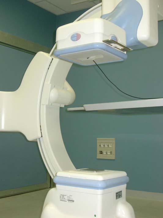
28 4 geometries Under couch Over couch C-arm C-arm-lat IAEA 27
 4.")
29 Under couch measurement of K i 1. Use anti-scatter grid if used in the clinical situation 2. The space between the couch and the phantom must be sufficient for positioning the detector 3. Position the detector in contact with the phantom and at the centre of its entrance surface (in the case of back shielded detector position it outside sensitive area of ABC) 4. Position the image intensifier at 100 mm from the exit surface of the phantom 5. Measure and record the focus to intensifier and focus to detector distances. 6. Expose the phantom under automatic brightness control and record the dosimeter reading, M, tube voltage, tube current and the exposure settings (FOV, image quality, pulse rate/countinous mode). Repeat the measurement three times 7. Repeat step 6 for all image intensifier field sizes, dose rates and automatic brightness control options in normal clinical use. 8. If a dosimeter with an ionization chamber is used, record the temperature and pressure. IAEA 28
30 Other geometries (differences) Over couch Set the focus to couch (table top) distance equal to that used in clinical practice. If a standard distance is to be used, set the focus to couch distance equal to 1000 mm C-arm & C-arm lateral proj Set the distance between the X ray focus and the image intensifier to 1000 mm (if this distance can be varied). IAEA 29

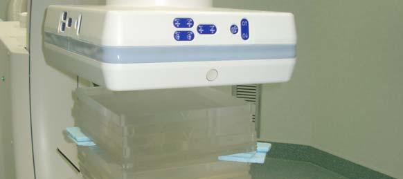
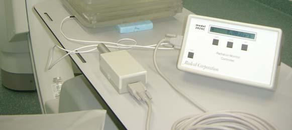
31 Example of under couch measuremetn set-up IAEA 30
32 Calculation Entrance surface air kerma rate Calculate the mean dosimeter reading from the measurements Calculate the entrance surface air kerma rate, K e, from the mean dosimeter reading _.. Ke = M N K, Q0 k TP is the correction factor for temperature and pressure N K,Q0 chamber calibration coefficient k Q factor to corrects for differences in the response of the dosimeter at the calibration quality, Q 0, and at the measurement quality, Q. K Q K TP T and P temperature and pressure (in o C and kpa) recorded during the measurement and T 0 and P 0 are their reference values for which N K,Q 0 is provided. IAEA 31
33 Calculation (cont.) If PMMA is used _.. BPMMA Ke = M N K, Q KQK 0 TP Bwater If a back shielded detector is used _.. Ke = M N K, Q0 K Q K TP B water If needed, the calculated value of Ke is corrected for a difference between the position of the reference point of the detector and the phantom surface using the inverse square law IAEA 32
34 IAEA 33
 70.0 60.0 50.0 40.0 30.0 20.0 10.0 Fluoro Low Fluoro Normal 0.")
35 Fluoroscopy mode: example air kerma rates Entrance surface air kerma for different fluoro modes and patient thickness Fluoroscopy modes. Innova 2000, FOV 17 cm 80.0 Entrance Surface Dose (mgy/min) Fluoro Low Fluoro Normal PMMA thickness (cm) Ionisation chamber to measure phantom entrance surface air kerma IAEA rate (K e ) 34
36 Uncertainties on K e Three scenarios which require, from scenario 1 to scenario 3, increasing attention to parameters of measurement. IAEA 35
37 Measurements on patients In examinations using fluoroscopy, irradiation geometry and time vary individually from patient to patient. Effects on patient exposures of these variations are captured by the air kerma area product (P KA ), KAP is easily measured using a flat transmission ionization chamber (KAPmeter) mounted on the collimator housing. The KAP meter does not disturb the examination and gives real time information. In the Code of Practice, measurement of the air kerma area product (P KA ) is recommended for monitoring patient exposures in examinations involving fluoroscopy IAEA 36

38 Air kerma-area product The air kerma-area product, P KA, is the integral of the air kerma over the area of the X ray beam in a plane perpendicular to the beam axis, thus P KA = A K( x, y)dxdy Unit: Gy m 2 P KA has the useful property that it is approximately invariant with distance from the X ray tube focus. when interactions in air and extra-focal radiation can be neglected And, the planes of measurement do not include a significant contribution from backscattered radiation from the patient or phantom. IAEA 37

39 KAP meters KAP meter with flat trasparent ionisation chamber In some systems: KAP is calculated from kv, filtration, mas, diaphragms positions IAEA 38
40 Measurement on patient 1. Mount the KAP meter on the exit surface of the collimator housing of the X ray tube. This step is omitted in the case of a built-in KAP meter. 2. Record, if possible, the tube voltage and any other machine parameters (e.g. operating mode chosen, tube current and pulse rate if appropriate) used during the examination. 3. Record the reading, M, of the KAP meter. 4. If the operating mode is changed during the procedure, it may be helpful to use recorded KAP meter readings (if available) and machine parameters for each stage. 5. Record the temperature and pressure. IAEA 39
41 IAEA Code of Practice: Patient selection It is important that the size of a sample of patients is sufficiently large as to avoid large statistical variations of the mean value of the measured quantity. Care has to be paid also to the selection of patients according to their anatomical parameters (e.g. weight). A range of patients for the sample size can be found in the literature. Selection of patients so that the mean weight of the sample lies within 5 kg of 70 kg or within 5 kg of 60 kg in some geographical regions has been shown to be sufficient IAEA 40
42 Calculation Air kerma-area product, P KA Calculate the P KA from the KAP meter reading P. KA _. = M N P ktp is the correction factor for temperature and pressure N PKA,Q0 chamber calibration coefficient k Q factor to corrects for differences in the response of the dosimeter at the calibration quality, Q 0, and at the measurement quality, Q. For a total filtration of up to about 3 mm aluminium, this quality can be indicated by the value of the HVL, irrespective of the X ray tube voltage. For beams with stronger filtrations, more comprehensive calibration of the KAP meter may be required., Q KA 0 K Q K TP IAEA 41
43 Uncertainties The uncertainty in the calibration coefficient when the tube voltage and filtration are known and the energy dependence accounted for can be reduced to about 6% at the 95% confidence level. IEC specifies acceptable limits of uncertainty in the response of KAP meters when individual exposure parameters (influence quantities) vary the estimated uncertainty of a measurement with KAP meters is 25% at the 95% confidence interval (k = 2) this corresponds to a single value for the calibration coefficient representing all factors, i.e. all possible doses, dose rates and X ray energies in clinical practice, P KA rate ( ) μgy m2 s 1 X ray spectrum (50 150) kv, total filtration 2.5 mm Al IAEA 42
44 Uncertainties If a calibration coefficient has only been established for an over couch situation, the insertion of a table with a mattress in the beam reduces the air kerma incident on the patient by up to 15 40%, depending on the HVL of the beam, beam angulations and table construction This has to be considered when using the KAP to estimate patient exposure IAEA 43
45 KAP meter calibration The KAP meters should be calibrated for each stand where they are used. Calibrations both in situ and at a standard laboratory are possible. Modern radiology departments usually possess a number of machines with KAPs. It is not realistic to calibrate each instrument at the SSDL and for built-in KAP meters this is not even possible. The calibration coefficient provided by the manufacturer should be checked before the instrument is used. IAEA 44


46 Udine Friuli-Venezia Giulia region Thank you! Mandi! IAEA 45
Calibration of KAP meters
 Calibration of KAP meters Alexandr Malusek! Division of Radiological Sciences Department of Medical and Health Sciences Linköping University! 2014-04-15 1 Outline 1. KAP meter construction 2. Air kerma-area
SPECIFICATION. Kilovoltage X-ray calibration system for protection and diagnostic level dosimetry. Prepared by
 SPECIFICATION Kilovoltage X-ray Prepared by Igor Gomola, Technical Officer, Project ECU6023, Date 2015-Oct-06 Revision Date Status Comments 0.1 2015-Oct-06 Draft Igor Gomola Page 1 of 12 1. Scope This
I. PERFORMANCE OF X-RAY PRODUCTION COMPONENTS FLUOROSCOPIC ACCEPTANCE TESTING: TEST PROCEDURES & PERFORMANCE CRITERIA
 FLUOROSCOPIC ACCEPTANCE TESTING: TEST PROCEDURES & PERFORMANCE CRITERIA EDWARD L. NICKOLOFF DEPARTMENT OF RADIOLOGY COLUMBIA UNIVERSITY NEW YORK, NY ACCEPTANCE TESTING GOALS PRIOR TO 1st CLINICAL USAGE
Key words: fluoroscopy, dose-area-product, kerma-area-product, calibration of KAP meters, patient exposure
 Accuracy and calibration of integrated radiation output indicators in diagnostic radiology: A report of the AAPM Imaging Physics Committee Task Group 190 Pei-Jan P. Lin a) Virginia Commonwealth University
Exposure Indices and Target Values in Radiography: What Are They and How Can You Use Them?
 Exposure Indices and Target Values in Radiography: What Are They and How Can You Use Them? Definition and Validation of Exposure Indices Ingrid Reiser, PhD DABR Department of Radiology University of Chicago
BASICS OF FLUOROSCOPY
 Medical Physics Residents Training Program BASICS OF FLUOROSCOPY Dr. Khalid Alyousef, PhD Department of Medical Imaging King Abdulaziz Medical City- Riyadh Edison examining the hand of Clarence Dally with
X-RAY FLUOROSCOPY IMAGING SYSTEMS. Dr Slavik Tabakov. Luminescence: Dept. Medical Eng. & Physics King s College London
 X-RAY FLUOROSCOPY IMAGING SYSTEMS Dr Slavik Tabakov OBJECTIVES - Image Intensifier construction - Input window - Accelerating and focusing electrodes - Output window - Conversion factor - II characteristics
2012 :15th SESSION of ESMP
 2012 :15th SESSION of ESMP Lecture presented in Archamps (Salève Building) by : Elly CASTELLANO (London) Patient dosimetry in x-ray imaging and CT Elly Castellano Objectives measurable dose quantities
Update on Fluoroscopy Physics AAPM MO-A-210A-1 Stephen Balter, Ph.D.
 Update on Fluoroscopy Physics Stephen Balter, PhD Columbia University Draft for MO-A-210A-1 2009 AAPM Educational objectives Understand dosimetric concepts relating to interventional fluoroscopy Characterize
Interventional Radiological Equipment selection and installation
 Interventional Radiological Equipment selection and installation Renato Padovani ICTP Learning objectives To understand the main components of an interventional radiology equipment To understand the relevance
X-RAY IMAGING EE 472 F2017. Prof. Yasser Mostafa Kadah
 X-RAY IMAGING EE 472 F2017 Prof. Yasser Mostafa Kadah www.k-space.org Recommended Textbook Stewart C. Bushong, Radiologic Science for Technologists: Physics, Biology, and Protection, 10 th ed., Mosby,
Dose Reduction and Image Preservation After the Introduction of a 0.1 mm Cu Filter into the LODOX Statscan unit above 110 kvp
 Dose Reduction and Image Preservation After the Introduction of a into the LODOX Statscan unit above 110 kvp Abstract: CJ Trauernicht 1, C Rall 1, T Perks 2, G Maree 1, E Hering 1, S Steiner 3 1) Division
Introduction. Chapter 16 Diagnostic Radiology. Primary radiological image. Primary radiological image
 Introduction Chapter 16 Diagnostic Radiology Radiation Dosimetry I Text: H.E Johns and J.R. Cunningham, The physics of radiology, 4 th ed. http://www.utoledo.edu/med/depts/radther In diagnostic radiology
X-ray Tube and Generator Basic principles and construction
 X-ray Tube and Generator Basic principles and construction Dr Slavik Tabakov - Production of X-rays and Patient Dose OBJECTIVES - X-ray tube construction - Anode - types, efficiency - Classical X-ray generator
CHAPTER 6 QC Test For Fluoroscopic Equipment. Prepared by:- Kamarul Amin bin Abu Bakar School of Medical Imaging KLMUC
 CHAPTER 6 QC Test For Fluoroscopic Equipment Prepared by:- Kamarul Amin bin Abdullah @ Abu Bakar School of Medical Imaging KLMUC Lesson Outcomes Describe the objectives of each QC test done. Identify QC
X-RAYS - NO UNAUTHORISED ENTRY
 Licencing of premises Premises Refer Guidelines A radiation warning sign and warning notice, X-RAYS - NO UNAUTHORISED ENTRY must be displayed at all entrances leading to the rooms where x-ray units are
Test Equipment for Radiology and CT Quality Control Contents
 Test Equipment for Radiology and CT Quality Control Contents Quality Control Testing...2 Photometers for Digital Clinical Display QC...3 Primary Workstations...3 Secondary Workstations...3 Testing of workstations...3
MEASURING CONDITIONS AND UNCERTAINTIES FOR THE COMPARISON AND CALIBRATION OF NATIONAL DOSIMETRIC STANDARDS AT THE BIPM *
 MEASURING CONDITIONS AND UNCERTAINTIES FOR THE COMPARISON AND CALIBRATION OF NATIONAL DOSIMETRIC STANDARDS AT THE BIPM * C. Kessler and D.T. Burns December 2018 BUREAU INTERNATIONAL DES POIDS ET MESURES
Half value layer and AEC receptor dose compliance survey in Estonia
 Half value layer and AEC receptor dose compliance survey in Estonia K. Kepler, A. Vladimirov Training Centre of Medical Physics, University of Tartu Testing Centre of the University of Tartu, Estonia E-mail:
Fluoroscopy - Chapter 9
 Fluoroscopy - Chapter 9 Kalpana Kanal, Ph.D., DABR Lecturer, Diagnostic Physics Dept. of Radiology UW Medicine a copy of this lecture may be found at: http://courses.washington.edu/radxphys/physicscourse04-05.html
FOUR CATEGORIES OF SAFETY
 OCTOBER 2013 FOUR CATEGORIES OF SAFETY DOSIMETRY PERSONAL SAFETY EQUIPMENT EQUIPMENT KNOWLEDGE PHYSICAL SAFETY DOSIMETRY THERMAL LUMINISCENT DEVICES AND FILM BADGES CNSC PERMISSIBLE DOSES WHOLE BODY DOSE
Estimation of signal transfer property for wireless digital detector in different measurement schemes
 Estimation of signal transfer property for wireless digital detector in different measurement schemes Anatoli Vladimirov, Kalle Kepler Training Centre of Medical Physics, University of Tartu, Estonia 11
Ch. 223 VETERINARY MEDICINE CHAPTER 223. VETERINARY MEDICINE GENERAL PROVISIONS X-RAYS RADIOACTIVE MATERIAL. Authority
 Ch. 223 VETERINARY MEDICINE 25 223.1 CHAPTER 223. VETERINARY MEDICINE Sec. 223.1. Purpose and scope. 223.2. [Reserved]. 223.2a. Definitions. 223.3 223.6. [Reserved]. 223.7. Structural shielding. 223.8.
10/15/2012 SECTION III - CHAPTER 6 DIGITAL FLUOROSCOPY RADT 3463 COMPUTERIZED IMAGING
 RADT 3463 - COMPUTERIZED IMAGING Section III: Chapter 6 RADT 3463 Computerized Imaging 1 SECTION III - CHAPTER 6 DIGITAL FLUOROSCOPY RADT 3463 COMPUTERIZED IMAGING Section III: Chapter 6 RADT 3463 Computerized
Renato Padovani, University Hospital, Udine, Italy
 DATAMED II Training Course Sofia, May 19-20, 2011 Patient dose assessment in fluoroscopy and interventional procedures Renato Padovani, University Hospital, Udine, Italy Content Fluoroscopy guided diagnostic
Preliminary studies of a new monitor ionization chamber
 1 Preliminary studies of a new monitor ionization chamber Maíra T. Yoshizumi, Vitor Vivolo and Linda V. E. Caldas Instituto de Pesquisas Energéticas e Nucleares (IPEN-CNEN) Comissão Nacional de Energia
Nuclear Associates
 Nuclear Associates 07-706 Patient Phantom/Penetrometer System Users Manual March 2005 Manual No. 07-706-1 Rev. 2 2004, 2005 Fluke Corporation, All rights reserved. Printed in U.S.A. All product names are
RULES OF TENNESSEE DEPARTMENT OF ENVIRONMENT AND CONSERVATION DIVISION OF RADIOLOGICAL HEALTH CHAPTER USE OF X-RAY APPARATUS
 RULES OF TENNESSEE DEPARTMENT OF ENVIRONMENT AND CONSERVATION DIVISION OF RADIOLOGICAL HEALTH CHAPTER 0400-20-06 USE OF X-RAY APPARATUS TABLE OF CONTENTS 0400-20-06-.01 Purpose 0400-20-06-.06 Veterinary
Teaching Digital Radiography and Fluoroscopic Radiation Protection
 Teaching Digital Radiography and Fluoroscopic Radiation Protection WCEC 20 th Student Educator Radiographer Conference Dennis Bowman, RT(R), CRT (R)(F) Community Hospital of the Monterey Peninsula (CHOMP)
The importance of radiation quality for optimisation in radiology
 Available online at http://www.biij.org/2007/2/e38 doi: 10.2349/biij.3.2.e38 biij Biomedical Imaging and Intervention Journal COMMENTARY The importance of radiation quality for optimisation in radiology
Nuclear Associates
 Nuclear Associates 76-700 Digital Subtraction Angiography Phantom Users Manual March 2005 Manual No. 76-700-1 Rev. 2 2004, 2005 Fluke Corporation, All rights reserved. Printed in U.S.A. All product names
SYLLABUS. 1. Identification of Subject:
 SYLLABUS Date/ Revision : 30 January 2017/1 Faculty : Life Sciences Approval : Dean, Faculty of Life Sciences SUBJECT : Biophysics 1. Identification of Subject: Name of Subject : Biophysics Code of Subject
Technical aspects on DAP calibration and CT calibration
 EURADOS Report 2015-03 European Radiation Dosimetry Group e. V. Braunschweig, August 2015 Technical aspects on DAP calibration and CT calibration H. Järvinen, O. Turak, M. Ginjaume, J. Daures, C. Hourdakis,
AIR KERMA RATES MEASUREMENT IN AN INTERVENTIONAL CARDIOLOGY SUITE
 AIR KRMA RATS MASURMNT IN AN INTRVNTIONAL ARIOLOGY SUIT anevaro, L.V. 1 ; Gamarra Sanchez, M..; Pereira, L.S.; Maurício,.L.P. Instituto de Radioproteção e osimetria. omissão Nacional de nergia Nuclear.
Overview of Safety Code 35
 Common Quality Control Procedures for All s Quality Control Procedures Film All s Daily Quality Control Tests Equipment Warm-up (D1) According to manufacturers instructions Can include auto calibration(d1)
Multi-Access Biplane Lab
 Multi-Access Biplane Lab Advanced technolo gies deliver optimized biplane imaging Designed in concert with leading physicians, the Infinix VF-i/BP provides advanced, versatile patient access to meet the
TESTING FLAT-PANEL IMAGING SYSTEMS: What the Medical Physicist Needs to Know. JAMES A. TOMLINSON, M.S., D.A.B.R. Diagnostic Radiological Physicist
 TESTING FLAT-PANEL IMAGING SYSTEMS: What the Medical Physicist Needs to Know JAMES A. TOMLINSON, M.S., D.A.B.R. Diagnostic Radiological Physicist Topics Image Uniformity and Artifacts Image Quality - Detail
1-1. GENERAL 1-2. DISCOVERY OF X-RAYS
 1-1. GENERAL Radiography is a highly technical field, indispensable to the modern dental practice, but presenting many potential hazards. The dental radiographic specialist must be thoroughly familiar
Exposure System Selection
 Principles of Imaging Science II (RAD120) Exposure Systems Exposure System Selection Radiographic exposure is a very complex process Best technique systems manipulate one variable while holding others
AN ABSTRACT OF THE THESIS OF. W. Scott Helms for the degree of Master of Science in Radiation Health Physics
 AN ABSTRACT OF THE THESIS OF W. Scott Helms for the degree of Master of Science in Radiation Health Physics presented on November 24, 2014 Title: A Quantitative Comparison of Cardiovascular Imaging Systems
The Current State of EPID-Based Linear Accelerator Quality Assurance. Disclosures. Purpose of this First Talk 8/3/2017
 The Current State of EPID-Based Linear Accelerator Quality Assurance Timothy Ritter, PhD, DABR, FAAPM 1 Disclosures Employed by the Veterans Health Administration Faculty appointment with the University
Disclosures. Outline 7/31/2017. Current Implementation Status of IEC Standard : Exposure Index (EI) for Digital Radiography
 for Digital Radiography") Current Implementation Status of IEC Standard 62494-1: Exposure Index (EI) for Digital Radiography July 31, 2017 Ryan Fisher, PhD, DABR Katie Hulme, MS, DABR None Disclosures Outline Review of IEC Standard
Current Implementation Status of IEC Standard 62494-1: Exposure Index (EI) for Digital Radiography July 31, 2017 Ryan Fisher, PhD, DABR Katie Hulme, MS, DABR None Disclosures Outline Review of IEC Standard
10/26/2015. Study Harder
 This presentation is a professional collaboration of development time prepared by: Rex Christensen Terri Jurkiewicz and Diane Kawamura Study Harder CR detection is inefficient, inferior to film screen
DETERMINATION OF INHERENT AND ADDITIONAL FILTRATION IN ORDER TO ESTABLISH RADIATION QUALITIES ACCORDING TO IEC 61267
 2009 International Nuclear Atlantic Conference - INAC 2009 Rio de Janeiro,RJ, Brazil, September27 to October 2, 2009 ASSOCIAÇÃO BRASILEIRA DE ENERGIA NUCLEAR - ABEN ISBN: 978-85-99141-03-8 DETERMINATION
INTERNATIONAL STANDARD
 INTERNATIONAL STANDARD IEC 62220-1 First edition 2003-10 Medical electrical equipment Characteristics of digital X-ray imaging devices Part 1: Determination of the detective quantum efficiency Appareils
Introduction of a Single Chip TLD System for Patient Dosimetry
 Introduction of a Single Chip TLD System for Patient Dosimetry C. Hranitzky a, M. Halda a, G. Müller a, B. Obryk b, H. Stadtmann a* a Austrian Research Centers GmbH ARC, 2444 Seibersdorf, Austria. b Institute
Overview. Professor Roentgen was a Physicist!!! The Physics of Radiation Oncology X-ray Imaging
 The Physics of Radiation Oncology X-ray Imaging Charles E. Willis, Ph.D. DABR Associate Professor Department of Imaging Physics The University of Texas M.D. Anderson Cancer Center Houston, Texas Overview
Response characteristics of a tandem ionization chamber in standard X-ray beams
 Applied Radiation and Isotopes 58 (2003) 495 500 Response characteristics of a tandem ionization chamber in standard X-ray beams Alessandro M. Costa*, Linda V.E. Caldas Instituto de Pesquisas Energ!eticas
EC Guideline 109: Guidance on Diagnostic Reference Levels REFERENCE LEVELS AT EUROPEAN LEVEL FOR CARDIAC INTERVENTIONAL PROCEDURES
 SENTINEL WORKSHOP Delft, - April 7 EC Guideline : Guidance on Diagnostic Reference Levels REFERENCE LEVELS AT EUROPEAN LEVEL FOR CARDIAC INTERVENTIONAL PROCEDURES R. Padovani, E. Vano, A. Trianni, C. Bokou,
Y11-DR Digital Radiography (DR) Image Quality
 Image Quality") Y11-DR Digital Radiography (DR) Image Quality Image quality is stressed for all systems in Safety Code 35. In the relevant sections Health Canada s advice is the manufacturer s recommended test procedures
Y11-DR Digital Radiography (DR) Image Quality Image quality is stressed for all systems in Safety Code 35. In the relevant sections Health Canada s advice is the manufacturer s recommended test procedures
Digital Radiography : Flat Panel
 Digital Radiography : Flat Panel Flat panels performances & operation How does it work? - what is a sensor? - ideal sensor Flat panels limits and solutions - offset calibration - gain calibration - non
Practical Medical Physics Session: TG-151 Dose Monitoring. August 5, 2013 Katie Hulme, M.S.
 Practical Medical Physics Session: TG-151 Dose Monitoring August 5, 2013 Katie Hulme, M.S. Digital Imaging and Dose Creep Images courtesy of Agfa Healthcare Under-Exposed Over-Exposed Freedman et al.,
DIAGNOSTIC ACCREDITATION PROGRAM. Radiology and CT Quality Control Procedures Workbook
 DIAGNOSTIC ACCREDITATION PROGRAM Radiology and CT Quality Control Procedures Workbook Quality Control Procedures Radiography/CR/DR Safety Code 35 Summary For more detail about each quality control (QC)
Multiple Choice Identify the letter of the choice that best completes the statement or answers the question.
 RA110 test 3 Multiple Choice Identify the letter of the choice that best completes the statement or answers the question. 1. An object 35 cm in width is radiographed at 100 cm SID and at a 50 cm SOD. What
10/3/2012. Study Harder
 This presentation is a professional collaboration of development time prepared by: Rex Christensen Terri Jurkiewicz and Diane Kawamura Study Harder CR detection is inefficient, inferior to film screen
Unfors EDD-30 Radiation Protection in Fluoroscopy
 Unfors EDD-30 Radiation Protection in Fluoroscopy Immediate Warning Decrease Your Dose Interventional radiology procedures are considered to be essential to medical diagnosis and treatment. It is recognized,
Cylindrical Ion Chambers Victoreen Model 550 Series
 Cylindrical Ion Chambers Victoreen Model 550 Series! Cylindrical Ion Chambers for use with Model 35040 and Model 530 electrometers! Wide range of applications in Diagnostic X-Ray and Radiation Oncology
Exposure in Dental Radiology: A Comparison Between Intra-oral, Panoramic and Tomographic Examinations
 Exposure in Dental Radiology: A Comparison Between Intra-oral, Panoramic and Tomographic Examinations S. Baechler 1, P. Monnin 1, A. Aroua 1, J.F. Valley 1, M. Perrier, P. Trueb 3, F.R. Verdun 1 1 University
X-ray Imaging. PHYS Lecture. Carlos Vinhais. Departamento de Física Instituto Superior de Engenharia do Porto
 X-ray Imaging PHYS Lecture Carlos Vinhais Departamento de Física Instituto Superior de Engenharia do Porto cav@isep.ipp.pt Overview Projection Radiography Anode Angle Focal Spot Magnification Blurring
Title: A COMPARISON OF Cs-137 AND X-RAY SOURCES AS CALIBRATION REFERENCES FOR THERMOLUMINESCENT DOSIMETER CHIPS
 Title: A COMPARISON OF Cs-137 AND X-RAY SOURCES AS CALIBRATION REFERENCES FOR THERMOLUMINESCENT DOSIMETER CHIPS By Aravind Ravichandran arr192@mail.usask.ca University of Saskatchewan Address: 2424 Cumberland
Sarah Hughes, MS, DABR Radiation Safety Officer
 Sarah Hughes, MS, DABR Radiation Safety Officer 502-852-6146 sarah.hughes@louisville.edu Mo my back is burnin!!! I got it MAG the cine! Sumthin s not right. Where s his heart? Fluoroscopy http://dccwww.bumc.bu.edu/fluoroscopy/def
INTRODUCTION TO FLEXIBLE BRONCHOSCOPY. Fluoroscopy Synopsis HENRI G COLT MD SECOND EDITION THE BRONCHOSCOPY EDUCATION PROJECT SERIES
 SECOND EDITION INTRODUCTION TO FLEXIBLE BRONCHOSCOPY Fluoroscopy Synopsis HENRI G COLT MD With contributions from Dr. S. Murgu THE BRONCHOSCOPY EDUCATION PROJECT SERIES FLUOROSCOPY SYNOPSIS The purpose
Clinical Experiences with a Patient Skin Dose Monitoring and Tracking Program
 Clinical Experiences with a Patient Skin Dose Monitoring and Tracking Program Allen R. Goode, MS, DABR Chief Diagnostic Medical Physicist Department of Radiology & Medical Imaging University of Virginia
Initial Survey Results on the Accuracy of Dose Data in the DICOM Objects
 Initial Survey Results on the Accuracy of Dose Data in the DICOM Objects Renato Padovani, Annalisa Trianni University Hospital S. Maria della Misericordia Udine, Italy INTRODUCTION The EC MED has forced
I. Introduction.
 JOURNAL OF APPLIED CLINICAL MEDICAL PHYSICS, VOLUME 15, NUMBER 1, 2014 Accuracy of measuring half- and quarter-value layers and appropriate aperture width of a convenient method using a lead-covered case
Minnesota Rules, Chapter 4732 X-ray Revision
 Minnesota Rules, Chapter 4732 X-ray Revision DRAFT FLUOROSCOPIC X-RAY SYSTEMS, 1.0 Subpart 1. Applicability. Subpart 2. Limitation of the useful beam. Subpart 3. Measuring compliance; primary protective
Breast Tomosynthesis. Bob Liu, Ph.D. Department of Radiology Massachusetts General Hospital And Harvard Medical School
 Breast Tomosynthesis Bob Liu, Ph.D. Department of Radiology Massachusetts General Hospital And Harvard Medical School Outline Physics aspects of breast tomosynthesis Quality control of breast tomosynthesis
Digital Imaging started in the 1972 with Digital subtraction angiography Clinical digital imaging was employed from the 1980 ~ 37 years ago Amount of
 Digital Imaging started in the 1972 with Digital subtraction angiography Clinical digital imaging was employed from the 1980 ~ 37 years ago Amount of radiation to the population due to Medical Imaging
Effect of patient support pads on image quality and dose in fluoroscopy a)
") Effect of patient support pads on image quality and dose in fluoroscopy a) William R. Geiser, Walter Huda, b) and Nikolaos A. Gkanatsios Department of Radiology, University of Florida, Gainesville, Florida
Effect of patient support pads on image quality and dose in fluoroscopy a) William R. Geiser, Walter Huda, b) and Nikolaos A. Gkanatsios Department of Radiology, University of Florida, Gainesville, Florida
SPRINGFIELD TECHNICAL COMMUNITY COLLEGE ACADEMIC AFFAIRS
 SPRINGFIELD TECHNICAL COMMUNITY COLLEGE ACADEMIC AFFAIRS Course Number: RADG 212 Department: Radiography Course Title: Equip. Operation & Maint. Semester: Spring Year: 1997 Objectives/ Unit One: The X-ray
New Detectors for X-Ray Metal Thickness Measuring
 ECNDT 2006 - Poster 132 New Detectors for X-Ray Metal Thickness Measuring Boris V. ARTEMIEV, Alexander I. MASLOV, Association SPEKTR- GROUP, Moscow, Russia Abstract. X-ray thickness measuring instruments
While digital techniques have the potential to reduce patient doses, they also have the potential to significantly increase them.
 In press 2004 1 2 Guest Editorial (F. Mettler, H. Ringertz and E. Vano) Guest Editorial (F. Mettler, H. Ringertz and E. Vano) Digital radiology An appropriate analogy that is easy for most people to understand
Photons interaction with matter
 ب س م هللا الر ح من الر حیم Photons interaction with matter Ionization Ionization is the process of removing an electron from an electrically neutral atom to produce an ion pair. An ion is an atom or subatomic
Ask EuroSafe Imaging Tips & Tricks. Paediatric Imaging Working Group. Dose Management in Digital Radiography
 Ask EuroSafe Imaging Tips & Tricks Paediatric Imaging Working Group Dose Management in Digital Radiography Raija Seuri (HUS Medical Imaging Center, FI) Cristina Almeida (Centro Hospitalar de Lisboa Central,
Nuclear Associates
 Nuclear Associates 07-649 CDRH Fluoroscopic Phantom Users Manual March 2005 Manual No. 07-649-1 Rev. 2 2004, 2005 Fluke Corporation, All rights reserved. Printed in U.S.A. All product names are trademarks
A. Trianni 1, D. Gasparini 2 and R. Padovani 1
 A. Trianni 1, D. Gasparini 2 and R. Padovani 1 1 Medical Physics Department, University Hospital, Udine, Italy 2 Radiology Department, University Hospital, Udine, Italy International Symposium on Standards,
RaySafe X2. Effortless measurements of X-ray
 RaySafe X2 Effortless measurements of X-ray At your fingertips We ve grown accustomed to intuitive interactions with our devices. After all, it s not the device that s most important, but what you can
Calculating the peak skin dose resulting from fluoroscopically guided interventions. Part I: Methods
 JOURNAL OF APPLIED CLINICAL MEDICAL PHYSICS, VOLUME 12, NUMBER 4, fall 2011 Calculating the peak skin dose resulting from fluoroscopically guided interventions. Part I: Methods A. Kyle Jones, 1a and Alexander
Version 1.0. TechnicVR. Student Guide
 Version 1.0 TechnicVR s h a d e r w a r e. c o m Student Guide TechnicVR s h a d e r w a r e. c o m Student Guide shaderware 2008 PO Box 103 Saltburn Cleveland TS12 1WP w w w. s h a d e r w a r e. c o
C-ARM FLUOROSCOPIC AND SPOT-FILM SYSTEMS
 PART X C-ARM FLUOROSCOPIC AND SPOT-FILM SYSTEMS FORM FD 3260 REPRINTED APRIL 2000 ROUTINE COMPLIANCE TESTING C-ARM FLUOROSCOPES (Test Procedure CFA - Use Form FDA 3260) 1.0 GENERAL GUIDANCE 1.1 This procedure
RAD 150 RADIOLOGIC EXPOSURE TECHNIQUE II
 RAD 150 RADIOLOGIC EXPOSURE TECHNIQUE II APPROVED 12/O2/2011 EFFECTIVE SPRING 2013-14 Prefix & Number RAD 150 Course Title: Radiologic Exposure Technique II & Lab Purpose of this submission: New Change/Updated
X-rays. X-rays are produced when electrons are accelerated and collide with a target. X-rays are sometimes characterized by the generating voltage
 X-rays Ouch! 1 X-rays X-rays are produced when electrons are accelerated and collide with a target Bremsstrahlung x-rays Characteristic x-rays X-rays are sometimes characterized by the generating voltage
Luminos RF Classic. Where value meets performance.
 Luminos RF Classic Where value meets performance www.siemens.com/healthcare What s good value in fluoroscopy? That s easy. Luminos RF Classic. 2 Whether for its handling convenience, outstanding image
Development of the Use of Amorphous Silicon (ASi) Electronic Portal Imaging Devices as a Physics Tool for Routine Linear Accelerator QA
 Electronic Portal Imaging Devices as a Physics Tool for Routine Linear Accelerator QA") Development of the Use of Amorphous Silicon (ASi) Electronic Portal Imaging Devices as a Physics Tool for Routine Linear Accelerator QA Gena M.A.H 1, Ahmed L.El-Attar 2, Elbadry M. Zahran 3, Hany El-Gamal
Development of the Use of Amorphous Silicon (ASi) Electronic Portal Imaging Devices as a Physics Tool for Routine Linear Accelerator QA Gena M.A.H 1, Ahmed L.El-Attar 2, Elbadry M. Zahran 3, Hany El-Gamal
New spectral benefi ts, proven low dose
 New spectral benefi ts, proven low dose Philips MicroDose mammography SI, technical data sheet Philips MicroDose SI with single-shot spectral imaging is a fullfi eld digital mammography solution that delivers
Amorphous Selenium Direct Radiography for Industrial Imaging
 DGZfP Proceedings BB 67-CD Paper 22 Computerized Tomography for Industrial Applications and Image Processing in Radiology March 15-17, 1999, Berlin, Germany Amorphous Selenium Direct Radiography for Industrial
Truly flexible to meet your clinical needs
 Truly flexible to meet your clinical needs 2 Adapting to meet your needs Flexible Fast and responsive Excellent image quality Designed with ergonomic efficiency Equipped with dose management tools 3 Three
Acceptance Testing of a Digital Breast Tomosynthesis Unit
 Acceptance Testing of a Digital Breast Tomosynthesis Unit 2012 AAPM Spring Clinical Meeting Jessica Clements, M.S., DABR Objectives Review of technology and clinical advantages Acceptance Testing Procedures
ARCO Rk.5 Digital Mobile C-Arm
 Product Data ARCO Rk.5 Digital Mobile C-Arm Product Data 1 Easy to move INTRODUCTION Designed with the latest technology throughout, the ARCO Rk.5 set new standards for the excellence image quality ease
Ludlum Medical Physics
 Ludlum Medical Physics Medical Imaging Radiology QA Test Tools NEW LUDLUM PRODUCT LINE Medical Physics Products Medical Physics Products What are they? Products used to measure radiation output and to
LECTURE 1 The Radiographic Image
 LECTURE 1 The Radiographic Image Prepared by:- KAMARUL AMIN ABDULLAH @ ABU BAKAR UiTM Faculty of Health Sciences Medical Imaging Department 11/23/2011 KAMARUL AMIN (C) 1 Lesson Objectives At the end of
Advanced digital image processing for clinical excellence in fluoroscopy
 Dynamic UNIQUE Digital fluoroscopy solutions Dynamic UNIQUE Advanced digital image processing for clinical excellence in fluoroscopy André Gooßen, PhD, Image Processing Specialist Dörte Hilcken, Clinical
CR Basics and FAQ. Overview. Historical Perspective
 Page: 1 of 6 CR Basics and FAQ Overview Computed Radiography is a term used to describe a system that electronically records a radiographic image. Computed Radiographic systems use unique image receptors
INTERNATIONAL STANDARD
 INTERNATIONAL STANDARD IEC 62220-1 First edition 2003-10 Medical electrical equipment Characteristics of digital X-ray imaging devices Part 1: Determination of the detective quantum efficiency Appareils
Predicted image quality of a CMOS APS X-ray detector across a range of mammographic beam qualities
 Journal of Physics: Conference Series PAPER OPEN ACCESS Predicted image quality of a CMOS APS X-ray detector across a range of mammographic beam qualities Recent citations - Resolution Properties of a
DIGITAL IMAGE PROCESSING IN X-RAY IMAGING
 DIGITAL IMAGE PROCESSING IN X-RAY IMAGING Shalini Kumari 1, Bachan Prasad 2,Aliya Nasim 3 Department of Electronics And Communication Engineering R.V.S College of Engineering & Technology, Jamshedpur,
3/31/2011. Objectives. Emory University. Historical Development. Historical Development. Historical Development
 Teaching Radiographic Technique in a Digital Imaging Paradigm Objectives 1. Discuss the historical development of digital imaging. Dawn Couch Moore, M.M.Sc., RT(R) Assistant Professor and Director Emory
ORAMED S MEASUREMENT AND SIMULATION CAMPAIGN FOR EXREMITY AND EYE LENS DOSES OF MEDICAL STAFF INVOLVED IN INTERVENTIONAL RADIOLOGY AND CARDIOLOGY
 ORAMED S MEASUREMENT AND SIMULATION CAMPAIGN FOR EXREMITY AND EYE LENS DOSES OF MEDICAL STAFF INVOLVED IN INTERVENTIONAL RADIOLOGY AND CARDIOLOGY Carinou E., Brodecki M.,Domienik J., Donadille L., Ferrari
QC Testing for Computed Tomography (CT) Scanner
 Scanner") QC Testing for Computed Tomography (CT) Scanner QA - Quality Assurance All planned and systematic actions needed to provide confidence on a structure, system or component. all-encompassing program, including
QC Testing for Computed Tomography (CT) Scanner QA - Quality Assurance All planned and systematic actions needed to provide confidence on a structure, system or component. all-encompassing program, including
RADIATION SAFETY DIRECTORATE
 RADIATION SAFETY DIRECTORATE Pursuant to Article 26-e, paragraph 1, item 13 of the Law on Ionising Radiation Protection and Radiation Safety (Official Gazette of the Republic of Macedonia No. 48/02 and