TESTING FLAT-PANEL IMAGING SYSTEMS: What the Medical Physicist Needs to Know. JAMES A. TOMLINSON, M.S., D.A.B.R. Diagnostic Radiological Physicist
|
|
|
- Gyles Robbins
- 6 years ago
- Views:
Transcription
1 TESTING FLAT-PANEL IMAGING SYSTEMS: What the Medical Physicist Needs to Know JAMES A. TOMLINSON, M.S., D.A.B.R. Diagnostic Radiological Physicist
2 Topics Image Uniformity and Artifacts Image Quality - Detail Image Quality - Contrast Detector Sensitivity
3 Image Uniformity - Setup PMMA 8 thick x 14 x17 Copper 2-3 mm x 14 x17 Typical radiographic technique or AEC
4 Image Uniformity - Analysis Visual inspection of image to look for shading or signal brightness variation Use typical display window, then narrow window Provides a quick review Comprehensive testing requires analytical tools to measure signal variation across image
5 Image Artifacts Inspect uniformity image for: signal voids or defects lines (banding) residual image burn CAUTION: Latent images can result from phosphor burn if detector dose is too high!
6 Image Quality - Detail Primarily controlled by sampling pitch (pixel size) and display pitch Use PSF, LSF, or ESF to generate MTF curve Line-pair test tool quickly evaluates cut-off frequency in the field
7 DF Detail Past experience using line-pair test tool with digital fluoro shows increasing detail with decreasing image size, because fluoroscopic image has more detail than converted digital matrix, up to 2048x2048. FOV (cm) lp/mm (1k)
 lp/mm (1k) 20 3.5 16 3.5 13 3.")
8 Flat Panel Detail Flat panel detectors often have the same cut-off frequency regardless of FOV. FOV (cm) lp/mm (1k)
9 Digital Zoom Because native detector element size is constant, it is limiting factor with detail When change FOV, the system does digital zoom, by overlaying original data pattern over new display matrix (like digital cameras) Since original pattern remains constant from sampling frequency, so does cut-off frequency
10 detector matrix test pattern
11 detector matrix
12 display matrix
13 zoom area detector matrix
14 zoom same cut-off frequency
15 Magnification If detector matrix ( ) display matrix (1024), image magnification (zoom) results in spread of data across pixels and no increased detail If detector matrix (2048) is > display matrix (1024), image magnification (zoom) results in increased detail displayed
16 Pixel Binning Some systems combine a group of detector element data together to form one pixel for display Results in larger than expected blur and low cut-off frequencies Used in some applications, such as cardiac cath, where data throughput is restricted at high frame rates
17 Pixel Binning (1k display) FOV lp/mm xlarge 1.7 large 1.7 medium 1.7 small 3.5
18 Sampling Pitch Size Matrix Pixel Pitch 3 MP 1500x mm 2 lp/mm 5 MP 2000x mm 3 lp/mm 9 MP 2700x mm 3.8 lp/mm
19 Aliasing To reduce aliasing errors with detector and display matrices, place test object at 45 0 to the matrix Underestimates cut-off frequency Divide observed frequency by sin 45 0 (0.7)
20 Typical DR Resolution 3-5 lp/mm Should use magnifying glasses, especially for old senior physicists, due to visual loss with age
21 Image Contrast Contrast sensitivity and resolution is much better for DR systems than screen/film systems Image should be viewed at a greater distance than for detail review.
22 Dynamic Range Signal DR S/F Log Exposure
 and size Typical")
23 Contrast Sensitivity Test tools commonly available to determine cutoff contrast level (%) and size Typical contrast = < 1%

24
25 Sensitivity 16% 4.5% 0.5% 1.5% 2%
26 10 mm Resolution 0.6 mm
27 Detector Radiation Sensitivity Measure radiation to the detector, required to produce acceptable image Clinical medical physicists need to know what the patient entrance radiation exposures are, for exposure control, organ dose calculations and fetal dose estimates
28 AEC Calibration To assess routine patient exposure, the ESE from AEC should be assessed with 4-10 inches of PMMA The kvp should be appropriate for the thickness We have found that for patient ESE: CR 2x > Screen/film 2x > DR
29
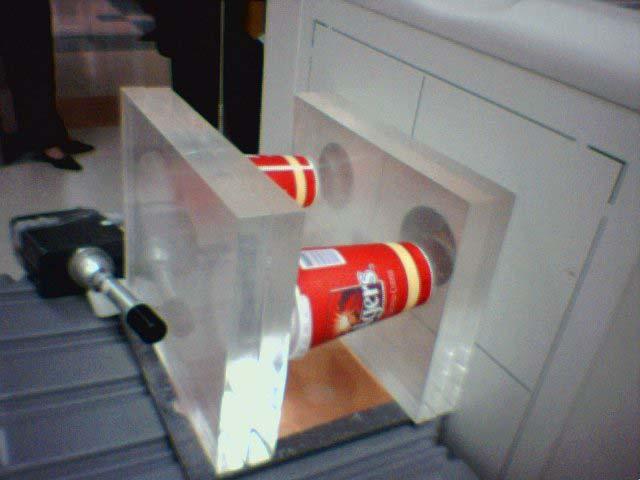
30 My Image Quality Phantom With readily available test tools, a combo phantom can be built to provide quick evaluation of detail and contrast. Considerable cost savings to other phantoms
31
32


33 1.0 % contrast


34


35 4.0 lp/mm
36 Name That Phantom D.R.I.Q. D.R.I.P. The TESTOOL M.I.Q.P. P.E.D.R.O.

37
I. PERFORMANCE OF X-RAY PRODUCTION COMPONENTS FLUOROSCOPIC ACCEPTANCE TESTING: TEST PROCEDURES & PERFORMANCE CRITERIA
 FLUOROSCOPIC ACCEPTANCE TESTING: TEST PROCEDURES & PERFORMANCE CRITERIA EDWARD L. NICKOLOFF DEPARTMENT OF RADIOLOGY COLUMBIA UNIVERSITY NEW YORK, NY ACCEPTANCE TESTING GOALS PRIOR TO 1st CLINICAL USAGE
Amorphous Selenium Direct Radiography for Industrial Imaging
 DGZfP Proceedings BB 67-CD Paper 22 Computerized Tomography for Industrial Applications and Image Processing in Radiology March 15-17, 1999, Berlin, Germany Amorphous Selenium Direct Radiography for Industrial
STEREOTACTIC BREAST BIOPSY EQUIPMENT SURVEYS
 STEREOTACTIC BREAST BIOPSY EQUIPMENT SURVEYS JAMES A. TOMLINSON, M.S. Diagnostic Radiological Physicist American Board of Radiology Certified Medical Physics Consultants, Inc. Bio 28 yrs experience 100%
CHAPTER 6 QC Test For Fluoroscopic Equipment. Prepared by:- Kamarul Amin bin Abu Bakar School of Medical Imaging KLMUC
 CHAPTER 6 QC Test For Fluoroscopic Equipment Prepared by:- Kamarul Amin bin Abdullah @ Abu Bakar School of Medical Imaging KLMUC Lesson Outcomes Describe the objectives of each QC test done. Identify QC
Features and Weaknesses of Phantoms for CR/DR System Testing
 Physics testing of image detectors Parameters to test Features and Weaknesses of Phantoms for CR/DR System Testing Spatial resolution Contrast resolution Uniformity/geometric distortion Dose response/signal
Breast Tomosynthesis. Bob Liu, Ph.D. Department of Radiology Massachusetts General Hospital And Harvard Medical School
 Breast Tomosynthesis Bob Liu, Ph.D. Department of Radiology Massachusetts General Hospital And Harvard Medical School Outline Physics aspects of breast tomosynthesis Quality control of breast tomosynthesis
Introduction. Chapter 16 Diagnostic Radiology. Primary radiological image. Primary radiological image
 Introduction Chapter 16 Diagnostic Radiology Radiation Dosimetry I Text: H.E Johns and J.R. Cunningham, The physics of radiology, 4 th ed. http://www.utoledo.edu/med/depts/radther In diagnostic radiology
10/3/2012. Study Harder
 This presentation is a professional collaboration of development time prepared by: Rex Christensen Terri Jurkiewicz and Diane Kawamura Study Harder CR detection is inefficient, inferior to film screen
10/26/2015. Study Harder
 This presentation is a professional collaboration of development time prepared by: Rex Christensen Terri Jurkiewicz and Diane Kawamura Study Harder CR detection is inefficient, inferior to film screen
7/24/2014. Image Quality for the Radiation Oncology Physicist: Review of the Fundamentals and Implementation. Disclosures. Outline
 Image Quality for the Radiation Oncology Physicist: Review of the Fundamentals and Implementation Image Quality Review I: Basics and Image Quality TH-A-16A-1 Thursday 7:30AM - 9:30AM Room: 16A J. Anthony
Digital Detector Array Image Quality for Various GOS Scintillators
 Digital Detector Array Image Quality for Various GOS Scintillators More info about this article: http://www.ndt.net/?id=22768 Brian S. White 1, Mark E. Shafer 2, William H. Russel 3, Eric Fallet 4, Jacques
COMPUTED RADIOGRAPHY CHAPTER 4 EFFECTIVE USE OF CR
 This presentation is a professional collaboration of development time prepared by: Rex Christensen Terri Jurkiewicz and Diane Kawamura New Technology https://www.youtube.com/watch?v=ptkzznazb 7U COMPUTED
X-RAY FLUOROSCOPY IMAGING SYSTEMS. Dr Slavik Tabakov. Luminescence: Dept. Medical Eng. & Physics King s College London
 X-RAY FLUOROSCOPY IMAGING SYSTEMS Dr Slavik Tabakov OBJECTIVES - Image Intensifier construction - Input window - Accelerating and focusing electrodes - Output window - Conversion factor - II characteristics
Exposure Indices and Target Values in Radiography: What Are They and How Can You Use Them?
 Exposure Indices and Target Values in Radiography: What Are They and How Can You Use Them? Definition and Validation of Exposure Indices Ingrid Reiser, PhD DABR Department of Radiology University of Chicago
Quality Control for Stereotactic Breast Biopsy. Robert J. Pizzutiello, Jr., F.A.C.M.P. Upstate Medical Physics, Inc
 Quality Control for Stereotactic Breast Biopsy Robert J. Pizzutiello, Jr., F.A.C.M.P. Upstate Medical Physics, Inc. 716-924-0350 Methods of Imaging Guided Breast Biopsy Ultrasound guided, hand-held needle
Acceptance Testing of a Digital Breast Tomosynthesis Unit
 Acceptance Testing of a Digital Breast Tomosynthesis Unit 2012 AAPM Spring Clinical Meeting Jessica Clements, M.S., DABR Objectives Review of technology and clinical advantages Acceptance Testing Procedures
Nuclear Associates
 Nuclear Associates 07-647 R/F QC Phantom Operators Manual March 2005 Manual No. 07-647-1 Rev. 2 2004, 2005 Fluke Corporation, All rights reserved. All product names are trademarks of their respective companies
Learning Objectives: What s my motivation? (unknown screen actor) Workshop Overview
 Workshop Overview") Practical Medical Physics Adapting Traditional Clinical Medical Physics to Digital Radiography Charles E. Willis, Ph.D., DABR Associate Professor Department of Imaging Physics The University of Texas M.D.
Practical Medical Physics Adapting Traditional Clinical Medical Physics to Digital Radiography Charles E. Willis, Ph.D., DABR Associate Professor Department of Imaging Physics The University of Texas M.D.
Investigation of Effective DQE (edqe) parameters for a flat panel detector
 parameters for a flat panel detector") Investigation of Effective DQE (edqe) parameters for a flat panel detector Poster No.: C-1892 Congress: ECR 2013 Type: Authors: Keywords: DOI: Scientific Exhibit D. Bor 1, S. Cubukcu 1, A. Yalcin 1, O.
Investigation of Effective DQE (edqe) parameters for a flat panel detector Poster No.: C-1892 Congress: ECR 2013 Type: Authors: Keywords: DOI: Scientific Exhibit D. Bor 1, S. Cubukcu 1, A. Yalcin 1, O.
Half value layer and AEC receptor dose compliance survey in Estonia
 Half value layer and AEC receptor dose compliance survey in Estonia K. Kepler, A. Vladimirov Training Centre of Medical Physics, University of Tartu Testing Centre of the University of Tartu, Estonia E-mail:
RAD 150 RADIOLOGIC EXPOSURE TECHNIQUE II
 RAD 150 RADIOLOGIC EXPOSURE TECHNIQUE II APPROVED 12/O2/2011 EFFECTIVE SPRING 2013-14 Prefix & Number RAD 150 Course Title: Radiologic Exposure Technique II & Lab Purpose of this submission: New Change/Updated
Exposure System Selection
 Principles of Imaging Science II (RAD120) Exposure Systems Exposure System Selection Radiographic exposure is a very complex process Best technique systems manipulate one variable while holding others
BASICS OF FLUOROSCOPY
 Medical Physics Residents Training Program BASICS OF FLUOROSCOPY Dr. Khalid Alyousef, PhD Department of Medical Imaging King Abdulaziz Medical City- Riyadh Edison examining the hand of Clarence Dally with
X-ray Imaging. PHYS Lecture. Carlos Vinhais. Departamento de Física Instituto Superior de Engenharia do Porto
 X-ray Imaging PHYS Lecture Carlos Vinhais Departamento de Física Instituto Superior de Engenharia do Porto cav@isep.ipp.pt Overview Projection Radiography Anode Angle Focal Spot Magnification Blurring
Fluoroscopy - Chapter 9
 Fluoroscopy - Chapter 9 Kalpana Kanal, Ph.D., DABR Lecturer, Diagnostic Physics Dept. of Radiology UW Medicine a copy of this lecture may be found at: http://courses.washington.edu/radxphys/physicscourse04-05.html
Test Equipment for Radiology and CT Quality Control Contents
 Test Equipment for Radiology and CT Quality Control Contents Quality Control Testing...2 Photometers for Digital Clinical Display QC...3 Primary Workstations...3 Secondary Workstations...3 Testing of workstations...3
Image Display and Perception
 Image Display and Perception J. Anthony Seibert, Ph.D. Department of Radiology UC Davis Medical Center Sacramento, California, USA Image acquisition, display, & interpretation X-rays kvp mas Tube filtration
Evaluation of a quality control phantom for digital chest radiography
 JOURNAL OF APPLIED CLINICAL MEDICAL PHYSICS, VOLUME 2, NUMBER 2, SPRING 2001 Evaluation of a quality control phantom for digital chest radiography Eugene Mah* Department of Radiology, Medical University
Y11-DR Digital Radiography (DR) Image Quality
 Image Quality") Y11-DR Digital Radiography (DR) Image Quality Image quality is stressed for all systems in Safety Code 35. In the relevant sections Health Canada s advice is the manufacturer s recommended test procedures
Y11-DR Digital Radiography (DR) Image Quality Image quality is stressed for all systems in Safety Code 35. In the relevant sections Health Canada s advice is the manufacturer s recommended test procedures
AUTOMATED AND QUANTITATIVE METHOD FOR QUALITY ASSURANCE OF DIGITAL RADIOGRAPHY IMAGING SYSTEMS
 International Workshop SMART MATERIALS, STRUCTURES & NDT in AEROSPACE Conference NDT in Canada 2011 2-4 November 2011, Montreal, Quebec, Canada AUTOMATED AND QUANTITATIVE METHOD FOR QUALITY ASSURANCE OF
SYLLABUS. TITLE: Equipment Operation I. DEPARTMENT: Radiologic Technology
 CODE: RADT 156 INSTITUTE: Health Science TITLE: Equipment Operation I DEPARTMENT: Radiologic Technology COURSE DESCRIPTION: This course covers the principles of equipment operation and maintenance of radiographic
Nuclear Associates
 Nuclear Associates 76-700 Digital Subtraction Angiography Phantom Users Manual March 2005 Manual No. 76-700-1 Rev. 2 2004, 2005 Fluke Corporation, All rights reserved. Printed in U.S.A. All product names
Essentials of Digital Imaging
 Essentials of Digital Imaging Module 7 Transcript 2016 ASRT. All rights reserved. Essentials of Digital Imaging Module 7 Quality 1. ASRT Animation 2. Welcome Welcome to the Essentials of Digital Imaging:
Overview of Safety Code 35
 Common Quality Control Procedures for All s Quality Control Procedures Film All s Daily Quality Control Tests Equipment Warm-up (D1) According to manufacturers instructions Can include auto calibration(d1)
X-ray Tube and Generator Basic principles and construction
 X-ray Tube and Generator Basic principles and construction Dr Slavik Tabakov - Production of X-rays and Patient Dose OBJECTIVES - X-ray tube construction - Anode - types, efficiency - Classical X-ray generator
Aspire HD. Program Manual. 2nd Edition - October 2012
 Quality Control 1 Aspire HD Quality Control Program Manual 2nd Edition - October 2012 Overview Installation of FDR Mammography QC Program Weekly Test 2 3 4 Quarterly Test 5 Semi-annual Test 6 Annual Test
Overview. Professor Roentgen was a Physicist!!! The Physics of Radiation Oncology X-ray Imaging
 The Physics of Radiation Oncology X-ray Imaging Charles E. Willis, Ph.D. DABR Associate Professor Department of Imaging Physics The University of Texas M.D. Anderson Cancer Center Houston, Texas Overview
Joint ICTP/IAEA Advanced School on Dosimetry in Diagnostic Radiology and its Clinical Implementation May 2009
 2033-6 Joint ICTP/IAEA Advanced School on Dosimetry in Diagnostic Radiology and its Clinical Implementation 11-15 May 2009 Dosimetry for Fluoroscopy Basics Renato Padovani EFOMP Joint ICTP-IAEA Advanced
Acquisition, Processing and Display
 Acquisition, Processing and Display Terri L. Fauber, R.T. (R)(M) Department of Radiation Sciences School of Allied Health Professions Virginia Commonwealth University Topics Image Characteristics Image
Preliminary Modulation Transfer Function Study on Amorphous Silicon Flat Panel System for Industrial Digital Radiography
 ECNDT 26 - Poster 17 Preliminary Modulation Transfer Function Study on Amorphous Silicon Flat Panel System for Industrial Digital Radiography Khairul Anuar MOHD SALLEH, Ab. Razak HAMZAH and Mohd Ashhar
Dose Reduction and Image Preservation After the Introduction of a 0.1 mm Cu Filter into the LODOX Statscan unit above 110 kvp
 Dose Reduction and Image Preservation After the Introduction of a into the LODOX Statscan unit above 110 kvp Abstract: CJ Trauernicht 1, C Rall 1, T Perks 2, G Maree 1, E Hering 1, S Steiner 3 1) Division
While digital techniques have the potential to reduce patient doses, they also have the potential to significantly increase them.
 In press 2004 1 2 Guest Editorial (F. Mettler, H. Ringertz and E. Vano) Guest Editorial (F. Mettler, H. Ringertz and E. Vano) Digital radiology An appropriate analogy that is easy for most people to understand
Nuclear Associates
 Nuclear Associates 07-649 CDRH Fluoroscopic Phantom Users Manual March 2005 Manual No. 07-649-1 Rev. 2 2004, 2005 Fluke Corporation, All rights reserved. Printed in U.S.A. All product names are trademarks
Mammography: Physics of Imaging
 Mammography: Physics of Imaging Robert G. Gould, Sc.D. Professor and Vice Chair Department of Radiology and Biomedical Imaging University of California San Francisco, California Mammographic Imaging: Uniqueness
10/15/2012 SECTION III - CHAPTER 6 DIGITAL FLUOROSCOPY RADT 3463 COMPUTERIZED IMAGING
 RADT 3463 - COMPUTERIZED IMAGING Section III: Chapter 6 RADT 3463 Computerized Imaging 1 SECTION III - CHAPTER 6 DIGITAL FLUOROSCOPY RADT 3463 COMPUTERIZED IMAGING Section III: Chapter 6 RADT 3463 Computerized
RaySafe X2. Effortless measurements of X-ray
 RaySafe X2 Effortless measurements of X-ray At your fingertips We ve grown accustomed to intuitive interactions with our devices. After all, it s not the device that s most important, but what you can
Computed Radiography
 BAM Berlin Computed Radiography --INDE 2007, Kalpakkam, India -- Uwe Zscherpel, Uwe Ewert BAM Berlin, Division VIII.3 Requests Requests and and information information to: to: Dr. Dr. U. U. Zscherpel Zscherpel
X-RAY MEDICAL EQUIPMENT
 X-RAY MEDICAL EQUIPMENT CHEST RADIOGRAPHY GENERAL RADIOGRAPHY & FLUOROSCOPY RADIOTHERAPY MOBILE HEALTHCARE MAMMOGRAPHY MAMMOSCAN FULL FIELD DIGITAL MAMMOGRAPHY SYSTEM Biopsy Attachment џ MAMMOSCAN an ADANI
Digital radiography (DR) post processing techniques for pediatric radiology
 post processing techniques for pediatric radiology") Digital radiography (DR) post processing techniques for pediatric radiology St Jude Children s Research Hospital Samuel Brady, MS PhD DABR samuel.brady@stjude.org Purpose Review common issues and solutions
Digital radiography (DR) post processing techniques for pediatric radiology St Jude Children s Research Hospital Samuel Brady, MS PhD DABR samuel.brady@stjude.org Purpose Review common issues and solutions
of sufficient quality and quantity
 of sufficient quality and quantity The patient s body attenuates the beam as it passes though the body More energy is deposited in organs located near the entry of the beam than near the exit of the beam
AN ABSTRACT OF THE THESIS OF. W. Scott Helms for the degree of Master of Science in Radiation Health Physics
 AN ABSTRACT OF THE THESIS OF W. Scott Helms for the degree of Master of Science in Radiation Health Physics presented on November 24, 2014 Title: A Quantitative Comparison of Cardiovascular Imaging Systems
THE ART OF THE IMAGE: IDENTIFICATION AND REMEDIATION OF IMAGE ARTIFACTS IN MAMMOGRAPHY
 THE ART OF THE IMAGE: IDENTIFICATION AND REMEDIATION OF IMAGE ARTIFACTS IN MAMMOGRAPHY William Geiser, MS DABR Senior Medical Physicist MD Anderson Cancer Center Houston, Texas wgeiser@mdanderson.org INTRODUCTION
2017 West Coast Educators Conference Orlando. Projection Geometry. 1. Review hierarchy of image qualities (amplified version):
:") Spatial Resolution in the Digital Age: NOTES Quinn B. Carroll, MEd, RT 2017 West Coast Educators Conference Orlando Projection Geometry 1. Review hierarchy of image qualities (amplified version): a. Maximum
Spatial Resolution in the Digital Age: NOTES Quinn B. Carroll, MEd, RT 2017 West Coast Educators Conference Orlando Projection Geometry 1. Review hierarchy of image qualities (amplified version): a. Maximum
Current technology in digital image production (CR/DR and other modalities) Jaroonroj Wongnil 25 Mar 2016
 Jaroonroj Wongnil 25 Mar 2016") Current technology in digital image production (CR/DR and other modalities) Jaroonroj Wongnil 25 Mar 2016 Current technology in digital image production (CR/DR and other modalities) 2/ Overview Digital
Current technology in digital image production (CR/DR and other modalities) Jaroonroj Wongnil 25 Mar 2016 Current technology in digital image production (CR/DR and other modalities) 2/ Overview Digital
Practical Aspects of Medical Physics Surveys of Mammography Equipment and Facilities
 Practical Aspects of Medical Physics Surveys of Mammography Equipment and Facilities Melissa Martin, M.S., FAAPM, FACR, FACMP AAPM Annual Meeting - Philadelphia July 19, 2010 MO-B-204C-1 Educational Objectives
YSF - 300/DAR i
 C506-E067 Integrated Digital R/F System YSF - 300/DAR - 8000i photo shown with optional I.I. cover A Fully Integrated Digital R/F System Expanding the Clinical Boundaries. With more than 130 years of dedication
Quality Control of Full Field Digital Mammography Units
 Quality Control of Full Field Digital Mammography Units Melissa C. Martin, M.S., FACMP, FACR, FAAPM Melissa@TherapyPhysics.com 310-612-8127 ACMP Annual Meeting Virginia Beach, VA May 2, 2009 History of
DISC QC/QA Program for Digital Imaging Systems using the DR Radchex Plus Meter
 DISC QC/QA Program for Digital Imaging Systems using the DR Radchex Plus Meter Revision Date: January 5th, 2017 www.disc-imaging.com Table of Contents Section A: Preliminary Setup Requirements... 4 Tools
History of digital imaging
 CR/QA RADCHEX History of digital imaging Early, crude digital detectors were developed in the 1970 s Image quality was problematic Processing time of digital images was untenable Viewing, transfer and
Estimation of signal transfer property for wireless digital detector in different measurement schemes
 Estimation of signal transfer property for wireless digital detector in different measurement schemes Anatoli Vladimirov, Kalle Kepler Training Centre of Medical Physics, University of Tartu, Estonia 11
Comparison of computed radiography and filmõscreen combination using a contrast-detail phantom
 JOURNAL OF APPLIED CLINICAL MEDICAL PHYSICS, VOLUME 4, NUMBER 1, WINTER 2003 Comparison of computed radiography and filmõscreen combination using a contrast-detail phantom Z. F. Lu,* E. L. Nickoloff, J.
diagnostic examination
 RADIOLOGICAL PHYSICS 2011 Raphex diagnostic examination Adel A. Mustafa, Ph.D., Editor PUBLISHED FOR: RAMPS (Radiological and Medical Physics Society of New York) preface The RAPHEX Diagnostic exam 2011
8/2/2017. Radiologist Responsibilities. Radiologist Responsibilities. Medical Physicist Mammography Equipment Evaluation and Annual Survey
 Implementation of the 2016 ACR Digital Mammography QC Manual Medical Physicist Mammography Equipment Evaluation and Annual Survey Eric A Berns, PhD, FACR Radiologist Responsibilities Radiologist Responsibilities
Outline. Digital Radiography. Understanding Digital Modalities: Image Quality and Dose. Image Quality. Dose Control
 Understanding Digital Modalities: Image Quality and Dose S. Jeff Shepard, M.S. University of Texas M. D. Anderson Cancer Center Houston, Texas Special Acknowledgement: Stephen K. Thompson, M.S. William
Update on Fluoroscopy Physics AAPM MO-A-210A-1 Stephen Balter, Ph.D.
 Update on Fluoroscopy Physics Stephen Balter, PhD Columbia University Draft for MO-A-210A-1 2009 AAPM Educational objectives Understand dosimetric concepts relating to interventional fluoroscopy Characterize
Digital Imaging started in the 1972 with Digital subtraction angiography Clinical digital imaging was employed from the 1980 ~ 37 years ago Amount of
 Digital Imaging started in the 1972 with Digital subtraction angiography Clinical digital imaging was employed from the 1980 ~ 37 years ago Amount of radiation to the population due to Medical Imaging
Outline ASRT Changes Impact on current curriculum Potential new courses WECM Changes Last update Resources and needs
 Change nd Annual Blinn College 2 nd Educator s Workshop For Radiologic Sciences July 28, 2007 Christi Carter, MSRS, RT(R) Outline ASRT Changes Impact on current curriculum Potential new courses WECM Changes
Investigation of the line-pair pattern method for evaluating mammographic focal spot performance
 Investigation of the line-pair pattern method for evaluating mammographic focal spot performance Mitchell M. Goodsitt, a) Heang-Ping Chan, and Bob Liu Department of Radiology, University of Michigan, Ann
Chromatic X-Ray imaging with a fine pitch CdTe sensor coupled to a large area photon counting pixel ASIC
 Chromatic X-Ray imaging with a fine pitch CdTe sensor coupled to a large area photon counting pixel ASIC R. Bellazzini a,b, G. Spandre a*, A. Brez a, M. Minuti a, M. Pinchera a and P. Mozzo b a INFN Pisa
Digital Radiography : Flat Panel
 Digital Radiography : Flat Panel Flat panels performances & operation How does it work? - what is a sensor? - ideal sensor Flat panels limits and solutions - offset calibration - gain calibration - non
Quality control of Gamma Camera. By Dr/ Ibrahim Elsayed Saad 242 NMT
 Quality control of Gamma Camera By Dr/ Ibrahim Elsayed Saad 242 NMT WHAT IS QUALITY? The quality of a practice is to fulfill the expectations and demands from: Patient Clinicain Your self Quality assurance
Practical Medical Physics Session: TG-151 Dose Monitoring. August 5, 2013 Katie Hulme, M.S.
 Practical Medical Physics Session: TG-151 Dose Monitoring August 5, 2013 Katie Hulme, M.S. Digital Imaging and Dose Creep Images courtesy of Agfa Healthcare Under-Exposed Over-Exposed Freedman et al.,
Digital Images & Image Quality
 Introduction to Medical Engineering (Medical Imaging) Suetens 1 Digital Images & Image Quality Ho Kyung Kim Pusan National University Radiation imaging DR & CT: x-ray Nuclear medicine: gamma-ray Ultrasound
Teaching Digital Radiography and Fluoroscopic Radiation Protection
 Teaching Digital Radiography and Fluoroscopic Radiation Protection WCEC 20 th Student Educator Radiographer Conference Dennis Bowman, RT(R), CRT (R)(F) Community Hospital of the Monterey Peninsula (CHOMP)
7/20/2014. Outline. Outline. Disclosures. Learning Objectives. SBB: Practical Aspects of ACR Accreditation, QC and ACR On Site Surveys
 Outline SBB: Practical Aspects of ACR Accreditation, QC and ACR On Site Surveys Robert J. Pizzutiello, MS, FACR, FAAPM, FAC Residency Program Director, Upstate Medical Physics, PC Senior Vice President,
Beam-Restricting Devices
 Beam-Restricting Devices Three factors contribute to an increase in scatter radiation: Increased kvp Increased Field Size Increased Patient or Body Part Size. X-ray Interactions a some interact with the
Ludlum Medical Physics
 Ludlum Medical Physics Medical Imaging Radiology QA Test Tools NEW LUDLUM PRODUCT LINE Medical Physics Products Medical Physics Products What are they? Products used to measure radiation output and to
ESTABLISHING A QUALITY ASSURANCE ROUTINE FOR DIGITAL IMAGING
 ESTABLISHING A QUALITY ASSURANCE ROUTINE FOR DIGITAL IMAGING A thesis submitted in partial fulfillment of the requirements for the degree of Master of Science By RANA AL SULAIMAN B.S. in Physics, King
Nuclear Associates
 Nuclear Associates 07-706 Patient Phantom/Penetrometer System Users Manual March 2005 Manual No. 07-706-1 Rev. 2 2004, 2005 Fluke Corporation, All rights reserved. Printed in U.S.A. All product names are
Appropriate Inspection Distance of Digital X-Ray Imaging Equipment for Diagnosis
 Indian Journal of Science and Technology Vol 8(S8), 380-386, April 2015 ISSN (Print) : 0974-6846 ISSN (Online) : 0974-5645 DOI: 10.17485/ijst/2015/v8iS8/70528 Appropriate Inspection Distance of Digital
Setting up digital imaging department!
 Outline Setting up digital imaging department! From screen/film to digital radiography PACS/Tele radiology Setting up digital department Digital Imaging Napapong Pongnapang, Ph.D. Department of Radiological
Mammography is a radiographic procedure specially designed for detecting breast pathology Approximately 1 woman in 8 will develop breast cancer over
 Mammography is a radiographic procedure specially designed for detecting breast pathology Approximately 1 woman in 8 will develop breast cancer over a lifetime Breast cancer screening programs rely on
The importance of radiation quality for optimisation in radiology
 Available online at http://www.biij.org/2007/2/e38 doi: 10.2349/biij.3.2.e38 biij Biomedical Imaging and Intervention Journal COMMENTARY The importance of radiation quality for optimisation in radiology
inspexio SMX-225CT FPD HR
 Microfocus X-Ray CT System C251-E029A Advanced Operability and Excellent Image Quality That Overturns Conventional Assumptions Microfocus X-Ray CT System The is a high-performance microfocus X-ray CT system
Multiple Choice Identify the letter of the choice that best completes the statement or answers the question.
 RA202 image production class two Multiple Choice Identify the letter of the choice that best completes the statement or answers the question. 1. What removes excess chemistry from the film prior to it
X-RAYS - NO UNAUTHORISED ENTRY
 Licencing of premises Premises Refer Guidelines A radiation warning sign and warning notice, X-RAYS - NO UNAUTHORISED ENTRY must be displayed at all entrances leading to the rooms where x-ray units are
Image Quality Artifacts in Digital Imaging
 MAHIDOL UNIVERSITY Wisdom of the Land Image Quality Artifacts in Digital Imaging Napapong Pongnapang, Ph.D. Department of Radiological Technology Faculty of Medical Technology Mahidol University, Bangkok,
Surveying and QC of Stereotactic Breast Biopsy Units for ACR Accreditation
 Surveying and QC of Stereotactic Breast Biopsy Units for ACR Accreditation AAPM Annual Clinical Meeting Indianapolis, IN August 5, 2013 Learning Objectives Become familiar with the recommendations and
Optimization of Digital Mammography Resolution Using Magnification Technique in Computed Radiography 1
 Optimization of Digital Mammography Resolution Using Magnification Technique in Computed Radiography 1 Gham Hur, M.D., Yoon Joon Hwang, M.D., Soon Joo Cha, M.D., Su Young Kim, M.D., Yong Hoon Kim, M.D.
CHAPTER 2 COMMISSIONING OF KILO-VOLTAGE CONE BEAM COMPUTED TOMOGRAPHY FOR IMAGE-GUIDED RADIOTHERAPY
 14 CHAPTER 2 COMMISSIONING OF KILO-VOLTAGE CONE BEAM COMPUTED TOMOGRAPHY FOR IMAGE-GUIDED RADIOTHERAPY 2.1 INTRODUCTION kv-cbct integrated with linear accelerators as a tool for IGRT, was developed to
DIGITAL IMAGE PROCESSING IN X-RAY IMAGING
 DIGITAL IMAGE PROCESSING IN X-RAY IMAGING Shalini Kumari 1, Bachan Prasad 2,Aliya Nasim 3 Department of Electronics And Communication Engineering R.V.S College of Engineering & Technology, Jamshedpur,
SECTION I - CHAPTER 2 DIGITAL IMAGING PROCESSING CONCEPTS
 RADT 3463 - COMPUTERIZED IMAGING Section I: Chapter 2 RADT 3463 Computerized Imaging 1 SECTION I - CHAPTER 2 DIGITAL IMAGING PROCESSING CONCEPTS RADT 3463 COMPUTERIZED IMAGING Section I: Chapter 2 RADT
Tomophan TSP004 Manual
 T h e P h a n t o m L a b o r a t o r y 1 Tomophan TSP004 Manual Copyright 2016 WARRANTY THE PHANTOM LABORATORY INCORPORATED ( Seller ) warrants that this product shall remain in good working order and
Introduction. Digital Mammography QA: Comparing the Manufacturers Recommendations. What is QC and why is it important? Review & compare QC tests
 Slide 1 Digital Mammography QA: Comparing the Manufacturers Recommendations Eric A. Berns, Ph.D. Slide 2 Introduction What is QC and why is it important? Review & compare QC tests Key take home points
Do you have any other questions? Please call us at (Toll Free) or , or
 or , or") INSTRUCTIONS Read the appropriate course/ textbook. This is an open book test. A score of 75% or higher is needed to receive CE credit. You will have a maximum of three attempts to pass this course. Please
INSTRUCTIONS Read the appropriate course/ textbook. This is an open book test. A score of 75% or higher is needed to receive CE credit. You will have a maximum of three attempts to pass this course. Please
Digital radiography: Practical advantages of Digital Radiography. Practical Advantages in image quality
 Digital radiography: Digital radiography is set to become the most common form of processing radiographic images in the next 10 years. This is due to a number of practical and image quality issues. Practical
Digital Image Management: the Basics
 Digital Image Management: the Basics Napapong Pongnapang, Ph.D. Department of Radiological Technology Faculty of Medical Technology Mahidol University Outline From screen/film to digital radiography PACS/Tele
Multi-Access Biplane Lab
 Multi-Access Biplane Lab Advanced technolo gies deliver optimized biplane imaging Designed in concert with leading physicians, the Infinix VF-i/BP provides advanced, versatile patient access to meet the
Photomultiplier Tube
 Nuclear Medicine Uses a device known as a Gamma Camera. Also known as a Scintillation or Anger Camera. Detects the release of gamma rays from Radionuclide. The radionuclide can be injected, inhaled or
Studies on reduction of exposure dose using digital scattered X-ray removal processing
 Studies on reduction of exposure dose using digital scattered X-ray removal processing Poster No.: C-1834 Congress: ECR 2015 Type: Scientific Exhibit Authors: K. Kashiyama, M. Funahashi, T. Nakaoka, T.