X-rays. X-rays are produced when electrons are accelerated and collide with a target. X-rays are sometimes characterized by the generating voltage
|
|
|
- Muriel Boyd
- 5 years ago
- Views:
Transcription

1 X-rays Ouch! 1
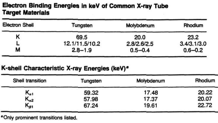
2 X-rays X-rays are produced when electrons are accelerated and collide with a target Bremsstrahlung x-rays Characteristic x-rays X-rays are sometimes characterized by the generating voltage kv soft x-rays kv diagnostic x-rays kv orthovoltage x-rays 300 kv 1 MV intermediate energy x-rays > 1MV megavoltage x-rays 2
3 Bremmstrahlung Bremsstrahlung x-rays occur when electrons are (de)accelerated in the Coulomb field of a nucleus 3
4 Bremsstrahlung 4
5 Bremsstrahlung The power radiated from an accelerating charge is given by Larmor s equation 2 2 P = 2 3 e c a 3 In the case of an electron in the Coulomb field of a nucleus F Ze a = = k ~ m r 2 m 2 Z 5
6 Bremsstrahlung The probability of bremsstrahlung goes as Z 2, hence high Z targets are more effective than low Z The energy of the x-rays varies from zero to the maximum kinetic energy of the electron (x-ray tube kvp) The energy spectrum from a thick target goes as 1/E but inherent (1mm Al eq) plus additional (few mm Al) filtration removes the lower energy x-rays Here I am referring to diagnostic x-rays 6
7 Bremsstrahlung The unfiltered energy spectrum is approximately given by Kramer s law which was an early application of quantum mechanics I ( ) ( ) E = KZ T E γ e γ 7
8 Bremsstrahlung 8

9 Characteristic x-rays After excitation, ions with a vacancy in their inner shell can de-excite Radiatively through x-ray fluorescence Non-radiatively through the emission of Auger electrons 9
10 Characteristic X-rays Thus an x-ray spectrum will also show characteristic x-rays arising from L to K and M to K transitions after ionization of a K electron Usually transitions to higher shells absorbed by the filtration or are not x-rays 10
11 Characteristic X-rays The probability of K shell fluorescence increases with Z 11
12 Characteristic X-rays 12
13 Characteristic X-rays Sometimes the characteristic x-rays are emphasized using the same material for target and filter Characteristic x-rays from molybdenum are effective in maximizing contrast in mammography 13
14 Characteristic X-rays Mo target, filter, and result 14
15 Directionality For MeV electrons, bremsstrahlung x- rays are preferentially emitted in the electron s direction For kev electrons, bremsstrahlung x- rays are emitted at larger angles Characteristic x-rays are emitted isotropically since there is no angular correlation between the incident electron that causes the ionization and the fluorescent photon 15
16 X-ray Tube A simplified x-ray tube (Coolidge type) shows the idea behind most x-ray tubes today 16
17 X-ray Tube In addition to bremsstrahlung and characteristic x-ray production, electrons also loose energy through collisions Collision losses dominate in this energy region radiation loss EZ ( E in MeV) collision loss 820 For 100 kev electrons in W radiation loss collision loss Thus >99% of the electron energy goes into heating the target rather than x-rays Removing heat from the anode in a vacuum is an issue = 17
18 X-ray Tube Efficiency of x-ray production depends on the tube voltage and the target material W (Z=74) in this example P P deposited radiated = VI = Efficiency ε = P P 9 ZV radiated deposited 2 I = ZV kvp (V) Heat (%) X- rays (%)

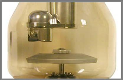
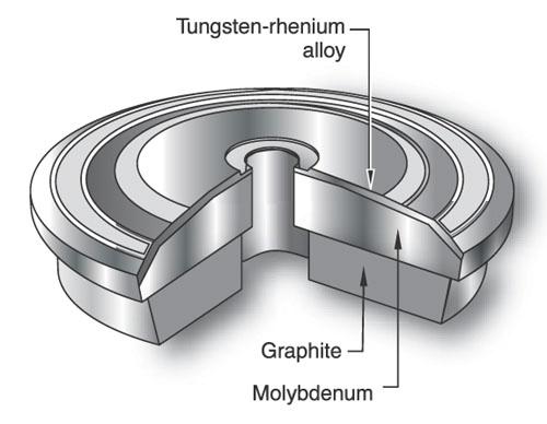
19 X-ray Tube X-ray tubes 19

20 X-ray Tube More detail 20
 and cooling")
21 X-ray Tube Housing for shielding (Pb) and cooling (oil) 21

22 X-ray Tube More detail 22
23 X-ray Tube The main parts of the x-ray tube are Cathode/filament Typical electron current is A for short exposures (< 100 ms) Anode/target Glass/metal envelope Accelerating voltage Typical voltage is kvp 23
24 Cathode consists of Cathode Low R tungsten wire for thermionic emission Tungsten has a high melting point (3370C) and minimum deposit on the glass tube Tube current is controlled by varying the filament current which is a few amps A focusing cup Uses electric field lines to focus the electrons Typically there are two filaments Long one: higher current, lower resolution Large focal spot Short one: lower current, higher resolution Small focal spot 24

25 Cathode Dual focus filament is common 25
26 Anode Usually made of tungsten in copper because of high Z and high melting point Molybdenum and rhodium used for soft tissue imaging Large rotating surface for heat distribution and radiative heat loss Rotation of 3k-10k revolutions/minute Resides in a vacuum (~10-6 torr) Thermally decoupled from motor to avoid overheating of the shaft Target is at an tilted angle with respect to axis Bremsstrahlung is emitted at ~ right angles for low energy electrons Determines focal spot size 26
27 Anode 27
28 Anode 28

29 Anode The heating of the anode limits the voltage, current, and exposure time An exposure rating chart gives these limits 29
 HU (Heating Unit) ~ J Damaged anodes")
30 Anode Power = V x I (watts) Energy = Power x time = V x I x s (joules) HU (Heating Unit) ~ J Damaged anodes 30
31 Anode The angle determines the projected focal spot The smaller the angle the better the resolution Typically 7-20 degrees θ Angle θ Angle Incident electron beam width Actual focal spot size Incident electron beam width Apparent focal spot size Actual focal spot size Increased apparent focal spot size Film Film 31
32 X-rays The energy of the photons depends on the electron energy (kvp) and the target atomic number Z The number of photons depends on the the electron energy (kvp), Z, and the beam current (ma) A typical number / area is ~ / m 2 About 1% will hit the film ~ / m 2 Absorption and detection efficiency will further reduce this number 32
33 Automatic Exposure Control X Ray tube Collimator Beam Air Soft tissue Bone Patient Table Grid AEC detectors Cassette AEC detectors can ionization chambers or solidstate detectors 33
34 Automatic Exposure Control Most modern x-rays machines are equipped with automatic exposure control also called a phototime The AEC sets the technical parameters of the machine (kv, ma, time, ) in order to avoid repeated exposures AEC is used to keep the radiographic quality (film density) equal on all patients AEC detectors can be ionization chambers or solid state detectors 34
35 Grid To reduce the number of secondary scattered photons making it to the film, a grid between the patient and film is used 35
36 Details Grid Grid bars are usually lead whereas the grid openings are usually made of aluminum or carbon Grid thickness is typically 3 mm Grid ratio is H/W and 10/1 is typical Grid frequency of 60 lines / cm is typical B/W/H on the figure might be 0.045, 0.120, 1.20 in mm The Bucky factor is the entrance exposure w/wo the grid while achieving the same film density 4 is average 36


37 Accelerating Voltage The potential difference between cathode and anode must be generated by 60 Hz 220V AC power High voltages are produced using a transformer 37
38 Accelerating Voltage Electrons are accelerated when the filament is at a negative potential with respect to the target Diode circuits can be used to provide rectification (AC to DC voltage) Three phase power (6 pulse or 12 pulse) can be used to reduce ripple Constant potential operation can be achieved by using constant potential (voltage regulations) or high frequency x- ray generators 38
39 Half-wave Rectifier Not very efficient 39


40 Full-wave Bridge Rectifier This circuit allows the entire input waveform to be used 40
41 Accelerating Voltage kv ripple (%) 100% 13% 4% Single phase single pulse Single phase 2-pulse Three phase 6-pulse Three phase 12-pulse Line voltage 0.01 s 0.02 s 41
Mammography is a radiographic procedure specially designed for detecting breast pathology Approximately 1 woman in 8 will develop breast cancer over
 Mammography is a radiographic procedure specially designed for detecting breast pathology Approximately 1 woman in 8 will develop breast cancer over a lifetime Breast cancer screening programs rely on
X-RAY IMAGING EE 472 F2017. Prof. Yasser Mostafa Kadah
 X-RAY IMAGING EE 472 F2017 Prof. Yasser Mostafa Kadah www.k-space.org Recommended Textbook Stewart C. Bushong, Radiologic Science for Technologists: Physics, Biology, and Protection, 10 th ed., Mosby,
X-ray Tube and Generator Basic principles and construction
 X-ray Tube and Generator Basic principles and construction Dr Slavik Tabakov - Production of X-rays OBJECTIVES - X-ray tube construction - Anode - types, efficiency - X-ray tube working characteristics
X-ray Tube and Generator Basic principles and construction
 X-ray Tube and Generator Basic principles and construction Dr Slavik Tabakov - Production of X-rays and Patient Dose OBJECTIVES - X-ray tube construction - Anode - types, efficiency - Classical X-ray generator
- KiloVoltage. Technique 101: Getting Back to Basics
 Why do I need to know technique? Technique 101: Getting Back to Basics Presented by: Thomas G. Sandridge, M.S., M.Ed., R.T.(R) Program Director Northwestern Memorial Hospital School of Radiography Chicago,
X-rays in medical diagnostics
 X-rays in medical diagnostics S.Dolanski Babić 2017/18. History W.C.Röntgen (1845-1923) discovered a new type of radiation Nature, Jan. 23. 1896.; Science, Feb.14. 1896. X- rays: Induced the ionization
Technical data CAMARGUE CS-VH50/300. VARIABLE Height Bucky Table With Ceiling Suspension
 Technical data VARIABLE Height Bucky Table With Ceiling Suspension Model Variations CAMARGUE FH (Fixed Height) CAMARGUE FH Tomo CAMARGUE FH Ceiling suspension CAMARGUE VH (Variable Height) CAMARGUE VH
Exposure System Selection
 Principles of Imaging Science II (RAD120) Exposure Systems Exposure System Selection Radiographic exposure is a very complex process Best technique systems manipulate one variable while holding others
DENTAL RADIOGRAPHY KAMARUL AMIN BIN ABU BAKAR
 DENTAL RADIOGRAPHY KAMARUL AMIN BIN ABDULLAH @ ABU BAKAR Components of the Dental X-Ray Machine Dental x-ray machines may vary somewhat in size and appearance, but all machines will have three primary
1. Carlton, Richard R., and Arlene M. Adler. Principles of Radiographic Imaging: An Art and a Science, 5th edition (2013).
.") CODE: RADT 151 INSTITUTE: Health Science TITLE: Radiographic Exposure DEPARTMENT: Radiologic Technology COURSE DESCRIPTION: This course covers the principles of radiographic exposure selection and manipulation
CODE: RADT 151 INSTITUTE: Health Science TITLE: Radiographic Exposure DEPARTMENT: Radiologic Technology COURSE DESCRIPTION: This course covers the principles of radiographic exposure selection and manipulation
RADIOGRAPHIC EXPOSURE
 RADIOGRAPHIC EXPOSURE Receptor Exposure Receptor Exposure the that interacts with the receptor. Computed Radiography ( ) requires a. Direct Digital Radiography (DR) requires a. Exposure Indicators Exposure
SPRINGFIELD TECHNICAL COMMUNITY COLLEGE ACADEMIC AFFAIRS
 SPRINGFIELD TECHNICAL COMMUNITY COLLEGE ACADEMIC AFFAIRS Course Number: RADG 212 Department: Radiography Course Title: Equip. Operation & Maint. Semester: Spring Year: 1997 Objectives/ Unit One: The X-ray
Introduction. Chapter 16 Diagnostic Radiology. Primary radiological image. Primary radiological image
 Introduction Chapter 16 Diagnostic Radiology Radiation Dosimetry I Text: H.E Johns and J.R. Cunningham, The physics of radiology, 4 th ed. http://www.utoledo.edu/med/depts/radther In diagnostic radiology
Partial Replication of Storms/Scanlan Glow Discharge Radiation
 Partial Replication of Storms/Scanlan Glow Discharge Radiation Rick Cantwell and Matt McConnell Coolescence, LLC March 2008 Introduction The Storms/Scanlan paper 1 presented at the 8 th international workshop
Mammography: Physics of Imaging
 Mammography: Physics of Imaging Robert G. Gould, Sc.D. Professor and Vice Chair Department of Radiology and Biomedical Imaging University of California San Francisco, California Mammographic Imaging: Uniqueness
PD233: Design of Biomedical Devices and Systems
 PD233: Design of Biomedical Devices and Systems (Lecture-8 Medical Imaging Systems) (Imaging Systems Basics, X-ray and CT) Dr. Manish Arora CPDM, IISc Course Website: http://cpdm.iisc.ac.in/utsaah/courses/
Beam-Restricting Devices
 Beam-Restricting Devices Three factors contribute to an increase in scatter radiation: Increased kvp Increased Field Size Increased Patient or Body Part Size. X-ray Interactions a some interact with the
Photons interaction with matter
 ب س م هللا الر ح من الر حیم Photons interaction with matter Ionization Ionization is the process of removing an electron from an electrically neutral atom to produce an ion pair. An ion is an atom or subatomic
Explain what is meant by a photon and state one of its main properties [2]
![Explain what is meant by a photon and state one of its main properties [2]](/thumbs/80/82516318.jpg "Explain what is meant by a photon and state one of its main properties [2]") 1 (a) A patient has an X-ray scan taken in hospital. The high-energy X-ray photons interact with the atoms inside the body of the patient. Explain what is meant by a photon and state one of its main properties....
1 (a) A patient has an X-ray scan taken in hospital. The high-energy X-ray photons interact with the atoms inside the body of the patient. Explain what is meant by a photon and state one of its main properties....
Maltase cross tube. D. Senthilkumar P a g e 1
 Thermionic Emission Maltase cross tube Definition: The emission of electrons when a metal is heated to a high temperature Explanation: In metals, there exist free electrons which are able to move around
MILADY. Product Data. Page 1 of 8
 Page 1 of 8 The MILADY Mammographic Unit offers the best quality-to-price ratio to our customers worldwide. The unit advanced technology together with the application of industrial production standards,
LECTURE 1 The Radiographic Image
 LECTURE 1 The Radiographic Image Prepared by:- KAMARUL AMIN ABDULLAH @ ABU BAKAR UiTM Faculty of Health Sciences Medical Imaging Department 11/23/2011 KAMARUL AMIN (C) 1 Lesson Objectives At the end of
80 Physics Essentials Workbook Stage 2 Physics
 80 Physics Essentials Workbook Stage 2 Physics the thickness of the tissue: Obviously, the thicker the tissue through which the X-rays have to pass the more they will be absorbed from the beam passing
X-RAYS - NO UNAUTHORISED ENTRY
 Licencing of premises Premises Refer Guidelines A radiation warning sign and warning notice, X-RAYS - NO UNAUTHORISED ENTRY must be displayed at all entrances leading to the rooms where x-ray units are
Seminar 8. Radiology S8 1
 Seminar 8 Radiology Medical imaging. X-ray image formation. Energizing and controlling the X-ray tube. Image detectors. The acquisition of analog and digital images. Digital image processing. Selected
Maximizing clinical outcomes
 Maximizing clinical outcomes Digital Tomosynthesis Dual Energy Subtraction Automated Long Length Imaging Improved image quality at a low dose Xray Xray Patented ISS capture technology promotes high sensitivity
P R E S E N T E D B Y. K A M A R U L A M I N A B D U L L A H Dip. MED. IMG., BSc. MED. IMG. (UiTM)
") + - P R E S E N T E D B Y K A M A R U L A M I N A B D U L L A H Dip. MED. IMG., BSc. MED. IMG. (UiTM) 1 I N T R O D U C T I O N : An x-ray generator is a device that Supplies electrical power to x-ray
+ - P R E S E N T E D B Y K A M A R U L A M I N A B D U L L A H Dip. MED. IMG., BSc. MED. IMG. (UiTM) 1 I N T R O D U C T I O N : An x-ray generator is a device that Supplies electrical power to x-ray
Using Carbon Nano-Tube Field Emitters to Miniaturize X-Ray Tubes
 Using Carbon Nano-Tube Field Emitters to Miniaturize X-Ray Tubes Authors: Martin Pesce, RT(R), Xiaohui Wang, PhD, Peter Rowland X-rays are produced by the impact of an accelerated electron beam on a tungsten
MXHF-1500RF is controlled by Digital key panel console that displays KV, ma and mas with APR menu programmed.
 R/F TV X-RAY SYSTEM DIAGNOSTIC RADIOGRAPHIC FLUOROSCOPIC TV SYSTEM MXHF-1500RF SYSTEM OUTLINE Product Data No. 041021-01 MXHF-1500RF is controlled by Digital key panel console that displays KV, ma and
1-1. GENERAL 1-2. DISCOVERY OF X-RAYS
 1-1. GENERAL Radiography is a highly technical field, indispensable to the modern dental practice, but presenting many potential hazards. The dental radiographic specialist must be thoroughly familiar
V SALAI SELVAM, AP & HOD, ECE, Sriram Engg. College, Perumalpattu 1 MEDICAL ELECTRONICS UNIT IV
 V SALAI SELVAM, AP & HOD, ECE, Sriram Engg. College, Perumalpattu 1 MEDICAL ELECTRONICS UNIT IV Ionizing and non-ionizing radiations: The radiation that ionizes the gases through which it travels is known
X-Rays and endoscopes
 X-Rays and endoscopes 1 What are X-rays? X-ray refers to electromagnetic radiation with a wavelength between 0.01nm - 10nm. increasing wavelength visible light ultraviolet x-ray increasing energy X-rays
Medical Imaging. X-rays, CT/CAT scans, Ultrasound, Magnetic Resonance Imaging
 Medical Imaging X-rays, CT/CAT scans, Ultrasound, Magnetic Resonance Imaging From: Physics for the IB Diploma Coursebook 6th Edition by Tsokos, Hoeben and Headlee And Higher Level Physics 2 nd Edition
New Detectors for X-Ray Metal Thickness Measuring
 ECNDT 2006 - Poster 132 New Detectors for X-Ray Metal Thickness Measuring Boris V. ARTEMIEV, Alexander I. MASLOV, Association SPEKTR- GROUP, Moscow, Russia Abstract. X-ray thickness measuring instruments
MaxRay Handheld X-ray Systems Operator Training Exam
 MaxRay Handheld X-ray Systems Operator Training Exam Employee: Instructor: ate: Score: Instructions Read each question carefully and choose the best answer. 1) LR is 2) 3) 4) a. a safety principle meant
Veterinary Science Preparatory Training for the Veterinary Assistant. Floron C. Faries, Jr., DVM, MS
 Veterinary Science Preparatory Training for the Veterinary Assistant Floron C. Faries, Jr., DVM, MS Radiology Floron C. Faries, Jr., DVM, MS Objectives Determine the appropriate machine settings for making
Optimization of Energy Modulation Filter for Dual Energy CBCT Using Geant4 Monte-Carlo Simulation
 Original Article PROGRESS in MEDICAL PHYSICS 27(3), Sept. 2016 http://dx.doi.org/10.14316/pmp.2016.27.3.125 pissn 2508-4445, eissn 2508-4453 Optimization of Energy Modulation Filter for Dual Energy CBCT
Beam Production, characteristics and shaping
 Kantonsspital Luzern Beam Production, characteristics and shaping Dr. Manfred Sassowsky Cantonal Hospital Lucerne (KSL) Institute for Radio-Oncology 3.9.2007 X-ray production 60 Co units Linear Accelerators
Development of a flat-panel x-ray source
 Scholars' Mine Doctoral Dissertations Student Theses and Dissertations 214 Development of a flat-panel x-ray source Edwin Joseph Grant Follow this and additional works at: http://scholarsmine.mst.edu/doctoral_dissertations
Atomic and nuclear physics
 Atomic and nuclear physics X-ray physics Physics of the atomic shell LEYBOLD Physics Leaflets Investigating the energy spectrum of an x-ray tube as a function of the high voltage and the emission current
X-ray tube with needle-like anode
 NUKLEONIKA 2002;47(3):101 105 ORIGINAL PAPER X-ray tube with needle-like anode Mieczys aw S apa, W odzimierz StraÊ, Marek Traczyk, Jerzy Dora, Miros aw Snopek, Ryszard Gutowski, Wojciech Drabik Abstract
Course Outline: At the completion of each chapter the student should be able to
 Radiographic Imaging Equipment (RADR 2309) Credit: 3 semester credit hours (3 hours lecture) Prerequisite: RADR 1313 Principles of Radiographic Imaging I Course Description: Equipment and physics of x-ray
SPECIFICATION. Kilovoltage X-ray calibration system for protection and diagnostic level dosimetry. Prepared by
 SPECIFICATION Kilovoltage X-ray Prepared by Igor Gomola, Technical Officer, Project ECU6023, Date 2015-Oct-06 Revision Date Status Comments 0.1 2015-Oct-06 Draft Igor Gomola Page 1 of 12 1. Scope This
Image Quality. HTC Grid High Transmission Cellular Grid provides higher contrast images
 B R E A S T I M A G I N G S O L U T I O N S Setting the benchmark for mammography M-IV Series Innovations in breast imaging The Lorad M-IV Series exemplifies Hologic's commitment to developing advanced
Visibility of Detail
 Visibility of Detail Radiographic Quality Quality radiographic images represents the, and information is for diagnosis. The of the anatomic structures and the accuracy of their ( ) determine the overall
AN ABSTRACT OF THE THESIS OF. W. Scott Helms for the degree of Master of Science in Radiation Health Physics
 AN ABSTRACT OF THE THESIS OF W. Scott Helms for the degree of Master of Science in Radiation Health Physics presented on November 24, 2014 Title: A Quantitative Comparison of Cardiovascular Imaging Systems
Rotating Anode X-ray Tube
 Datasheet RADII KL74-1.0/2.0-125 Rotating Anode X-ray Tube Rotating anode X-ray tube for the purpose of general diagnostic X-ray procedures. Specially processed Tungsten faced molybdenum target of 74 mm
X-ray Imaging. PHYS Lecture. Carlos Vinhais. Departamento de Física Instituto Superior de Engenharia do Porto
 X-ray Imaging PHYS Lecture Carlos Vinhais Departamento de Física Instituto Superior de Engenharia do Porto cav@isep.ipp.pt Overview Projection Radiography Anode Angle Focal Spot Magnification Blurring
MC SIMULATION OF SCATTER INTENSITIES IN A CONE-BEAM CT SYSTEM EMPLOYING A 450 kv X-RAY TUBE
 MC SIMULATION OF SCATTER INTENSITIES IN A CONE-BEAM CT SYSTEM EMPLOYING A 450 kv X-RAY TUBE A. Miceli ab, R. Thierry a, A. Flisch a, U. Sennhauser a, F. Casali b a Empa - Swiss Federal Laboratories for
Beam Production, Characteristics and Shaping
 Beam Production, Characteristics and Shaping Dr. Manfred Sassowsky Outline X-ray production 60 Co units Linear Accelerators Beam characteristics Beam shaping Beam Production, Characteristics and Shaping
ANALYTICAL MICRO X-RAY FLUORESCENCE SPECTROMETER
 Copyright(c)JCPDS-International Centre for Diffraction Data 2001,Advances in X-ray Analysis,Vol.44 325 ANALYTICAL MICRO X-RAY FLUORESCENCE SPECTROMETER ABSTRACT William Chang, Jonathan Kerner, and Edward
Multiple Choice Identify the letter of the choice that best completes the statement or answers the question.
 RA110 test 3 Multiple Choice Identify the letter of the choice that best completes the statement or answers the question. 1. An object 35 cm in width is radiographed at 100 cm SID and at a 50 cm SOD. What
CIRCLEX 0.3/0.8P324&0.6/1.2P324DK-85
 PD53-012 p Rotating Anode X-ray tube Assembly 0.3/0.8P32&0.6/1.2P32DK-85 GENERAL The Shimadzu 0.3/0.8P32DK-85 & 0.6/1.2P32DK-85, Rotating Anode X-ray tube assemblies are rated to 150kV and feature a 100mm
Spectroscopy in the UV and Visible: Instrumentation. Spectroscopy in the UV and Visible: Instrumentation
 Spectroscopy in the UV and Visible: Instrumentation Typical UV-VIS instrument 1 Source - Disperser Sample (Blank) Detector Readout Monitor the relative response of the sample signal to the blank Transmittance
Physics Laboratory Scattering of Photons from Electrons: Compton Scattering
 RR Oct 2001 SS Dec 2001 MJ Oct 2009 Physics 34000 Laboratory Scattering of Photons from Electrons: Compton Scattering Objective: To measure the energy of high energy photons scattered from electrons in
Test Equipment for Radiology and CT Quality Control Contents
 Test Equipment for Radiology and CT Quality Control Contents Quality Control Testing...2 Photometers for Digital Clinical Display QC...3 Primary Workstations...3 Secondary Workstations...3 Testing of workstations...3
(12) Patent Application Publication (10) Pub. No.: US 2005/ A1
 Patent Application Publication (10) Pub. No.: US 2005/ A1") (19) United States US 20050069086A1 (12) Patent Application Publication (10) Pub. No.: US 2005/0069086 A1 Deych et al. (43) Pub. Date: Mar. 31, 2005 (54) DYNAMIC EXPOSURE CONTROL IN RADIOGRAPHY (75) Inventors:
(19) United States US 20050069086A1 (12) Patent Application Publication (10) Pub. No.: US 2005/0069086 A1 Deych et al. (43) Pub. Date: Mar. 31, 2005 (54) DYNAMIC EXPOSURE CONTROL IN RADIOGRAPHY (75) Inventors:
X-RAY. Lecture No.4. Image Characteristics:
 Lecture No.4 X-RAY أ.م.د. اسامة مراد ابراهيم Image Characteristics: *Radiographic density: It s the degree of blackness of the film. when a film is exposed by an x-ray beam (or by light in case of screenfilm
Imaging Technique Optimization of Tungsten Anode FFDM System
 Imaging Technique Optimization of Tungsten Anode FFDM System Biao Chen a*, Andrew P. Smith b, Zhenxue Jing a, Elena Ingal a a Hologic, Inc. 600 Technology Drive, DE 1970 b Hologic, Inc. 35 Crosby Drive,
I. PERFORMANCE OF X-RAY PRODUCTION COMPONENTS FLUOROSCOPIC ACCEPTANCE TESTING: TEST PROCEDURES & PERFORMANCE CRITERIA
 FLUOROSCOPIC ACCEPTANCE TESTING: TEST PROCEDURES & PERFORMANCE CRITERIA EDWARD L. NICKOLOFF DEPARTMENT OF RADIOLOGY COLUMBIA UNIVERSITY NEW YORK, NY ACCEPTANCE TESTING GOALS PRIOR TO 1st CLINICAL USAGE
Digital Imaging started in the 1972 with Digital subtraction angiography Clinical digital imaging was employed from the 1980 ~ 37 years ago Amount of
 Digital Imaging started in the 1972 with Digital subtraction angiography Clinical digital imaging was employed from the 1980 ~ 37 years ago Amount of radiation to the population due to Medical Imaging
Investigation of the line-pair pattern method for evaluating mammographic focal spot performance
 Investigation of the line-pair pattern method for evaluating mammographic focal spot performance Mitchell M. Goodsitt, a) Heang-Ping Chan, and Bob Liu Department of Radiology, University of Michigan, Ann
Scintillation Counters
 PHY311/312 Detectors for Nuclear and Particle Physics Dr. C.N. Booth Scintillation Counters Unlike many other particle detectors, which exploit the ionisation produced by the passage of a charged particle,
Scanning electron microscope
 Scanning electron microscope 5 th CEMM workshop Maja Koblar, Sc. Eng. Physics Outline The basic principle? What is an electron? Parts of the SEM Electron gun Electromagnetic lenses Apertures Detectors
Image Quality. HTC Grid High Transmission Cellular Grid provides higher contrast images
 B R E A S T I M A G I N G S O L U T I O N S Setting the benchmark for mammography M-IV Series Innovations in breast imaging The Lorad M-IV Series exemplifies Hologic s commitment to developing advanced
TECHNICAL DATA. GIOTTO IMAGE SDL/W is pre-arranged for Full Field Digital Biopsy examination with the patient in prone position.
 Ver. 01/06/07 TECHNICAL DATA GIOTTO IMAGE SDL/W LOW DOSE, FULL FIELD DIGITAL MAMMOGRAPHY UNIT USING AMORPHOUS SELENIUM (a-se) TECHNOLOGY DETECTOR (pre-arranged for stereotactic biopsy with the same digital
MWPC Gas Gain with Argon-CO 2 80:20 Gas Mixture
 IMA Journal of Mathematical Control and Information Page 1 of 10 doi:10.1093/imamci/dri000 1. Principles of Operation MWPC Gas Gain with Argon-CO 2 80:20 Gas Mixture Michael Roberts A multi-wire proportional
Physics 4BL: Electricity and Magnetism Lab manual. UCLA Department of Physics and Astronomy
 Physics 4BL: Electricity and Magnetism Lab manual UCLA Department of Physics and Astronomy Last revision April 16, 2017 1 Lorentz Force Laboratory 2: Lorentz Force In 1897, only 120 years ago, J.J. Thomson
Zaidi Embong and Husin Wagiran Physics Department, University Of Technology Malaysia, P.O Box 791, 80990, Johor Baharu
 MY9800971 Optimization of a Spectrometry for Energy -Dispersive X-ray Fluorescence Analysis by X-ray Tube in Combination with Secondary Target for Multielements Determination of Sediment Samples. Zaidi
ABOVETABLE X-RAY SOURCE FLUOROSCOPIC AND SPOT-FILM SYSTEMS
 PART VI ABOVETABLE X-RAY SOURCE FLUOROSCOPIC AND SPOT-FILM SYSTEMS FORM FDA 3069 REPRINTED April 2000 ROUTINE COMPLIANCE TESTING ABOVETABLE X-RAY SOURCE FLUOROSCOPIC AND SPOT-FILM SYSTEMS (Test Procedure
OPTI510R: Photonics. Khanh Kieu College of Optical Sciences, University of Arizona Meinel building R.626
 OPTI510R: Photonics Khanh Kieu College of Optical Sciences, University of Arizona kkieu@optics.arizona.edu Meinel building R.626 Photodetectors Introduction Most important characteristics Photodetector
PRACTICAL CONSIDERATIONS AND EFFECTS OF METALLIC SCREEN FLUORESCENCE AND BACKSCATTER CONTROL IN GAMMA COMPUTED RADIOGRAPHY
 19 th World Conference on Non-Destructive Testing 2016 PRACTICAL CONSIDERATIONS AND EFFECTS OF METALLIC SCREEN FLUORESCENCE AND BACKSCATTER CONTROL IN GAMMA COMPUTED RADIOGRAPHY Steven MANGO 1 1 Carestream
X-ray Imaging (XRI) Lectures on Medical Biophysics. Dept. Biophysics, Medical Faculty, Masaryk University in Brno. X-ray Imaging (XRI)
 Lectures on Medical Biophysics. Dept. Biophysics, Medical Faculty, Masaryk University in Brno. X-ray Imaging (XRI)") Lectures on Medical Biophysics Dept. Biophysics, Medical Faculty, Masaryk University in Brno Wilhelm Conrad Roentgen 1845-1923 Godfrey N. Hounsfield 1919-2004 X-Ray Imaging X-ray imaging (XRI) is still
Lectures on Medical Biophysics Dept. Biophysics, Medical Faculty, Masaryk University in Brno Wilhelm Conrad Roentgen 1845-1923 Godfrey N. Hounsfield 1919-2004 X-Ray Imaging X-ray imaging (XRI) is still
2014 CINDE Toronto Portable X-ray & X-Ray Production
 2014 CINDE Toronto Portable X-ray & X-Ray Production Nils Hase, Business Development Manager Portable X-Ray business unit Copenhagen and USA Technology with Passion 3 COMET Headquarters Flamatt Switzerland
CR Basics and FAQ. Overview. Historical Perspective
 Page: 1 of 6 CR Basics and FAQ Overview Computed Radiography is a term used to describe a system that electronically records a radiographic image. Computed Radiographic systems use unique image receptors
Version 1.0. TechnicVR. Student Guide
 Version 1.0 TechnicVR s h a d e r w a r e. c o m Student Guide TechnicVR s h a d e r w a r e. c o m Student Guide shaderware 2008 PO Box 103 Saltburn Cleveland TS12 1WP w w w. s h a d e r w a r e. c o
Principle of X-Ray Systems
 Principle of X-Ray Systems Hossein Ebrahimi Nasab PHYSICS OF X-RAYS Nature of X-rays Energy unit Interaction with matter INTERACTION WITH THE MATTER In vacuum: photon move along a straight line In materials,
Rotating Anode X-Ray Tube Housing Assembly. General Data
 Display Devices & Components Company TECHNICAL DATA ROTANODE TM E7252X Rotating Anode X-Ray Tube Housing Assembly! High speed rotating anode X-ray tube housing assembly for high energy radiographic and
Ludlum Medical Physics
 Ludlum Medical Physics Medical Imaging Radiology QA Test Tools NEW LUDLUM PRODUCT LINE Medical Physics Products Medical Physics Products What are they? Products used to measure radiation output and to
Scanning electron microscope
 Scanning electron microscope 6 th CEMM workshop Maja Koblar, Sc. Eng. Physics Outline The basic principle? What is an electron? Parts of the SEM Electron gun Electromagnetic lenses Apertures Chamber and
Terahertz Radiation of a Low-inductance Discharge in Vacuum with Laser-plasma Initiation
 VII International Conference on Photonics and Information Optics Volume 2018 Conference Paper Terahertz Radiation of a Low-inductance Discharge in Vacuum with Laser-plasma Initiation K. I. Kozlovskii,
Tomosynthesis. Energy Subtraction. Long View Imaging
 Freedom and Flexibility in Imaging Experience a wide range of applications targeted to improve diagnostic capabilities combined with a precise design that facilitates imaging. With unique image processing
New spectral benefi ts, proven low dose
 New spectral benefi ts, proven low dose Philips MicroDose mammography SI, technical data sheet Philips MicroDose SI with single-shot spectral imaging is a fullfi eld digital mammography solution that delivers
Human Retina. Sharp Spot: Fovea Blind Spot: Optic Nerve
 I am Watching YOU!! Human Retina Sharp Spot: Fovea Blind Spot: Optic Nerve Human Vision Optical Antennae: Rods & Cones Rods: Intensity Cones: Color Energy of Light 6 10 ev 10 ev 4 1 2eV 40eV KeV MeV Energy
Dose Reduction and Image Preservation After the Introduction of a 0.1 mm Cu Filter into the LODOX Statscan unit above 110 kvp
 Dose Reduction and Image Preservation After the Introduction of a into the LODOX Statscan unit above 110 kvp Abstract: CJ Trauernicht 1, C Rall 1, T Perks 2, G Maree 1, E Hering 1, S Steiner 3 1) Division
Digital Radiography System
 www.drgem.co.kr GXR-SD SERIES GXR-SD SERIES Digital Radiography System GXR-SD Series www.drgem.co.kr High Performance and Reliability The Optimal System for Digital Solution The GXR-SD Series is a diagnostic
Effect of pressure, temperature and humidity in air on photon fluence and air kerma values at low photon energies
 ARTICLE IN PRESS Radiation Physics and Chemistry 68 (2003) 707 720 Effect of pressure, temperature and humidity in air on photon fluence and air kerma values at low photon energies M. Assiamah, D. Mavunda,
Ph 3324 The Scintillation Detector and Gamma Ray Spectroscopy
 Ph 3324 The Scintillation Detector and Gamma Ray Spectroscopy Required background reading Attached are several pages from an appendix on the web for Tipler-Llewellyn Modern Physics. Read the section on
SCANNING ELECTRON MICROSCOPY AND X-RAY MICROANALYSIS
 SCANNING ELECTRON MICROSCOPY AND X-RAY MICROANALYSIS Robert Edward Lee Electron Microscopy Center Department of Anatomy and Neurobiology Colorado State University P T R Prentice Hall, Englewood Cliffs,
Study of increased radiation when an x-ray tube is placed in a strong magnetic field
 Study of increased radiation when an x-ray tube is placed in a strong magnetic field Zhifei Wen Departments of Radiology and Physics, Stanford University, Stanford, California 94305 Norbert J. Pelc Departments
Nuclear medicine imaging has been an integral component
 CONTINUING EDUCATION X-Ray Imaging Physics for Nuclear Medicine Technologists. Part 1: Basic Principles of X-Ray Production* J. Anthony Seibert, PhD Department of Radiology, Imaging Research Center, University
Diffraction-enhanced X-ray Imaging (DEXI) Medical Solutions. More information using less radiation
 Medical Solutions. More information using less radiation") Diffraction-enhanced X-ray Imaging (DEXI) Medical Solutions More information using less radiation Medical Small Animal Security NDE/NDT Diffraction-Enhanced X-ray Imaging Medical Solutions Safe non-invasive
Diffraction-enhanced X-ray Imaging (DEXI) Medical Solutions More information using less radiation Medical Small Animal Security NDE/NDT Diffraction-Enhanced X-ray Imaging Medical Solutions Safe non-invasive
Rotating Anode X-Ray Tube Housing Assembly. General Data. IEC Classification... Class I
 Technical Data TD ROTANODE E7239X E7239FX E7239GX 197 Rotating Anode X-Ray Tube Housing Assembly Rotating anode X-ray tube housing assembly for the purpose of general diagnostic X-ray procedures. Specially
Overview. Professor Roentgen was a Physicist!!! The Physics of Radiation Oncology X-ray Imaging
 The Physics of Radiation Oncology X-ray Imaging Charles E. Willis, Ph.D. DABR Associate Professor Department of Imaging Physics The University of Texas M.D. Anderson Cancer Center Houston, Texas Overview
MODULE I SCANNING ELECTRON MICROSCOPE (SEM)
") MODULE I SCANNING ELECTRON MICROSCOPE (SEM) Scanning Electron Microscope (SEM) Initially, the plan of SEM was offered by H. Stintzing in 1927 (a German patent application). His suggested procedure was
MODULE I SCANNING ELECTRON MICROSCOPE (SEM) Scanning Electron Microscope (SEM) Initially, the plan of SEM was offered by H. Stintzing in 1927 (a German patent application). His suggested procedure was
I. Introduction.
 JOURNAL OF APPLIED CLINICAL MEDICAL PHYSICS, VOLUME 15, NUMBER 1, 2014 Accuracy of measuring half- and quarter-value layers and appropriate aperture width of a convenient method using a lead-covered case
Radiology. Radiograph: Is the image of an object made with use of X- ray instead of light.
 Radiology د. اريج Lec. 3 X Ray Films Radiograph: Is the image of an object made with use of X- ray instead of light. Dental x- ray film: Is a recording media on which image of the object was made by exposing
Spectrophotometer. An instrument used to make absorbance, transmittance or emission measurements is known as a spectrophotometer :
 Spectrophotometer An instrument used to make absorbance, transmittance or emission measurements is known as a spectrophotometer : Spectrophotometer components Excitation sources Deuterium Lamp Tungsten
KENYA NATIONAL EXAMINATION COUNCIL REVISION MOCK EXAMS 2016 TOP NATIONAL SCHOOLS
 KENYA NATIONAL EXAMINATION COUNCIL REVISION MOCK EXAMS 2016 TOP NATIONAL SCHOOLS PRECIOUS BLOOD HIGH SCHOOL 232/1 PHYSICS PAPER 2 SCHOOLS NET KENYA Osiligi House, Opposite KCB, Ground Floor Off Magadi
Components of Optical Instruments
 Components of Optical Instruments General Design of Optical Instruments Sources of Radiation Wavelength Selectors (Filters, Monochromators, Interferometers) Sample Containers Radiation Transducers (Detectors)