Seminar 8. Radiology S8 1
|
|
|
- Frederick Lawrence
- 5 years ago
- Views:
Transcription
1 Seminar 8 Radiology Medical imaging. X-ray image formation. Energizing and controlling the X-ray tube. Image detectors. The acquisition of analog and digital images. Digital image processing. Selected radiological methods. S8 1
 of the product of the absorption coefficient and the thickness of the patient body S8")
2 X-ray image formation X-ray image reflects the 2-dimensional distribution (projection of 3-dimensional object on a plane) of the product of the absorption coefficient and the thickness of the patient body S8 2
3 Biophysical background Microscopic description Two effects are mostly responsible for the attenuation of the X-ray beam interaction with atomic electrons 1) Photoelectric effect (A) 2) Compton (incoherent) scattering (C and D) Remark: There are also rays (B, E) that penetrate human body without interaction Remark: The rays recorded by the detector (B, C, E) responsible for image formation S8 3
4 Macroscopic description Attenuation of the X-ray radiation in the investigated object human body the law of absorption I(x) = I(0)*exp( -µ*x) µ linear absorption coefficient x thickness Linear absorption coefficient depends on the X-ray energy and chemical composition (effective atomic number Zeff) of the object µ = const(x-ray energy)*ne NE number of electrons/cm 3 = ρ N A v Z e A f f Material Zeff Density (g/cm 3 ) Relative µ (H2O = 1) Water (blood) Muscle Fat Air Bone Iodine Differentiation Water(blood)/muscle impossible Air/muscle possible Bone/muscle possible Remark: There is possibility to optimize X-ray energy S8 4
 Cooling HT high voltage S8 5")
5 X-ray tube X-ray tube housing 1) Absorbs radiation, except for the radiation that passes through the window as X-ray beam 2) Cooling HT high voltage S8 5
6 X-ray tube = energy converter electrical energy X- ray radiation and heat Airtight envelope (glass, ceramic, metal) support and electrical insulation maintain a vacuum in the tube electric current flows as the electron beam Cathode emits electrons and focuses them at the anode filament like in a light bulb Electric DC potential (U) between anode and cathode 40 kv 160 kv Electron energy = e*u Anode is the component in which the X-rays and heat are produced X-rays are produced when moving electrons are suddenly stopped Focal spot X-rays are produced in a very small area on the surface of the anode known as the focal spot rectangular(circular) shape (0.1 mm 2.0 mm) S8 6


7 View of a modern X-ray tube S8 7
8 X-ray production 2 processes 1) Bremsstrahlung production electron passes close to the atom electron is deflected and slowed down by the electrostatic force from the anode atoms the energy lost by the electrons appears in the form of X-ray photons (EM radiation) 2) Characteristic X-rays production orbital electron ejected form the target atom (photoelectric effect) a vacancy is created energy will be released when the vacancy is filled photon energy is characteristic for the target (anode) atom X-ray tube energy spectrum S8 8
9 Maximum energy energy of the electron beam (kvp p peak) Minimum energy applied filter (Al, Cu thickness ~mm) S8 9
10 Power supply DC generator (high voltage generator) Three parameters are adjusted 1) Voltage (kvp) 2) Current (ma) 3) Time of operation exposure time (s) Total intensity of the X-ray beam (total number of photons) as well as the amount of produced heat is proportional to the product of the tube current and the exposure time to describe the X-ray tube operation the product of current and time is commonly used (so called charge mas) Two parameters (kvp and mas) are used to characterize X- ray tube operation in the medical examination S8 10

11 Scattered radiation and contrast Contrast general definition C = I 1 I 1 I 2 S8 11
 are responsible for production of scattered radiation S8 12")
12 Contrast reduction scattered radiation Remark: Rays C and D (Fig. p. 3) are responsible for production of scattered radiation S8 12
13 Radiographic detectors 1) Conventional radiography a film to create the image 2) Computed radiography (CR) an image plate (IP) made of photo-stimulable phosphor is used to create the image 3) Direct radiography (DR) captures the image directly onto a flat panel detector (FPD) A scintillator made from cesium iodide (CsI) combined with silicon photo-diode or charge coupled device CCD 4) Fluoroscopy a continuous beam of radiation and the images appear on the screen like on a TV (real time imaging) S8 13
14 1) Conventional radiographic detector X-ray film Film structure Base clear polyester material Emulsion silver bromide crystals (grains) suspended in gelatine (G) each grain contains ~10 9 silver atoms The photographic process Formation of a film image two-step process 1) Exposure of the film to radiation which forms an invisible latent image 2) Chemical process (development) that converts the latent image into a visible image S8 14
15 Single grain A) Unexposed film AgBr grain contains Ag + and Br - ions and a place, so called sensitive speck (crystal structure imperfection) B) Exposure absorption of light photons by Br - frees the extra electrons the free electrons move to the sensitive speck, causing it to become negatively charged silver ion reaches the speck and is neutralized an atom of black metallic silver is deposited C) The process is repeated several times (depends on the number of photons that reach grain) and finally a grain of metallic silver is produced latent image (Fig. C) D) Development (automatic processor) a few steps to fix the metallic silver grains and to remove all unexposed grains E) Visible image metallic silver S8 15
 I 1 I 0 Optical density at point 2 D2 = log( ) I 2")
16 How to describe quantitatively the visible image optical density I 0 Optical density at point 1 D1 = log( ) I 1 I 0 Optical density at point 2 D2 = log( ) I 2 S8 16
17 Film contrast characteristic The characteristic curve (H-D curve Hurter & Driffield) Three characteristic regions 1) Low exposure (toe) white image no contrast 2) Linear region it is desirable to expose film within this region the highest contrast 3) High exposure (shoulder) dark image no contrast S8 17


18 In radiology charge is used to characterize exposure since both quantities are linearly related Charge = 3 mas Charge = 12 mas S8 Charge = 48 mas 18


19 Example of the X-ray examination mammography S8 19
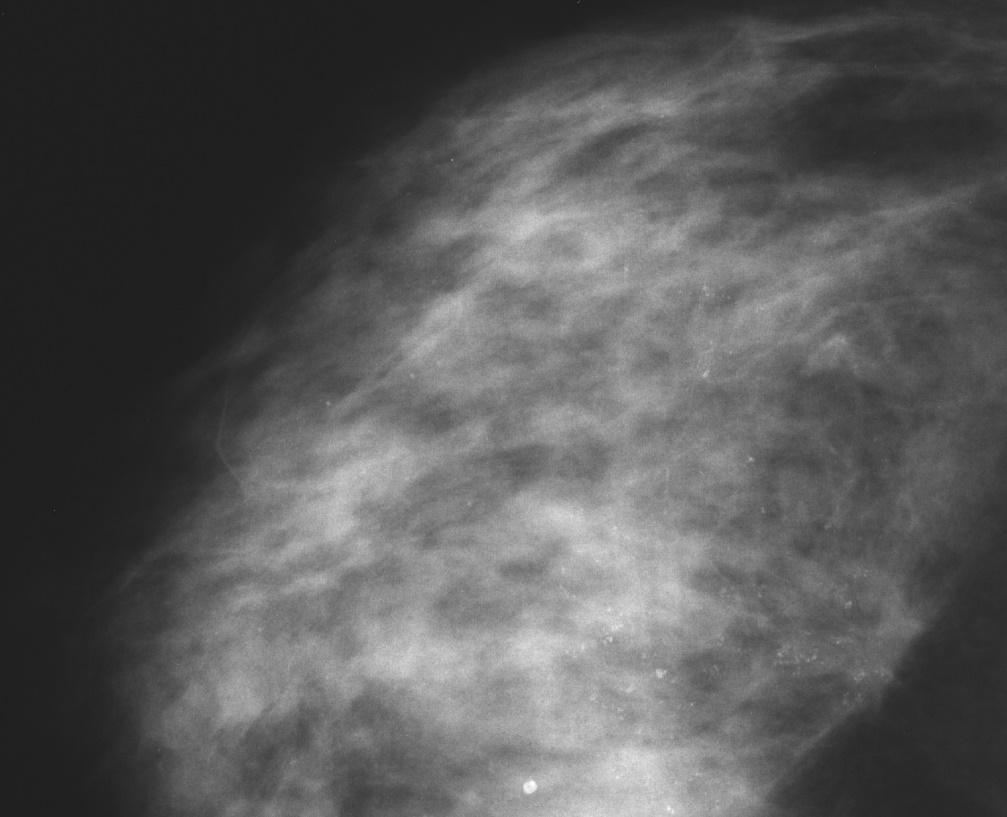
20 Left breast micro-calcification cancer Lcc Lb S8 20


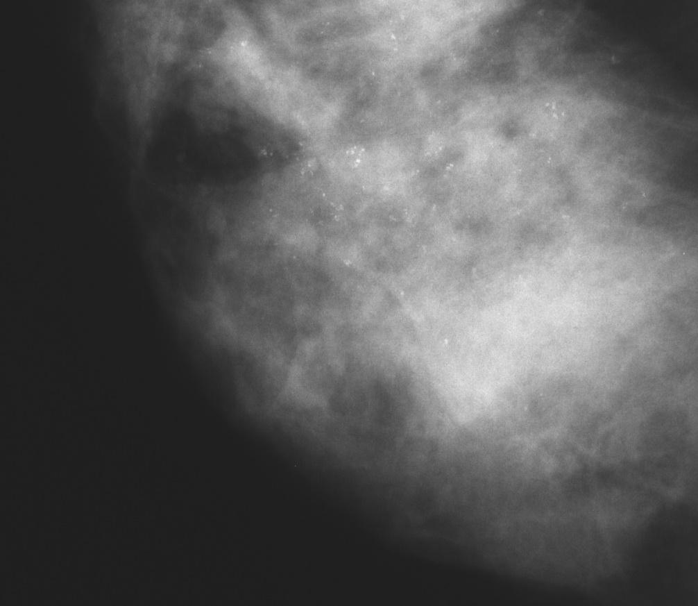
21 Right breast Rcc Rb S8 21
 and digitizes the image.")
22 2) Computed radiography (CR) The image plate (IP) is used instead of the X-ray film and the x-ray exposure is made. Next, the IP is run through a special laser scanner that reads (photo-stimulated luminescence - PSL) and digitizes the image. The digital image can then be treated using conventional digital image-processing software. Principle of IP operation A foil (thickness = ~0.2 mm) composed of Ba compounds (BaFX, X = Cl, Br, I) with small amount of Eu 2+ ions (BaFCl:Eu 2+ ) The mechanism of the PSL is illustrated by the energy level diagram of BaFBr:Eu 2+ S8 22
 reads and")

23 Laser scanner (He-Ne 633 nm) reads and digitizes the image Laser scanner operation S8 23
24 Advantages of image plate in comparison with X-ray film 1) Characteristic curve a wider dynamic range 2) One IP may be used many times 3) Digital image computed radiography (CR) S8 24


25 Analog and digital signals Digital image Digital image 3-dimensional matrix 2 dimensions are used to describe position + 3 rd dimension is used to give the values of the parameter Quantization Pixel = picture element total number of pixels = 512*512, 1024*1024, 2048*2048 (HR) Digitalization Remark: Analog image may be considered as the digital image characterize by the pixel size 0 and the qualitative value of the parameter Remark: Scanner is used to transforms analog to digital image S8 25
26 Remark: In the digital imaging the binary numerical system is commonly used. The binary (base 2) numerical system has 2 possible values, often represented as 0 or 1, for each place-value. In contrast, the decimal (base 10) numeral system has 10 possible values (0,1,2,3,4,5,6,7,8, or 9) for each place-value A bit is the smallest possible piece of information. A bit can have only two possible states: 1 or 0. We may find it useful to think of this in terms of "yes or no", "true or false", "on or off". By gathering groups of bits together, we can make much more complicated information. A byte is simply a group of 8 bits. 8 bits (1 byte) is equivalent to 2 8 = 256 possibilities. Intensity within each pixel = (8 14) bits Photon counting Poisson distribution Mean value = N N Standard Deviation = Final result of photon counting N ± N S8 26
27 The transformation between white and black can be realized with different number of intermediate grey scale levels. Practically it is difficult to distinguish between the first three bars despite the fact that the number of used grey level differs considerably (the numbers describe numbers of used grey levels) S8 27


28 3) Direct radiography DR typically captures the image directly onto a flat panel detector. Image processing or enhancement can be applied on DR images as well as CR images due to the digital format of each. A scintillator in the detector s outer layer, which is made from cesium iodide (CsI), converts X-ray to light S8 28
.")
29 The light is then channeled through the photodiode layer where it is converted to a digital output signal. Another possibility, the light is read out by optical fibers connected with charged couple device (CCD). S8 29
 cm 2 ).")

30 Charge Coupled Device Silicon wafer (thickness = ~0.3 mm, maximum dimension ~(5*5) cm 2 ). Maximum ~(5000*5000) electrodes are evaporated on the silicon surface each electrode is polarized and forms an electric potential well S8 30


 (30*40")
31 Direct radiology CCD detector (24*36 mm 2 ) (30*40 mm 2 ) S8 31
32 S8 32

 radiology")
33 Comparison of conventional (a) and direct (b) radiology S8 33
 fluoroscopic")

34 4) Fluoroscopic imaging system Disadvantage of the conventional (film as detector) radiography time consuming procedure to produce an instantaneous and continuous image (real time image) fluoroscopic imaging system C-arm system Modern fluorosope uses a device called an image intensifier to enhance the analog output of the real time x-ray image, where it is picked up by either a video or CCD camera. S8 34
35 Mechanism of action of an intensifier tube Four steps: 1) X-ray photons (~50 kev) many light photons (~2.5 ev) 2) Light photons emission of electrons (WOCs, WOBa) 3) DC voltage (~20 kv) accelerates towards the output screen 4) Output screen converts electron energy into bright visible image Net result of operation a much brighter image at the output screen that at the input phosphor gain of the intensifier tube ~10000 Remark: An increase of the number of light photons is achieved by the increase of the energy of electrons S8 35
 ln( 0 ) = µx I Before contrast injection M = µsds + µbdb + µbldn After contrast injection L = µsds + µbdb +")
36 Clinical application contrast radiology Contrast radiology physical background ds The law of absorption I I(x) = I0exp(-µx) ln( 0 ) = µx I Before contrast injection M = µsds + µbdb + µbldn After contrast injection L = µsds + µbdb + µjdn S8 36
 Routine contrast radiology coronarography Catheter femoral artery contrast medium")
37 Two possibilities: 1. µjdn >> µsds + µbdb routine contrast radiology 2. µjdn µsds + µbdb DSA (digital subtraction angiography) Routine contrast radiology coronarography Catheter femoral artery contrast medium (I Z = 53, Ba Z = 56 compounds) image of contrast distribution S8 37

38 S06_01 Remark: White-black reverse in comparison with X-ray film intensifier tube operation S06_14 S8 38
39 Digital subtraction angiography DSA O = L - M = (µj - µbl)dn µjdn S8 39
Unit thickness. Unit area. σ = NΔX = ΔI / I 0
 Unit thickness I 0 ΔI I σ = ΔI I 0 NΔX = ΔI / I 0 NΔX Unit area Δx Average probability of reaction with atom for the incident photons at unit area with the thickness of Delta-X Atom number at unit area
Introduction. Chapter 16 Diagnostic Radiology. Primary radiological image. Primary radiological image
 Introduction Chapter 16 Diagnostic Radiology Radiation Dosimetry I Text: H.E Johns and J.R. Cunningham, The physics of radiology, 4 th ed. http://www.utoledo.edu/med/depts/radther In diagnostic radiology
X-ray Imaging. PHYS Lecture. Carlos Vinhais. Departamento de Física Instituto Superior de Engenharia do Porto
 X-ray Imaging PHYS Lecture Carlos Vinhais Departamento de Física Instituto Superior de Engenharia do Porto cav@isep.ipp.pt Overview Projection Radiography Anode Angle Focal Spot Magnification Blurring
Radiology. Radiograph: Is the image of an object made with use of X- ray instead of light.
 Radiology د. اريج Lec. 3 X Ray Films Radiograph: Is the image of an object made with use of X- ray instead of light. Dental x- ray film: Is a recording media on which image of the object was made by exposing
X-ray Tube and Generator Basic principles and construction
 X-ray Tube and Generator Basic principles and construction Dr Slavik Tabakov - Production of X-rays and Patient Dose OBJECTIVES - X-ray tube construction - Anode - types, efficiency - Classical X-ray generator
BASICS OF FLUOROSCOPY
 Medical Physics Residents Training Program BASICS OF FLUOROSCOPY Dr. Khalid Alyousef, PhD Department of Medical Imaging King Abdulaziz Medical City- Riyadh Edison examining the hand of Clarence Dally with
X-ray Tube and Generator Basic principles and construction
 X-ray Tube and Generator Basic principles and construction Dr Slavik Tabakov - Production of X-rays OBJECTIVES - X-ray tube construction - Anode - types, efficiency - X-ray tube working characteristics
Acquisition, Processing and Display
 Acquisition, Processing and Display Terri L. Fauber, R.T. (R)(M) Department of Radiation Sciences School of Allied Health Professions Virginia Commonwealth University Topics Image Characteristics Image
Overview. Professor Roentgen was a Physicist!!! The Physics of Radiation Oncology X-ray Imaging
 The Physics of Radiation Oncology X-ray Imaging Charles E. Willis, Ph.D. DABR Associate Professor Department of Imaging Physics The University of Texas M.D. Anderson Cancer Center Houston, Texas Overview
X-rays in medical diagnostics
 X-rays in medical diagnostics S.Dolanski Babić 2017/18. History W.C.Röntgen (1845-1923) discovered a new type of radiation Nature, Jan. 23. 1896.; Science, Feb.14. 1896. X- rays: Induced the ionization
X-RAY IMAGING EE 472 F2017. Prof. Yasser Mostafa Kadah
 X-RAY IMAGING EE 472 F2017 Prof. Yasser Mostafa Kadah www.k-space.org Recommended Textbook Stewart C. Bushong, Radiologic Science for Technologists: Physics, Biology, and Protection, 10 th ed., Mosby,
Photons interaction with matter
 ب س م هللا الر ح من الر حیم Photons interaction with matter Ionization Ionization is the process of removing an electron from an electrically neutral atom to produce an ion pair. An ion is an atom or subatomic
CR Basics and FAQ. Overview. Historical Perspective
 Page: 1 of 6 CR Basics and FAQ Overview Computed Radiography is a term used to describe a system that electronically records a radiographic image. Computed Radiographic systems use unique image receptors
10/15/2012 SECTION III - CHAPTER 6 DIGITAL FLUOROSCOPY RADT 3463 COMPUTERIZED IMAGING
 RADT 3463 - COMPUTERIZED IMAGING Section III: Chapter 6 RADT 3463 Computerized Imaging 1 SECTION III - CHAPTER 6 DIGITAL FLUOROSCOPY RADT 3463 COMPUTERIZED IMAGING Section III: Chapter 6 RADT 3463 Computerized
10/26/2015. Study Harder
 This presentation is a professional collaboration of development time prepared by: Rex Christensen Terri Jurkiewicz and Diane Kawamura Study Harder CR detection is inefficient, inferior to film screen
10/3/2012. Study Harder
 This presentation is a professional collaboration of development time prepared by: Rex Christensen Terri Jurkiewicz and Diane Kawamura Study Harder CR detection is inefficient, inferior to film screen
SECTION I - CHAPTER 1 DIGITAL RADIOGRAPHY: AN OVERVIEW OF THE TEXT. Exam Content Specifications 8/22/2012 RADT 3463 COMPUTERIZED IMAGING
 RADT 3463 - COMPUTERIZED IMAGING Section I: Chapter 1 RADT 3463 Computerized Imaging 1 SECTION I - CHAPTER 1 DIGITAL RADIOGRAPHY: AN OVERVIEW OF THE TEXT RADT 3463 COMPUTERIZED IMAGING Section I: Chapter
Setting up digital imaging department!
 Outline Setting up digital imaging department! From screen/film to digital radiography PACS/Tele radiology Setting up digital department Digital Imaging Napapong Pongnapang, Ph.D. Department of Radiological
X-rays. X-rays are produced when electrons are accelerated and collide with a target. X-rays are sometimes characterized by the generating voltage
 X-rays Ouch! 1 X-rays X-rays are produced when electrons are accelerated and collide with a target Bremsstrahlung x-rays Characteristic x-rays X-rays are sometimes characterized by the generating voltage
PD233: Design of Biomedical Devices and Systems
 PD233: Design of Biomedical Devices and Systems (Lecture-8 Medical Imaging Systems) (Imaging Systems Basics, X-ray and CT) Dr. Manish Arora CPDM, IISc Course Website: http://cpdm.iisc.ac.in/utsaah/courses/
Explain what is meant by a photon and state one of its main properties [2]
![Explain what is meant by a photon and state one of its main properties [2]](/thumbs/80/82516318.jpg "Explain what is meant by a photon and state one of its main properties [2]") 1 (a) A patient has an X-ray scan taken in hospital. The high-energy X-ray photons interact with the atoms inside the body of the patient. Explain what is meant by a photon and state one of its main properties....
1 (a) A patient has an X-ray scan taken in hospital. The high-energy X-ray photons interact with the atoms inside the body of the patient. Explain what is meant by a photon and state one of its main properties....
Radiographic Testing (RT) [10]
![Radiographic Testing (RT) [10]](/thumbs/74/70215439.jpg "Radiographic Testing (RT) [10]") Radiographic Testing (RT) [10] Definition: An NDT method that utilizes x-rays or gamma radiation to detect discontinuities in materials, and to present their images on recording medium. 1> Electromagnetic
Radiographic Testing (RT) [10] Definition: An NDT method that utilizes x-rays or gamma radiation to detect discontinuities in materials, and to present their images on recording medium. 1> Electromagnetic
Amorphous Selenium Direct Radiography for Industrial Imaging
 DGZfP Proceedings BB 67-CD Paper 22 Computerized Tomography for Industrial Applications and Image Processing in Radiology March 15-17, 1999, Berlin, Germany Amorphous Selenium Direct Radiography for Industrial
Moving from film to digital: A study of digital x-ray benefits, challenges and best practices
 Moving from film to digital: A study of digital x-ray benefits, challenges and best practices H.U. Pöhler 1 and N. D Ademo 2 DÜRR NDT GmbH & Co. KG, Höpfigheimer Straße 22, Bietigheim-Bissingen, 74321,
DIGITAL IMAGE PROCESSING IN X-RAY IMAGING
 DIGITAL IMAGE PROCESSING IN X-RAY IMAGING Shalini Kumari 1, Bachan Prasad 2,Aliya Nasim 3 Department of Electronics And Communication Engineering R.V.S College of Engineering & Technology, Jamshedpur,
DALLA LUCE VISIBILE AI RAGGI X: NUOVI RIVELATORI DI IMMAGINI PER RAGGI X A DISCRIMINAZIONE IN ENERGIA ED APPLICAZIONI
 DALLA LUCE VISIBILE AI RAGGI X: NUOVI RIVELATORI DI IMMAGINI PER RAGGI X A DISCRIMINAZIONE IN ENERGIA ED APPLICAZIONI D. Pacella ENEA - Frascati LIMS, Frascati 14-15 ottobre 2015 Come per la fotografia:
Do you have any other questions? Please call us at (Toll Free) or , or
 or , or") INSTRUCTIONS Read the appropriate course/ textbook. This is an open book test. A score of 75% or higher is needed to receive CE credit. You will have a maximum of three attempts to pass this course. Please
INSTRUCTIONS Read the appropriate course/ textbook. This is an open book test. A score of 75% or higher is needed to receive CE credit. You will have a maximum of three attempts to pass this course. Please
LECTURE 1 The Radiographic Image
 LECTURE 1 The Radiographic Image Prepared by:- KAMARUL AMIN ABDULLAH @ ABU BAKAR UiTM Faculty of Health Sciences Medical Imaging Department 11/23/2011 KAMARUL AMIN (C) 1 Lesson Objectives At the end of
Current technology in digital image production (CR/DR and other modalities) Jaroonroj Wongnil 25 Mar 2016
 Jaroonroj Wongnil 25 Mar 2016") Current technology in digital image production (CR/DR and other modalities) Jaroonroj Wongnil 25 Mar 2016 Current technology in digital image production (CR/DR and other modalities) 2/ Overview Digital
Current technology in digital image production (CR/DR and other modalities) Jaroonroj Wongnil 25 Mar 2016 Current technology in digital image production (CR/DR and other modalities) 2/ Overview Digital
PRACTICAL CONSIDERATIONS AND EFFECTS OF METALLIC SCREEN FLUORESCENCE AND BACKSCATTER CONTROL IN GAMMA COMPUTED RADIOGRAPHY
 19 th World Conference on Non-Destructive Testing 2016 PRACTICAL CONSIDERATIONS AND EFFECTS OF METALLIC SCREEN FLUORESCENCE AND BACKSCATTER CONTROL IN GAMMA COMPUTED RADIOGRAPHY Steven MANGO 1 1 Carestream
Examination of Pipe Welds by Image Plate Based Computed Radiography System
 Examination of Pipe Welds by Image Plate Based Computed Radiography System Sanjoy Das, M.S.Rana, Benny Sebastian, D. Mukherjee and K.K. Abdulla Atomic Fuels Division Bhabha Atomic Research Centre Mumbai
X-RAY FLUOROSCOPY IMAGING SYSTEMS. Dr Slavik Tabakov. Luminescence: Dept. Medical Eng. & Physics King s College London
 X-RAY FLUOROSCOPY IMAGING SYSTEMS Dr Slavik Tabakov OBJECTIVES - Image Intensifier construction - Input window - Accelerating and focusing electrodes - Output window - Conversion factor - II characteristics
Mammography is a radiographic procedure specially designed for detecting breast pathology Approximately 1 woman in 8 will develop breast cancer over
 Mammography is a radiographic procedure specially designed for detecting breast pathology Approximately 1 woman in 8 will develop breast cancer over a lifetime Breast cancer screening programs rely on
3/31/2011. Objectives. Emory University. Historical Development. Historical Development. Historical Development
 Teaching Radiographic Technique in a Digital Imaging Paradigm Objectives 1. Discuss the historical development of digital imaging. Dawn Couch Moore, M.M.Sc., RT(R) Assistant Professor and Director Emory
Medical Imaging. X-rays, CT/CAT scans, Ultrasound, Magnetic Resonance Imaging
 Medical Imaging X-rays, CT/CAT scans, Ultrasound, Magnetic Resonance Imaging From: Physics for the IB Diploma Coursebook 6th Edition by Tsokos, Hoeben and Headlee And Higher Level Physics 2 nd Edition
Computed Radiography
 BAM Berlin Computed Radiography --INDE 2007, Kalpakkam, India -- Uwe Zscherpel, Uwe Ewert BAM Berlin, Division VIII.3 Requests Requests and and information information to: to: Dr. Dr. U. U. Zscherpel Zscherpel
MC SIMULATION OF SCATTER INTENSITIES IN A CONE-BEAM CT SYSTEM EMPLOYING A 450 kv X-RAY TUBE
 MC SIMULATION OF SCATTER INTENSITIES IN A CONE-BEAM CT SYSTEM EMPLOYING A 450 kv X-RAY TUBE A. Miceli ab, R. Thierry a, A. Flisch a, U. Sennhauser a, F. Casali b a Empa - Swiss Federal Laboratories for
AN ABSTRACT OF THE THESIS OF. W. Scott Helms for the degree of Master of Science in Radiation Health Physics
 AN ABSTRACT OF THE THESIS OF W. Scott Helms for the degree of Master of Science in Radiation Health Physics presented on November 24, 2014 Title: A Quantitative Comparison of Cardiovascular Imaging Systems
ABSORBED DOSE DISTRIBUTIONS USING THE ISODENSITOMETRIC METHOD FOR EXPOSURES WITH FILTER EMPLOYED FOR MAMMOGRAPHIES
 Romanian Reports in Physics, Vol. 65, No. 1, P. 168 177, 213 ABSORBED DOSE DISTRIBUTIONS USING THE ISODENSITOMETRIC METHOD FOR EXPOSURES WITH FILTER EMPLOYED FOR MAMMOGRAPHIES F. SCARLAT 1, A. SCARISOREANU
Radiographic sensitivity improved by optimized high resolution X -ray detector design.
 DIR 2007 - International Symposium on Digital industrial Radiology and Computed Tomography, June 25-27, 2007, Lyon, France Radiographic sensitivity improved by optimized high resolution X -ray detector
PERFORMANCE CHARACTERIZATION OF AMORPHOUS SILICON DIGITAL DETECTOR ARRAYS FOR GAMMA RADIOGRAPHY
 12 th A-PCNDT 2006 Asia-Pacific Conference on NDT, 5 th 10 th Nov 2006, Auckland, New Zealand PERFORMANCE CHARACTERIZATION OF AMORPHOUS SILICON DIGITAL DETECTOR ARRAYS FOR GAMMA RADIOGRAPHY Rajashekar
X-RAY. Lecture No.4. Image Characteristics:
 Lecture No.4 X-RAY أ.م.د. اسامة مراد ابراهيم Image Characteristics: *Radiographic density: It s the degree of blackness of the film. when a film is exposed by an x-ray beam (or by light in case of screenfilm
LlIGHT REVIEW PART 2 DOWNLOAD, PRINT and submit for 100 points
 WRITE ON SCANTRON WITH NUMBER 2 PENCIL DO NOT WRITE ON THIS TEST LlIGHT REVIEW PART 2 DOWNLOAD, PRINT and submit for 100 points Multiple Choice Identify the choice that best completes the statement or
Radiology Physics Lectures: Digital Radiography. Digital Radiography. D. J. Hall, Ph.D. x20893
 Digital Radiography D. J. Hall, Ph.D. x20893 djhall@ucsd.edu Background Common Digital Modalities Digital Chest Radiograph - 4096 x 4096 x 12 bit CT - 512 x 512 x 12 bit SPECT - 128 x 128 x 8 bit MRI -
DIGITAL RADIOGRAPHY. Digital radiography is a film-less technology used to record radiographic images.
 DIGITAL RADIOGRAPHY Digital radiography is a film-less technology used to record radiographic images. 1 The purpose of digital imaging is to generate images that can be used in the diagnosis and assessment
Chapters 1-3. Chapter 1: Introduction and applications of photogrammetry Chapter 2: Electro-magnetic radiation. Chapter 3: Basic optics
 Chapters 1-3 Chapter 1: Introduction and applications of photogrammetry Chapter 2: Electro-magnetic radiation Radiation sources Classification of remote sensing systems (passive & active) Electromagnetic
RADIOGRAPHY TERMS TO KNOW SELF STUDY DENTALELLE TUTORING
 RADIOGRAPHY TERMS TO KNOW SELF STUDY DENTALELLE TUTORING PLEASE NOTE You DO NOT need to study these for the board exam if this is why you bought our Radiography course, however if you come across any terms
Nuclear Associates
 Nuclear Associates 76-700 Digital Subtraction Angiography Phantom Users Manual March 2005 Manual No. 76-700-1 Rev. 2 2004, 2005 Fluke Corporation, All rights reserved. Printed in U.S.A. All product names
Veterinary Science Preparatory Training for the Veterinary Assistant. Floron C. Faries, Jr., DVM, MS
 Veterinary Science Preparatory Training for the Veterinary Assistant Floron C. Faries, Jr., DVM, MS Radiology Floron C. Faries, Jr., DVM, MS Objectives Determine the appropriate machine settings for making
Medical Imaging: A Look inside. Medical Imaging. Medical Imaging. Visible Human Project
 Medical Imaging: A Look inside Medical Imaging Allows physicians to see what had previously been unseeable: internal organs and tissues, bones, a beating heart, etc. Allows physicians to: detect brain
The importance of radiation quality for optimisation in radiology
 Available online at http://www.biij.org/2007/2/e38 doi: 10.2349/biij.3.2.e38 biij Biomedical Imaging and Intervention Journal COMMENTARY The importance of radiation quality for optimisation in radiology
Mammography: Physics of Imaging
 Mammography: Physics of Imaging Robert G. Gould, Sc.D. Professor and Vice Chair Department of Radiology and Biomedical Imaging University of California San Francisco, California Mammographic Imaging: Uniqueness
Photomultiplier Tube
 Nuclear Medicine Uses a device known as a Gamma Camera. Also known as a Scintillation or Anger Camera. Detects the release of gamma rays from Radionuclide. The radionuclide can be injected, inhaled or
Film Replacement in Radiographic Weld Inspection The New ISO Standard
 BAM Berlin Film Replacement in Radiographic Weld Inspection The New ISO Standard 17636-2 Uwe Ewert, Uwe Zscherpel, Mirko Jechow Requests and information to: uwez@bam.de 1 Outline - The 3 essential parameters
Multiple Choice Identify the letter of the choice that best completes the statement or answers the question.
 RA110 test 3 Multiple Choice Identify the letter of the choice that best completes the statement or answers the question. 1. An object 35 cm in width is radiographed at 100 cm SID and at a 50 cm SOD. What
Partial Replication of Storms/Scanlan Glow Discharge Radiation
 Partial Replication of Storms/Scanlan Glow Discharge Radiation Rick Cantwell and Matt McConnell Coolescence, LLC March 2008 Introduction The Storms/Scanlan paper 1 presented at the 8 th international workshop
Fluoroscopy - Chapter 9
 Fluoroscopy - Chapter 9 Kalpana Kanal, Ph.D., DABR Lecturer, Diagnostic Physics Dept. of Radiology UW Medicine a copy of this lecture may be found at: http://courses.washington.edu/radxphys/physicscourse04-05.html
IBEX MATERIALS DETECTION TECHNOLOGY
 WHITE PAPER: IBEX MATERIALS DETECTION TECHNOLOGY IBEX Innovations Ltd. Registered in England and Wales: 07208355 Address: Discovery 2, NETPark, William Armstrong Way, Sedgefield, TS21 3FH, UK Patents held
1. Carlton, Richard R., and Arlene M. Adler. Principles of Radiographic Imaging: An Art and a Science, 5th edition (2013).
.") CODE: RADT 151 INSTITUTE: Health Science TITLE: Radiographic Exposure DEPARTMENT: Radiologic Technology COURSE DESCRIPTION: This course covers the principles of radiographic exposure selection and manipulation
CODE: RADT 151 INSTITUTE: Health Science TITLE: Radiographic Exposure DEPARTMENT: Radiologic Technology COURSE DESCRIPTION: This course covers the principles of radiographic exposure selection and manipulation
- KiloVoltage. Technique 101: Getting Back to Basics
 Why do I need to know technique? Technique 101: Getting Back to Basics Presented by: Thomas G. Sandridge, M.S., M.Ed., R.T.(R) Program Director Northwestern Memorial Hospital School of Radiography Chicago,
STUDENT REVIEW QUESTION SET K CR/DR CONTENT AREA
 STUDENT REVIEW QUESTION SET K CR/DR CONTENT AREA RADT 2913 COMPREHENSIVE REVIEW 1 The CR cassette is backed by aluminum that: A. reflects x-rays B. absorbs x-rays C. captures the image D. transmits x-rays
Phase Imaging Using Focused Polycapillary Optics
 Phase Imaging Using Focused Polycapillary Optics Sajid Bashir, Sajjad Tahir, Jonathan C. Petruccelli, C.A. MacDonald Dept. of Physics, University at Albany, Albany, New York Abstract Contrast in conventional
Computed Radiography of Resistance Temperature Sensor for Indian PHWR
 National Seminar & Exhibition on Non-Destructive Evaluation, NDE 2014, Pune, December 4-6, 2014 (NDE-India 2014) Vol.20 No.6 (June 2015) - The e-journal of Nondestructive Testing - ISSN 1435-4934 www.ndt.net/?id=17831
1-1. GENERAL 1-2. DISCOVERY OF X-RAYS
 1-1. GENERAL Radiography is a highly technical field, indispensable to the modern dental practice, but presenting many potential hazards. The dental radiographic specialist must be thoroughly familiar
SPRINGFIELD TECHNICAL COMMUNITY COLLEGE ACADEMIC AFFAIRS
 SPRINGFIELD TECHNICAL COMMUNITY COLLEGE ACADEMIC AFFAIRS Course Number: RADG 212 Department: Radiography Course Title: Equip. Operation & Maint. Semester: Spring Year: 1997 Objectives/ Unit One: The X-ray
Visibility of Detail
 Visibility of Detail Radiographic Quality Quality radiographic images represents the, and information is for diagnosis. The of the anatomic structures and the accuracy of their ( ) determine the overall
Chapters 1-3. Chapter 1: Introduction and applications of photogrammetry Chapter 2: Electro-magnetic radiation. Chapter 3: Basic optics
 Chapters 1-3 Chapter 1: Introduction and applications of photogrammetry Chapter 2: Electro-magnetic radiation Radiation sources Classification of remote sensing systems (passive & active) Electromagnetic
V SALAI SELVAM, AP & HOD, ECE, Sriram Engg. College, Perumalpattu 1 MEDICAL ELECTRONICS UNIT IV
 V SALAI SELVAM, AP & HOD, ECE, Sriram Engg. College, Perumalpattu 1 MEDICAL ELECTRONICS UNIT IV Ionizing and non-ionizing radiations: The radiation that ionizes the gases through which it travels is known
COMPUTED TOMOGRAPHY 1
 COMPUTED TOMOGRAPHY 1 Why CT? Conventional X ray picture of a chest 2 Introduction Why CT? In a normal X-ray picture, most soft tissue doesn't show up clearly. To focus in on organs, or to examine the
Today s Outline - January 25, C. Segre (IIT) PHYS Spring 2018 January 25, / 26
 PHYS Spring 2018 January 25, / 26") Today s Outline - January 25, 2018 C. Segre (IIT) PHYS 570 - Spring 2018 January 25, 2018 1 / 26 Today s Outline - January 25, 2018 HW #2 C. Segre (IIT) PHYS 570 - Spring 2018 January 25, 2018 1 / 26 Today
Today s Outline - January 25, 2018 C. Segre (IIT) PHYS 570 - Spring 2018 January 25, 2018 1 / 26 Today s Outline - January 25, 2018 HW #2 C. Segre (IIT) PHYS 570 - Spring 2018 January 25, 2018 1 / 26 Today
Digital Imaging started in the 1972 with Digital subtraction angiography Clinical digital imaging was employed from the 1980 ~ 37 years ago Amount of
 Digital Imaging started in the 1972 with Digital subtraction angiography Clinical digital imaging was employed from the 1980 ~ 37 years ago Amount of radiation to the population due to Medical Imaging
NPTEL NPTEL ONLINE COURSE. NPTEL Online Certification Course (NOC) NPTEL. Theory and Practice of Non Destructive Testing
 NPTEL. Theory and Practice of Non Destructive Testing") NPTEL NPTEL ONLINE COURSE NPTEL Online Certification Course (NOC) NPTEL Theory and Practice of Non Destructive Testing Dr. Ranjit Bauri Dept. of Metallurgical & Materials Engineering IIT Madras, Chennai
NPTEL NPTEL ONLINE COURSE NPTEL Online Certification Course (NOC) NPTEL Theory and Practice of Non Destructive Testing Dr. Ranjit Bauri Dept. of Metallurgical & Materials Engineering IIT Madras, Chennai
Gamma Spectrometer Initial Project Proposal
 Gamma Spectrometer Initial Project Proposal Group 9 Aman Kataria Johnny Klarenbeek Dean Sullivan David Valentine Introduction There are currently two main types of gamma radiation detectors used for gamma
RAD 150 RADIOLOGIC EXPOSURE TECHNIQUE II
 RAD 150 RADIOLOGIC EXPOSURE TECHNIQUE II APPROVED 12/O2/2011 EFFECTIVE SPRING 2013-14 Prefix & Number RAD 150 Course Title: Radiologic Exposure Technique II & Lab Purpose of this submission: New Change/Updated
New Detectors for X-Ray Metal Thickness Measuring
 ECNDT 2006 - Poster 132 New Detectors for X-Ray Metal Thickness Measuring Boris V. ARTEMIEV, Alexander I. MASLOV, Association SPEKTR- GROUP, Moscow, Russia Abstract. X-ray thickness measuring instruments
Digital Detector Array Image Quality for Various GOS Scintillators
 Digital Detector Array Image Quality for Various GOS Scintillators More info about this article: http://www.ndt.net/?id=22768 Brian S. White 1, Mark E. Shafer 2, William H. Russel 3, Eric Fallet 4, Jacques
Essentials of Digital Imaging
 Essentials of Digital Imaging Module 1 Transcript 2016 ASRT. All rights reserved. Essentials of Digital Imaging Module 1 Fundamentals 1. ASRT Animation 2. Welcome Welcome to Essentials of Digital Imaging
OPTI510R: Photonics. Khanh Kieu College of Optical Sciences, University of Arizona Meinel building R.626
 OPTI510R: Photonics Khanh Kieu College of Optical Sciences, University of Arizona kkieu@optics.arizona.edu Meinel building R.626 Photodetectors Introduction Most important characteristics Photodetector
Chemistry Instrumental Analysis Lecture 7. Chem 4631
 Chemistry 4631 Instrumental Analysis Lecture 7 UV to IR Components of Optical Basic components of spectroscopic instruments: stable source of radiant energy transparent container to hold sample device
80 Physics Essentials Workbook Stage 2 Physics
 80 Physics Essentials Workbook Stage 2 Physics the thickness of the tissue: Obviously, the thicker the tissue through which the X-rays have to pass the more they will be absorbed from the beam passing
X-RAYS - NO UNAUTHORISED ENTRY
 Licencing of premises Premises Refer Guidelines A radiation warning sign and warning notice, X-RAYS - NO UNAUTHORISED ENTRY must be displayed at all entrances leading to the rooms where x-ray units are
Gas scintillation Glass GEM detector for high-resolution X-ray imaging and CT
 Gas scintillation Glass GEM detector for high-resolution X-ray imaging and CT Takeshi Fujiwara 1, Yuki Mitsuya 2, Hiroyuki Takahashi 2, and Hiroyuki Toyokawa 2 1 National Institute of Advanced Industrial
Beam-Restricting Devices
 Beam-Restricting Devices Three factors contribute to an increase in scatter radiation: Increased kvp Increased Field Size Increased Patient or Body Part Size. X-ray Interactions a some interact with the
SYLLABUS. 1. Identification of Subject:
 SYLLABUS Date/ Revision : 30 January 2017/1 Faculty : Life Sciences Approval : Dean, Faculty of Life Sciences SUBJECT : Biophysics 1. Identification of Subject: Name of Subject : Biophysics Code of Subject
Real Time Linear Array Imaging. Brian Caccamise
 Real Time Linear Array Imaging Brian Caccamise 1 Real Time Linear Array Imaging What is Real Time Linear Array Imaging? Or Real Time Radiography (RTR)? 2 Real Time Linear Array Imaging It s Not This! Shoe
NDE SOLUTIONS RADIOGRAPHY COURSE OUTLINE
 NDE SOLUTIONS RADIOGRAPHY COURSE OUTLINE 80 Hour Course Length 1.0 NDT Qualification and Introduction (3 Hours) 1.1 NDT Introduction 1.2 NDT Qualification and Certification 1.2.1 NAS 410 1.2.2 SNT-TC-1A
SECTION I - CHAPTER 2 DIGITAL IMAGING PROCESSING CONCEPTS
 RADT 3463 - COMPUTERIZED IMAGING Section I: Chapter 2 RADT 3463 Computerized Imaging 1 SECTION I - CHAPTER 2 DIGITAL IMAGING PROCESSING CONCEPTS RADT 3463 COMPUTERIZED IMAGING Section I: Chapter 2 RADT
STEREOTACTIC BREAST BIOPSY EQUIPMENT SURVEYS
 STEREOTACTIC BREAST BIOPSY EQUIPMENT SURVEYS JAMES A. TOMLINSON, M.S. Diagnostic Radiological Physicist American Board of Radiology Certified Medical Physics Consultants, Inc. Bio 28 yrs experience 100%
High Performance. Image Intensifiers
 High Performance Image Intensifiers Image Intensifier Diodes PROXIFIER and MCP Image Intensifiers MCP-PROXIFIER Features Outstanding gain up to > 10 8 W/W High Quantum Efficiency up to 35 % Excellent Resolution
Studies on reduction of exposure dose using digital scattered X-ray removal processing
 Studies on reduction of exposure dose using digital scattered X-ray removal processing Poster No.: C-1834 Congress: ECR 2015 Type: Scientific Exhibit Authors: K. Kashiyama, M. Funahashi, T. Nakaoka, T.
Studies on reduction of exposure dose using digital scattered X-ray removal processing
 Studies on reduction of exposure dose using digital scattered X-ray removal processing Poster No.: C-1834 Congress: ECR 2015 Type: Scientific Exhibit Authors: K. Kashiyama, M. Funahashi, T. Nakaoka, T.
Calibration of KAP meters
 Calibration of KAP meters Alexandr Malusek! Division of Radiological Sciences Department of Medical and Health Sciences Linköping University! 2014-04-15 1 Outline 1. KAP meter construction 2. Air kerma-area
Dose Reduction and Image Preservation After the Introduction of a 0.1 mm Cu Filter into the LODOX Statscan unit above 110 kvp
 Dose Reduction and Image Preservation After the Introduction of a into the LODOX Statscan unit above 110 kvp Abstract: CJ Trauernicht 1, C Rall 1, T Perks 2, G Maree 1, E Hering 1, S Steiner 3 1) Division
Chapter 23 Study Questions Name: Class:
 Chapter 23 Study Questions Name: Class: Multiple Choice Identify the letter of the choice that best completes the statement or answers the question. 1. When you look at yourself in a plane mirror, you
Digital Imaging Rochester Institute of Technology
 Digital Imaging 1999 Rochester Institute of Technology So Far... camera AgX film processing image AgX photographic film captures image formed by the optical elements (lens). Unfortunately, the processing
Components of Optical Instruments
 Components of Optical Instruments General Design of Optical Instruments Sources of Radiation Wavelength Selectors (Filters, Monochromators, Interferometers) Sample Containers Radiation Transducers (Detectors)
EE119 Introduction to Optical Engineering Fall 2009 Final Exam. Name:
 EE119 Introduction to Optical Engineering Fall 2009 Final Exam Name: SID: CLOSED BOOK. THREE 8 1/2 X 11 SHEETS OF NOTES, AND SCIENTIFIC POCKET CALCULATOR PERMITTED. TIME ALLOTTED: 180 MINUTES Fundamental
Spectroscopy in the UV and Visible: Instrumentation. Spectroscopy in the UV and Visible: Instrumentation
 Spectroscopy in the UV and Visible: Instrumentation Typical UV-VIS instrument 1 Source - Disperser Sample (Blank) Detector Readout Monitor the relative response of the sample signal to the blank Transmittance
Multiple Choice Identify the letter of the choice that best completes the statement or answers the question.
 RA202 image production class two Multiple Choice Identify the letter of the choice that best completes the statement or answers the question. 1. What removes excess chemistry from the film prior to it
Basis of Computed Radiography & PACS
 Basis of Computed Radiography & PACS Slavik Tabakov Computed Radiography (CR) refers to new types of X-ray detectors (i.e. replaces the X-ray Film) The CR output media is a digital image, which can be