Radiology Physics Lectures: Digital Radiography. Digital Radiography. D. J. Hall, Ph.D. x20893
|
|
|
- Angel Robinson
- 6 years ago
- Views:
Transcription
1 Digital Radiography D. J. Hall, Ph.D. x20893
2 Background Common Digital Modalities Digital Chest Radiograph x 4096 x 12 bit CT x 512 x 12 bit SPECT x 128 x 8 bit MRI x 256 x 8 bit US x 512 x 8 or 24 bits Viewing Station 2k x 2k x 12 bits
3 Computed Radiography Photostimulable phosphor Energy trapped on plate Readout at later time BaFBr or BaFI Flexible plate stored in cassette Exposed to x-rays like film Processed in special reader
4 Computed Radiography Photostimulable phosphor - readout

5 Computed Radiography Reader
6 Computed Radiography - emission wavelengths
7 Computed Radiography - How it works!
8 Computed Radiography - Dynamic Range
9 Charge Coupled Devices Make images from visible light Made of Silicon Visible light liberates electrons Electrons accumulate in individual pixel cells Accumulated charge readout pixel by pixel Requires coupling between light source and CCD Used for fluoroscopy and cine-angiography Large FOV imaging loses light Proportional to areas of CCD and light source
10 Charge Coupled Devices - readout

11 Charge Coupled Device - with Image Intensifier


12 Charge Coupled Device - with fiberoptic coupling

13 Charge Coupled Device - with lens coupling
14 Flat Panel Detectors Photodetector sensitive to visible light Coupled to x-ray intensifying screen Gd 2 O 2 S or CsI CsI grown in columnar crystals to improve efficiency X-rays absorbed in screen give off visible light Visible light absorbed in photodetector Fill factor determines efficiency Detector size determines best spatial resolution 125 μm -> 4 cycles/mm 100 μm -> 5 cycles/mm

15 Flat Panel Detectors - general configuration

16 Flat Panel Detectors - fill factor

17 Flat Panel Detectors - readout process
18 Direct detection flat panel systems Photoconductor layer on TFT array Direct detection of x-rays - x-ray to electron Not x-ray to light to electron Typically Selenium Electrons follow E-field lines Relatively thick detectors
19 Direct detection flat panel systems
20 Digital mammography Full-field digital mammography Mosaic of CCD detectors TFT flat panel detectors Slot-scan detector 1D detector array

21 Digital mammography - slot scan detector


22 Radiology Physics Lectures: Digital Radiography Digital Mammography - FFDM Digital Imaging Detector Large dynamic range Reasonable spatial resolution (300 μm) Digital image -> input to CAD system Expensive ~ $300k


23 Digital Mammography - FFDM Digital Detector Film-Screen

24 Digital mammography - slot scan detector readout
25 Patient Dose Considerations - Film vs. Digital Need comparable DQE Detected Quantum Efficiency CR ~ equivalent to 200 speed film system Flat Panel Detectors ~ 2x - 3x faster than CR
26 Hard Copy vs. Soft Copy Resolution varies with modality: SPECT & PET x 128 pixels MRI x 256 pixels US x 512 pixels CT x 512 pixels Digital angio x 1024 pixels Digital chest x 4300 pixels Digital mammo x 4000 pixels Monitor must reproduce data fidelity
27 Image Processing Correct for flaw in image Dead pixels Non-linearities in transfer function Dark noise Non-uniformities across field Store flat-field image for corrections


28 Image Processing - correction raw corrected


29 Image Processing - global adjustment wide narrow inverted
30 Image Processing - convolution Mathematical process Permits image enhancement Computationally efficient Various operations (kernels): Soft tissue - smoothing Bone - edge enhancement
31 Image Processing - convolution

32 Image Processing - convolution original 11x11 smooth edge enhance edge - smooth

33 Image Processing - histogram equalization original equalized
34 Contrast versus Spatial Resolution
35 Geometric Tomography

36 Geometric Tomography
37 Geometric Tomography - slice sensitivity profile
38 Digital Tomosynthesis
39 Temporal Subtraction
40 Dual Energy Subtraction One-pulse sandwiched detector Two-pulse single detector

41 Dual Energy Subtraction Low High Bone Soft tissue
42 Review Question
43 Review Question
44 Review Question
45 Review Question
46 Review Question


47 Review Question
Current technology in digital image production (CR/DR and other modalities) Jaroonroj Wongnil 25 Mar 2016
 Jaroonroj Wongnil 25 Mar 2016") Current technology in digital image production (CR/DR and other modalities) Jaroonroj Wongnil 25 Mar 2016 Current technology in digital image production (CR/DR and other modalities) 2/ Overview Digital
Current technology in digital image production (CR/DR and other modalities) Jaroonroj Wongnil 25 Mar 2016 Current technology in digital image production (CR/DR and other modalities) 2/ Overview Digital
10/26/2015. Study Harder
 This presentation is a professional collaboration of development time prepared by: Rex Christensen Terri Jurkiewicz and Diane Kawamura Study Harder CR detection is inefficient, inferior to film screen
10/3/2012. Study Harder
 This presentation is a professional collaboration of development time prepared by: Rex Christensen Terri Jurkiewicz and Diane Kawamura Study Harder CR detection is inefficient, inferior to film screen
Digital radiography: Practical advantages of Digital Radiography. Practical Advantages in image quality
 Digital radiography: Digital radiography is set to become the most common form of processing radiographic images in the next 10 years. This is due to a number of practical and image quality issues. Practical
Setting up digital imaging department!
 Outline Setting up digital imaging department! From screen/film to digital radiography PACS/Tele radiology Setting up digital department Digital Imaging Napapong Pongnapang, Ph.D. Department of Radiological
Introduction. Chapter 16 Diagnostic Radiology. Primary radiological image. Primary radiological image
 Introduction Chapter 16 Diagnostic Radiology Radiation Dosimetry I Text: H.E Johns and J.R. Cunningham, The physics of radiology, 4 th ed. http://www.utoledo.edu/med/depts/radther In diagnostic radiology
Do you have any other questions? Please call us at (Toll Free) or , or
 or , or") INSTRUCTIONS Read the appropriate course/ textbook. This is an open book test. A score of 75% or higher is needed to receive CE credit. You will have a maximum of three attempts to pass this course. Please
INSTRUCTIONS Read the appropriate course/ textbook. This is an open book test. A score of 75% or higher is needed to receive CE credit. You will have a maximum of three attempts to pass this course. Please
Digital Image Management: the Basics
 Digital Image Management: the Basics Napapong Pongnapang, Ph.D. Department of Radiological Technology Faculty of Medical Technology Mahidol University Outline From screen/film to digital radiography PACS/Tele
SECTION I - CHAPTER 1 DIGITAL RADIOGRAPHY: AN OVERVIEW OF THE TEXT. Exam Content Specifications 8/22/2012 RADT 3463 COMPUTERIZED IMAGING
 RADT 3463 - COMPUTERIZED IMAGING Section I: Chapter 1 RADT 3463 Computerized Imaging 1 SECTION I - CHAPTER 1 DIGITAL RADIOGRAPHY: AN OVERVIEW OF THE TEXT RADT 3463 COMPUTERIZED IMAGING Section I: Chapter
X-Ray Medical Imaging and Pixel detectors
 X-Ray Medical Imaging and Pixel detectors PIXEL 2000 Genova, June 5-8 th 2000 J.P.Moy, TRI XELL, Moirans, France 1 OUTLINE - X-ray medical imaging. The requirements, some particular features - Present
Overview. Professor Roentgen was a Physicist!!! The Physics of Radiation Oncology X-ray Imaging
 The Physics of Radiation Oncology X-ray Imaging Charles E. Willis, Ph.D. DABR Associate Professor Department of Imaging Physics The University of Texas M.D. Anderson Cancer Center Houston, Texas Overview
Mammography: Physics of Imaging
 Mammography: Physics of Imaging Robert G. Gould, Sc.D. Professor and Vice Chair Department of Radiology and Biomedical Imaging University of California San Francisco, California Mammographic Imaging: Uniqueness
Amorphous Selenium Direct Radiography for Industrial Imaging
 DGZfP Proceedings BB 67-CD Paper 22 Computerized Tomography for Industrial Applications and Image Processing in Radiology March 15-17, 1999, Berlin, Germany Amorphous Selenium Direct Radiography for Industrial
Basis of Computed Radiography & PACS
 Basis of Computed Radiography & PACS Slavik Tabakov Computed Radiography (CR) refers to new types of X-ray detectors (i.e. replaces the X-ray Film) The CR output media is a digital image, which can be
3/31/2011. Objectives. Emory University. Historical Development. Historical Development. Historical Development
 Teaching Radiographic Technique in a Digital Imaging Paradigm Objectives 1. Discuss the historical development of digital imaging. Dawn Couch Moore, M.M.Sc., RT(R) Assistant Professor and Director Emory
Acquisition, Processing and Display
 Acquisition, Processing and Display Terri L. Fauber, R.T. (R)(M) Department of Radiation Sciences School of Allied Health Professions Virginia Commonwealth University Topics Image Characteristics Image
SYLLABUS. 1. Identification of Subject:
 SYLLABUS Date/ Revision : 30 January 2017/1 Faculty : Life Sciences Approval : Dean, Faculty of Life Sciences SUBJECT : Biophysics 1. Identification of Subject: Name of Subject : Biophysics Code of Subject
Features and Weaknesses of Phantoms for CR/DR System Testing
 Physics testing of image detectors Parameters to test Features and Weaknesses of Phantoms for CR/DR System Testing Spatial resolution Contrast resolution Uniformity/geometric distortion Dose response/signal
SYLLABUS. TITLE: Equipment Operation I. DEPARTMENT: Radiologic Technology
 CODE: RADT 156 INSTITUTE: Health Science TITLE: Equipment Operation I DEPARTMENT: Radiologic Technology COURSE DESCRIPTION: This course covers the principles of equipment operation and maintenance of radiographic
SECTION I - CHAPTER 2 DIGITAL IMAGING PROCESSING CONCEPTS
 RADT 3463 - COMPUTERIZED IMAGING Section I: Chapter 2 RADT 3463 Computerized Imaging 1 SECTION I - CHAPTER 2 DIGITAL IMAGING PROCESSING CONCEPTS RADT 3463 COMPUTERIZED IMAGING Section I: Chapter 2 RADT
Digital Imaging Considerations Computed Radiography
 Digital Imaging Considerations Digital Radiography Computed Radiography o Cassette based Direct or Indirect Digital Radiography o Cassetteless Computed Radiography 1 CR Image Acquisition Most like conventional
Unit thickness. Unit area. σ = NΔX = ΔI / I 0
 Unit thickness I 0 ΔI I σ = ΔI I 0 NΔX = ΔI / I 0 NΔX Unit area Δx Average probability of reaction with atom for the incident photons at unit area with the thickness of Delta-X Atom number at unit area
COMPUTED TOMOGRAPHY 1
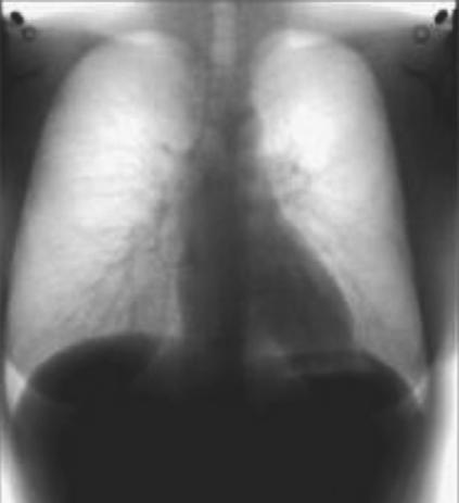
 COMPUTED TOMOGRAPHY 1 Why CT? Conventional X ray picture of a chest 2 Introduction Why CT? In a normal X-ray picture, most soft tissue doesn't show up clearly. To focus in on organs, or to examine the
Essentials of Digital Imaging
 Essentials of Digital Imaging Module 1 Transcript 2016 ASRT. All rights reserved. Essentials of Digital Imaging Module 1 Fundamentals 1. ASRT Animation 2. Welcome Welcome to Essentials of Digital Imaging
RADIOGRAPHIC EXPOSURE
 RADIOGRAPHIC EXPOSURE Receptor Exposure Receptor Exposure the that interacts with the receptor. Computed Radiography ( ) requires a. Direct Digital Radiography (DR) requires a. Exposure Indicators Exposure
A Comprehensive Review of Image Production
 A Comprehensive Review of Image Production Presented by: John Fleming, M.Ed., RT(R)(MR)(CT) St. Petersburg College Office: (727) 341-3758 E-mail: flemingj@spcollege.edu Lesson Objectives: ARRT Content
Veterinary Science Preparatory Training for the Veterinary Assistant. Floron C. Faries, Jr., DVM, MS
 Veterinary Science Preparatory Training for the Veterinary Assistant Floron C. Faries, Jr., DVM, MS Radiology Floron C. Faries, Jr., DVM, MS Objectives Determine the appropriate machine settings for making
CR Basics and FAQ. Overview. Historical Perspective
 Page: 1 of 6 CR Basics and FAQ Overview Computed Radiography is a term used to describe a system that electronically records a radiographic image. Computed Radiographic systems use unique image receptors
BASICS OF FLUOROSCOPY
 Medical Physics Residents Training Program BASICS OF FLUOROSCOPY Dr. Khalid Alyousef, PhD Department of Medical Imaging King Abdulaziz Medical City- Riyadh Edison examining the hand of Clarence Dally with
X-ray Imaging. PHYS Lecture. Carlos Vinhais. Departamento de Física Instituto Superior de Engenharia do Porto
 X-ray Imaging PHYS Lecture Carlos Vinhais Departamento de Física Instituto Superior de Engenharia do Porto cav@isep.ipp.pt Overview Projection Radiography Anode Angle Focal Spot Magnification Blurring
DELWORKS DR MEDICAL. take the next step
 DELWORKS DR MEDICAL take the next step DELWORKS MEDICAL DR If you are thinking of taking the next step to digital radiography, consider a DelWorks Medical DR Retrofit Package, the easy and affordable way
X-ray light valve (XLV): a novel detectors technology for digital mammography*
: a novel detectors technology for digital mammography*") X-ray light valve (XLV): a novel detectors technology for digital mammography* Sorin Marcovici, Vlad Sukhovatkin, Peter Oakham XLV Diagnostics Inc., Thunder Bay, ON P7A 7T1, Canada ABSTRACT A novel method,
X-ray light valve (XLV): a novel detectors technology for digital mammography* Sorin Marcovici, Vlad Sukhovatkin, Peter Oakham XLV Diagnostics Inc., Thunder Bay, ON P7A 7T1, Canada ABSTRACT A novel method,
Small Animal Radiographic Techniques and Positioning COPYRIGHTED MATERIAL
 Small Animal Radiographic Techniques and Positioning COPYRIGHTED MATERIAL Section 1 Theory and Equipment 1 Introduction to Digital Imaging Small animal radiography has changed dramatically in the past
DIGITAL IMAGE PROCESSING IN X-RAY IMAGING
 DIGITAL IMAGE PROCESSING IN X-RAY IMAGING Shalini Kumari 1, Bachan Prasad 2,Aliya Nasim 3 Department of Electronics And Communication Engineering R.V.S College of Engineering & Technology, Jamshedpur,
High Energy Digital Radiography & 3D-CT for Industrial Systems
 DIR 2007 - International Symposium on Digital industrial Radiology and Computed Tomography, June 25-27, 2007, Lyon, France High Energy Digital Radiography & 3D-CT for Industrial Systems Non-Destructive
X-ray detectors in healthcare and their applications
 X-ray detectors in healthcare and their applications Pixel 2012, Inawashiro September 4th, 2012 Martin Spahn, PhD Clinical applications of X-ray imaging Current X-ray detector technology (case study radiography
X-RAY MEDICAL EQUIPMENT
 X-RAY MEDICAL EQUIPMENT CHEST RADIOGRAPHY GENERAL RADIOGRAPHY & FLUOROSCOPY RADIOTHERAPY MOBILE HEALTHCARE MAMMOGRAPHY MAMMOSCAN FULL FIELD DIGITAL MAMMOGRAPHY SYSTEM Biopsy Attachment џ MAMMOSCAN an ADANI
Examination of Pipe Welds by Image Plate Based Computed Radiography System
 Examination of Pipe Welds by Image Plate Based Computed Radiography System Sanjoy Das, M.S.Rana, Benny Sebastian, D. Mukherjee and K.K. Abdulla Atomic Fuels Division Bhabha Atomic Research Centre Mumbai
PERFORMANCE CHARACTERIZATION OF AMORPHOUS SILICON DIGITAL DETECTOR ARRAYS FOR GAMMA RADIOGRAPHY
 12 th A-PCNDT 2006 Asia-Pacific Conference on NDT, 5 th 10 th Nov 2006, Auckland, New Zealand PERFORMANCE CHARACTERIZATION OF AMORPHOUS SILICON DIGITAL DETECTOR ARRAYS FOR GAMMA RADIOGRAPHY Rajashekar
NON-DESTRUCTIVE EVALUATION UTILIZING IMAGING PLATES FOR FIELD RADIOGRAPHY APPLICATIONS
 19 th World Conference on Non-Destructive Testing 2016 NON-DESTRUCTIVE EVALUATION UTILIZING IMAGING PLATES FOR FIELD RADIOGRAPHY APPLICATIONS Brian S. WHITE 1 1 Carestream NDT, 1049 Ridge Road West, Rochester,
INNOVATION BY DESIGN. Toshiba A History of Leadership REMOTE CONTROL R/F SYSTEM
 INNOVATION BY DESIGN For over 130 years, Toshiba has led the world in developing technology to improve the quality of life. This Made for Life TM commitment is reflected in our family of leading-edge imaging
Basis of Computed Radiography & PACS
 Basis of Computed Radiography & PACS Slavik Tabakov slavik.tabakov@emerald2.co.uk Digital Film-screen Image comparison and image transfer through various systems 1 Source: A. Pascoal CR system using laser
Overview of Safety Code 35
 Common Quality Control Procedures for All s Quality Control Procedures Film All s Daily Quality Control Tests Equipment Warm-up (D1) According to manufacturers instructions Can include auto calibration(d1)
X-Ray-Based Medical Imaging and Resolution
 Residents Section Physics Minimodule Huda and Abrahams Resolution on Radiographs Residents Section Physics Minimodule Residents inradiology Walter Huda 1 R. Brad Abrahams 2 Huda W, Abrahams RB Keywords:
Dental Radiography. One of the problems of dental radiography is having different dimensions than normal.
 The prototype receptor (the recording medium) most commonly used in dental radiography is the radiographic film. However, there are many other new more efficient receptors than the formed one that can
Breast Tomosynthesis. Bob Liu, Ph.D. Department of Radiology Massachusetts General Hospital And Harvard Medical School
 Breast Tomosynthesis Bob Liu, Ph.D. Department of Radiology Massachusetts General Hospital And Harvard Medical School Outline Physics aspects of breast tomosynthesis Quality control of breast tomosynthesis
ACQUISITION HARDWARE FOR DIGITAL IMAGING
 ACQUISITION HARDWARE FOR DIGITAL IMAGING WILLIAM R. WIDMER Use of digital radiography is growing rapidly in veterinary medicine. Two basic digital imaging systems are available, computed radiography (CR)
Film Replacement in Radiographic Weld Inspection The New ISO Standard
 BAM Berlin Film Replacement in Radiographic Weld Inspection The New ISO Standard 17636-2 Uwe Ewert, Uwe Zscherpel, Mirko Jechow Requests and information to: uwez@bam.de 1 Outline - The 3 essential parameters
REAL-TIME X-RAY IMAGE PROCESSING; TECHNIQUES FOR SENSITIVITY
 REAL-TIME X-RAY IMAGE PROCESSING; TECHNIQUES FOR SENSITIVITY IMPROVEMENT USING LOW-COST EQUIPMENT R.M. Wallingford and J.N. Gray Center for Aviation Systems Reliability Iowa State University Ames,IA 50011
STUDENT REVIEW QUESTION SET K CR/DR CONTENT AREA
 STUDENT REVIEW QUESTION SET K CR/DR CONTENT AREA RADT 2913 COMPREHENSIVE REVIEW 1 The CR cassette is backed by aluminum that: A. reflects x-rays B. absorbs x-rays C. captures the image D. transmits x-rays
Quality Control of Full Field Digital Mammography Units
 Quality Control of Full Field Digital Mammography Units Melissa C. Martin, M.S., FACMP, FACR, FAAPM Melissa@TherapyPhysics.com 310-612-8127 ACMP Annual Meeting Virginia Beach, VA May 2, 2009 History of
DIGITAL RADIOGRAPHY. Digital radiography is a film-less technology used to record radiographic images.
 DIGITAL RADIOGRAPHY Digital radiography is a film-less technology used to record radiographic images. 1 The purpose of digital imaging is to generate images that can be used in the diagnosis and assessment
Digital Imaging CT & MR
 Digital Imaging CT & MR January 22, 2008 Digital Radiography, CT and MRI generate images in a digital format What is a Digital Image? A digital image is made up of picture elements, pixels row by column
ON THE WAY TO DIGITAL RADIOGRAPHY
 The 14 th International Conference of the Slovenian Society for Non-Destructive Testing»Application of Contemporary Non-Destructive Testing in Engineering«September 4-6, 2017, Bernardin, Slovenia More
Digital Detector Array Image Quality for Various GOS Scintillators
 Digital Detector Array Image Quality for Various GOS Scintillators More info about this article: http://www.ndt.net/?id=22768 Brian S. White 1, Mark E. Shafer 2, William H. Russel 3, Eric Fallet 4, Jacques
Alma Mater Studiorum Università di Bologna
 Alma Mater Studiorum Università di Bologna DOTTORATO DI RICERCA IN FISICA Ciclo XXIII Settore scientifico-disciplinare: Scienze matematiche fisiche ed astronomiche FIS/07: FISICA APPLICATA Ph.D. Thesis
STATUS AND PROSPECTS OF DIGITAL DETECTOR TECHNOLOGY FOR CR AND DR Ulrich Neitzel Philips Medical Systems, Röntgenstrasse 24, D Hamburg, Germany
 Radiation Protection Dosimetry (2005), Vol. 114, Nos 1-3, pp. 32 38 doi:10.1093/rpd/nch532 INVITED PAPER STATUS AND PROSPECTS OF DIGITAL DETECTOR TECHNOLOGY FOR CR AND DR Ulrich Neitzel Philips Medical
Essentials of Digital Imaging
 Essentials of Digital Imaging Module 2 Transcript 2016 ASRT. All rights reserved. Essentials of Digital Imaging Module 2 Processing 1. ASRT Animation 2. Welcome Welcome to Essentials of Digital Imaging
X-RAY DIAGNOSTIC EQUIPMENT
 More than 20 years NP JSC AMICO offers high quality radiological equipment for hospitals. Factories located in the nearest suburb of Moscow manufacture up-to-date X-ray equipment conforming all standards.
Properties of a Detector
 Properties of a Detector Quantum Efficiency fraction of photons detected wavelength and spatially dependent Dynamic Range difference between lowest and highest measurable flux Linearity detection rate
Digital Industrial Radiography
 Digital Industrial Radiography Dr. Helmut Wolf, Anna University Chennai Dr.Theobald Fuchs, Fraunhofer Development Center X-ray Technology, Fuerth, Germany 1. Introduction In the previous issues, the physical
Radiographic sensitivity improved by optimized high resolution X -ray detector design.
 DIR 2007 - International Symposium on Digital industrial Radiology and Computed Tomography, June 25-27, 2007, Lyon, France Radiographic sensitivity improved by optimized high resolution X -ray detector
Medical Imaging. X-rays, CT/CAT scans, Ultrasound, Magnetic Resonance Imaging
 Medical Imaging X-rays, CT/CAT scans, Ultrasound, Magnetic Resonance Imaging From: Physics for the IB Diploma Coursebook 6th Edition by Tsokos, Hoeben and Headlee And Higher Level Physics 2 nd Edition
Gamex CR 2.0 Program description and operating manual
 Gamex CR 2.0 Program description and operating manual Issue No. : 2.0 Date of Issue : Jan. 2013 Z.U.T. NDT SOFT http://www.ndtsoft.eu Copyright (c) 2013 by Z.U.T. NDT SOFT All Rights Reserved Disclaimer
Photomultiplier Tube
 Nuclear Medicine Uses a device known as a Gamma Camera. Also known as a Scintillation or Anger Camera. Detects the release of gamma rays from Radionuclide. The radionuclide can be injected, inhaled or
COMPUTED RADIOGRAPHY (CR)
") COMPUTED RADIOGRAPHY (CR) Moving with the time Avi Avner BVSc BSc CVR DVDI MRCVS CR-Basics A five step process: 1. X-ray image received on phosphor plate 2. Image extracted from phosphor plate by Laser
COMPUTED RADIOGRAPHY (CR) Moving with the time Avi Avner BVSc BSc CVR DVDI MRCVS CR-Basics A five step process: 1. X-ray image received on phosphor plate 2. Image extracted from phosphor plate by Laser
Radionuclide Imaging MII Single Photon Emission Computed Tomography (SPECT)
") Radionuclide Imaging MII 3073 Single Photon Emission Computed Tomography (SPECT) Single Photon Emission Computed Tomography (SPECT) The successful application of computer algorithms to x-ray imaging in
Radionuclide Imaging MII 3073 Single Photon Emission Computed Tomography (SPECT) Single Photon Emission Computed Tomography (SPECT) The successful application of computer algorithms to x-ray imaging in
RADIOGRAPHY TERMS TO KNOW SELF STUDY DENTALELLE TUTORING
 RADIOGRAPHY TERMS TO KNOW SELF STUDY DENTALELLE TUTORING PLEASE NOTE You DO NOT need to study these for the board exam if this is why you bought our Radiography course, however if you come across any terms
X-RAY DIAGNOSTIC EQUIPMENT
 More than 20 years NP JSC AMICO offers high quality radiological equipment for hospitals. Factories located in the nearest suburb of Moscow manufacture up-to-date X-ray equipment conforming all standards.
X-RAY IMAGING EE 472 F2017. Prof. Yasser Mostafa Kadah
 X-RAY IMAGING EE 472 F2017 Prof. Yasser Mostafa Kadah www.k-space.org Recommended Textbook Stewart C. Bushong, Radiologic Science for Technologists: Physics, Biology, and Protection, 10 th ed., Mosby,
I. PERFORMANCE OF X-RAY PRODUCTION COMPONENTS FLUOROSCOPIC ACCEPTANCE TESTING: TEST PROCEDURES & PERFORMANCE CRITERIA
 FLUOROSCOPIC ACCEPTANCE TESTING: TEST PROCEDURES & PERFORMANCE CRITERIA EDWARD L. NICKOLOFF DEPARTMENT OF RADIOLOGY COLUMBIA UNIVERSITY NEW YORK, NY ACCEPTANCE TESTING GOALS PRIOR TO 1st CLINICAL USAGE
Photostimulable phosphor plates (PSPs)
") DIGITAL IMAGING Digital imaging Photostimulable phosphor plates (PSPs) Indirect digital PSPs are composed of a polyester base with a phosphor layer (europium activated barium fluorohalide) on one side.
DIGITAL IMAGING Digital imaging Photostimulable phosphor plates (PSPs) Indirect digital PSPs are composed of a polyester base with a phosphor layer (europium activated barium fluorohalide) on one side.
INTERNATIONAL STANDARD
 INTERNATIONAL STANDARD IEC 62220-1 First edition 2003-10 Medical electrical equipment Characteristics of digital X-ray imaging devices Part 1: Determination of the detective quantum efficiency Appareils
Seminar 8. Radiology S8 1
 Seminar 8 Radiology Medical imaging. X-ray image formation. Energizing and controlling the X-ray tube. Image detectors. The acquisition of analog and digital images. Digital image processing. Selected
X-ray Tube and Generator Basic principles and construction
 X-ray Tube and Generator Basic principles and construction Dr Slavik Tabakov - Production of X-rays and Patient Dose OBJECTIVES - X-ray tube construction - Anode - types, efficiency - Classical X-ray generator
radiography detector
 Clinical evaluation of a full field digital projection radiography detector Gary S. Shaber'1, Denny L. Leeb, Jeffrey Belib, Gregory Poweii1', Andrew D.A. Maidment'1 a Thomas Jefferson University Hospital,
X-RAYS - NO UNAUTHORISED ENTRY
 Licencing of premises Premises Refer Guidelines A radiation warning sign and warning notice, X-RAYS - NO UNAUTHORISED ENTRY must be displayed at all entrances leading to the rooms where x-ray units are
DEVELOPMENT OF A FLAT PANEL DETECTOR WITH AVALANCHE GAIN FOR INTERVENTIONAL RADIOLOGY MATTHEW M. WRONSKI
 DEVELOPMENT OF A FLAT PANEL DETECTOR WITH AVALANCHE GAIN FOR INTERVENTIONAL RADIOLOGY by MATTHEW M. WRONSKI A thesis submitted in conformity with the requirements for the degree of Doctor of Philosophy
Mammography Solution. AMULET Innovality. The new leader in the AMULET series. Tomosynthesis, 3D mammography and biopsy are all available.
 Mammography Solution AMULET Innovality The new leader in the AMULET series. Tomosynthesis, 3D mammography and biopsy are all available. FUJIFILM supports the Pink Ribbon Campaign for early detection of
10/15/2012 SECTION III - CHAPTER 6 DIGITAL FLUOROSCOPY RADT 3463 COMPUTERIZED IMAGING
 RADT 3463 - COMPUTERIZED IMAGING Section III: Chapter 6 RADT 3463 Computerized Imaging 1 SECTION III - CHAPTER 6 DIGITAL FLUOROSCOPY RADT 3463 COMPUTERIZED IMAGING Section III: Chapter 6 RADT 3463 Computerized
Minimum Requirements for Digital Radiography Equipment and Measurement Procedures by Different Industries and Standard Organizations
 uwe.ewert@bam.de Minimum Requirements for Digital Radiography Equipment and Measurement Procedures by Different Industries and Standard Organizations Uwe Ewert and Uwe Zscherpel BAM Federal Institute for
We are IntechOpen, the first native scientific publisher of Open Access books. International authors and editors. Our authors are among the TOP 1%
 We are IntechOpen, the first native scientific publisher of Open Access books 3,350 108,000 1.7 M Open access books available International authors and editors Downloads Our authors are among the 151 Countries
Medical Imaging: A Look inside. Medical Imaging. Medical Imaging. Visible Human Project
 Medical Imaging: A Look inside Medical Imaging Allows physicians to see what had previously been unseeable: internal organs and tissues, bones, a beating heart, etc. Allows physicians to: detect brain
While digital techniques have the potential to reduce patient doses, they also have the potential to significantly increase them.
 In press 2004 1 2 Guest Editorial (F. Mettler, H. Ringertz and E. Vano) Guest Editorial (F. Mettler, H. Ringertz and E. Vano) Digital radiology An appropriate analogy that is easy for most people to understand
Radiographic Testing (RT) [10]
![Radiographic Testing (RT) [10]](/thumbs/74/70215439.jpg "Radiographic Testing (RT) [10]") Radiographic Testing (RT) [10] Definition: An NDT method that utilizes x-rays or gamma radiation to detect discontinuities in materials, and to present their images on recording medium. 1> Electromagnetic
Radiographic Testing (RT) [10] Definition: An NDT method that utilizes x-rays or gamma radiation to detect discontinuities in materials, and to present their images on recording medium. 1> Electromagnetic
Advances in High Energy X-ray Digital Detector Arrays
 19 th World Conference on Non-Destructive Testing 2016 Advances in High Energy X-ray Digital Detector Arrays Clifford BUENO 1, William ROSS 1, Jeffrey SHAW 1, Joshua SALISBURY 1, Edward J NIETERS 1, Forrest
Digital radiography (DR) post processing techniques for pediatric radiology
 post processing techniques for pediatric radiology") Digital radiography (DR) post processing techniques for pediatric radiology St Jude Children s Research Hospital Samuel Brady, MS PhD DABR samuel.brady@stjude.org Purpose Review common issues and solutions
Digital radiography (DR) post processing techniques for pediatric radiology St Jude Children s Research Hospital Samuel Brady, MS PhD DABR samuel.brady@stjude.org Purpose Review common issues and solutions
Exposure Indices and Target Values in Radiography: What Are They and How Can You Use Them?
 Exposure Indices and Target Values in Radiography: What Are They and How Can You Use Them? Definition and Validation of Exposure Indices Ingrid Reiser, PhD DABR Department of Radiology University of Chicago
Outline ASRT Changes Impact on current curriculum Potential new courses WECM Changes Last update Resources and needs
 Change nd Annual Blinn College 2 nd Educator s Workshop For Radiologic Sciences July 28, 2007 Christi Carter, MSRS, RT(R) Outline ASRT Changes Impact on current curriculum Potential new courses WECM Changes
Medical Images Analysis and Processing
 Medical Images Analysis and Processing - 25642 Emad Course Introduction Course Information: Type: Graduated Credits: 3 Prerequisites: Digital Image Processing Course Introduction Reference(s): Insight
DIGITAL IMAGING Recognise the importance of quality assurance
 DIGITAL IMAGING Recognise the importance of quality assurance There are two types of digital image receptor both of which capture a two dimensional image of the three dimensional patient. These are Computed
Moving from film to digital: A study of digital x-ray benefits, challenges and best practices
 Moving from film to digital: A study of digital x-ray benefits, challenges and best practices H.U. Pöhler 1 and N. D Ademo 2 DÜRR NDT GmbH & Co. KG, Höpfigheimer Straße 22, Bietigheim-Bissingen, 74321,
Enhanced Functionality of High-Speed Image Processing Engine SUREengine PRO. Sharpness (spatial resolution) Graininess (noise intensity)
 Graininess (noise intensity)") Vascular Enhanced Functionality of High-Speed Image Processing Engine SUREengine PRO Medical Systems Division, Shimadzu Corporation Yoshiaki Miura 1. Introduction In recent years, digital cardiovascular
Vascular Enhanced Functionality of High-Speed Image Processing Engine SUREengine PRO Medical Systems Division, Shimadzu Corporation Yoshiaki Miura 1. Introduction In recent years, digital cardiovascular
LARGE FORMATTED AND HIGH RESOLUTION CMOS FLAT PANEL SENSORS FOR X-RAY
 The 8 th International Conference of the Slovenian Society for Non-Destructive Testing»Application of Contemporary Non-Destructive Testing in Engineering«September 1-3, 2005, Portorož, Slovenia, pp. 165-172
RaySafe X2. Effortless measurements of X-ray
 RaySafe X2 Effortless measurements of X-ray At your fingertips We ve grown accustomed to intuitive interactions with our devices. After all, it s not the device that s most important, but what you can
Experiences of users in Digital Radiography
 Computed Radiography Products & Applications Experiences of users in Digital Radiography Jimmy Opdekamp May Jimmy 2006Opdekamp Global Product Manager CR Int l Workshop Imaging NDT Chennai, 25-28 April
9/10/2012. Computed Radiography Chapter 3 Physics and Technology. What is Computed Radiography?
 Computed Radiography Chapter 3 Physics and Technology This presentation is a professional collaboration of development time prepared by: Rex Christensen Terri Jurkiewicz and Diane Kawamura Today s Humor:
Chromatic X-Ray imaging with a fine pitch CdTe sensor coupled to a large area photon counting pixel ASIC
 Chromatic X-Ray imaging with a fine pitch CdTe sensor coupled to a large area photon counting pixel ASIC R. Bellazzini a,b, G. Spandre a*, A. Brez a, M. Minuti a, M. Pinchera a and P. Mozzo b a INFN Pisa
Fluoroscopy - Chapter 9
 Fluoroscopy - Chapter 9 Kalpana Kanal, Ph.D., DABR Lecturer, Diagnostic Physics Dept. of Radiology UW Medicine a copy of this lecture may be found at: http://courses.washington.edu/radxphys/physicscourse04-05.html
DALLA LUCE VISIBILE AI RAGGI X: NUOVI RIVELATORI DI IMMAGINI PER RAGGI X A DISCRIMINAZIONE IN ENERGIA ED APPLICAZIONI
 DALLA LUCE VISIBILE AI RAGGI X: NUOVI RIVELATORI DI IMMAGINI PER RAGGI X A DISCRIMINAZIONE IN ENERGIA ED APPLICAZIONI D. Pacella ENEA - Frascati LIMS, Frascati 14-15 ottobre 2015 Come per la fotografia: