X-RAY FLUOROSCOPY IMAGING SYSTEMS. Dr Slavik Tabakov. Luminescence: Dept. Medical Eng. & Physics King s College London
|
|
|
- Terence Skinner
- 5 years ago
- Views:
Transcription

 - PM detectors; II input screens (CsI:Tl) Phosphorescence - emitting broad light spectrum (light continues")
 2. I a. T = (C. Z). 2. mas Radiography of the lumbar spine (with parameters 80, 30 mas): X = k. 80.80.30 = k.")
1 X-RAY FLUOROSCOPY IMAGING SYSTEMS Dr Slavik Tabakov OBJECTIVES - Image Intensifier construction - Input window - Accelerating and focusing electrodes - Output window - Conversion factor - II characteristics - Modulation Transfer function -Digital fluoroscopy Dept. Medical Eng. & Physics King s College London slavik.tabakov@kcl.ac.uk, slavik.tabakov@emerald2.co.uk Fluoroscopy delivers very high patient dose. This can be illustrated with an example: The electrical energy imparted to the anode during an exposure is A = C 1. U a. I a. T The X-ray tube anode efficiency is E = C 2. Z. U a Luminescence: Fluorescence - emitting narrow light spectrum (very short afterglow ~nsec) - PM detectors; II input screens (CsI:Tl) Phosphorescence - emitting broad light spectrum (light continues after radiation) - monitor screens, II output screens (ZnCdS:Ag) The old fluoroscopic screens are no longer used due to high dose and low resolution From the two equations follows that the energy produced in a single exposure will be X = C. A. E = C. Z. (U a ) 2. I a. T = (C. Z). 2. mas Radiography of the lumbar spine (with parameters 80, 30 mas): X = k = k. 192,000 Fluoroscopy - 3 minutes Barium meal (with parameters 80, 1mA) X = k = k. 1,152,000 In this example fluoroscopy delivers approx. 6 times more X-ray energy (dose)
 or ZnS (old) phosphor - Photocathode (a layer of CsSb 3 ) - Accelerating")
 - II housing (mu-metal) - Output coupling to the TV camera II Input screen: Columnar crystals of CsI")

 Coupling: fibre optic or tandem optic")
2 Basic Components of an Image Intensifier - Input window (Ti or Al) 95% transmission - Input screen: CsI (new) or ZnS (old) phosphor - Photocathode (a layer of CsSb 3 ) - Accelerating electrodes zoom (e.g. 30/23/15 cm) - Output screen (2.5 cm) - II housing (mu-metal) - Output coupling to the TV camera II Input screen: Columnar crystals of CsI which reduces dispertion (collimation); absorbs approx. 60% of X-rays Photocathode applied directly to CsI both light spectrum match very well II Accelerating electrodes II Output screen: Phosphor (ZnCdS:Ag) on glass base The accelerated e - produce multiple light photons; thin Al foil prevent return of light (veiling glare) Coupling: fibre optic or tandem optic Conversion factor ~ (cd.m -2 /μgy.s -1 ) = (output phosphor light / input screen dose rate) Total gain (out. light photons /inp. X photons )
 1 X-ray photon >> 1000 light photons (input screen) >> >>50 photo e - >> 3000 light photons (output screen) in the case above the total gain is 3000 MTF of II depending on zoom")
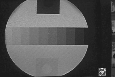
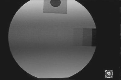
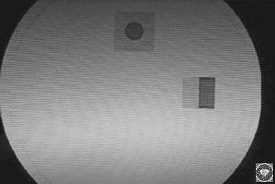
3 Total gain (out. light photons /inp. X photons ) 1 X-ray photon >> 1000 light photons (input screen) >> >>50 photo e - >> 3000 light photons (output screen) in the case above the total gain is 3000 MTF of II depending on zoom (magnification) Some II Characteristics: Minification gain -D m -inp./output diam. (D inp / D out ) 2 Flux gain -F x (approx ): Out.scr. light photons / inp. ligh photons to photocath. Brightness gain -G B G B = D m x F x * Zooming increases the resolution, but requires higher dose rate!! Contrast Ratio -X-ray scatter at input window, input phosphor -Light scatter within phosphor, not-absorbed light by phosphor -Back scatter from output phosphor (to photocathode), at output window L c light intensity at centre of image (pure white) Cont. Ratio (C v )= L c /L d : ideally max/0 ; in reality approx. 30/1 L d - light intensity at centre of image (cover with Pb) II field size 40 cm (16 ) 32 cm (12.5 ) 20 cm (8 ) 15 cm (6 ) Resolution (Lp/mm) Contr. ratio 20:1 25:1 30:1 35:1 Automatic Brightness Control System (ABS) - produces images with constant brightness by keeping constant entrance dose rate to the II The feedback C1 have two options - taking signal from D1 (dosimeter) or D2 (photometer). * II entr. dose rate is approx. 1 μgy/sec and should not exceeds 2 μgy/sec. * The maximal patient entrance skin dose should not exceed 0.01 Gy/min). Convers. Factor (cd/m / mr/s) Distortion (pincushion %) Dose (relative) Table from: D.Dowsett, P.Kenny, E.Johnston - different types and characteristic curves of changing the /ma Graph from: E Krestel (SIEMENS)
,")


 Modulation Transfer Function MTF=(recorded signal")
; also MTF(f) = FT{LSF(x)} MTF~m = sin π.u f / π.")
4 TV camera types: Vidicon - gamma 0.7; slow response, some contrast loss (light integration), high dark current, but low noise - suitable for organs Plumbicon - gamma 1; quick response, small dark current, but high noise - suitable for cardiac examinations Modulation Transfer Function and Contrast Transfer Function 10% - cut-off frequency (lim. sp. res.) Modulation Transfer Function MTF=(recorded signal f)/(origin. signal f); also MTF(f) = FT{LSF(x)} MTF~m = sin π.u f / π.u f,where u f = f/l * (M-1)/2M,where M-magnif.; f focal spot; L period of the structure (~ to spatial frequency)


")

")

5 Overall II-TV system MTF = MTF 1 x MTF 2 x x MTF n Dynamic range of II -much larger than this of radiographic film (output luminance per dose unit) Resolution and Magnification of II - electronic zoom up to 4 times (lp/mm) Digital Fluoroscopy Digital subtraction and unsharp masking
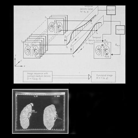
6 Mathematical operation in DSA: Functional imaging; Logarithmic & Square Root Subtraction, etc. Functional Imaging II contrast with different (constant ma)
X-ray Tube and Generator Basic principles and construction
 X-ray Tube and Generator Basic principles and construction Dr Slavik Tabakov - Production of X-rays and Patient Dose OBJECTIVES - X-ray tube construction - Anode - types, efficiency - Classical X-ray generator
BASICS OF FLUOROSCOPY
 Medical Physics Residents Training Program BASICS OF FLUOROSCOPY Dr. Khalid Alyousef, PhD Department of Medical Imaging King Abdulaziz Medical City- Riyadh Edison examining the hand of Clarence Dally with
Introduction. Chapter 16 Diagnostic Radiology. Primary radiological image. Primary radiological image
 Introduction Chapter 16 Diagnostic Radiology Radiation Dosimetry I Text: H.E Johns and J.R. Cunningham, The physics of radiology, 4 th ed. http://www.utoledo.edu/med/depts/radther In diagnostic radiology
Fluoroscopy - Chapter 9
 Fluoroscopy - Chapter 9 Kalpana Kanal, Ph.D., DABR Lecturer, Diagnostic Physics Dept. of Radiology UW Medicine a copy of this lecture may be found at: http://courses.washington.edu/radxphys/physicscourse04-05.html
10/15/2012 SECTION III - CHAPTER 6 DIGITAL FLUOROSCOPY RADT 3463 COMPUTERIZED IMAGING
 RADT 3463 - COMPUTERIZED IMAGING Section III: Chapter 6 RADT 3463 Computerized Imaging 1 SECTION III - CHAPTER 6 DIGITAL FLUOROSCOPY RADT 3463 COMPUTERIZED IMAGING Section III: Chapter 6 RADT 3463 Computerized
Joint ICTP/IAEA Advanced School on Dosimetry in Diagnostic Radiology and its Clinical Implementation May 2009
 2033-6 Joint ICTP/IAEA Advanced School on Dosimetry in Diagnostic Radiology and its Clinical Implementation 11-15 May 2009 Dosimetry for Fluoroscopy Basics Renato Padovani EFOMP Joint ICTP-IAEA Advanced
I. PERFORMANCE OF X-RAY PRODUCTION COMPONENTS FLUOROSCOPIC ACCEPTANCE TESTING: TEST PROCEDURES & PERFORMANCE CRITERIA
 FLUOROSCOPIC ACCEPTANCE TESTING: TEST PROCEDURES & PERFORMANCE CRITERIA EDWARD L. NICKOLOFF DEPARTMENT OF RADIOLOGY COLUMBIA UNIVERSITY NEW YORK, NY ACCEPTANCE TESTING GOALS PRIOR TO 1st CLINICAL USAGE
TESTING FLAT-PANEL IMAGING SYSTEMS: What the Medical Physicist Needs to Know. JAMES A. TOMLINSON, M.S., D.A.B.R. Diagnostic Radiological Physicist
 TESTING FLAT-PANEL IMAGING SYSTEMS: What the Medical Physicist Needs to Know JAMES A. TOMLINSON, M.S., D.A.B.R. Diagnostic Radiological Physicist Topics Image Uniformity and Artifacts Image Quality - Detail
DIGITAL IMAGE PROCESSING IN X-RAY IMAGING
 DIGITAL IMAGE PROCESSING IN X-RAY IMAGING Shalini Kumari 1, Bachan Prasad 2,Aliya Nasim 3 Department of Electronics And Communication Engineering R.V.S College of Engineering & Technology, Jamshedpur,
10/26/2015. Study Harder
 This presentation is a professional collaboration of development time prepared by: Rex Christensen Terri Jurkiewicz and Diane Kawamura Study Harder CR detection is inefficient, inferior to film screen
Beam-Restricting Devices
 Beam-Restricting Devices Three factors contribute to an increase in scatter radiation: Increased kvp Increased Field Size Increased Patient or Body Part Size. X-ray Interactions a some interact with the
LECTURE 1 The Radiographic Image
 LECTURE 1 The Radiographic Image Prepared by:- KAMARUL AMIN ABDULLAH @ ABU BAKAR UiTM Faculty of Health Sciences Medical Imaging Department 11/23/2011 KAMARUL AMIN (C) 1 Lesson Objectives At the end of
10/3/2012. Study Harder
 This presentation is a professional collaboration of development time prepared by: Rex Christensen Terri Jurkiewicz and Diane Kawamura Study Harder CR detection is inefficient, inferior to film screen
Amorphous Selenium Direct Radiography for Industrial Imaging
 DGZfP Proceedings BB 67-CD Paper 22 Computerized Tomography for Industrial Applications and Image Processing in Radiology March 15-17, 1999, Berlin, Germany Amorphous Selenium Direct Radiography for Industrial
X-ray Imaging. PHYS Lecture. Carlos Vinhais. Departamento de Física Instituto Superior de Engenharia do Porto
 X-ray Imaging PHYS Lecture Carlos Vinhais Departamento de Física Instituto Superior de Engenharia do Porto cav@isep.ipp.pt Overview Projection Radiography Anode Angle Focal Spot Magnification Blurring
Seminar 8. Radiology S8 1
 Seminar 8 Radiology Medical imaging. X-ray image formation. Energizing and controlling the X-ray tube. Image detectors. The acquisition of analog and digital images. Digital image processing. Selected
X-RAYS - NO UNAUTHORISED ENTRY
 Licencing of premises Premises Refer Guidelines A radiation warning sign and warning notice, X-RAYS - NO UNAUTHORISED ENTRY must be displayed at all entrances leading to the rooms where x-ray units are
Basis of Computed Radiography & PACS
 Basis of Computed Radiography & PACS Slavik Tabakov Computed Radiography (CR) refers to new types of X-ray detectors (i.e. replaces the X-ray Film) The CR output media is a digital image, which can be
Do you have any other questions? Please call us at (Toll Free) or , or
 or , or") INSTRUCTIONS Read the appropriate course/ textbook. This is an open book test. A score of 75% or higher is needed to receive CE credit. You will have a maximum of three attempts to pass this course. Please
INSTRUCTIONS Read the appropriate course/ textbook. This is an open book test. A score of 75% or higher is needed to receive CE credit. You will have a maximum of three attempts to pass this course. Please
Dose Reduction and Image Preservation After the Introduction of a 0.1 mm Cu Filter into the LODOX Statscan unit above 110 kvp
 Dose Reduction and Image Preservation After the Introduction of a into the LODOX Statscan unit above 110 kvp Abstract: CJ Trauernicht 1, C Rall 1, T Perks 2, G Maree 1, E Hering 1, S Steiner 3 1) Division
Multiple Choice Identify the letter of the choice that best completes the statement or answers the question.
 RA110 test 3 Multiple Choice Identify the letter of the choice that best completes the statement or answers the question. 1. An object 35 cm in width is radiographed at 100 cm SID and at a 50 cm SOD. What
Photons interaction with matter
 ب س م هللا الر ح من الر حیم Photons interaction with matter Ionization Ionization is the process of removing an electron from an electrically neutral atom to produce an ion pair. An ion is an atom or subatomic
X-ray Tube and Generator Basic principles and construction
 X-ray Tube and Generator Basic principles and construction Dr Slavik Tabakov - Production of X-rays OBJECTIVES - X-ray tube construction - Anode - types, efficiency - X-ray tube working characteristics
AN ABSTRACT OF THE THESIS OF. W. Scott Helms for the degree of Master of Science in Radiation Health Physics
 AN ABSTRACT OF THE THESIS OF W. Scott Helms for the degree of Master of Science in Radiation Health Physics presented on November 24, 2014 Title: A Quantitative Comparison of Cardiovascular Imaging Systems
X-Ray Medical Imaging and Pixel detectors
 X-Ray Medical Imaging and Pixel detectors PIXEL 2000 Genova, June 5-8 th 2000 J.P.Moy, TRI XELL, Moirans, France 1 OUTLINE - X-ray medical imaging. The requirements, some particular features - Present
Basis of Computed Radiography & PACS
 Basis of Computed Radiography & PACS Slavik Tabakov slavik.tabakov@emerald2.co.uk Digital Film-screen Image comparison and image transfer through various systems 1 Source: A. Pascoal CR system using laser
Distributed by. Ecotron Anyview Series. The Intelligent C-Arm System
 Distributed by Ecotron Anyview Series The Intelligent C-Arm System The Ecotron Anyview Series is a new standard for C-arm imaging Excellent radiographic imaging at an economical price Advanced X-ray Generator
Nuclear Associates
 Nuclear Associates 76-700 Digital Subtraction Angiography Phantom Users Manual March 2005 Manual No. 76-700-1 Rev. 2 2004, 2005 Fluke Corporation, All rights reserved. Printed in U.S.A. All product names
Acquisition, Processing and Display
 Acquisition, Processing and Display Terri L. Fauber, R.T. (R)(M) Department of Radiation Sciences School of Allied Health Professions Virginia Commonwealth University Topics Image Characteristics Image
Radiology. Radiograph: Is the image of an object made with use of X- ray instead of light.
 Radiology د. اريج Lec. 3 X Ray Films Radiograph: Is the image of an object made with use of X- ray instead of light. Dental x- ray film: Is a recording media on which image of the object was made by exposing
Radiographic sensitivity improved by optimized high resolution X -ray detector design.
 DIR 2007 - International Symposium on Digital industrial Radiology and Computed Tomography, June 25-27, 2007, Lyon, France Radiographic sensitivity improved by optimized high resolution X -ray detector
Exposure System Selection
 Principles of Imaging Science II (RAD120) Exposure Systems Exposure System Selection Radiographic exposure is a very complex process Best technique systems manipulate one variable while holding others
siemens.com/luminos-fusion Luminos Fusion The 2-in-1 system that fits your needs and fits your budget
 siemens.com/luminos-fusion Luminos Fusion The 2-in-1 system that fits your needs and fits your budget Luminos Fusion The 2-in-1 system that fits your needs and fits your budget Luminos Fusion provides
SPRINGFIELD TECHNICAL COMMUNITY COLLEGE ACADEMIC AFFAIRS
 SPRINGFIELD TECHNICAL COMMUNITY COLLEGE ACADEMIC AFFAIRS Course Number: RADG 112 Department: Radiography Course Title: Image Production & Eval. Semester: Spring Year: 1997 Objectives/ Unit One: Introduction
CHAPTER 10. DIGITAL SUBTRACTION ANGIOGRAPHY (DSA) 10.1 Introduction
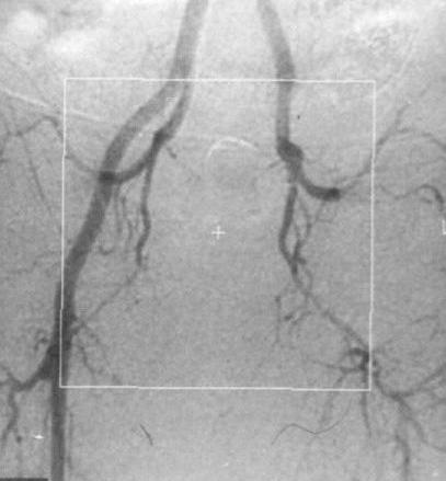
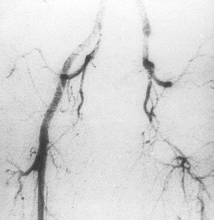
 10.1 Introduction") PHYSICS OF MEDICAL X-RAY IMAGING (1)Chapter 10 CHAPTER 10. DIGITAL SUBTRACTION ANGIOGRAPHY (DSA) 10.1 Introduction In this chapter we will discuss a technique that is known as digital subtraction angiography
PHYSICS OF MEDICAL X-RAY IMAGING (1)Chapter 10 CHAPTER 10. DIGITAL SUBTRACTION ANGIOGRAPHY (DSA) 10.1 Introduction In this chapter we will discuss a technique that is known as digital subtraction angiography
CHAPTER 6 QC Test For Fluoroscopic Equipment. Prepared by:- Kamarul Amin bin Abu Bakar School of Medical Imaging KLMUC
 CHAPTER 6 QC Test For Fluoroscopic Equipment Prepared by:- Kamarul Amin bin Abdullah @ Abu Bakar School of Medical Imaging KLMUC Lesson Outcomes Describe the objectives of each QC test done. Identify QC
Radiology Physics Lectures: Digital Radiography. Digital Radiography. D. J. Hall, Ph.D. x20893
 Digital Radiography D. J. Hall, Ph.D. x20893 djhall@ucsd.edu Background Common Digital Modalities Digital Chest Radiograph - 4096 x 4096 x 12 bit CT - 512 x 512 x 12 bit SPECT - 128 x 128 x 8 bit MRI -
Nuclear Associates
 Nuclear Associates 07-649 CDRH Fluoroscopic Phantom Users Manual March 2005 Manual No. 07-649-1 Rev. 2 2004, 2005 Fluke Corporation, All rights reserved. Printed in U.S.A. All product names are trademarks
SPRINGFIELD TECHNICAL COMMUNITY COLLEGE ACADEMIC AFFAIRS
 SPRINGFIELD TECHNICAL COMMUNITY COLLEGE ACADEMIC AFFAIRS Course Number: RADG 212 Department: Radiography Course Title: Equip. Operation & Maint. Semester: Spring Year: 1997 Objectives/ Unit One: The X-ray
X-rays. X-rays are produced when electrons are accelerated and collide with a target. X-rays are sometimes characterized by the generating voltage
 X-rays Ouch! 1 X-rays X-rays are produced when electrons are accelerated and collide with a target Bremsstrahlung x-rays Characteristic x-rays X-rays are sometimes characterized by the generating voltage
Photomultiplier Tube
 Nuclear Medicine Uses a device known as a Gamma Camera. Also known as a Scintillation or Anger Camera. Detects the release of gamma rays from Radionuclide. The radionuclide can be injected, inhaled or
Mammography: Physics of Imaging
 Mammography: Physics of Imaging Robert G. Gould, Sc.D. Professor and Vice Chair Department of Radiology and Biomedical Imaging University of California San Francisco, California Mammographic Imaging: Uniqueness
1-1. GENERAL 1-2. DISCOVERY OF X-RAYS
 1-1. GENERAL Radiography is a highly technical field, indispensable to the modern dental practice, but presenting many potential hazards. The dental radiographic specialist must be thoroughly familiar
X-rays in medical diagnostics
 X-rays in medical diagnostics S.Dolanski Babić 2017/18. History W.C.Röntgen (1845-1923) discovered a new type of radiation Nature, Jan. 23. 1896.; Science, Feb.14. 1896. X- rays: Induced the ionization
Fluoroscopy & Tomography. Rhodes
 Fluoroscopy & Tomography Rhodes 10-526-194 1 Historical Development Dynamic examination Active Diagnosis Domain of radiologist Fluoroscope Invented by Edison in 1896 2 Fluoroscopic Imaging Chain Components
Version 1.0. TechnicVR. Student Guide
 Version 1.0 TechnicVR s h a d e r w a r e. c o m Student Guide TechnicVR s h a d e r w a r e. c o m Student Guide shaderware 2008 PO Box 103 Saltburn Cleveland TS12 1WP w w w. s h a d e r w a r e. c o
Unit thickness. Unit area. σ = NΔX = ΔI / I 0
 Unit thickness I 0 ΔI I σ = ΔI I 0 NΔX = ΔI / I 0 NΔX Unit area Δx Average probability of reaction with atom for the incident photons at unit area with the thickness of Delta-X Atom number at unit area
Mammography is a radiographic procedure specially designed for detecting breast pathology Approximately 1 woman in 8 will develop breast cancer over
 Mammography is a radiographic procedure specially designed for detecting breast pathology Approximately 1 woman in 8 will develop breast cancer over a lifetime Breast cancer screening programs rely on
X-RAY IMAGING EE 472 F2017. Prof. Yasser Mostafa Kadah
 X-RAY IMAGING EE 472 F2017 Prof. Yasser Mostafa Kadah www.k-space.org Recommended Textbook Stewart C. Bushong, Radiologic Science for Technologists: Physics, Biology, and Protection, 10 th ed., Mosby,
Essentials of Digital Imaging
 Essentials of Digital Imaging Module 7 Transcript 2016 ASRT. All rights reserved. Essentials of Digital Imaging Module 7 Quality 1. ASRT Animation 2. Welcome Welcome to the Essentials of Digital Imaging:
ProX Intraoral X-ray. PLANMECA is proud to introduce a new intraoral X-ray unit to its comprehensive collection of imaging products- the ProX.
 The premium intraoral X-ray unit... ProX Intraoral X-ray PLANMECA is proud to introduce a new intraoral X-ray unit to its comprehensive collection of imaging products- the ProX. This advanced unit provides
NDE SOLUTIONS RADIOGRAPHY COURSE OUTLINE
 NDE SOLUTIONS RADIOGRAPHY COURSE OUTLINE 80 Hour Course Length 1.0 NDT Qualification and Introduction (3 Hours) 1.1 NDT Introduction 1.2 NDT Qualification and Certification 1.2.1 NAS 410 1.2.2 SNT-TC-1A
Standard Guide for Radioscopy 1
 Designation: 98 An American National Standard Standard Guide for Radioscopy 1 This standard is issued under the fixed designation ; the number immediately following the designation indicates the year of
X-RAY. Lecture No.4. Image Characteristics:
 Lecture No.4 X-RAY أ.م.د. اسامة مراد ابراهيم Image Characteristics: *Radiographic density: It s the degree of blackness of the film. when a film is exposed by an x-ray beam (or by light in case of screenfilm
1. Carlton, Richard R., and Arlene M. Adler. Principles of Radiographic Imaging: An Art and a Science, 5th edition (2013).
.") CODE: RADT 151 INSTITUTE: Health Science TITLE: Radiographic Exposure DEPARTMENT: Radiologic Technology COURSE DESCRIPTION: This course covers the principles of radiographic exposure selection and manipulation
CODE: RADT 151 INSTITUTE: Health Science TITLE: Radiographic Exposure DEPARTMENT: Radiologic Technology COURSE DESCRIPTION: This course covers the principles of radiographic exposure selection and manipulation
PLD5600A High Frequency Digital Gastrointestinal &DR System(630mA)
") PLD5600A High Frequency Digital Gastrointestinal &DR System(630mA) Application: Full support perspective, gastrointestinal spot film, GI (barium meal, barium enema), orthopedic photography, pediatrics
PLD5600A High Frequency Digital Gastrointestinal &DR System(630mA) Application: Full support perspective, gastrointestinal spot film, GI (barium meal, barium enema), orthopedic photography, pediatrics
Digital Detector Array Image Quality for Various GOS Scintillators
 Digital Detector Array Image Quality for Various GOS Scintillators More info about this article: http://www.ndt.net/?id=22768 Brian S. White 1, Mark E. Shafer 2, William H. Russel 3, Eric Fallet 4, Jacques
Effect of patient support pads on image quality and dose in fluoroscopy a)
") Effect of patient support pads on image quality and dose in fluoroscopy a) William R. Geiser, Walter Huda, b) and Nikolaos A. Gkanatsios Department of Radiology, University of Florida, Gainesville, Florida
Effect of patient support pads on image quality and dose in fluoroscopy a) William R. Geiser, Walter Huda, b) and Nikolaos A. Gkanatsios Department of Radiology, University of Florida, Gainesville, Florida
Luminos drf Max. Taking 2-in-1 to the MAX in radiography and fluoroscopy. siemens.com/luminos-drf-max
 Luminos drf Max Taking 2-in-1 to the MAX in radiography and fluoroscopy siemens.com/luminos-drf-max 2 It s more. It s MAX. MAX Multiple Advances in X-ray It s more than just single features or functions.
V SALAI SELVAM, AP & HOD, ECE, Sriram Engg. College, Perumalpattu 1 MEDICAL ELECTRONICS UNIT IV
 V SALAI SELVAM, AP & HOD, ECE, Sriram Engg. College, Perumalpattu 1 MEDICAL ELECTRONICS UNIT IV Ionizing and non-ionizing radiations: The radiation that ionizes the gases through which it travels is known
COMPUTED RADIOGRAPHY CHAPTER 4 EFFECTIVE USE OF CR
 This presentation is a professional collaboration of development time prepared by: Rex Christensen Terri Jurkiewicz and Diane Kawamura New Technology https://www.youtube.com/watch?v=ptkzznazb 7U COMPUTED
QC in Diagnostic Radiology. Main steps for a QC survey in Diagnostic Radiology
 EVALUATING X-RAY TUBE AND GENERATOR PERFORMANCE : DEMO for PRACTICAL QUALITY CONTROL (QC) Dr Slavik Tabakov Dept. Medical Eng. & Physics, King's College London slavik.tabakov@kcl.ac.uk QC in Diagnostic
Luminos RF Classic. Where value meets performance.
 Luminos RF Classic Where value meets performance www.siemens.com/healthcare What s good value in fluoroscopy? That s easy. Luminos RF Classic. 2 Whether for its handling convenience, outstanding image
NPTEL NPTEL ONLINE COURSE. NPTEL Online Certification Course (NOC) NPTEL. Theory and Practice of Non Destructive Testing
 NPTEL. Theory and Practice of Non Destructive Testing") NPTEL NPTEL ONLINE COURSE NPTEL Online Certification Course (NOC) NPTEL Theory and Practice of Non Destructive Testing Dr. Ranjit Bauri Dept. of Metallurgical & Materials Engineering IIT Madras, Chennai
NPTEL NPTEL ONLINE COURSE NPTEL Online Certification Course (NOC) NPTEL Theory and Practice of Non Destructive Testing Dr. Ranjit Bauri Dept. of Metallurgical & Materials Engineering IIT Madras, Chennai
X-RAY TUBE ARCING: MANIFESTATION AND DETECTION DURING QUALUTY CONTROL
 X-RAY TUBE ARCING: MANIFESTATION AND DETECTION DURING QUALUTY CONTROL S. Tabakov 1,2 1 Dept. Medical Eng. and Physics, King s College London, SE5 9RS, UK; 2 President IOMP, York YO24 1ES, UK Abstract X-ray
CR Basics and FAQ. Overview. Historical Perspective
 Page: 1 of 6 CR Basics and FAQ Overview Computed Radiography is a term used to describe a system that electronically records a radiographic image. Computed Radiographic systems use unique image receptors
Film Replacement in Radiographic Weld Inspection The New ISO Standard
 BAM Berlin Film Replacement in Radiographic Weld Inspection The New ISO Standard 17636-2 Uwe Ewert, Uwe Zscherpel, Mirko Jechow Requests and information to: uwez@bam.de 1 Outline - The 3 essential parameters
Contrast. Contrast: the difference in density on adjacent areas of a radiograph or other image receptor. Subjective. Long Scale (Low Contrast)
") Contrast Contrast: the difference in density on adjacent areas of a radiograph or other image receptor. Subject Subjective Radiographic Long Scale (Low Contrast) Short Scale (High Contrast) Factors affecting
Contrast Contrast: the difference in density on adjacent areas of a radiograph or other image receptor. Subject Subjective Radiographic Long Scale (Low Contrast) Short Scale (High Contrast) Factors affecting
Nuclear Associates
 Nuclear Associates 07-591 Focal Spot Test Tool Users Manual February 2005 Manual No. 07-591-1 Rev. 2 2004, 2005 Fluke Corporation, All rights reserved. Printed in U.S.A. All product names are trademarks
Setting up digital imaging department!
 Outline Setting up digital imaging department! From screen/film to digital radiography PACS/Tele radiology Setting up digital department Digital Imaging Napapong Pongnapang, Ph.D. Department of Radiological
SYLLABUS. 1. Identification of Subject:
 SYLLABUS Date/ Revision : 30 January 2017/1 Faculty : Life Sciences Approval : Dean, Faculty of Life Sciences SUBJECT : Biophysics 1. Identification of Subject: Name of Subject : Biophysics Code of Subject
Overview. Professor Roentgen was a Physicist!!! The Physics of Radiation Oncology X-ray Imaging
 The Physics of Radiation Oncology X-ray Imaging Charles E. Willis, Ph.D. DABR Associate Professor Department of Imaging Physics The University of Texas M.D. Anderson Cancer Center Houston, Texas Overview
Breast Tomosynthesis. Bob Liu, Ph.D. Department of Radiology Massachusetts General Hospital And Harvard Medical School
 Breast Tomosynthesis Bob Liu, Ph.D. Department of Radiology Massachusetts General Hospital And Harvard Medical School Outline Physics aspects of breast tomosynthesis Quality control of breast tomosynthesis
Mobile digital radiography system for nondestructive testing of large diameter pipelines
 18th World Conference on Nondestructive Testing, 16-20 April 2012, Durban, South Africa Mobile digital radiography system for nondestructive testing of large diameter pipelines Vasily A. KLIMENOV, Aleksey
Overview of Safety Code 35
 Common Quality Control Procedures for All s Quality Control Procedures Film All s Daily Quality Control Tests Equipment Warm-up (D1) According to manufacturers instructions Can include auto calibration(d1)
RADIATION SAFETY DIRECTORATE
 RADIATION SAFETY DIRECTORATE Pursuant to Article 26-e, paragraph 1, item 13 of the Law on Ionising Radiation Protection and Radiation Safety (Official Gazette of the Republic of Macedonia No. 48/02 and
ISO INTERNATIONAL STANDARD
 INTERNATIONAL STANDARD ISO 16371-1 First edition 2011-10-01 Non-destructive testing Industrial computed radiography with storage phosphor imaging plates Part 1: Classification of systems Essais non destructifs
A Comprehensive Review of Image Production
 A Comprehensive Review of Image Production Presented by: John Fleming, M.Ed., RT(R)(MR)(CT) St. Petersburg College Office: (727) 341-3758 E-mail: flemingj@spcollege.edu Lesson Objectives: ARRT Content
Examination, TEN1, in courses SK2500/SK2501, Physics of Biomedical Microscopy,
 KTH Applied Physics Examination, TEN1, in courses SK2500/SK2501, Physics of Biomedical Microscopy, 2009-06-05, 8-13, FB51 Allowed aids: Compendium Imaging Physics (handed out) Compendium Light Microscopy
Medical Imaging. X-rays, CT/CAT scans, Ultrasound, Magnetic Resonance Imaging
 Medical Imaging X-rays, CT/CAT scans, Ultrasound, Magnetic Resonance Imaging From: Physics for the IB Diploma Coursebook 6th Edition by Tsokos, Hoeben and Headlee And Higher Level Physics 2 nd Edition
PROCEEDINGS OF A SYMPOSIUM HELD AT THE CAVENDISH LABORATORY, CAMBRIDGE, Edited by
 X - R A Y M I C R O S C O P Y A N D M I C R O R A D I O G R A P H Y PROCEEDINGS OF A SYMPOSIUM HELD AT THE CAVENDISH LABORATORY, CAMBRIDGE, 1956 Edited by V. E. COSSLETT Cavendish Laboratory, University
Estimation of signal transfer property for wireless digital detector in different measurement schemes
 Estimation of signal transfer property for wireless digital detector in different measurement schemes Anatoli Vladimirov, Kalle Kepler Training Centre of Medical Physics, University of Tartu, Estonia 11
COPYRIGHT 2002 by Srinivasan Vedantham ALL RIGHTS RESERVED. Use or inclusion of any portion of this document in another work intended for
 COPYRIGHT 00 by Srinivasan Vedantham ALL RIGHTS RESERVED Use or inclusion of any portion of this document in another work intended for commercial use will require permission from the copyright owner. ii
Phase Imaging Using Focused Polycapillary Optics
 Phase Imaging Using Focused Polycapillary Optics Sajid Bashir, Sajjad Tahir, Jonathan C. Petruccelli, C.A. MacDonald Dept. of Physics, University at Albany, Albany, New York Abstract Contrast in conventional
Nuclear Associates
 Nuclear Associates 07-647 R/F QC Phantom Operators Manual March 2005 Manual No. 07-647-1 Rev. 2 2004, 2005 Fluke Corporation, All rights reserved. All product names are trademarks of their respective companies
CPSC 4040/6040 Computer Graphics Images. Joshua Levine
 CPSC 4040/6040 Computer Graphics Images Joshua Levine levinej@clemson.edu Lecture 04 Displays and Optics Sept. 1, 2015 Slide Credits: Kenny A. Hunt Don House Torsten Möller Hanspeter Pfister Agenda Open
BCA 9SK. 60 Hz on request Absorbed current on Stationary Vac and Vac in fluoro mode
 ELECTRICAL CHARACTERISTICS Voltage 230 Vac ±10% monophase standard 105 / 115 / 125 / 220 / 240 Vac ±10% monophase on request Frequency 50 Hz standard 60 Hz on request Absorbed current on Stationary 4.5
Teaching Digital Radiography and Fluoroscopic Radiation Protection
 Teaching Digital Radiography and Fluoroscopic Radiation Protection WCEC 20 th Student Educator Radiographer Conference Dennis Bowman, RT(R), CRT (R)(F) Community Hospital of the Monterey Peninsula (CHOMP)
Essentials of Digital Imaging
 Essentials of Digital Imaging Module 1 Transcript 2016 ASRT. All rights reserved. Essentials of Digital Imaging Module 1 Fundamentals 1. ASRT Animation 2. Welcome Welcome to Essentials of Digital Imaging
PRACTICAL CONSIDERATIONS AND EFFECTS OF METALLIC SCREEN FLUORESCENCE AND BACKSCATTER CONTROL IN GAMMA COMPUTED RADIOGRAPHY
 19 th World Conference on Non-Destructive Testing 2016 PRACTICAL CONSIDERATIONS AND EFFECTS OF METALLIC SCREEN FLUORESCENCE AND BACKSCATTER CONTROL IN GAMMA COMPUTED RADIOGRAPHY Steven MANGO 1 1 Carestream
BCA 9RK. 60 Hz on request Absorbed current on Stationary Vac and Vac in fluoro mode
 ELECTRICAL CHARACTERISTICS RADIOLOGICAL DATA MONOBLOC Voltage 230 Vac ±10% monophase standard 105 / 115 / 125 / 220 / 240 Vac ±10% monophase on request Frequency 50 Hz standard 60 Hz on request Absorbed
Performance of Image Intensifiers in Radiographic Systems
 DOE/NV/11718--396 LA-UR-00-211 Performance of Image Intensifiers in Radiographic Systems Stuart A. Baker* a, Nicholas S. P. King b, Wilfred Lewis a, Stephen S. Lutz c, Dane V. Morgan a, Tim Schaefer a,
PD233: Design of Biomedical Devices and Systems
 PD233: Design of Biomedical Devices and Systems (Lecture-8 Medical Imaging Systems) (Imaging Systems Basics, X-ray and CT) Dr. Manish Arora CPDM, IISc Course Website: http://cpdm.iisc.ac.in/utsaah/courses/
Technical information. X-RAY PRODUCTS/Integrated solutions
 X-RAY PRODUCTS/Integrated solutions MTHF C-arm mobile Units with image intensifier Mobile C-arm units for fluoroscopy and radiography, with high frequency generator. Complete with digital image memories.
Real Time Linear Array Imaging. Brian Caccamise
 Real Time Linear Array Imaging Brian Caccamise 1 Real Time Linear Array Imaging What is Real Time Linear Array Imaging? Or Real Time Radiography (RTR)? 2 Real Time Linear Array Imaging It s Not This! Shoe
DENTAL RADIOGRAPHY KAMARUL AMIN BIN ABU BAKAR
 DENTAL RADIOGRAPHY KAMARUL AMIN BIN ABDULLAH @ ABU BAKAR Components of the Dental X-Ray Machine Dental x-ray machines may vary somewhat in size and appearance, but all machines will have three primary
SECTION I - CHAPTER 2 DIGITAL IMAGING PROCESSING CONCEPTS
 RADT 3463 - COMPUTERIZED IMAGING Section I: Chapter 2 RADT 3463 Computerized Imaging 1 SECTION I - CHAPTER 2 DIGITAL IMAGING PROCESSING CONCEPTS RADT 3463 COMPUTERIZED IMAGING Section I: Chapter 2 RADT
Essential Parameters for the Visibility of IQIs and Small Indications in Digital Radiography
 7 th European-American Workshop on Reliability of NDE Essential Parameters for the Visibility of IQIs and Small Indications in Digital Radiography Uwe EWERT, Uwe ZSCHERPEL, Justus VOGEL, Fangzhou ZHANG
Test Equipment for Radiology and CT Quality Control Contents
 Test Equipment for Radiology and CT Quality Control Contents Quality Control Testing...2 Photometers for Digital Clinical Display QC...3 Primary Workstations...3 Secondary Workstations...3 Testing of workstations...3
Examination of Pipe Welds by Image Plate Based Computed Radiography System
 Examination of Pipe Welds by Image Plate Based Computed Radiography System Sanjoy Das, M.S.Rana, Benny Sebastian, D. Mukherjee and K.K. Abdulla Atomic Fuels Division Bhabha Atomic Research Centre Mumbai