CHAPTER 6 QC Test For Fluoroscopic Equipment. Prepared by:- Kamarul Amin bin Abu Bakar School of Medical Imaging KLMUC
|
|
|
- Curtis Peters
- 6 years ago
- Views:
Transcription
1 CHAPTER 6 QC Test For Fluoroscopic Equipment Prepared by:- Kamarul Amin bin Abu Bakar School of Medical Imaging KLMUC
2 Lesson Outcomes Describe the objectives of each QC test done. Identify QC tools and equipments involved in each QC test. Discuss the analysis of result of QC.
3 Contents of QC Fluoroscopic A. Exposure rate B. Beam alignment C. Source-to-skin distance limits D. Intensifier viewing system high contrast E. Intensifier viewing system low contrast F. TV monitors and recorders G. Automatic brightness control
4 EXPOSURE RATE
5 Objective and Equipments To determine the variation in reproducibility over a number of exposures at the same generator setting. To determine the variation in average kv over a number of exposures at the same generator setting. To evaluate the linearity of generator for commonly used exposure settings. Phantom, dosimeter, stopwatch, digital kv meter, energized fluoroscopic unit
6 Reproducibility (analysis)
7 kv Accuracy (Analysis) To calculate the kilovolt variance, we used this equation : kvp variance : (kvpmax -kvpmin) (kvpmax+ kvpmin) The kv variance should be less equal to 0.05 The measure kv and the value indicated on the control should coincide within + 5%.
8 Milliampere Linearity (Analysis) To calculate the ma linearity,we must used this equation: Linearity variance: (mr/masmax - mr/masmin 2) (mr/masaverage) The linearity variance should within 0.1 (10%)
9 BEAM ALIGNMENT
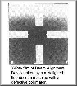
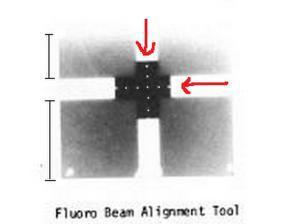
10 Objective and Equipments To evaluate the x-ray beam alignment of fluoroscopy unit in ensuring it is not larger than normal collimation. Fluoroscopic beam alignment device, x-ray film, protection shielding, energized fluoro unit.
11 Analysis Theedgesofthebeamfieldarenotinalign,thereforeb eamfieldandimagereceptoraremisaligned. The center of the field is indicated by the central dot and any misalignment of the beam can be checked by counting the number of dots visible in each channel. Supposedly, the dot at the intersection of the two lines should be at the center of the image There should be an equal number of dots visible on each of the channels when counted from the centre.
12
13 SOURCE TO SKIN DISTANCE
14 Objective and Equipments To reduce the skin exposure to the patient as much as possible while still assuring that sufficient x-ray output is obtainable to perform the diagnostic procedure. Energized fluoro unit, measuring tape.
15 Analysis Standard result: the source to skin distance must be not less than 15 inches (38cm) for stationary fluoroscopy for mobile fluoroscopy must be not less than 12 inches (30cm)
16 INTENSIFIER VIEWING SYSTEM HIGH CONTRAST
17 Objective and Equipments
18
 / size of FOV used) = minimum number of")
19 Analysis The minimum number of lp/mm that must be resolved by the system is given by the equation: 2 lp/mm x (6 inches (15cm) / size of FOV used) = minimum number of lp/mm.
20 INTENSIFIER VIEWING SYSTEM LOW CONTRAST
21 Objective and Equipments To resolve relatively large objects that differ slightly in radiolucency from the surrounding area. Energized fluoroscopic unit, low contrast resolution test tool
22
23 Analysis When the plates are sandwiched together and imaged on a fluoroscopic system, low contrast evaluation can be made based on the shallowest visible hole. With the low contrast resolution test tool, the contrast between the holes and the surrounding area is 2%. with CDRH phantom, the two deepest holes should be visible clearly. Better system are able to visualize the smaller holes on the test tools or the shallower holes on the CDRH phantom.
24 TV MONITORS AND RECORDERS
25 Objective and Equipments To assure that the TV monitor and recorder are functioning properly. Energized fluoro unit, phantom, test tool
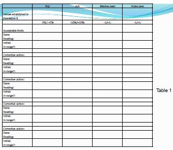
26 For kvp and ma changes: Analysis If the indicated kvp differs from the baseline value by more than 5% or the ma by more than 10%, recheck the setup of the phantom and fluoro system. If the setup is correct and the changes persists, then contact service personnel or the medical physicist. Decrease in number of meshes seen: If one mesh group is lost and the kvp value is the same then try adjusting the monitor brightness if possible to optimize visualization. If no improvement is obtained then contact service or medical physicist to perform a fluoroscopic resolution test and system focusing adjustment. Decrease number of holes seen: If one hole is lost and the kvp value is the same then try adjusting the monitor brightness if possible to optimize visualization. Ensure that you are viewing the monitor from a distance of 4 feet. If no improvement is obtained then contact service or medical physicist to perform a fluoroscopic resolution test and system focusing adjustment.
27 TEST TOOL
28
29 AUTOMATIC BRIGHTNESS CONTROL
30 Objective and Equipments To assure that the automatic brightness control are functioning properly. Energized fluoro unit, lead apron, phantom
31 Analysis As the thickness of structure increases, the brightness or contrast remain the same. ABC adjusts the contrast automatically. Allows the fluoroscopic unit to automatically maintain the brightness level of the image (variations of thickness and attenuation)
Nuclear Associates
 Nuclear Associates 07-649 CDRH Fluoroscopic Phantom Users Manual March 2005 Manual No. 07-649-1 Rev. 2 2004, 2005 Fluke Corporation, All rights reserved. Printed in U.S.A. All product names are trademarks
I. PERFORMANCE OF X-RAY PRODUCTION COMPONENTS FLUOROSCOPIC ACCEPTANCE TESTING: TEST PROCEDURES & PERFORMANCE CRITERIA
 FLUOROSCOPIC ACCEPTANCE TESTING: TEST PROCEDURES & PERFORMANCE CRITERIA EDWARD L. NICKOLOFF DEPARTMENT OF RADIOLOGY COLUMBIA UNIVERSITY NEW YORK, NY ACCEPTANCE TESTING GOALS PRIOR TO 1st CLINICAL USAGE
Nuclear Associates
 Nuclear Associates 07-647 R/F QC Phantom Operators Manual March 2005 Manual No. 07-647-1 Rev. 2 2004, 2005 Fluke Corporation, All rights reserved. All product names are trademarks of their respective companies
Nuclear Associates
 Nuclear Associates 07-706 Patient Phantom/Penetrometer System Users Manual March 2005 Manual No. 07-706-1 Rev. 2 2004, 2005 Fluke Corporation, All rights reserved. Printed in U.S.A. All product names are
TESTING FLAT-PANEL IMAGING SYSTEMS: What the Medical Physicist Needs to Know. JAMES A. TOMLINSON, M.S., D.A.B.R. Diagnostic Radiological Physicist
 TESTING FLAT-PANEL IMAGING SYSTEMS: What the Medical Physicist Needs to Know JAMES A. TOMLINSON, M.S., D.A.B.R. Diagnostic Radiological Physicist Topics Image Uniformity and Artifacts Image Quality - Detail
C-ARM FLUOROSCOPIC AND SPOT-FILM SYSTEMS
 PART X C-ARM FLUOROSCOPIC AND SPOT-FILM SYSTEMS FORM FD 3260 REPRINTED APRIL 2000 ROUTINE COMPLIANCE TESTING C-ARM FLUOROSCOPES (Test Procedure CFA - Use Form FDA 3260) 1.0 GENERAL GUIDANCE 1.1 This procedure
Nuclear Associates
 Nuclear Associates 76-700 Digital Subtraction Angiography Phantom Users Manual March 2005 Manual No. 76-700-1 Rev. 2 2004, 2005 Fluke Corporation, All rights reserved. Printed in U.S.A. All product names
Joint ICTP/IAEA Advanced School on Dosimetry in Diagnostic Radiology and its Clinical Implementation May 2009
 2033-6 Joint ICTP/IAEA Advanced School on Dosimetry in Diagnostic Radiology and its Clinical Implementation 11-15 May 2009 Dosimetry for Fluoroscopy Basics Renato Padovani EFOMP Joint ICTP-IAEA Advanced
LECTURE 1 The Radiographic Image
 LECTURE 1 The Radiographic Image Prepared by:- KAMARUL AMIN ABDULLAH @ ABU BAKAR UiTM Faculty of Health Sciences Medical Imaging Department 11/23/2011 KAMARUL AMIN (C) 1 Lesson Objectives At the end of
ABOVETABLE X-RAY SOURCE FLUOROSCOPIC AND SPOT-FILM SYSTEMS
 PART VI ABOVETABLE X-RAY SOURCE FLUOROSCOPIC AND SPOT-FILM SYSTEMS FORM FDA 3069 REPRINTED April 2000 ROUTINE COMPLIANCE TESTING ABOVETABLE X-RAY SOURCE FLUOROSCOPIC AND SPOT-FILM SYSTEMS (Test Procedure
Y11-DR Digital Radiography (DR) Image Quality
 Image Quality") Y11-DR Digital Radiography (DR) Image Quality Image quality is stressed for all systems in Safety Code 35. In the relevant sections Health Canada s advice is the manufacturer s recommended test procedures
Y11-DR Digital Radiography (DR) Image Quality Image quality is stressed for all systems in Safety Code 35. In the relevant sections Health Canada s advice is the manufacturer s recommended test procedures
Introduction. Chapter 16 Diagnostic Radiology. Primary radiological image. Primary radiological image
 Introduction Chapter 16 Diagnostic Radiology Radiation Dosimetry I Text: H.E Johns and J.R. Cunningham, The physics of radiology, 4 th ed. http://www.utoledo.edu/med/depts/radther In diagnostic radiology
NEMA. Cardiology Phantom Nuclear Associates Model Diagnostic Imaging. Introduction. Features. Applications
 NEMA Cardiology Phantom Nuclear Associates Model 07-680 Diagnostic Imaging! Phantom and test procedures simulate a range of fluoroscopically-guided invasive and interventional procedures! Provides simultaneous
DENTAL RADIOGRAPHY KAMARUL AMIN BIN ABU BAKAR
 DENTAL RADIOGRAPHY KAMARUL AMIN BIN ABDULLAH @ ABU BAKAR Components of the Dental X-Ray Machine Dental x-ray machines may vary somewhat in size and appearance, but all machines will have three primary
NJDEP Medical Physicist s Radiographic QC Survey Registration Number:
 Facility Name NJDEP ID # NJDEP Medical Physicist s Radiographic QC Survey PLEASE PRINT Facility Information Unit Information Manufacturer Model Console Model # Console serial # Tube serial # Location (room)
Minnesota Rules, Chapter 4732 X-ray Revision
 Minnesota Rules, Chapter 4732 X-ray Revision DRAFT FLUOROSCOPIC X-RAY SYSTEMS, 1.0 Subpart 1. Applicability. Subpart 2. Limitation of the useful beam. Subpart 3. Measuring compliance; primary protective
Test Equipment for Radiology and CT Quality Control Contents
 Test Equipment for Radiology and CT Quality Control Contents Quality Control Testing...2 Photometers for Digital Clinical Display QC...3 Primary Workstations...3 Secondary Workstations...3 Testing of workstations...3
FOUR CATEGORIES OF SAFETY
 OCTOBER 2013 FOUR CATEGORIES OF SAFETY DOSIMETRY PERSONAL SAFETY EQUIPMENT EQUIPMENT KNOWLEDGE PHYSICAL SAFETY DOSIMETRY THERMAL LUMINISCENT DEVICES AND FILM BADGES CNSC PERMISSIBLE DOSES WHOLE BODY DOSE
X-RAYS - NO UNAUTHORISED ENTRY
 Licencing of premises Premises Refer Guidelines A radiation warning sign and warning notice, X-RAYS - NO UNAUTHORISED ENTRY must be displayed at all entrances leading to the rooms where x-ray units are
1-1. GENERAL 1-2. DISCOVERY OF X-RAYS
 1-1. GENERAL Radiography is a highly technical field, indispensable to the modern dental practice, but presenting many potential hazards. The dental radiographic specialist must be thoroughly familiar
Beam-Restricting Devices
 Beam-Restricting Devices Three factors contribute to an increase in scatter radiation: Increased kvp Increased Field Size Increased Patient or Body Part Size. X-ray Interactions a some interact with the
INTRODUCTION TO FLEXIBLE BRONCHOSCOPY. Fluoroscopy Synopsis HENRI G COLT MD SECOND EDITION THE BRONCHOSCOPY EDUCATION PROJECT SERIES
 SECOND EDITION INTRODUCTION TO FLEXIBLE BRONCHOSCOPY Fluoroscopy Synopsis HENRI G COLT MD With contributions from Dr. S. Murgu THE BRONCHOSCOPY EDUCATION PROJECT SERIES FLUOROSCOPY SYNOPSIS The purpose
Nuclear Associates
 Nuclear Associates 07-644 Grid Alignment Test Tool Users Manual March 2005 Manual No. 07-644-1 Rev. 2 2004, 2005 Fluke Corporation, All rights reserved. Printed in U.S.A. All product names are trademarks
DIAGNOSTIC ACCREDITATION PROGRAM. Radiology and CT Quality Control Procedures Workbook
 DIAGNOSTIC ACCREDITATION PROGRAM Radiology and CT Quality Control Procedures Workbook Quality Control Procedures Radiography/CR/DR Safety Code 35 Summary For more detail about each quality control (QC)
QC Testing for Computed Tomography (CT) Scanner
 Scanner") QC Testing for Computed Tomography (CT) Scanner QA - Quality Assurance All planned and systematic actions needed to provide confidence on a structure, system or component. all-encompassing program, including
QC Testing for Computed Tomography (CT) Scanner QA - Quality Assurance All planned and systematic actions needed to provide confidence on a structure, system or component. all-encompassing program, including
Nuclear Associates
 Nuclear Associates 07-591 Focal Spot Test Tool Users Manual February 2005 Manual No. 07-591-1 Rev. 2 2004, 2005 Fluke Corporation, All rights reserved. Printed in U.S.A. All product names are trademarks
Fluoroscopy - Chapter 9
 Fluoroscopy - Chapter 9 Kalpana Kanal, Ph.D., DABR Lecturer, Diagnostic Physics Dept. of Radiology UW Medicine a copy of this lecture may be found at: http://courses.washington.edu/radxphys/physicscourse04-05.html
SPRINGFIELD TECHNICAL COMMUNITY COLLEGE ACADEMIC AFFAIRS
 SPRINGFIELD TECHNICAL COMMUNITY COLLEGE ACADEMIC AFFAIRS Course Number: RADG 212 Department: Radiography Course Title: Equip. Operation & Maint. Semester: Spring Year: 1997 Objectives/ Unit One: The X-ray
Acceptance Testing of a Digital Breast Tomosynthesis Unit
 Acceptance Testing of a Digital Breast Tomosynthesis Unit 2012 AAPM Spring Clinical Meeting Jessica Clements, M.S., DABR Objectives Review of technology and clinical advantages Acceptance Testing Procedures
RULES OF TENNESSEE DEPARTMENT OF ENVIRONMENT AND CONSERVATION DIVISION OF RADIOLOGICAL HEALTH CHAPTER USE OF X-RAY APPARATUS
 RULES OF TENNESSEE DEPARTMENT OF ENVIRONMENT AND CONSERVATION DIVISION OF RADIOLOGICAL HEALTH CHAPTER 0400-20-06 USE OF X-RAY APPARATUS TABLE OF CONTENTS 0400-20-06-.01 Purpose 0400-20-06-.06 Veterinary
MAN Revision 002. InSight Mini C-arm Imaging System Technical Reference Manual
 MAN-00668 Revision 002 InSight Mini C-arm Imaging System Technical Reference Manual April 2007 The information contained in this Manual is confidential and proprietary to Hologic, Inc. This information
Unfors EDD-30 Radiation Protection in Fluoroscopy
 Unfors EDD-30 Radiation Protection in Fluoroscopy Immediate Warning Decrease Your Dose Interventional radiology procedures are considered to be essential to medical diagnosis and treatment. It is recognized,
STEREOTACTIC BREAST BIOPSY EQUIPMENT SURVEYS
 STEREOTACTIC BREAST BIOPSY EQUIPMENT SURVEYS JAMES A. TOMLINSON, M.S. Diagnostic Radiological Physicist American Board of Radiology Certified Medical Physics Consultants, Inc. Bio 28 yrs experience 100%
Essentials of Digital Imaging
 Essentials of Digital Imaging Module 7 Transcript 2016 ASRT. All rights reserved. Essentials of Digital Imaging Module 7 Quality 1. ASRT Animation 2. Welcome Welcome to the Essentials of Digital Imaging:
GE AMX 4+ Portable X-Ray
 GE AMX 4+ Portable X-Ray Typical Manufacturer s Picture GE Healthcare s AMX-4+ analog X-ray system provides high-performance in a compact, easy-to-maneuver package. The rotating arm and tube simplify positioning
Ludlum Medical Physics
 Ludlum Medical Physics Medical Imaging Radiology QA Test Tools NEW LUDLUM PRODUCT LINE Medical Physics Products Medical Physics Products What are they? Products used to measure radiation output and to
Collimation Assessment Using GAFCHROMIC XR-M2
 Collimation Assessment Using GAFCHROMIC XR-M2 I. Introduction A method of collimation assessment for GE Senographe full-field digital mammography (FFDM) systems is described that uses a self-developing
Overview of Safety Code 35
 Common Quality Control Procedures for All s Quality Control Procedures Film All s Daily Quality Control Tests Equipment Warm-up (D1) According to manufacturers instructions Can include auto calibration(d1)
10/3/2012. Study Harder
 This presentation is a professional collaboration of development time prepared by: Rex Christensen Terri Jurkiewicz and Diane Kawamura Study Harder CR detection is inefficient, inferior to film screen
Visualization of sources of scattered radiation from x-ray equipment used for interventional radiology
 Visualization of sources of scattered radiation from x-ray equipment used for interventional radiology Poster No.: C-1190 Congress: ECR 2011 Type: Scientific Exhibit Authors: K. Chida, T. Takahashi, D.
10/26/2015. Study Harder
 This presentation is a professional collaboration of development time prepared by: Rex Christensen Terri Jurkiewicz and Diane Kawamura Study Harder CR detection is inefficient, inferior to film screen
COMPUTED RADIOGRAPHY CHAPTER 4 EFFECTIVE USE OF CR
 This presentation is a professional collaboration of development time prepared by: Rex Christensen Terri Jurkiewicz and Diane Kawamura New Technology https://www.youtube.com/watch?v=ptkzznazb 7U COMPUTED
NATIONWIDE EVALUATION OF X-RAY TRENDS (NEXT) SUMMARY OF 2003 FLUOROSCOPY SURVEY
 SUMMARY OF 2003 FLUOROSCOPY SURVEY") CRCPD Publication E-09-5 Available Online at No Charge NATIONWIDE EVALUATION OF X-RAY TRENDS (NEXT) SUMMARY OF 2003 FLUOROSCOPY SURVEY September 2009 Published by Conference of Radiation Control Program
CRCPD Publication E-09-5 Available Online at No Charge NATIONWIDE EVALUATION OF X-RAY TRENDS (NEXT) SUMMARY OF 2003 FLUOROSCOPY SURVEY September 2009 Published by Conference of Radiation Control Program
HIGH-RESOLUTION CORE FLUOROSCOPY, AN IMPORTANT TOOL FOR CORE ANALYSIS
 SCA2007-59 1/6 HIGH-RESOLUTION CORE FLUOROSCOPY, AN IMPORTANT TOOL FOR CORE ANALYSIS Christopher M. Prince VP Imaging Technology, PTS Laboratories, Inc., Houston, TX This paper was prepared for presentation
Multiple Choice Identify the letter of the choice that best completes the statement or answers the question.
 RA110 test 3 Multiple Choice Identify the letter of the choice that best completes the statement or answers the question. 1. An object 35 cm in width is radiographed at 100 cm SID and at a 50 cm SOD. What
Investigation of the line-pair pattern method for evaluating mammographic focal spot performance
 Investigation of the line-pair pattern method for evaluating mammographic focal spot performance Mitchell M. Goodsitt, a) Heang-Ping Chan, and Bob Liu Department of Radiology, University of Michigan, Ann
BASICS OF FLUOROSCOPY
 Medical Physics Residents Training Program BASICS OF FLUOROSCOPY Dr. Khalid Alyousef, PhD Department of Medical Imaging King Abdulaziz Medical City- Riyadh Edison examining the hand of Clarence Dally with
MXHF-1500RF is controlled by Digital key panel console that displays KV, ma and mas with APR menu programmed.
 R/F TV X-RAY SYSTEM DIAGNOSTIC RADIOGRAPHIC FLUOROSCOPIC TV SYSTEM MXHF-1500RF SYSTEM OUTLINE Product Data No. 041021-01 MXHF-1500RF is controlled by Digital key panel console that displays KV, ma and
Quality Control for Stereotactic Breast Biopsy. Robert J. Pizzutiello, Jr., F.A.C.M.P. Upstate Medical Physics, Inc
 Quality Control for Stereotactic Breast Biopsy Robert J. Pizzutiello, Jr., F.A.C.M.P. Upstate Medical Physics, Inc. 716-924-0350 Methods of Imaging Guided Breast Biopsy Ultrasound guided, hand-held needle
Ch. 223 VETERINARY MEDICINE CHAPTER 223. VETERINARY MEDICINE GENERAL PROVISIONS X-RAYS RADIOACTIVE MATERIAL. Authority
 Ch. 223 VETERINARY MEDICINE 25 223.1 CHAPTER 223. VETERINARY MEDICINE Sec. 223.1. Purpose and scope. 223.2. [Reserved]. 223.2a. Definitions. 223.3 223.6. [Reserved]. 223.7. Structural shielding. 223.8.
Sarah Hughes, MS, DABR Radiation Safety Officer
 Sarah Hughes, MS, DABR Radiation Safety Officer 502-852-6146 sarah.hughes@louisville.edu Mo my back is burnin!!! I got it MAG the cine! Sumthin s not right. Where s his heart? Fluoroscopy http://dccwww.bumc.bu.edu/fluoroscopy/def
Surveying and QC of Stereotactic Breast Biopsy Units for ACR Accreditation
 Surveying and QC of Stereotactic Breast Biopsy Units for ACR Accreditation AAPM Annual Clinical Meeting Indianapolis, IN August 5, 2013 Learning Objectives Become familiar with the recommendations and
Diagnostic x-ray equipment compliance and facility survey
 Canada Health Canada Canada CA9600871 CA9600871 Diagnostic x-ray equipment compliance and facility survey Diagnostic x-ray equipment compliance and facility survey Recommended procedures for equipment
Nuclear Associates EZ CR-DIN Phantoms
 Nuclear Associates 07-605-7777 EZ CR-DIN Phantoms Users Manual August 2006 Manual No. 07-605-7777-1 Rev. 4 2006 Fluke Corporation, All rights reserved. Printed in U.S.A. All product names are trademarks
CHAPTER 2 COMMISSIONING OF KILO-VOLTAGE CONE BEAM COMPUTED TOMOGRAPHY FOR IMAGE-GUIDED RADIOTHERAPY
 14 CHAPTER 2 COMMISSIONING OF KILO-VOLTAGE CONE BEAM COMPUTED TOMOGRAPHY FOR IMAGE-GUIDED RADIOTHERAPY 2.1 INTRODUCTION kv-cbct integrated with linear accelerators as a tool for IGRT, was developed to
Fluke199XRAY. Users Manual. Medical ScopeMeter
 Fluke199XRAY Medical ScopeMeter Users Manual 4822 872 30791 August 2006 2006 Fluke Corporation, All rights reserved. All product names are trademarks of their respective companies. Table of Contents Title
Dedicated Veterinary Imaging Solutions Digital, CR and Analog Imaging Solutions for any size patient and any size budget.
 by Dedicated Veterinary Imaging Solutions Digital, CR and Analog Imaging Solutions for any size patient and any size budget. Serving the Veterinary Profession for Over 75 Years. ... We See Things Differently
Key words: fluoroscopy, dose-area-product, kerma-area-product, calibration of KAP meters, patient exposure
 Accuracy and calibration of integrated radiation output indicators in diagnostic radiology: A report of the AAPM Imaging Physics Committee Task Group 190 Pei-Jan P. Lin a) Virginia Commonwealth University
RAD 150 RADIOLOGIC EXPOSURE TECHNIQUE II
 RAD 150 RADIOLOGIC EXPOSURE TECHNIQUE II APPROVED 12/O2/2011 EFFECTIVE SPRING 2013-14 Prefix & Number RAD 150 Course Title: Radiologic Exposure Technique II & Lab Purpose of this submission: New Change/Updated
Visibility of Detail
 Visibility of Detail Radiographic Quality Quality radiographic images represents the, and information is for diagnosis. The of the anatomic structures and the accuracy of their ( ) determine the overall
Safelight Fog does what to contrast and density on film?
 Terri Jurkiewicz Safelight Fog does what to contrast and density on film? ANSWER INCREASES DENSITY DECREASES CONTRAST Explain how you determine if the focal spot size is within appropriate limits.
Evaluation of a quality control phantom for digital chest radiography
 JOURNAL OF APPLIED CLINICAL MEDICAL PHYSICS, VOLUME 2, NUMBER 2, SPRING 2001 Evaluation of a quality control phantom for digital chest radiography Eugene Mah* Department of Radiology, Medical University
Assessment of Beam Alignment, Collimation and Half Value Layer of Some Selected X-Ray Machines in Plateau State, Nigeria
 International Journal of Innovative Scientific & Engineering Technologies Research 5(4):-5, Oct.-Dec., 07 SEAHI PUBLICATIONS, 07 www.seahipaj.org ISSN: 60-896X Assessment of Beam Alignment, Collimation
X-RAY FLUOROSCOPY IMAGING SYSTEMS. Dr Slavik Tabakov. Luminescence: Dept. Medical Eng. & Physics King s College London
 X-RAY FLUOROSCOPY IMAGING SYSTEMS Dr Slavik Tabakov OBJECTIVES - Image Intensifier construction - Input window - Accelerating and focusing electrodes - Output window - Conversion factor - II characteristics
Veterinary Science Preparatory Training for the Veterinary Assistant. Floron C. Faries, Jr., DVM, MS
 Veterinary Science Preparatory Training for the Veterinary Assistant Floron C. Faries, Jr., DVM, MS Radiology Floron C. Faries, Jr., DVM, MS Objectives Determine the appropriate machine settings for making
Digital Imaging started in the 1972 with Digital subtraction angiography Clinical digital imaging was employed from the 1980 ~ 37 years ago Amount of
 Digital Imaging started in the 1972 with Digital subtraction angiography Clinical digital imaging was employed from the 1980 ~ 37 years ago Amount of radiation to the population due to Medical Imaging
Overview. Professor Roentgen was a Physicist!!! The Physics of Radiation Oncology X-ray Imaging
 The Physics of Radiation Oncology X-ray Imaging Charles E. Willis, Ph.D. DABR Associate Professor Department of Imaging Physics The University of Texas M.D. Anderson Cancer Center Houston, Texas Overview
QUALITY CONTROL TESTS IN SOME DIAGNOSTIC X-RAY UNITS IN BANGLADESH
 Bangladesh Journal of Medical Physics Vol. 4, No.1, 2011 QUALITY CONTROL TESTS IN SOME DIAGNOSTIC X-RAY UNITS IN BANGLADESH M. Begum 1, A. S. Mollah 2, M. A. Zaman 3 and A. K. M. M. Rahman 4 1 Health Physics
Essentials of Digital Imaging
 Essentials of Digital Imaging Module 6 Transcript 2016 ASRT. All rights reserved. Essentials of Digital Imaging Module 6 Dose Reduction and Patient Safety 1. ASRT Animation 2. Welcome Welcome to Essentials
X-ray Tube and Generator Basic principles and construction
 X-ray Tube and Generator Basic principles and construction Dr Slavik Tabakov - Production of X-rays and Patient Dose OBJECTIVES - X-ray tube construction - Anode - types, efficiency - Classical X-ray generator
SPECIFICATION. Kilovoltage X-ray calibration system for protection and diagnostic level dosimetry. Prepared by
 SPECIFICATION Kilovoltage X-ray Prepared by Igor Gomola, Technical Officer, Project ECU6023, Date 2015-Oct-06 Revision Date Status Comments 0.1 2015-Oct-06 Draft Igor Gomola Page 1 of 12 1. Scope This
Half value layer and AEC receptor dose compliance survey in Estonia
 Half value layer and AEC receptor dose compliance survey in Estonia K. Kepler, A. Vladimirov Training Centre of Medical Physics, University of Tartu Testing Centre of the University of Tartu, Estonia E-mail:
Dose Reduction and Image Preservation After the Introduction of a 0.1 mm Cu Filter into the LODOX Statscan unit above 110 kvp
 Dose Reduction and Image Preservation After the Introduction of a into the LODOX Statscan unit above 110 kvp Abstract: CJ Trauernicht 1, C Rall 1, T Perks 2, G Maree 1, E Hering 1, S Steiner 3 1) Division
Ansur TNT Users Manual. Plug-In
 Ansur TNT 12000 Plug-In Users Manual August 2009, Rev. 2, 12/09 2009 Fluke Corporation. All rights reserved. Specifications are subject to change without notice. All product names are trademarks of their
Title: A COMPARISON OF Cs-137 AND X-RAY SOURCES AS CALIBRATION REFERENCES FOR THERMOLUMINESCENT DOSIMETER CHIPS
 Title: A COMPARISON OF Cs-137 AND X-RAY SOURCES AS CALIBRATION REFERENCES FOR THERMOLUMINESCENT DOSIMETER CHIPS By Aravind Ravichandran arr192@mail.usask.ca University of Saskatchewan Address: 2424 Cumberland
REQUIREMENTS FOR LICENCE HOLDERS WITH RESPECT TO QUALITY CONTROL TESTS FOR DIAGNOSTIC X-RAY IMAGING SYSTEMS
 REQUIREMENTS FOR LICENCE HOLDERS WITH RESPECT TO QUALITY CONTROL TESTS FOR DIAGNOSTIC X-RAY IMAGING SYSTEMS DEPARTMENT OF HEALTH DIRECTORATE: RADIATION CONTROL Implementation date: 31 March 2009 Contents
Notice of Rulemaking Hearing
 Department of State Division of Publications For Department of State Use Only 312 Rosa L. Parks, 8th Floor SnodgrassfTN Tower Sequence Number: Oil -Cf) - (r Nashville, TN 37243 Phone: 615.741.2650 Notice
Nuclear Associates &
 Nuclear Associates 06-526 & 06-526-2200 RAD-CHECK PLUS Operators Manual March 2005 Manual No. 136201 Rev. 5 2003, 2005 Fluke Corporation, All rights reserved. Printed in U.S.A. All product names are trademarks
IChapter Number,ChapterTiiie I0400: _OJ TJse of X-Ray Apparatus! Rule Number Rule Title
 Department of State Division of Publications 312 Rosa L. Parks Avenue, 8t11 Floor SnodgrassfTN Tower Nashville, TN 37243 Phone: 615-741-2650 Fax: 615-741-5133 Email: register.information@tn.gov For Department
Exposure System Selection
 Principles of Imaging Science II (RAD120) Exposure Systems Exposure System Selection Radiographic exposure is a very complex process Best technique systems manipulate one variable while holding others
Teaching Digital Radiography and Fluoroscopic Radiation Protection
 Teaching Digital Radiography and Fluoroscopic Radiation Protection WCEC 20 th Student Educator Radiographer Conference Dennis Bowman, RT(R), CRT (R)(F) Community Hospital of the Monterey Peninsula (CHOMP)
SYLLABUS. TITLE: Equipment Operation I. DEPARTMENT: Radiologic Technology
 CODE: RADT 156 INSTITUTE: Health Science TITLE: Equipment Operation I DEPARTMENT: Radiologic Technology COURSE DESCRIPTION: This course covers the principles of equipment operation and maintenance of radiographic
The X-ray circuit: part II
 The X-ray circuit: part II By Dr. Mohsen Dashti 357 Radiologic Processing & Procedure Lecture notes #2 Key issues Types of x-ray equipment. Power for x-ray generator. A basic x-ray circuit. Generators.
Learning Objectives: What s my motivation? (unknown screen actor) Workshop Overview
 Workshop Overview") Practical Medical Physics Adapting Traditional Clinical Medical Physics to Digital Radiography Charles E. Willis, Ph.D., DABR Associate Professor Department of Imaging Physics The University of Texas M.D.
Practical Medical Physics Adapting Traditional Clinical Medical Physics to Digital Radiography Charles E. Willis, Ph.D., DABR Associate Professor Department of Imaging Physics The University of Texas M.D.
Features and Weaknesses of Phantoms for CR/DR System Testing
 Physics testing of image detectors Parameters to test Features and Weaknesses of Phantoms for CR/DR System Testing Spatial resolution Contrast resolution Uniformity/geometric distortion Dose response/signal
X-ray Equipment in Medical Diagnosis Part A: Recommended Safety Procedures for Installation and Use
 Health Canada Santé Canada X-ray Equipment in Medical Diagnosis Part A: Recommended Safety Procedures for Installation and Use Safety Code 20A X-ray Equipment in Medical Diagnosis Part A: Recommended Safety
ProX Intraoral X-ray. PLANMECA is proud to introduce a new intraoral X-ray unit to its comprehensive collection of imaging products- the ProX.
 The premium intraoral X-ray unit... ProX Intraoral X-ray PLANMECA is proud to introduce a new intraoral X-ray unit to its comprehensive collection of imaging products- the ProX. This advanced unit provides
(CR) PORTABLE CHEST EXAM Baseline Definition Worksheet
 PORTABLE CHEST EXAM Baseline Definition Worksheet") Supertech P.O. Box 186. Elkhart, N 46515-0186. Phone: 1-800-654-1054 or 1-574-264-4310 Fax: 574-264-9551. Web: www.supertechx-ray.com. E-mail: sales@supertechx-ray.com (CR PORTABLE CHEST EXAM Baseline
Supertech P.O. Box 186. Elkhart, N 46515-0186. Phone: 1-800-654-1054 or 1-574-264-4310 Fax: 574-264-9551. Web: www.supertechx-ray.com. E-mail: sales@supertechx-ray.com (CR PORTABLE CHEST EXAM Baseline
Nuclear Associates , , CT Head and Body Dose Phantom
 Nuclear Associates 76-414,76-414-4150,76-415 CT Head and Body Dose Phantom Users Manual March 2005 Manual No. 76-414-1 Rev. 2 2004, 2005 Fluke Corporation, All rights reserved. Printed in U.S.A. All product
CHAPTER 40 - PROFESSIONAL LICENSING AND FACILITY REGULATION. PART 4 - Diagnostic X-Rays and Associated Imaging Systems in the Healing Arts
 216-RICR-40-20-4 TITLE 216 - DEPARTMENT OF HEALTH CHAPTER 40 - PROFESSIONAL LICENSING AND FACILITY REGULATION SUBCHAPTER 20 - RADIATION PART 4 - Diagnostic X-Rays and Associated Imaging Systems in the
Version 1.0. TechnicVR. Student Guide
 Version 1.0 TechnicVR s h a d e r w a r e. c o m Student Guide TechnicVR s h a d e r w a r e. c o m Student Guide shaderware 2008 PO Box 103 Saltburn Cleveland TS12 1WP w w w. s h a d e r w a r e. c o
Quality control of Gamma Camera. By Dr/ Ibrahim Elsayed Saad 242 NMT
 Quality control of Gamma Camera By Dr/ Ibrahim Elsayed Saad 242 NMT WHAT IS QUALITY? The quality of a practice is to fulfill the expectations and demands from: Patient Clinicain Your self Quality assurance
Clinical Experiences with a Patient Skin Dose Monitoring and Tracking Program
 Clinical Experiences with a Patient Skin Dose Monitoring and Tracking Program Allen R. Goode, MS, DABR Chief Diagnostic Medical Physicist Department of Radiology & Medical Imaging University of Virginia
Do you have any other questions? Please call us at (Toll Free) or , or
 or , or") INSTRUCTIONS Read the appropriate course/ textbook. This is an open book test. A score of 75% or higher is needed to receive CE credit. You will have a maximum of three attempts to pass this course. Please
INSTRUCTIONS Read the appropriate course/ textbook. This is an open book test. A score of 75% or higher is needed to receive CE credit. You will have a maximum of three attempts to pass this course. Please
Luminos RF Classic. Where value meets performance.
 Luminos RF Classic Where value meets performance www.siemens.com/healthcare What s good value in fluoroscopy? That s easy. Luminos RF Classic. 2 Whether for its handling convenience, outstanding image
X-ray Imaging. PHYS Lecture. Carlos Vinhais. Departamento de Física Instituto Superior de Engenharia do Porto
 X-ray Imaging PHYS Lecture Carlos Vinhais Departamento de Física Instituto Superior de Engenharia do Porto cav@isep.ipp.pt Overview Projection Radiography Anode Angle Focal Spot Magnification Blurring
Interventional Radiological Equipment selection and installation
 Interventional Radiological Equipment selection and installation Renato Padovani ICTP Learning objectives To understand the main components of an interventional radiology equipment To understand the relevance
Breast Tomosynthesis. Bob Liu, Ph.D. Department of Radiology Massachusetts General Hospital And Harvard Medical School
 Breast Tomosynthesis Bob Liu, Ph.D. Department of Radiology Massachusetts General Hospital And Harvard Medical School Outline Physics aspects of breast tomosynthesis Quality control of breast tomosynthesis
AN ABSTRACT OF THE THESIS OF. W. Scott Helms for the degree of Master of Science in Radiation Health Physics
 AN ABSTRACT OF THE THESIS OF W. Scott Helms for the degree of Master of Science in Radiation Health Physics presented on November 24, 2014 Title: A Quantitative Comparison of Cardiovascular Imaging Systems
2012 :15th SESSION of ESMP
 2012 :15th SESSION of ESMP Lecture presented in Archamps (Salève Building) by : Elly CASTELLANO (London) Patient dosimetry in x-ray imaging and CT Elly Castellano Objectives measurable dose quantities