Quality control of Gamma Camera. By Dr/ Ibrahim Elsayed Saad 242 NMT
|
|
|
- Marsha Bailey
- 5 years ago
- Views:
Transcription


1 Quality control of Gamma Camera By Dr/ Ibrahim Elsayed Saad 242 NMT
2 WHAT IS QUALITY? The quality of a practice is to fulfill the expectations and demands from: Patient Clinicain Your self
3 Quality assurance and quality control The concept of quality in the term quality assurance expresses the closeness with which the outcome of a given procedure approaches some ideal, free from all errors and artifacts (Whole procedure). The term quality control is used in reference to the specific measures taken to ensure that one particular aspect of the procedure is satisfactory (Single step in the procedure).
4 NUCLEAR MEDICINE SERVICE facilities patient care waiting time staff reporting competence experience optimisation radiopharmaceuticals methods examination technique instrumentation etc etc Primary service Secondary service Nuclear medicine examination or treatment

5 QA-PROGRAMME OBJECTIVES * Improvement in the quality of the diagnostic information. * Use of minimum amount of radionuclide activity to ensure the production of the desired diagnostic information. * Effective use of available resources
6 QA Medical exposure Choice of examination Determination of technical parameters Optimization of administered activity Methods of reducing the absorbed dose Quality control of equipment and radiopharmaceutical Quality assurance of methods Safe routines to avoid misadministration

7 Gamma Camera Used to measure the spatial and temporal distribution of a radiopharmaceutical
8 Test Schedule for Gamma Camera System Test Frequency in routine testing Acceptance Referenc e once per Quarter Week Half year 1-Physical Inspection A 2-Test of Intrinsic Flood-field Uniformity 3- Test of Intrinsic Spatial Resolution 4- Test of Intrinsic Count-rate Performance 5- Test of Spatial Linearity and Spatial Resolution A R Q W A R H A R H A 6- Test of Basic Computer Timing A R H 7- Test of Computer Timing in Dynamic Acquisition R A R H 8- Test of camera Sensitivity A R W


9 SOURCES FOR QC OF GAMMA- CAMERAS <1 mm Point source Collimated line source Line source Flood source Tc99m, Co57, Ga67


10 Phantoms for QC of gamma-cameras Bar phantom Slit phantom Orthogonal hole phantom Total performance phantom

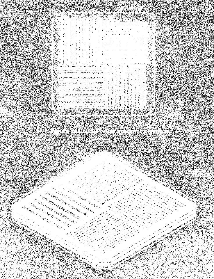
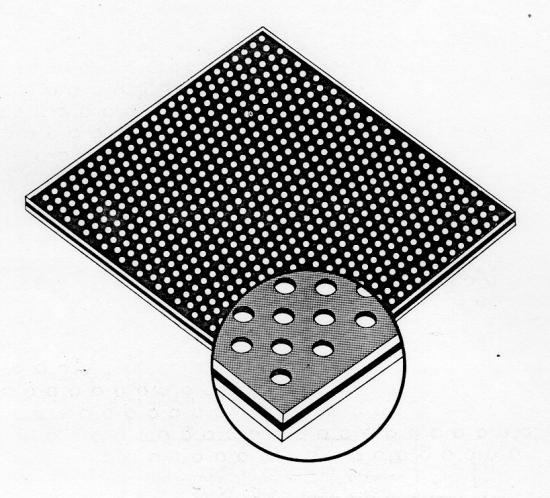
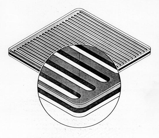
11 Phantoms for QC of gamma-cameras
12 Physical Inspection The Purpose of this test is to inspect a cameracomputer system for shipping damage and production flaws. Procedure: 1-Image Display Devices 2-Image Recording Devices 3-Electrical Connections, Fuses and Cables 4-Operation and Service Manuals 5-Check for controls that are difficult to operate or are noisy and switches that do not throw securely. Inspect keyboards for damage.
13 Special definitions for quality control System: a term used to refer to the performance of the camera detector, as it would be used in a clinical environment including components such as a collimator and supporting gantry. Energy window: A range of gamma and X-ray energies, which are to be accepted and processed. The window is expressed as a range of energies or as a percentage of expected peak energy. When expressed as percentage the peak energy must always be specified, and the window is always symmetrical about the peak energy value. Photopeak: the characteristic energy distribution resulting from the collection of total photon energy absorbed by the detector.
14 Differential uniformity: The amount of count density change per defined unit distance when the detector s incident gamma radiation is a homogenous flux over the field of measurement. Integral uniformity: A measure of the maximum count density variation over a defined large area of the scintillation detector for a uniform input gamma flux to the Useful Field of View of the camera. Pixel: a picture element used to store a value in a digital memory. It represents an area defined by dimensions in X and Y directions at a known position defined by X and Y coordinates.
15 Scatter: photons that have lost part of their energy due to interaction with a medium, such as water, plastic, or tissue. Sensitivity: The observed count rate per unit of radioactivity at a specified distance. Spectrum: a plot of the number of detected gamma rays versus the measured energy of the gamma rays.
16 Energy resolution: A term used to characterize the ability of a gamma camera to distinguish between photons of different energies Spatial resolution: A term that characterizes the scintillation camera s ability to accurately resolve spatially separated radioactive sources.
17 Full Width at Half Maximum (FWHM): The measure of the spread of a point or line spread function measured between locations 50 %down on each side from the peak amplitude. Full Width at Tenth Maximum (FWTM): The measure of the spread of a point or line response function measured between locations 90 % down on each side from the peak amplitude.
18 Differential linearity: The amount of positional distortion or displacement over a limited distance Useful Field of View (UFOV): The area of the detector, which is used for imaging gamma rays and x- rays. It is defined by a dimensioned figure supplied by the manufacturer. Central field of view (CFOV): The area defined by scaling all linear dimensions of the useful field of view (UFOV) by a factor of 75%.
19 Intrinsic: A term used to describe performance characteristics of a scintillation camera that exclude external variables, which affect these specifications such as collimators or display devices. Extrinsic: A term that is used to describe the total system performance including photons attenuated throw passing into the collimator, so that it includes all external factors such as collimator.
20 EnergyWindow Peaking 15 % window 25 % window 35 % window

21 140 KeV 137 KeV Wrong setting of energy window
22 1) Test of Intrinsic Flood-field Uniformity The purpose of this test is to test the combined intrinsic response of a cameracomputer system to a spatially uniform flux of incident gamma radiation over the camera field-of-view. Materials used are: Point source consisting of MBq ( mci) Tc-99m, giving a count rate not greater than c /s with a 20% PHA window. Source mounting for point source.
23 Procedure 1. Remove the collimator from the detector head. Align the head and the source mounting. 2. Mount the source in the source mounting with distance equal to 5 times the average detector diameter, which is (220 cm). 3. Centre a 20% PHA window on the Photopeak (Check of Energy Calibration of PHA). 4. Acquire an image on the cameras display device at a preset count of 15 x 10 6 using a 512X512 matrix. 6. Record the collection time for the camera. 7. Remove the source and mount the collimator.
24 Data Analysis: Determine the maximum (Max) and minimum (Min) counts in the pixels lying within the UFOV and the CFOV. The integral uniformity, IU, is then given by: IU = 100 * Max - Min Max + Min Determine for each row or column of pixels in the X and Y directions within the UFOV and the CFOV, the maximum count difference in any 6 contiguous pixels. Determine the highest value of this maximum count difference in the sets of rows and columns. The differential uniformity, DU, is then given by: DU =100* Hi ' Low Hi + Low
25 2) Test of Intrinsic Spatial Resolution The purpose of this test is to test the combined intrinsic spatial resolution of a camera- computer system in terms of the full width at half-maximum (FWHM) and full width at tenth-maximum (FWTM). Procedures: 1-The collimator is removed from the detector face. 2- Position the intrinsic-resolution phantom ( slit phantom) and then adjust the source on the source holder to be in front of the detector face with distance equal to 5 times the average detector diameter, which is (220 cm). 3-The acquisition parameters for this test is adjusted as follows: a)15000 K. counts. b)512x512x16 acquisition matrix. c)20 % PHA window on the photopeak which is 140 KeV for Tc-99m.
26 4-Acquire an image for the camera device at the preset count of 15 x By using the software provided on the processing computer connected to the camera, by which we can draw a line on a single slit of the slits in the phantom image, this will give us a profile which indicate to the peak from which we can calculate the FWHM in Pixels. And then by knowing the matrix size used while acquiring the image we can calculate the FWHM in millimeters.
27 6-Then the estimation of the intrinsic spatial resolutions is done in terms of FWHM of the line spread function. counts Pixels
28 At acceptance testing, the calculated values of FWHM in the X and Y directions should be compared with the manufacturer's worst-case values. At routine testing, the calculated values should be compared with the reference values.
29 3) Test of Intrinsic Spatial Linearity The same procedures for testing the intrinsic resolution is prepared. The resulted peaks for the lines separating the lead sheets is plotted on a graph and then the spatial distortion (linearity) is measured.

30 Test of Intrinsic Spatial Linearity
31 Test of Intrinsic Spatial Linearity Count pixels
32 Then the calculation of the distance between each adjacent peaks is done, and calculating the difference between the maximum distance and the minimum distance this will indicate the shifting between the parallel slits, this is directly the spatial linearity.


33
Radionuclide Imaging MII 3073 RADIONUCLIDE IMAGING SYSTEM
 Radionuclide Imaging MII 3073 RADIONUCLIDE IMAGING SYSTEM Preamplifiers and amplifiers The current from PMT must be further amplified before it can be processed and counted (the number of electrons yielded
Robert Pagnanelli BSRT(R)(N), CNMT, NCT, FASNC Chief Technologist, Nuclear Imaging Duke University Medical Center. Thursday September 8, 2011
(N), CNMT, NCT, FASNC Chief Technologist, Nuclear Imaging Duke University Medical Center. Thursday September 8, 2011") Robert Pagnanelli BSRT(R)(N), CNMT, NCT, FASNC Chief Technologist, Nuclear Imaging Duke University Medical Center Thursday September 8, 2011 Quality Control Quality control should be performed because:
Robert Pagnanelli BSRT(R)(N), CNMT, NCT, FASNC Chief Technologist, Nuclear Imaging Duke University Medical Center Thursday September 8, 2011 Quality Control Quality control should be performed because:
GS Introduction to Medical Physics IV Laboratory 5 Gamma Camera Characteristics
 GS02 0193 Introduction to Medical Physics IV Laboratory 5 Gamma Camera Characteristics Purpose: To introduce some of the basic characteristics of a gamma camera. This lab will introduce gamma camera QC
NM Module Section 2 6 th Edition Christian, Ch. 3
 NM 4303 Module Section 2 6 th Edition Christian, Ch. 3 Gas Filled Chamber Voltage Gas filled chamber uses Hand held detectors cutie pie Geiger counter Dose calibrators Cutie pie Chamber voltage in Ionization
Parameters Affecting on Intrinsic Uniformity Test For MEDISO
 ISPUB.COM The Internet Journal of Nuclear Medicine Volume 5 Number 2 Parameters Affecting on Intrinsic Uniformity Test For MEDISO S Zobly, A Osman Citation S Zobly, A Osman. Parameters Affecting on Intrinsic
CHAPTER 8 GENERIC PERFORMANCE MEASURES
 GENERIC PERFORMANCE MEASURES M.E. DAUBE-WITHERSPOON Department of Radiology, University of Pennsylvania, Philadelphia, Pennsylvania, United States of America 8.1. INTRINSIC AND EXTRINSIC MEASURES 8.1.1.
Acceptance Testing and Annual Physics Survey Recommendations for Gamma Camera, SPECT, and SPECT/CT Systems
 AAPM REPORT NO. 177 Acceptance Testing and Annual Physics Survey Recommendations for Gamma Camera, SPECT, and SPECT/CT Systems The Report of AAPM Task Group 177 February 2019 DISCLAIMER: This publication
Initial Certification
 Initial Certification Nuclear Medical Physics (NMP) Study Guide Part 2 Content Guide and Sample Questions The content of all ABR exams is determined by a panel of experts who select the items based on
Intrinsic and Tomographic Evaluation of Siemens e.cam SPECT System at the Korle-Bu Teaching Hospital (Ghana)
") Research Journal of Applied Sciences, Engineering and Technology 3(10): 1152-1158, 2011 ISSN: 2040-7467 Maxwell Scientific Organization, 2011 Submitted: July 17, 2011 Accepted: September 05, 2011 Published:
Research Journal of Applied Sciences, Engineering and Technology 3(10): 1152-1158, 2011 ISSN: 2040-7467 Maxwell Scientific Organization, 2011 Submitted: July 17, 2011 Accepted: September 05, 2011 Published:
Introduction. Chapter 16 Diagnostic Radiology. Primary radiological image. Primary radiological image
 Introduction Chapter 16 Diagnostic Radiology Radiation Dosimetry I Text: H.E Johns and J.R. Cunningham, The physics of radiology, 4 th ed. http://www.utoledo.edu/med/depts/radther In diagnostic radiology
International Journal of Scientific & Engineering Research, Volume 4, Issue 9, September ISSN
 International Journal of Scientific & Engineering Research, Volume 4, Issue 9, September-013 06 Evaluating the effect of acquisition parameters on image quality and acquisition time with SPECT using collimator
Quality control in dual head γ- cameras: Comparison between methods and softwares used for image analysis
 Quality control in dual head γ- cameras: Comparison between methods and softwares used for image analysis Abdalrhman Nayl Edam 1, *, Maria Rosa Fornasier 2, Mario de Denaro 2 Abdelmoneim Sulieman 3, Mohammed
Investigation of Multiple Head Registration / Center of Rotation for SPECT Gamma Cameras
 Egyptian J. Nucl. Med., Vol 2, No. 2, Dec. 2009 82 PHYSICS, Original Artical Investigation of Multiple Head Registration / Center of Rotation for SPECT Gamma Cameras Abdelsattar, M.B. Ph.D.; BuHumaid,
QUALITY CONTROL OF IMAGING DEVICES. P.S. Soni
 XA9847605 Chapter 8 P.S. Soni Quality Assurance and Quality Control in Nuclear Medicine Quality assurance in nuclear medicine refers collectively to all aspects of a nuclear medicine service. It would
CHAPTER 15 DEVICES FOR EVALUATING IMAGING SYSTEMS
 DEVICES FOR EVALUATING IMAGING SYSTEMS O. DEMIRKAYA, R. AL-MAZROU Department of Biomedical Physics, King Faisal Specialist Hospital and Research Centre, Riyadh, Saudi Arabia 15.1. DEVELOPING A QUALITY
Nuclear Associates , &
 Nuclear Associates 76-810, 76-814 76-815 & 76-818 Bar Phantoms and Test Patterns Operators Manual March 2005 Manual No. 76-810-1 Rev. 2 2004, 2005 Fluke Corporation, All rights reserved. Printed in U.S.A.
A new operative gamma camera for Sentinel Lymph Node procedure
 A new operative gamma camera for Sentinel Lymph Node procedure A physicist device for physicians Samuel Salvador, Virgile Bekaert, Carole Mathelin and Jean-Louis Guyonnet 12/06/2007 e-mail: samuel.salvador@ires.in2p3.fr
How Gamma Camera s Head-Tilts Affect Image Quality of a Nuclear Scintigram?
 November 2014, Volume 1, Number 4 How Gamma Camera s Head-Tilts Affect Image Quality of a Nuclear Scintigram? Hojjat Mahani 1,2, Alireza Kamali-Asl 3, *, Mohammad Reza Ay 2, 4 1. Radiation Application
Photomultiplier Tube
 Nuclear Medicine Uses a device known as a Gamma Camera. Also known as a Scintillation or Anger Camera. Detects the release of gamma rays from Radionuclide. The radionuclide can be injected, inhaled or
ACR Update in Nuclear Medicine Accreditation
 Disclaimer ACR Update in Nuclear Medicine Accreditation Beth A. Harkness, MS, DABR, FACR Henry Ford Health System Detroit, MI ACR physics subcommittee for nuclear medicine accreditation. My facility is
QC Testing for Computed Tomography (CT) Scanner
 Scanner") QC Testing for Computed Tomography (CT) Scanner QA - Quality Assurance All planned and systematic actions needed to provide confidence on a structure, system or component. all-encompassing program, including
QC Testing for Computed Tomography (CT) Scanner QA - Quality Assurance All planned and systematic actions needed to provide confidence on a structure, system or component. all-encompassing program, including
J of Nuclear Medicine Technology, first published online July 27, 2011 as doi: /jnmt
 J of Nuclear Medicine Technology, first published online July 27, 2011 as doi:10.2967/jnmt.110.084814 Extrinsic Versus Intrinsic Uniformity Correction for g-cameras Randy Bolstad 1, Jody Brown, RT(N),
Factors Affecting the resolution of SPECT Imaging. h.
 Factors Affecting the resolution of SPECT Imaging H. E. Mostafa *1, H. A. Ayoub 2 and Sh.Magraby 1 1 Kasr El-Ini Center for Oncology, Cairo University, 2 Faculty of Science, Suez Canal University hayamayoub@yahoo.com
T h e P h a n t o m L a b o r a t o r y
 T h e P h a n t o m L a b o r a t o r y 1 ECTphan Phantom SMR330 M a n u a l Copyright 2015 WARNING The use of this phantom requires radioactive fill solutions. Only people trained in the safe handling
Y11-DR Digital Radiography (DR) Image Quality
 Image Quality") Y11-DR Digital Radiography (DR) Image Quality Image quality is stressed for all systems in Safety Code 35. In the relevant sections Health Canada s advice is the manufacturer s recommended test procedures
Y11-DR Digital Radiography (DR) Image Quality Image quality is stressed for all systems in Safety Code 35. In the relevant sections Health Canada s advice is the manufacturer s recommended test procedures
CZT Technology: Fundamentals and Applications
 GE Healthcare CZT Technology: Fundamentals and Applications White Paper Abstract Nuclear Medicine traces its technology roots to the 1950 s, and while it has continued to evolve since the invention of
Evaluation of XRI-UNO CdTe detector for nuclear medical imaging
 Home Search Collections Journals About Contact us My IOPscience Evaluation of XRI-UNO CdTe detector for nuclear medical imaging This content has been downloaded from IOPscience. Please scroll down to see
2/14/2019. Nuclear Medicine Artifacts. Symmetric energy windows
 Nuclear Medicine Artifacts SCPMG Medical Imaging Technology & Informatics Medical Physics Group Brian Helbig, MS, DABR 1 2 Symmetric energy windows 3 1 Dynamic clinical study Energy peak shift Electrical
I. PERFORMANCE OF X-RAY PRODUCTION COMPONENTS FLUOROSCOPIC ACCEPTANCE TESTING: TEST PROCEDURES & PERFORMANCE CRITERIA
 FLUOROSCOPIC ACCEPTANCE TESTING: TEST PROCEDURES & PERFORMANCE CRITERIA EDWARD L. NICKOLOFF DEPARTMENT OF RADIOLOGY COLUMBIA UNIVERSITY NEW YORK, NY ACCEPTANCE TESTING GOALS PRIOR TO 1st CLINICAL USAGE
Results of the Measurement of the Collimator Hole Angulation for Different Collimators of SPECT with Adaptive Quality Control Phantom
 Modern Instrumentation, 2012, 1, 4953 http://dx.doi.org/10.4236/mi.2012.14007 Published Online October 2012 (http://www.scirp.org/journal/mi) Results of the Measurement of the Collimator Hole Angulation
Nuclear Associates
 Nuclear Associates 07-649 CDRH Fluoroscopic Phantom Users Manual March 2005 Manual No. 07-649-1 Rev. 2 2004, 2005 Fluke Corporation, All rights reserved. Printed in U.S.A. All product names are trademarks
PHYSICS ADVANCED LABORATORY I COMPTON SCATTERING Spring 2002
 PHYSICS 334 - ADVANCED LABORATORY I COMPTON SCATTERING Spring 00 Purposes: Demonstrate the phenomena associated with Compton scattering and the Klein-Nishina formula. Determine the mass of the electron.
NON-UNIFORM ATTENUATION CORRECTION USING SIMULTANEOUS TRANSMISSION AND EMISSION CONVERGING TOMOGRAPHY
 1134 IEEE TRANSACTIONS ON NUCLEAR SCIENCE, VOL. 39, NO. 4,1992 NON-UNIFORM ATTENUATION CORRECTION USING SIMULTANEOUS TRANSMISSION AND EMISSION CONVERGING TOMOGRAPHY C-H Tung, G. T. Gullberg, G. L. Zeng,
CyberKnife Iris Beam QA using Fluence Divergence
 CyberKnife Iris Beam QA using Fluence Divergence Ronald Berg, Ph.D., Jesse McKay, M.S. and Brett Nelson, M.S. Erlanger Medical Center and Logos Systems, Scotts Valley, CA Introduction The CyberKnife radiosurgery
SPECT Reconstruction & Filtering
 SPECT Reconstruction & Filtering Goals Understand the basics of SPECT Reconstruction Filtered Backprojection Iterative Reconstruction Make informed choices on filter selection and settings Pre vs. Post
Digital Radiography : Flat Panel
 Digital Radiography : Flat Panel Flat panels performances & operation How does it work? - what is a sensor? - ideal sensor Flat panels limits and solutions - offset calibration - gain calibration - non
Radionuclide Imaging MII Single Photon Emission Computed Tomography (SPECT)
") Radionuclide Imaging MII 3073 Single Photon Emission Computed Tomography (SPECT) Single Photon Emission Computed Tomography (SPECT) The successful application of computer algorithms to x-ray imaging in
Radionuclide Imaging MII 3073 Single Photon Emission Computed Tomography (SPECT) Single Photon Emission Computed Tomography (SPECT) The successful application of computer algorithms to x-ray imaging in
Performance evaluation of two CZT gamma ray imaging systems
 Louisiana State University LSU Digital Commons LSU Master's Theses Graduate School 2005 Performance evaluation of two CZT gamma ray imaging systems Laurie Kelly Louisiana State University and Agricultural
Chiara Secco. PET Performance measurements of the new LSO-Based Whole Body PET/CT. Scanner biograph 16 HI-REZ using the NEMA NU Standard.
 Chiara Secco PET Performance measurements of the new LSO-Based Whole Body PET/CT Scanner biograph 16 HI-REZ using the NEMA NU 2-2001 Standard. INTRODUCTION Since its introduction, CT has become a fundamental
... In vivo imaging in Nuclear Medicine. 1957: Anger camera (X;Y) X Y
 X Y") József Varga, PhD EMISSION IMAGING BASICS OF QUANTIFICATION Imaging devices Aims of image processing Reconstruction University of Debrecen Department of Nuclear Medicine. In vivo imaging in Nuclear Medicine
József Varga, PhD EMISSION IMAGING BASICS OF QUANTIFICATION Imaging devices Aims of image processing Reconstruction University of Debrecen Department of Nuclear Medicine. In vivo imaging in Nuclear Medicine
Breast Tomosynthesis. Bob Liu, Ph.D. Department of Radiology Massachusetts General Hospital And Harvard Medical School
 Breast Tomosynthesis Bob Liu, Ph.D. Department of Radiology Massachusetts General Hospital And Harvard Medical School Outline Physics aspects of breast tomosynthesis Quality control of breast tomosynthesis
Pinhole collimator design for nuclear survey system
 Annals of Nuclear Energy 29 (2002) 2029 2040 www.elsevier.com/locate/anucene Pinhole collimator design for nuclear survey system Wanno Lee*, Gyuseong Cho Department of Nuclear Engineering, Korea Advanced
Deadtime correction for two multihead Anger cameras in 131 I dual-energywindow-acquisition
 Deadtime correction for two multihead Anger cameras in 131 I dual-energywindow-acquisition mode Kenneth F. Koral, Kenneth R. Zasadny, Robert J. Ackermann, and Edward P. Ficaro Internal Medicine, Division
SPECIFICATION. Kilovoltage X-ray calibration system for protection and diagnostic level dosimetry. Prepared by
 SPECIFICATION Kilovoltage X-ray Prepared by Igor Gomola, Technical Officer, Project ECU6023, Date 2015-Oct-06 Revision Date Status Comments 0.1 2015-Oct-06 Draft Igor Gomola Page 1 of 12 1. Scope This
Primer on molecular imaging technology
 Primer on molecular imaging technology Craig S. Levin Division of Nuclear Medicine, Department of Radiology and Molecular Imaging Program at Stanford (MIPS), Stanford University School of Medicine, 300
Test Equipment for Radiology and CT Quality Control Contents
 Test Equipment for Radiology and CT Quality Control Contents Quality Control Testing...2 Photometers for Digital Clinical Display QC...3 Primary Workstations...3 Secondary Workstations...3 Testing of workstations...3
Ergo TM Imaging System
 Ergo TM Imaging System Unparalleled Clinical Flexibility and Imaging Quality The Ergo Imaging System is Digirad s advanced solid-state large field-of-view (LFOV) general purpose nuclear medicine camera.
Application of the HICAM Camera for Imaging of Prompt Gamma Rays in Measurements of Proton Beam Range
 Application of the HICAM Camera for Imaging of Prompt Gamma Rays in Measurements of Proton Beam Range R. Peloso 1, P. Busca 1, A.Celani 2, C. Fiorini 1, I.Perali 1, M. Basilavecchia 2, T. Frizzi 2, J.
Nuclear medicine is critically dependent on the accurate,
 CONTINUING EDUCATION Routine Quality Control of Clinical Nuclear Medicine Instrumentation: A Brief Review* Pat Zanzonico Departments of Medical Physics and Radiology, Memorial Sloan-Kettering Cancer Center,
Features and Weaknesses of Phantoms for CR/DR System Testing
 Physics testing of image detectors Parameters to test Features and Weaknesses of Phantoms for CR/DR System Testing Spatial resolution Contrast resolution Uniformity/geometric distortion Dose response/signal
Contrast Evaluation for a Dual Head SPECT System with Different Energy Peaking- Drift
 PHYSICS, ORIGINAL ARTICLE Contrast Evaluation for a Dual Head SPECT System with Different Energy Peaking- Drift Saad, I. *, Mattar, E. **, Ashour, A. *** * Department of Clinical oncology and Nuclear Medicine,
Exprerimental Evaluation of a Dedicated Pinhole SPECT System for Small Animal Imaging and Scintimammography
 ETASR - Engineering, Technology & Applied Science Research Vol. 1, o. 1, 211, 17-22 17 Exprerimental Evaluation of a Dedicated Pinhole SPECT System for Small Animal Imaging and Scintimammography S. David
Electronic Instrumentation for Radiation Detection Systems
 Electronic Instrumentation for Radiation Detection Systems January 23, 2018 Joshua W. Cates, Ph.D. and Craig S. Levin, Ph.D. Course Outline Lecture Overview Brief Review of Radiation Detectors Detector
Appendix I: Artefacts and Trouble-shooting
 Appendix I: Artefacts and Trouble-shooting Slide set of 101 slides based on the chapter authored by E. Busemann Sokole, N.J. Forwood of the publication (ISBN 978 92 0 143810 2): Nuclear Medicine Physics:
A high energy gamma camera using a multiple hole collimator
 ELSEVIER Nuclear Instruments and Methods in Physics Research A 353 (1994) 328-333 A high energy gamma camera using a multiple hole collimator and PSPMT SV Guru *, Z He, JC Ferreria, DK Wehe, G F Knoll
3D Diode Array Commissioning: Building Confidence in 3D QA Technology
 3D Diode Array Commissioning: Building Confidence in 3D QA Technology Caroline Yount, MS CANCER CENTER 3D QA The complex three-dimensional (3D) shapes of intensity modulated radiation therapy (IMRT) dose
10/3/2012. Study Harder
 This presentation is a professional collaboration of development time prepared by: Rex Christensen Terri Jurkiewicz and Diane Kawamura Study Harder CR detection is inefficient, inferior to film screen
Master of Science Thesis. SIMIND Based Pinhole Imaging
 Master of Science Thesis SIMIND Based Pinhole Imaging * Development and Validation Kurt Sundin Supervisor: Michael Ljungberg, PhD Medical Radiation Physics Clinical Sciences, Lund Lund University, 2006
7/24/2014. Image Quality for the Radiation Oncology Physicist: Review of the Fundamentals and Implementation. Disclosures. Outline
 Image Quality for the Radiation Oncology Physicist: Review of the Fundamentals and Implementation Image Quality Review I: Basics and Image Quality TH-A-16A-1 Thursday 7:30AM - 9:30AM Room: 16A J. Anthony
10/26/2015. Study Harder
 This presentation is a professional collaboration of development time prepared by: Rex Christensen Terri Jurkiewicz and Diane Kawamura Study Harder CR detection is inefficient, inferior to film screen
Reconstruction Filtering in Industrial gamma-ray CT Application
 Reconstruction Filtering in Industrial gamma-ray CT Application Lakshminarayana Yenumula *, Rajesh V Acharya, Umesh Kumar, and Ashutosh Dash Industrial Tomography and Instrumentation Section, Isotope Production
Industry Breakthrough
 Industry Breakthrough Dynamic SPECT Acquisition Quantifying Myocardial Blood Flow Nuclear Cardiology in the 21st Century In the 21st century, most nuclear cameras are still relying on a technology invented
RAD 150 RADIOLOGIC EXPOSURE TECHNIQUE II
 RAD 150 RADIOLOGIC EXPOSURE TECHNIQUE II APPROVED 12/O2/2011 EFFECTIVE SPRING 2013-14 Prefix & Number RAD 150 Course Title: Radiologic Exposure Technique II & Lab Purpose of this submission: New Change/Updated
CHAPTER 2 COMMISSIONING OF KILO-VOLTAGE CONE BEAM COMPUTED TOMOGRAPHY FOR IMAGE-GUIDED RADIOTHERAPY
 14 CHAPTER 2 COMMISSIONING OF KILO-VOLTAGE CONE BEAM COMPUTED TOMOGRAPHY FOR IMAGE-GUIDED RADIOTHERAPY 2.1 INTRODUCTION kv-cbct integrated with linear accelerators as a tool for IGRT, was developed to
AUTOMATED AND QUANTITATIVE METHOD FOR QUALITY ASSURANCE OF DIGITAL RADIOGRAPHY IMAGING SYSTEMS
 International Workshop SMART MATERIALS, STRUCTURES & NDT in AEROSPACE Conference NDT in Canada 2011 2-4 November 2011, Montreal, Quebec, Canada AUTOMATED AND QUANTITATIVE METHOD FOR QUALITY ASSURANCE OF
The Current State of EPID-Based Linear Accelerator Quality Assurance. Disclosures. Purpose of this First Talk 8/3/2017
 The Current State of EPID-Based Linear Accelerator Quality Assurance Timothy Ritter, PhD, DABR, FAAPM 1 Disclosures Employed by the Veterans Health Administration Faculty appointment with the University
Development of the Use of Amorphous Silicon (ASi) Electronic Portal Imaging Devices as a Physics Tool for Routine Linear Accelerator QA
 Electronic Portal Imaging Devices as a Physics Tool for Routine Linear Accelerator QA") Development of the Use of Amorphous Silicon (ASi) Electronic Portal Imaging Devices as a Physics Tool for Routine Linear Accelerator QA Gena M.A.H 1, Ahmed L.El-Attar 2, Elbadry M. Zahran 3, Hany El-Gamal
Development of the Use of Amorphous Silicon (ASi) Electronic Portal Imaging Devices as a Physics Tool for Routine Linear Accelerator QA Gena M.A.H 1, Ahmed L.El-Attar 2, Elbadry M. Zahran 3, Hany El-Gamal
QUALITY CONTROL PHANTOMS FOR RADIOTHERAPY AND MEDICAL IMAGING
 1 QUALITY CONTROL PHANTOMS FOR RADIOTHERAPY AND MEDICAL IMAGING QualiFormeD Phantoms A selection of test objects facilitating regulatory quality controls in radiation therapy and medical imaging Practical,
First Applications of the YAPPET Small Animal Scanner
 First Applications of the YAPPET Small Animal Scanner Guido Zavattini Università di Ferrara CALOR2 Congress, Annecy - FRANCE YAP-PET scanner Scintillator: YAP:Ce Size: matrix of 2x2 match like crystals
CHAPTER 9 POSITION SENSITIVE PHOTOMULTIPLIER TUBES
 CHAPTER 9 POSITION SENSITIVE PHOTOMULTIPLIER TUBES The current multiplication mechanism offered by dynodes makes photomultiplier tubes ideal for low-light-level measurement. As explained earlier, there
Ph 3324 The Scintillation Detector and Gamma Ray Spectroscopy
 Ph 3324 The Scintillation Detector and Gamma Ray Spectroscopy Required background reading Attached are several pages from an appendix on the web for Tipler-Llewellyn Modern Physics. Read the section on
K-edge subtraction X-ray imaging with a pixellated spectroscopic detector
 K-edge subtraction X-ray imaging with a pixellated spectroscopic detector Silvia Pani Department of Physics, University of Surrey Summary Hyperspectral imaging K-edge subtraction X-ray imaging for mammography
IBEX TECHNOLOGY APPLIED TO DIGITAL RADIOGRAPHY
 WHITE PAPER: IBEX TECHNOLOGY APPLIED TO DIGITAL RADIOGRAPHY IBEX Innovations Ltd. Registered in England and Wales: 07208355 Address: Discovery 2, NETPark, William Armstrong Way, Sedgefield, UK Patents:
A Fast Monolithic System for Proton Imaging. Fritz DeJongh ProtonVDA Inc October 2017
 A Fast Monolithic System for Proton Imaging Fritz DeJongh ProtonVDA Inc October 2017 Disclosures I am a cofounder and co-owner of ProtonVDA Inc We hold intellectual property rights on our proton imaging
Acceptance Testing of a Digital Breast Tomosynthesis Unit
 Acceptance Testing of a Digital Breast Tomosynthesis Unit 2012 AAPM Spring Clinical Meeting Jessica Clements, M.S., DABR Objectives Review of technology and clinical advantages Acceptance Testing Procedures
TESTING FLAT-PANEL IMAGING SYSTEMS: What the Medical Physicist Needs to Know. JAMES A. TOMLINSON, M.S., D.A.B.R. Diagnostic Radiological Physicist
 TESTING FLAT-PANEL IMAGING SYSTEMS: What the Medical Physicist Needs to Know JAMES A. TOMLINSON, M.S., D.A.B.R. Diagnostic Radiological Physicist Topics Image Uniformity and Artifacts Image Quality - Detail
Nuclear Associates , , CT Head and Body Dose Phantom
 Nuclear Associates 76-414,76-414-4150,76-415 CT Head and Body Dose Phantom Users Manual March 2005 Manual No. 76-414-1 Rev. 2 2004, 2005 Fluke Corporation, All rights reserved. Printed in U.S.A. All product
Design Optimization of a Small-animal SPECT System Using LGSO Continuous Crystals and Micro Parallel-hole Collimators
 Journal of the Korean Physical Society, Vol. 67, No. 1, July 2015, pp. 224 231 Design Optimization of a Small-animal SPECT System Using LGSO Continuous Crystals and Micro Parallel-hole Collimators Joong
Industry Breakthrough
 Industry Breakthrough Dynamic SPECT Acquisition Quantifying Myocardial Blood Flow D-S P EC T Cardiac Imaging System Nuclear Cardiology in the 21st Century In the 21st century, most nuclear cameras are
Amorphous Selenium Direct Radiography for Industrial Imaging
 DGZfP Proceedings BB 67-CD Paper 22 Computerized Tomography for Industrial Applications and Image Processing in Radiology March 15-17, 1999, Berlin, Germany Amorphous Selenium Direct Radiography for Industrial
Chromatic X-Ray imaging with a fine pitch CdTe sensor coupled to a large area photon counting pixel ASIC
 Chromatic X-Ray imaging with a fine pitch CdTe sensor coupled to a large area photon counting pixel ASIC R. Bellazzini a,b, G. Spandre a*, A. Brez a, M. Minuti a, M. Pinchera a and P. Mozzo b a INFN Pisa
Hardware for High Energy Applications 30 October 2009
 Paper No. 003 09 Hardware for High Energy Applications 30 October 2009 This document was created by the Federal Working Group on Industrial Digital Radiography. Reproduction is authorized. Federal Working
Maximizing clinical outcomes
 Maximizing clinical outcomes Digital Tomosynthesis Dual Energy Subtraction Automated Long Length Imaging Improved image quality at a low dose Xray Xray Patented ISS capture technology promotes high sensitivity
Backgrounds in DMTPC. Thomas Caldwell. Massachusetts Institute of Technology DMTPC Collaboration
 Backgrounds in DMTPC Thomas Caldwell Massachusetts Institute of Technology DMTPC Collaboration Cygnus 2009 June 12, 2009 Outline Expected backgrounds for surface run Detector operation Characteristics
LaBr 3 :Ce, the latest crystal for nuclear medicine
 10th Topical Seminar on Innovative Particle and Radiation Detectors 1-5 October 2006 Siena, Italy LaBr 3 :Ce, the latest crystal for nuclear medicine Roberto Pani On behalf of SCINTIRAD Collaboration INFN
DALLA LUCE VISIBILE AI RAGGI X: NUOVI RIVELATORI DI IMMAGINI PER RAGGI X A DISCRIMINAZIONE IN ENERGIA ED APPLICAZIONI
 DALLA LUCE VISIBILE AI RAGGI X: NUOVI RIVELATORI DI IMMAGINI PER RAGGI X A DISCRIMINAZIONE IN ENERGIA ED APPLICAZIONI D. Pacella ENEA - Frascati LIMS, Frascati 14-15 ottobre 2015 Come per la fotografia:
ipix Gamma Imager Product Introduction Steve Laskos Product Management Director
 ipix Gamma Imager Product Introduction Steve Laskos Product Management Director ipix: The Next Generation Gamma Imaging System ipix the new generation of gamma camera Simple, easy to use for experts and
SUSPENSION CRITERIA FOR IMAGE MONITORS AND VIEWING BOXES.
 SUSPENSION CRITERIA FOR IMAGE MONITORS AND VIEWING BOXES. Tingberg, Anders Published in: Radiation Protection Dosimetry DOI: 10.1093/rpd/ncs302 Published: 2013-01-01 Link to publication Citation for published
Prompt-Gamma Based Range Verification in Particle Therapy: New prospects (also) for 4D?
 for 4D?") Prompt-Gamma Based Range Verification in Particle Therapy: New prospects (also) for 4D? Guntram Pausch 1-3 1 OncoRay National Center for Radiation Research in Oncology, Dresden 2 Technische Universität
Prompt-Gamma Based Range Verification in Particle Therapy: New prospects (also) for 4D? Guntram Pausch 1-3 1 OncoRay National Center for Radiation Research in Oncology, Dresden 2 Technische Universität
HISTORY. CT Physics with an Emphasis on Application in Thoracic and Cardiac Imaging SUNDAY. Shawn D. Teague, MD
 CT Physics with an Emphasis on Application in Thoracic and Cardiac Imaging Shawn D. Teague, MD DISCLOSURES 3DR- advisory committee CT PHYSICS WITH AN EMPHASIS ON APPLICATION IN THORACIC AND CARDIAC IMAGING
Development of an innovative LSO-SiPM detector module for high-performance Positron Emission Tomography
 Development of an innovative LSO-SiPM detector module for high-performance Positron Emission Tomography Maria Leonor Trigo Franco Frazão leonorfrazao@ist.utl.pt Instituto Superior Técnico, Lisboa, Portugal
Changing the Shape of Nuclear Medicine
 TRUTH IN IMAGING Changing the Shape of Nuclear Medicine Multi-Purpose SPECT Scanner Nothing Gets Closer Introducing 360 Body Contour Scanning With 360 degree detector coverage, and unique proximity sensors
BASICS OF FLUOROSCOPY
 Medical Physics Residents Training Program BASICS OF FLUOROSCOPY Dr. Khalid Alyousef, PhD Department of Medical Imaging King Abdulaziz Medical City- Riyadh Edison examining the hand of Clarence Dally with
PET/CT Instrumentation Basics
 / Instrumentation Basics 1. Motivations for / imaging 2. What is a / Scanner 3. Typical Protocols 4. Attenuation Correction 5. Problems and Challenges with / 6. Examples Motivations for / Imaging Desire
PERFORMANCE CHARACTERIZATION OF AMORPHOUS SILICON DIGITAL DETECTOR ARRAYS FOR GAMMA RADIOGRAPHY
 12 th A-PCNDT 2006 Asia-Pacific Conference on NDT, 5 th 10 th Nov 2006, Auckland, New Zealand PERFORMANCE CHARACTERIZATION OF AMORPHOUS SILICON DIGITAL DETECTOR ARRAYS FOR GAMMA RADIOGRAPHY Rajashekar
IBEX MATERIALS DETECTION TECHNOLOGY
 WHITE PAPER: IBEX MATERIALS DETECTION TECHNOLOGY IBEX Innovations Ltd. Registered in England and Wales: 07208355 Address: Discovery 2, NETPark, William Armstrong Way, Sedgefield, TS21 3FH, UK Patents held
FRCR Nuclear Medicine
 FRCR Nuclear Medicine FRCR LECTURES Lecture I 20/09/2016: Nuclear Medicine and Image Formation Lecture II 22/09/2016: Positron Emission Tomography & QA Lecture III 27/09/2016: Radiation Detectors - Radiation
Dose Reduction and Image Preservation After the Introduction of a 0.1 mm Cu Filter into the LODOX Statscan unit above 110 kvp
 Dose Reduction and Image Preservation After the Introduction of a into the LODOX Statscan unit above 110 kvp Abstract: CJ Trauernicht 1, C Rall 1, T Perks 2, G Maree 1, E Hering 1, S Steiner 3 1) Division
Design Optimization of a Small-animal SPECT System Using LGSO Continuous. Crystal and a Micro Parallel-hole Collimator
 1 Design Optimization of a Small-animal SPECT System Using LGSO Continuous Crystal and a Micro Parallel-hole Collimator 1 Joong Hyun Kim, 2 Mikiko Ito, 2 Soo Mee Kim, 3 Seong Jong Hong, 2,4 Jae Sung Lee,
PET Detectors. William W. Moses Lawrence Berkeley National Laboratory March 26, 2002
 PET Detectors William W. Moses Lawrence Berkeley National Laboratory March 26, 2002 Step 1: Inject Patient with Radioactive Drug Drug is labeled with positron (β + ) emitting radionuclide. Drug localizes