Disclosures. Outline 7/31/2017. Current Implementation Status of IEC Standard : Exposure Index (EI) for Digital Radiography
|
|
|
- Joella Adams
- 5 years ago
- Views:
Transcription
1 Current Implementation Status of IEC Standard : Exposure Index (EI) for Digital Radiography July 31, 2017 Ryan Fisher, PhD, DABR Katie Hulme, MS, DABR None Disclosures Outline Review of IEC Standard Vendor-specifics of verifying compliance with IEC Standard Vendor Implementation of IEC Standard Determination of: Value of Interest Implications for calculated Exposure Index Deviation Index and Target Exposure Index Values 1

 Exposure Index The exposure index (EI) shall be related to the value of interest (V) according to the formula: EI = c 0 g(v) c 0 = 100")
2 TG An Exposure Indicator for Digital Radiography Appendix C (p42) One true index to unite them all A unified EXPOSURE INDEX for all digital radiography systems is needed to simplify its usage, e.g. for the establishment of exposure guidelines, particularly when systems of different manufacturers are used within the same department. This standard defines such a concept of the EXPOSURE INDEX. IEC (page 5) Exposure Index The exposure index (EI) shall be related to the value of interest (V) according to the formula: EI = c 0 g(v) c 0 = 100 μgy -1 g(v) is the equipment-specific inverse calibration function 2
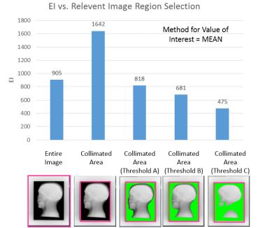
3 IEC ORIGINAL DATA The following corrections shall be made to the RAW DATA as in normal clinical use: Bad or defective pixels Flat-field corrections Corrections for geometric distortion IEC Determine the ORIGINAL DATA Image Segmentation Histogram Based Other IEC Determine the Image Segmentation Histogram Based Other The determination of the RELEVANT IMAGE REGION should be done by methods that identify the attenuated regions of the beam that are relevant to the diagnostic purpose of the acquired image. NOTE 2 While it is understood that the selection of the RELEVANT IMAGE REGION is an important step in the generation of the EXPOSURE INDEX and that a single unified method may be desirable, it is not feasible at this time. Future version of the standard may address this issue. IEC (page 9) 3
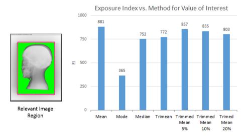
4 IEC Determine the V Mean Median Mode Trimmed mean Trimean Other Measure of Central Tendency IEC Determine the V Exposure Calculate the Index EI = c 0 g(v) Exposure Index Calibration The EI is to be calibrated such that under calibration conditions: EI = c 0 K CAL c 0 = 100 μgy -1 K CAL is the image receptor air kerma in μgy under the calibration conditions 4

 Calibration Conditions Homogenous irradiation of the effective image receptor area using FIXED")
5 Calibration Conditions Fixed beam quality (~RQA5): HVL of 6.8 ± 0.3 mm Al Added filtrations of either: 21 mm Aluminum 0.5 mm Copper and 2mm Aluminum X-ray tube voltage 66kVp 74 kvp Adjust tube voltage to obtain target HVL Calibration Conditions Homogenous irradiation of the effective image receptor area Measurement of image receptor air kerma should be made freein-air (no backscatter) Calibration Conditions Homogenous irradiation of the effective image receptor area using FIXED BEAM QUALITY 5
6 Calibration Conditions Determine the Central 10% of image area Calibration Conditions Determine the V CAL Mean Median Mode Trimmed mean Trimean Other Measure of Central Tendency Calibration Conditions Determine the V CAL Calculate K CAL K CAL = g(v CAL ) EI = c 0 K CAL 6
7 Inverse Calibration Function g(v CAL ) is the inverse calibration function: K CAL = f 1 V CAL = g(v CAL ) The specified inverse calibration function shall have an uncertainty of less than 20% under calibration conditions Beam Quality Dependence Calibration Condition 21 mm Al CsI data acquired on GE Flashpad DR Detector GoS data acquired on Fuji FDR D Evo II DR Detector Deviation Index If target exposure index (EI T ) values are provided by the system, the deviation index (DI) shall be automatically calculated according to: DI = 10 log 10 EI EI T NOTE 1 For this purpose, the TARGET EXPOSURE INDEX values for different examinations/applications need to be available on the digital x-ray imaging system, e.g. in a data base. Such values may be established by professional societies or by the responsible organization. IEC (page 12) 7
8 Summary IEC The IEC Exposure index (EI) is linear with incident detector air kerma The standard explicitly defines the conditions under which the EI shall be calibrated Relationship between EI and incident detector air kerma will vary with beam quality Summary IEC The standard does not define the method by which the vendor: Determines the Calculates the Value of Interest Summary IEC The standard does not attempt to establish Target Exposure Index (EI T ) values Leaves this to professional societies and responsible organizations States that they should be available on the system for the purposes of calculating the Deviation Index (DI) Acknowledges that EI T values may depend on: Type of detector Type of examination Diagnostic question 8

9 Implementation Status Who has implemented IEC ? What do you need to know for verifying compliance with IEC ? Differences in vendor approaches to implementation? Who has implemented IEC ? Vendor Legacy Exposure Indicator IEC Compliant Exposure Index Agfa lgm (No longer in use) EI Canon REX EI Carestream EI* EI Fuji S EI GE DEI (No longer in use) EI Konica S EI Philips EI* EI_s Siemens EXI Clinical EXI* *Not to be confused with the IEC exposure index (EI) **The Clinical EXI is normalized to calibration conditions (Physical EXI is NOT, unless requested by site), note, however, that the Clinical EXI is determined from histogram analysis as opposed to a fixed ROI Vendor Implementation The IEC standard explicitly states that the following items shall be documented: The filter and x-ray tube voltage used for calibration The method used for the selection of the relevant image region The method used to calculate the value of interest The inverse calibration function and the range of image receptor air kerma for which the inverse calibration function can be used to calculate the image receptor air kerma from the value of interest under calibration conditions 9
10 Vendor Questionnaire What is the inverse calibration function and range of image receptor air kerma for which the inverse calibration function can be used to calculate the image receptor air kerma from the value of interest under calibration conditions? What are the vendor s calibration conditions for verifying accuracy of the IEC exposure index (EI)? Recommended beam quality (filter, HVL, kvp) and SID? What exam tag(s), image processing parameters, and/or tools should be used to ensure a fixed region of interest at the center of the image is used when verifying accuracy of the EI? Size of VOI used to determine EI under these conditions? What is the vendor s tolerance for accuracy of the EI? If EI is out of tolerance, what is the methodology for calibrating the EI to bring back it in tolerance (if it exists)? For clinical images, how is the relevant image region and value of interest (VOI) determined? Are there specific assumptions made that the user should be aware of that can affect the EI that is displayed? Does the vendor recommend Target Exposure Index values for various exams / anatomical regions? If so, do these come pre-programmed and can they be modified? Vendor Implementation Filter and beam quality used for calibration verification Vendor Calibration Filter Beam Quality Agfa 21 mm Al or 0.5 mm Cu+ 2 mm Al (recommended but not provided) RQA5 (HVL of 6.8± 0.3 mm Al) Canon 21 mm Al (recommended but not provided) ~RQA5 (HVL of 7.1± 0.1 mm Al Carestream 21 mm Al or 0.5 mm Cu + 2 mm Al (recommended but 0.5 mm Cu & 1 mm Al provided) RQA5 (HVL of 6.8± 0.3 mm Al) Fuji 21 mm Al or 0.5 mm Cu + 2 mm Al (recommended but not provided) RQA5 (HVL of 6.8± 0.3 mm Al) GE 20 mm Al (filter provided by manufacturer) RQA5 (HVL of 6.8± 0.3 mm Al) Konica 21 mm Al or 0.5 mm Cu+ 2 mm Al (recommended but not provided) RQA5 (HVL of 6.8± 0.3 mm Al) Philips 21 mm Al (filter provided by manufacturer) RQA5 (HVL of 6.8± 0.3 mm Al) Siemens 0.6 mm Cu (insert internal filtration) ~RQA5 (HVL of 6.8± 0.3 mm Al) Vendor Implementation Method used for the selection of the relevant image region* *For Verification of EI Under Calibration Conditions Vendor Exam Tag How to Define a FIXED ROI Size of ROI Agfa System Diagnosis with Flat Field Use manual ROI tool User defined Canon Service Tag AEC (used for AEC calibration) n/a (determine by exam tag) 10cm x 10cm (center of detector) Carestream Pattern Use manual ROI tool User defined Fuji Sensitivity n/a (determine by exam tag) 10cm x 10cm (center of detector) GE n/a Enable Technical Mode 10cm x 10cm (center of detector) Konica S-Value n/a (determine by exam tag) 10cm x 10cm (center of detector) Philips n/a Use Simple Ranger Tool User defined Siemens n/a Physical EXI* According to DIN 6868 Part 58 (center of detector) *The Clinical EXI is normalized to calibration conditions (Physical EXI is NOT, unless requested by site), note, however, that the Clinical EXI is determined from histogram analysis as opposed to a fixed ROI 10
 No (mobile units) Yes (formerly No) ACCEPTANCE TESTING IS IMPORTANT No Yes* *Separate calibration factors for Clinical EXI and Physical EXI, vendor procedures only")
11 Vendor Agfa Canon Vendor Implementation What if my EI is out of tolerance? Carestream Fuji GE Konica Philips Siemens Tolerance on EI Accuracy ± 20% (per IEC) Method to Re-Calibrate EI? No No Yes Yes Yes (fixed rooms) No (mobile units) Yes (formerly No) ACCEPTANCE TESTING IS IMPORTANT No Yes* *Separate calibration factors for Clinical EXI and Physical EXI, vendor procedures only address calibration of Clinical EXI Vendor Implementation V? Relevant Image Region Value of Interest EI = c 0 g(v) 11


12 Value of Interest Calculation 12

 Exposure field identification Histogram")
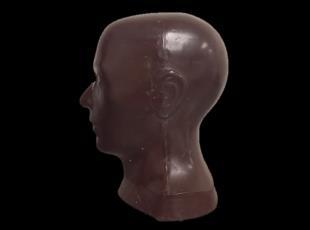
13 Agfa Canon Carestream Fuji GE Konica Philips Siemens Vendor Implementation Method used to calculate the value of interest Vendor Determination (Clinical) Value of Interest Image Pixel Histogram Clinical features Geometric location & exposure level Noise, signal & tissue variation measurements Calculation Method Median Exposure field identification Histogram analysis Mean Exposure field identification Histogram analysis Clinical Features Trimmed Mean Exposure field identification Histogram analysis Mean Exposure field identification Histogram analysis Median Uses the same ROI as used in automatic gradation processing Split pattern recognition Exposure field identification Histogram analysis 2-point average of Reference Values (L and H) Exposure field identification Histogram analysis Trimean Exposure field identification Histogram analysis Mean Multi-Vendor Head Phantom Study Imaged head phantom on various DR panels 8 x10 10 x12 14 x17 Consistent positioning and collimation Same detector dose in all images Recorded Exposure Index values scaled and corrected for input dose & panel sensitivity Theoretically, differences in EI should be mostly due to differences in vendor approach EI (average) = 576 STD = 117 COV = 20% 70 13
14 70 Target EI Factory preset EI T? Vendor Vendor EI T values Implementation of DI Agfa Recommended Values Configured by Apps (editable) Yes Canon Not currently provided (editable) Yes Carestream Preprogramed (editable) Yes Fuji Preprogramed (editable) Yes GE Preprogramed (editable) Yes Konica Preprogramed (editable) Yes Philips No No Siemens Preprogramed (editable)* Yes* Conclusions IEC Standard is widely implemented by vendors Sometimes in tandem with legacy dose indicators Vendor documentation can be hard to come by Differences in vendor methods of implementation can lead to appreciable variation in EI values Range of more than 2.5x with head phantom for same input AK Differences in vendors likely to be exam specific as well Shows need for vendor specific Target Expose Indexes at this time Panel EI sensitivity may need to be accounted for Some vendors don t allow recalibration Variation in implementation can make quality control efforts difficult 14

15 15
Exposure Indices and Target Values in Radiography: What Are They and How Can You Use Them?
 Exposure Indices and Target Values in Radiography: What Are They and How Can You Use Them? Definition and Validation of Exposure Indices Ingrid Reiser, PhD DABR Department of Radiology University of Chicago
Practical Medical Physics Session: TG-151 Dose Monitoring. August 5, 2013 Katie Hulme, M.S.
 Practical Medical Physics Session: TG-151 Dose Monitoring August 5, 2013 Katie Hulme, M.S. Digital Imaging and Dose Creep Images courtesy of Agfa Healthcare Under-Exposed Over-Exposed Freedman et al.,
Digital radiography (DR) post processing techniques for pediatric radiology
 post processing techniques for pediatric radiology") Digital radiography (DR) post processing techniques for pediatric radiology St Jude Children s Research Hospital Samuel Brady, MS PhD DABR samuel.brady@stjude.org Purpose Review common issues and solutions
Digital radiography (DR) post processing techniques for pediatric radiology St Jude Children s Research Hospital Samuel Brady, MS PhD DABR samuel.brady@stjude.org Purpose Review common issues and solutions
Estimation of signal transfer property for wireless digital detector in different measurement schemes
 Estimation of signal transfer property for wireless digital detector in different measurement schemes Anatoli Vladimirov, Kalle Kepler Training Centre of Medical Physics, University of Tartu, Estonia 11
Y11-DR Digital Radiography (DR) Image Quality
 Image Quality") Y11-DR Digital Radiography (DR) Image Quality Image quality is stressed for all systems in Safety Code 35. In the relevant sections Health Canada s advice is the manufacturer s recommended test procedures
Y11-DR Digital Radiography (DR) Image Quality Image quality is stressed for all systems in Safety Code 35. In the relevant sections Health Canada s advice is the manufacturer s recommended test procedures
Ask EuroSafe Imaging Tips & Tricks. Paediatric Imaging Working Group. Dose Management in Digital Radiography
 Ask EuroSafe Imaging Tips & Tricks Paediatric Imaging Working Group Dose Management in Digital Radiography Raija Seuri (HUS Medical Imaging Center, FI) Cristina Almeida (Centro Hospitalar de Lisboa Central,
COMPUTED RADIOGRAPHY CHAPTER 4 EFFECTIVE USE OF CR
 This presentation is a professional collaboration of development time prepared by: Rex Christensen Terri Jurkiewicz and Diane Kawamura New Technology https://www.youtube.com/watch?v=ptkzznazb 7U COMPUTED
I. PERFORMANCE OF X-RAY PRODUCTION COMPONENTS FLUOROSCOPIC ACCEPTANCE TESTING: TEST PROCEDURES & PERFORMANCE CRITERIA
 FLUOROSCOPIC ACCEPTANCE TESTING: TEST PROCEDURES & PERFORMANCE CRITERIA EDWARD L. NICKOLOFF DEPARTMENT OF RADIOLOGY COLUMBIA UNIVERSITY NEW YORK, NY ACCEPTANCE TESTING GOALS PRIOR TO 1st CLINICAL USAGE
3/31/2011. Objectives. Emory University. Historical Development. Historical Development. Historical Development
 Teaching Radiographic Technique in a Digital Imaging Paradigm Objectives 1. Discuss the historical development of digital imaging. Dawn Couch Moore, M.M.Sc., RT(R) Assistant Professor and Director Emory
Features and Weaknesses of Phantoms for CR/DR System Testing
 Physics testing of image detectors Parameters to test Features and Weaknesses of Phantoms for CR/DR System Testing Spatial resolution Contrast resolution Uniformity/geometric distortion Dose response/signal
An Exposure Indicator for Digital Radiography. Report of AAPM Task Group 116
 AAPM REPORT NO. 116 An Exposure Indicator for Digital Radiography Report of AAPM Task Group 116 July 2009 DISCLAIMER: This publication is based on sources and information believed to be reliable, but the
Acquisition, Processing and Display
 Acquisition, Processing and Display Terri L. Fauber, R.T. (R)(M) Department of Radiation Sciences School of Allied Health Professions Virginia Commonwealth University Topics Image Characteristics Image
Learning Objectives: What s my motivation? (unknown screen actor) Workshop Overview
 Workshop Overview") Practical Medical Physics Adapting Traditional Clinical Medical Physics to Digital Radiography Charles E. Willis, Ph.D., DABR Associate Professor Department of Imaging Physics The University of Texas M.D.
Practical Medical Physics Adapting Traditional Clinical Medical Physics to Digital Radiography Charles E. Willis, Ph.D., DABR Associate Professor Department of Imaging Physics The University of Texas M.D.
of sufficient quality and quantity
 of sufficient quality and quantity The patient s body attenuates the beam as it passes though the body More energy is deposited in organs located near the entry of the beam than near the exit of the beam
2217 US Highway 70 East Garner, NC Main: Fax:
 Viztek is committed to providing the highest image quality possible in our CR & DR product lines. There are several factors that directly affect the overall quality of CR & DR based images. The eposure
SPECIFICATION. Kilovoltage X-ray calibration system for protection and diagnostic level dosimetry. Prepared by
 SPECIFICATION Kilovoltage X-ray Prepared by Igor Gomola, Technical Officer, Project ECU6023, Date 2015-Oct-06 Revision Date Status Comments 0.1 2015-Oct-06 Draft Igor Gomola Page 1 of 12 1. Scope This
Key words: fluoroscopy, dose-area-product, kerma-area-product, calibration of KAP meters, patient exposure
 Accuracy and calibration of integrated radiation output indicators in diagnostic radiology: A report of the AAPM Imaging Physics Committee Task Group 190 Pei-Jan P. Lin a) Virginia Commonwealth University
New Exposure Indicators for Digital Radiography Simplified for Radiologists and Technologists
 Medical Physics and Informatics Technical Innovation Don et al. New Simplified Exposure Indicators Medical Physics and Informatics Technical Innovation Steven Don 1 ruce R. Whiting 2 Lois Jo Rutz 3 ruce
Quality assurance: a comparison study of radiographic exposure for neonatal chest radiographs at 4 academic hospitals
 DOI 10.1007/s00247-011-2290-1 ORIGINAL ARTICLE Quality assurance: a comparison study of radiographic exposure for neonatal chest radiographs at 4 academic hospitals Mervyn D. Cohen & Richard Markowitz
Outline. Digital Radiography. Understanding Digital Modalities: Image Quality and Dose. Image Quality. Dose Control
 Understanding Digital Modalities: Image Quality and Dose S. Jeff Shepard, M.S. University of Texas M. D. Anderson Cancer Center Houston, Texas Special Acknowledgement: Stephen K. Thompson, M.S. William
- KiloVoltage. Technique 101: Getting Back to Basics
 Why do I need to know technique? Technique 101: Getting Back to Basics Presented by: Thomas G. Sandridge, M.S., M.Ed., R.T.(R) Program Director Northwestern Memorial Hospital School of Radiography Chicago,
Breast Tomosynthesis. Bob Liu, Ph.D. Department of Radiology Massachusetts General Hospital And Harvard Medical School
 Breast Tomosynthesis Bob Liu, Ph.D. Department of Radiology Massachusetts General Hospital And Harvard Medical School Outline Physics aspects of breast tomosynthesis Quality control of breast tomosynthesis
NEMA XR X-ray Equipment for Interventional Procedures User Quality Control Mode
 NEMA XR 27-2012 X-ray Equipment for Interventional Procedures User Quality Control Mode Published by: National Electrical Manufacturers Association 1300 North 17th Street, Suite 1752 Rosslyn, Virginia
A study of exposure index value fluctuations in computed radiography and direct digital radiography using multiple manufacturers
 A study of exposure index value fluctuations in computed radiography and direct digital radiography using multiple manufacturers Poster No.: C-3011 Congress: ECR 2010 Type: Topic: Authors: Scientific Exhibit
Teaching Digital Radiography and Fluoroscopic Radiation Protection
 Teaching Digital Radiography and Fluoroscopic Radiation Protection WCEC 20 th Student Educator Radiographer Conference Dennis Bowman, RT(R), CRT (R)(F) Community Hospital of the Monterey Peninsula (CHOMP)
Digital Imaging Considerations Computed Radiography
 Digital Imaging Considerations Digital Radiography Computed Radiography o Cassette based Direct or Indirect Digital Radiography o Cassetteless Computed Radiography 1 CR Image Acquisition Most like conventional
DISC QC/QA Program for Digital Imaging Systems using the DR Radchex Plus Meter
 DISC QC/QA Program for Digital Imaging Systems using the DR Radchex Plus Meter Revision Date: January 5th, 2017 www.disc-imaging.com Table of Contents Section A: Preliminary Setup Requirements... 4 Tools
Mammography: Physics of Imaging
 Mammography: Physics of Imaging Robert G. Gould, Sc.D. Professor and Vice Chair Department of Radiology and Biomedical Imaging University of California San Francisco, California Mammographic Imaging: Uniqueness
Joint ICTP/IAEA Advanced School on Dosimetry in Diagnostic Radiology and its Clinical Implementation May 2009
 2033-6 Joint ICTP/IAEA Advanced School on Dosimetry in Diagnostic Radiology and its Clinical Implementation 11-15 May 2009 Dosimetry for Fluoroscopy Basics Renato Padovani EFOMP Joint ICTP-IAEA Advanced
Acceptance Testing of a Digital Breast Tomosynthesis Unit
 Acceptance Testing of a Digital Breast Tomosynthesis Unit 2012 AAPM Spring Clinical Meeting Jessica Clements, M.S., DABR Objectives Review of technology and clinical advantages Acceptance Testing Procedures
Half value layer and AEC receptor dose compliance survey in Estonia
 Half value layer and AEC receptor dose compliance survey in Estonia K. Kepler, A. Vladimirov Training Centre of Medical Physics, University of Tartu Testing Centre of the University of Tartu, Estonia E-mail:
The effect of compensating filter on image quality in lateral projection of thoraco lumbar radiography
 Journal of Physics: Conference Series OPEN ACCESS The effect of compensating filter on image quality in lateral projection of thoraco lumbar radiography To cite this article: N A A Daud et al 2014 J. Phys.:
Evaluation of cassette-based digital radiography detectors using standardized image quality metrics: AAPM TG-150 Draft Image Detector Tests
 JOURNAL OF APPLIED CLINICAL MEDICAL PHYSICS, VOLUME 17, NUMBER 5, 2016 Evaluation of cassette-based digital radiography detectors using standardized image quality metrics: AAPM TG-150 Draft Image Detector
Update on Fluoroscopy Physics AAPM MO-A-210A-1 Stephen Balter, Ph.D.
 Update on Fluoroscopy Physics Stephen Balter, PhD Columbia University Draft for MO-A-210A-1 2009 AAPM Educational objectives Understand dosimetric concepts relating to interventional fluoroscopy Characterize
Digital Imaging started in the 1972 with Digital subtraction angiography Clinical digital imaging was employed from the 1980 ~ 37 years ago Amount of
 Digital Imaging started in the 1972 with Digital subtraction angiography Clinical digital imaging was employed from the 1980 ~ 37 years ago Amount of radiation to the population due to Medical Imaging
History of digital imaging
 CR/QA RADCHEX History of digital imaging Early, crude digital detectors were developed in the 1970 s Image quality was problematic Processing time of digital images was untenable Viewing, transfer and
1. Patient size AEC. Large Patient High ma. Small Patient Low ma
 Comparison of the function and performance of CT AEC systems CTUG meeting by Emily Field Trainee clinical scientist 14 th th Breakdown CT Automatic Exposure Control (AEC) Background Project Description
Unfors EDD-30 Radiation Protection in Fluoroscopy
 Unfors EDD-30 Radiation Protection in Fluoroscopy Immediate Warning Decrease Your Dose Interventional radiology procedures are considered to be essential to medical diagnosis and treatment. It is recognized,
CR Basics and FAQ. Overview. Historical Perspective
 Page: 1 of 6 CR Basics and FAQ Overview Computed Radiography is a term used to describe a system that electronically records a radiographic image. Computed Radiographic systems use unique image receptors
STEREOTACTIC BREAST BIOPSY EQUIPMENT SURVEYS
 STEREOTACTIC BREAST BIOPSY EQUIPMENT SURVEYS JAMES A. TOMLINSON, M.S. Diagnostic Radiological Physicist American Board of Radiology Certified Medical Physics Consultants, Inc. Bio 28 yrs experience 100%
Automated dose control in multi-slice CT. Nicholas Keat Formerly ImPACT, St George's Hospital, London
 Automated dose control in multi-slice CT Nicholas Keat Formerly ImPACT, St George's Hospital, London Introduction to presentation CT contributes ~50+ % of all medical radiation dose Ideally all patients
GE AMX 4+ Portable X-Ray
 GE AMX 4+ Portable X-Ray Typical Manufacturer s Picture GE Healthcare s AMX-4+ analog X-ray system provides high-performance in a compact, easy-to-maneuver package. The rotating arm and tube simplify positioning
Calibration of KAP meters
 Calibration of KAP meters Alexandr Malusek! Division of Radiological Sciences Department of Medical and Health Sciences Linköping University! 2014-04-15 1 Outline 1. KAP meter construction 2. Air kerma-area
A Practical Overview of the Clinical and Operational Impact of Computed Radiography(CR) Implementations. Shirley Weddle, RT(R)(M), CIIP, BBA
 Implementations. Shirley Weddle, RT(R)(M), CIIP, BBA") A Practical Overview of the Clinical and Operational Impact of Computed Radiography(CR) Implementations Shirley Weddle, RT(R)(M), CIIP, BBA OBJECTIVES Define Computed Radiography (CR) Discuss CR vendor
A Practical Overview of the Clinical and Operational Impact of Computed Radiography(CR) Implementations Shirley Weddle, RT(R)(M), CIIP, BBA OBJECTIVES Define Computed Radiography (CR) Discuss CR vendor
Learning Objectives. Outline. Getting Started with CR. Converting the Radiology Department from Film-Screen to Digital: Making the Transition
 Converting the Radiology Department from Film-Screen to Digital: Making the Transition S. Jeff Shepard, MS, DABR University of Texas M. D. Anderson Cancer Center Houston, Texas jshepard@di.mdacc.tmc.edu
DRX Plus Detectors: Going from Good to Great
 DRX Plus Detectors: Going from Good to Great Authors: Karin Töpfer, Tim Wojcik Introduction Carestream s introduction in 2009 of the world s first portable, wireless, cassette-sized detector the CARESTREAM
Tailoring automatic exposure control toward constant detectability in digital mammography
 Tailoring automatic exposure control toward constant detectability in digital mammography Elena Salvagnini a) Department of Imaging and Pathology, Medical Physics and Quality Assessment, KUL, Herestraat
Quality Control of Full Field Digital Mammography Units
 Quality Control of Full Field Digital Mammography Units Melissa C. Martin, M.S., FACMP, FACR, FAAPM Melissa@TherapyPhysics.com 310-612-8127 ACMP Annual Meeting Virginia Beach, VA May 2, 2009 History of
Aspire HD. Program Manual. 2nd Edition - October 2012
 Quality Control 1 Aspire HD Quality Control Program Manual 2nd Edition - October 2012 Overview Installation of FDR Mammography QC Program Weekly Test 2 3 4 Quarterly Test 5 Semi-annual Test 6 Annual Test
Appropriate Inspection Distance of Digital X-Ray Imaging Equipment for Diagnosis
 Indian Journal of Science and Technology Vol 8(S8), 380-386, April 2015 ISSN (Print) : 0974-6846 ISSN (Online) : 0974-5645 DOI: 10.17485/ijst/2015/v8iS8/70528 Appropriate Inspection Distance of Digital
Fully Automatic X-ray meter kvp, dose, rate, HVL and time Extremely easy to use. Unfors ThinX RAD for Radiographic Quality Control
 Fully Automatic X-ray meter kvp, dose, rate, HVL and time Extremely easy to use Unfors ThinX RAD for Radiographic Quality Control Unfors ThinX RAD No keys! No menus! Just position and expose! Unfors ThinX
Diagnostic Imaging Specialists Corporation 163 St.Malo Street St.Malo, MB Canada R0A1T0
 Diagnostic Imaging Specialists Corporation 163 St.Malo Street St.Malo, MB Canada R0A1T0 Instruction Manual for DISC CR Radchex Meter (Wireless Version) Revision L: May 22 nd, 2007 CONTACT INFORMATION:
RAD 150 RADIOLOGIC EXPOSURE TECHNIQUE II
 RAD 150 RADIOLOGIC EXPOSURE TECHNIQUE II APPROVED 12/O2/2011 EFFECTIVE SPRING 2013-14 Prefix & Number RAD 150 Course Title: Radiologic Exposure Technique II & Lab Purpose of this submission: New Change/Updated
SYLLABUS. TITLE: Equipment Operation I. DEPARTMENT: Radiologic Technology
 CODE: RADT 156 INSTITUTE: Health Science TITLE: Equipment Operation I DEPARTMENT: Radiologic Technology COURSE DESCRIPTION: This course covers the principles of equipment operation and maintenance of radiographic
Overview of Safety Code 35
 Common Quality Control Procedures for All s Quality Control Procedures Film All s Daily Quality Control Tests Equipment Warm-up (D1) According to manufacturers instructions Can include auto calibration(d1)
2012 :15th SESSION of ESMP
 2012 :15th SESSION of ESMP Lecture presented in Archamps (Salève Building) by : Elly CASTELLANO (London) Patient dosimetry in x-ray imaging and CT Elly Castellano Objectives measurable dose quantities
Introduction. Digital Mammography QA: Comparing the Manufacturers Recommendations. What is QC and why is it important? Review & compare QC tests
 Slide 1 Digital Mammography QA: Comparing the Manufacturers Recommendations Eric A. Berns, Ph.D. Slide 2 Introduction What is QC and why is it important? Review & compare QC tests Key take home points
Practical Aspects of Medical Physics Surveys of Mammography Equipment and Facilities
 Practical Aspects of Medical Physics Surveys of Mammography Equipment and Facilities Melissa Martin, M.S., FAAPM, FACR, FACMP AAPM Annual Meeting - Philadelphia July 19, 2010 MO-B-204C-1 Educational Objectives
Dosepix Detector as kvp-meter in Radiology and Mammography: First steps
 Dosepix Detector as kvp-meter in Radiology and Mammography: First steps F.Bisello, I.Ritter, F.Tennert, A.Zang MediPix Collaboration Meeting, 19th February 2014, CERN Protect, Enhance, and Save Lives -
Nuclear Associates EZ CR-DIN Phantoms
 Nuclear Associates 07-605-7777 EZ CR-DIN Phantoms Users Manual August 2006 Manual No. 07-605-7777-1 Rev. 4 2006 Fluke Corporation, All rights reserved. Printed in U.S.A. All product names are trademarks
Minnesota Rules, Chapter 4732 X-ray Revision
 Minnesota Rules, Chapter 4732 X-ray Revision DRAFT FLUOROSCOPIC X-RAY SYSTEMS, 1.0 Subpart 1. Applicability. Subpart 2. Limitation of the useful beam. Subpart 3. Measuring compliance; primary protective
DIAGNOSTIC ACCREDITATION PROGRAM. Radiology and CT Quality Control Procedures Workbook
 DIAGNOSTIC ACCREDITATION PROGRAM Radiology and CT Quality Control Procedures Workbook Quality Control Procedures Radiography/CR/DR Safety Code 35 Summary For more detail about each quality control (QC)
DRAFT Technical evaluation of Philips Microdose SI digital mammography system
 DRAFT Technical evaluation of Philips Microdose SI digital mammography system NHSBSP Equipment Report 1310 August 2013 About the NHS Cancer Screening Programmes The national office of the NHS Cancer Screening
Clinical Experiences with a Patient Skin Dose Monitoring and Tracking Program
 Clinical Experiences with a Patient Skin Dose Monitoring and Tracking Program Allen R. Goode, MS, DABR Chief Diagnostic Medical Physicist Department of Radiology & Medical Imaging University of Virginia
Nathan Childress, Ph.D., DABR
 Nathan Childress, Ph.D., DABR Introduction TG-142 is a comprehensive QA protocol Covers nearly every aspect of machine and safety QA Recommends quantitative results Recommends high testing frequencies
Test Equipment for Radiology and CT Quality Control Contents
 Test Equipment for Radiology and CT Quality Control Contents Quality Control Testing...2 Photometers for Digital Clinical Display QC...3 Primary Workstations...3 Secondary Workstations...3 Testing of workstations...3
An Introduction to TG-142 Imaging QA Using Standard Imaging Products. Mark Wiesmeyer, PhD, DABR Technical Product Manager Standard Imaging, Inc.
 An Introduction to TG-142 Imaging QA Using Standard Imaging Products Mark Wiesmeyer, PhD, DABR Technical Product Manager Standard Imaging, Inc. Goals Understand the nature and intent of TG 142 imaging
Collimation Assessment Using GAFCHROMIC XR-M2
 Collimation Assessment Using GAFCHROMIC XR-M2 I. Introduction A method of collimation assessment for GE Senographe full-field digital mammography (FFDM) systems is described that uses a self-developing
Surveying and QC of Stereotactic Breast Biopsy Units for ACR Accreditation
 Surveying and QC of Stereotactic Breast Biopsy Units for ACR Accreditation AAPM Annual Clinical Meeting Indianapolis, IN August 5, 2013 Learning Objectives Become familiar with the recommendations and
Digital Detector Array Image Quality for Various GOS Scintillators
 Digital Detector Array Image Quality for Various GOS Scintillators More info about this article: http://www.ndt.net/?id=22768 Brian S. White 1, Mark E. Shafer 2, William H. Russel 3, Eric Fallet 4, Jacques
Enhanced Functionality of High-Speed Image Processing Engine SUREengine PRO. Sharpness (spatial resolution) Graininess (noise intensity)
 Graininess (noise intensity)") Vascular Enhanced Functionality of High-Speed Image Processing Engine SUREengine PRO Medical Systems Division, Shimadzu Corporation Yoshiaki Miura 1. Introduction In recent years, digital cardiovascular
Vascular Enhanced Functionality of High-Speed Image Processing Engine SUREengine PRO Medical Systems Division, Shimadzu Corporation Yoshiaki Miura 1. Introduction In recent years, digital cardiovascular
Outline ASRT Changes Impact on current curriculum Potential new courses WECM Changes Last update Resources and needs
 Change nd Annual Blinn College 2 nd Educator s Workshop For Radiologic Sciences July 28, 2007 Christi Carter, MSRS, RT(R) Outline ASRT Changes Impact on current curriculum Potential new courses WECM Changes
The Evaluation of Collimator Alignment of Diagnostic X-ray Tube Using Computed Radiography System
 The Evaluation of Collimator Alignment of Diagnostic X-ray Tube Using Computed Radiography System The Evaluation of Collimator Alignment of Diagnostic X-ray Tube Using Computed Radiography System Manus
DOSELAB TOMOTHERAPY TG-148 QA QUICK GUIDE TG-148 RECOMMENDED TESTS 1. V.B.1.C. - Y-JAW DIVERGENCE/BEAM CENTERING
 DOSELAB TOMOTHERAPY TG-148 QA QUICK GUIDE Rev. 1.0 DOSELAB TOMOTHERAPY TG-148 QA QUICK GUIDE DoseLab users may reference the following instructions to perform Tomotherapy Quality Assurance tests as recommended
New spectral benefi ts, proven low dose
 New spectral benefi ts, proven low dose Philips MicroDose mammography SI, technical data sheet Philips MicroDose SI with single-shot spectral imaging is a fullfi eld digital mammography solution that delivers
ELECTRONIC CONTROL CONCEPTS 160 Partition Street Saugerties, NY or local phone
 ELECTRONIC CONTROL CONCEPTS 160 Partition Street Saugerties, NY 12477 (800)VIP-XRAY (845)247-9028 Fax or 800-847-9729 local phone 845-246-9013 http://www.eccxray.com sales@eccxray.com INSTRUCTION MANUAL
Investigation of the line-pair pattern method for evaluating mammographic focal spot performance
 Investigation of the line-pair pattern method for evaluating mammographic focal spot performance Mitchell M. Goodsitt, a) Heang-Ping Chan, and Bob Liu Department of Radiology, University of Michigan, Ann
INTERNATIONAL STANDARD
 INTERNATIONAL STANDARD IEC 62220-1 First edition 2003-10 Medical electrical equipment Characteristics of digital X-ray imaging devices Part 1: Determination of the detective quantum efficiency Appareils
Overview. Professor Roentgen was a Physicist!!! The Physics of Radiation Oncology X-ray Imaging
 The Physics of Radiation Oncology X-ray Imaging Charles E. Willis, Ph.D. DABR Associate Professor Department of Imaging Physics The University of Texas M.D. Anderson Cancer Center Houston, Texas Overview
X-RAY IMAGING EE 472 F2017. Prof. Yasser Mostafa Kadah
 X-RAY IMAGING EE 472 F2017 Prof. Yasser Mostafa Kadah www.k-space.org Recommended Textbook Stewart C. Bushong, Radiologic Science for Technologists: Physics, Biology, and Protection, 10 th ed., Mosby,
X-RAYS - NO UNAUTHORISED ENTRY
 Licencing of premises Premises Refer Guidelines A radiation warning sign and warning notice, X-RAYS - NO UNAUTHORISED ENTRY must be displayed at all entrances leading to the rooms where x-ray units are
Beam-Restricting Devices
 Beam-Restricting Devices Three factors contribute to an increase in scatter radiation: Increased kvp Increased Field Size Increased Patient or Body Part Size. X-ray Interactions a some interact with the
Protocol for the Quality Control of the Physical and Technical Aspects of Digital Breast Tomosynthesis Systems
 Protocol for the Quality Control of the Physical and Technical Aspects of Digital Breast Tomosynthesis Systems Draft version 0.15 January 2014 European Reference Organisation for Quality Assured Breast
Exposure System Selection
 Principles of Imaging Science II (RAD120) Exposure Systems Exposure System Selection Radiographic exposure is a very complex process Best technique systems manipulate one variable while holding others
AUTOMATED AND QUANTITATIVE METHOD FOR QUALITY ASSURANCE OF DIGITAL RADIOGRAPHY IMAGING SYSTEMS
 International Workshop SMART MATERIALS, STRUCTURES & NDT in AEROSPACE Conference NDT in Canada 2011 2-4 November 2011, Montreal, Quebec, Canada AUTOMATED AND QUANTITATIVE METHOD FOR QUALITY ASSURANCE OF
A Comprehensive Review of Image Production
 A Comprehensive Review of Image Production Presented by: John Fleming, M.Ed., RT(R)(MR)(CT) St. Petersburg College Office: (727) 341-3758 E-mail: flemingj@spcollege.edu Lesson Objectives: ARRT Content
DigiMam Conformance Statement for DICOM V3.0
 DigiMam Conformance Statement for DICOM V3.0 Copyright 2004 by I.M.S. s.r.l. DOCUMENT VERSIONS Version Date Author Changes 1.00 15-Feb-05 IMS s.r.l. First Version DOCUMENT VERSIONS Page 2 of 29 TABLE OF
ESTABLISHING A QUALITY ASSURANCE ROUTINE FOR DIGITAL IMAGING
 ESTABLISHING A QUALITY ASSURANCE ROUTINE FOR DIGITAL IMAGING A thesis submitted in partial fulfillment of the requirements for the degree of Master of Science By RANA AL SULAIMAN B.S. in Physics, King
CT radiation profile width measurement using CR imaging plate raw data
 JOURNAL OF APPLIED CLINICAL MEDICAL PHYSICS, VOLUME 16, NUMBER 6, 2015 CT radiation profile width measurement using CR imaging plate raw data Thorarin A Bjarnason, 1,2,3a Chang-Ying Joseph Yang 3,4 Diagnostic
Quality Control for Stereotactic Breast Biopsy. Robert J. Pizzutiello, Jr., F.A.C.M.P. Upstate Medical Physics, Inc
 Quality Control for Stereotactic Breast Biopsy Robert J. Pizzutiello, Jr., F.A.C.M.P. Upstate Medical Physics, Inc. 716-924-0350 Methods of Imaging Guided Breast Biopsy Ultrasound guided, hand-held needle
Research Support. Dual-Source CT: What is it and How Do I Test it? Cynthia H. McCollough, Ph.D.
 Dual-Source CT: What is it and How Do I Test it? Cynthia H. McCollough, Ph.D. CT Clinical Innovation Center Department of Radiology Mayo Clinic College of Medicine Rochester, MN Research Support National
KODAK DIRECTVIEW CR Mammography Feature User s Guide
 KODAK DIRECTVIEW CR Mammography Feature User s Guide 17 September 2010 9G3741 Version 1.0 Carestream Health, Inc. 150 Verona Street Rochester, NY 14608 CARESTREAM, DIRECTVIEW, and DRYVIEW are trademarks
Nuclear Associates
 Nuclear Associates 07-649 CDRH Fluoroscopic Phantom Users Manual March 2005 Manual No. 07-649-1 Rev. 2 2004, 2005 Fluke Corporation, All rights reserved. Printed in U.S.A. All product names are trademarks
TESTING FLAT-PANEL IMAGING SYSTEMS: What the Medical Physicist Needs to Know. JAMES A. TOMLINSON, M.S., D.A.B.R. Diagnostic Radiological Physicist
 TESTING FLAT-PANEL IMAGING SYSTEMS: What the Medical Physicist Needs to Know JAMES A. TOMLINSON, M.S., D.A.B.R. Diagnostic Radiological Physicist Topics Image Uniformity and Artifacts Image Quality - Detail
Comparison of computed radiography and filmõscreen combination using a contrast-detail phantom
 JOURNAL OF APPLIED CLINICAL MEDICAL PHYSICS, VOLUME 4, NUMBER 1, WINTER 2003 Comparison of computed radiography and filmõscreen combination using a contrast-detail phantom Z. F. Lu,* E. L. Nickoloff, J.
Assessment of Beam Alignment, Collimation and Half Value Layer of Some Selected X-Ray Machines in Plateau State, Nigeria
 International Journal of Innovative Scientific & Engineering Technologies Research 5(4):-5, Oct.-Dec., 07 SEAHI PUBLICATIONS, 07 www.seahipaj.org ISSN: 60-896X Assessment of Beam Alignment, Collimation
A comparative study of several digital flat panel X-ray units: patients doses and image quality in chest radiography
 A comparative study of several digital flat panel X-ray units: patients doses and image quality in chest radiography Torres Cabrera R. 1, España López M.L. 2 Ruiz Manzano P. 3, Sastre Aguado J.M. 4,, Rivas
Protocol for the Quality Control of the Physical and Technical Aspects of Digital Breast Tomosynthesis Systems
 Protocol for the Quality Control of the Physical and Technical Aspects of Digital Breast Tomosynthesis Systems Draft version 0.10 February 2013 European Reference Organisation for Quality Assured Breast
Determination of the tube voltage from clinic mammographic system using two types of detectors
 BJRS BRAZILIAN JOURNAL OF RADIATION SCIENCES 03-1A (2015) 01-06 Determination of the tube voltage from clinic mammographic system using two types of detectors J. S. Barreira a ; V. Vivolo a a Gerência
Mammography is a radiographic procedure specially designed for detecting breast pathology Approximately 1 woman in 8 will develop breast cancer over
 Mammography is a radiographic procedure specially designed for detecting breast pathology Approximately 1 woman in 8 will develop breast cancer over a lifetime Breast cancer screening programs rely on