1. Queries are issued to the image archive for information about computed tomographic (CT)
|
|
|
- Marian Hart
- 5 years ago
- Views:
Transcription
 (Fig E1) are identified from these query results on the basis of a combination of dose screen image type and any expected")
1 Appendix E1 Exposure Extraction Method examinations. 1. Queries are issued to the image archive for information about computed tomographic (CT) 2. Potential dose report screen captures (hereafter, dose screens) (Fig E1) are identified from these query results on the basis of a combination of dose screen image type and any expected manufacturer-specific series numbers or series descriptions. E1a. Page 1 of 13

2 E1b. E1c. Page 2 of 13

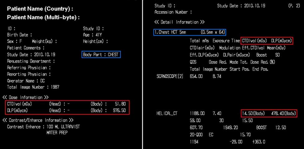
3 E1d. Figure E1: Sample screen capture dose reports for (a) GE Healthcare, (b) Siemens Healthcare, (c) Toshiba Medical Systems, and (d) Philips Healthcare. Contents and formatting differ, but all contain the x-ray tube output metrics CTDI vol and DLP, outlined in red boxes. Each separate row containing values of these metrics represents a distinct dose event, while all of the component dose events on a dose screen belong to the same CT encounter. The blue boxes contain series or scan descriptions that may aid anatomy determination. In d, the dose report content within a private DICOM attribute is included to the right. 3. Potential dose screens and the first image in every non dose screen series are retrieved. 4. Optical character recognition (OCR) is used to convert dose screen images to text. Manufacturerdependent regular expressions and logic rules are applied to capture exposure metrics, including dose-length product (DLP) and volume CT dose index (CTDI vol ), and table start and stop positions. 5. Additional potentially useful image-level information is gathered from the Digital Imaging and Communications in Medicine (DICOM) attributes, including protocol name and series description. These image-level data are matched to dose events. 6. A combination of data from steps four and five is used to assign anatomic regions to dose events by using a manufacturer-dependent strategy that is described later in this Appendix. Page 3 of 13
4 7. Dose information and image level data are stored in a relational database (SQL Sever 2005; Microsoft, Redmond, Wash) for subsequent analysis. Anatomy Assignment Strategy The algorithms used to assign anatomy are complex, not only because of the extremely variable format and content of exposure events reported by the different manufacturers, but also because of the variable fidelity of potentially available anatomic descriptors. The basic strategy used is to favor use of more specific dose event level descriptions, when available, instead of the typically less specific protocol-level descriptions. The eventlevel descriptions correspond to the series description on the Philips Healthcare (Best, the Netherlands) private DICOM dose sequences and the range name on the Siemens Healthcare (Forcheim, Germany) dose screens. Dose screens from GE Healthcare (Piscataway, NJ) and Toshiba Medical Systems (Tokyo, Japan) do not include explicit event-level descriptions, so protocol descriptions are used with additional anatomy mapping definitions used for the most common protocols comprising multiple dose events. For example, the entire scanning range is divided equally into thirds for common chest/abdomen/pelvis studies. These definitions and additional logic rules assign anatomy based on available table positions or relative scan lengths. It is also necessary to match dose events to images to use the image- or series-level anatomic descriptions or other DICOM attributes found only at the image level (as opposed to the dose screens). The anatomy assignment algorithms are described later in the Appendix for each dose screen manufacturer. Each line represents a matching attempt that uses a string of text from the specified source to match anatomic concepts in a DICOM CT anatomy dictionary, which are defined by the Systemized Nomenclature of Medicine Reference Terminology, or SNOMED-RT. Most concepts in the dictionary contain a list of synonyms, including commonly used abbreviations and other variants (eg, CAP for chest/abdomen/pelvis). The longest string is favored during matching. The matching algorithm is terminated once a match is found. Page 4 of 13

5 Two manufacturers dose screens (Philips Healthcare and Siemens Healthcare) often contain useful dose event level descriptions, in which case anatomy matching is attempted directly at the dose event level. In contrast, the other two manufacturers (GE Healthcare and Toshiba Medical Systems) have no dose event level descriptions, so matching is initially performed by using protocol-level descriptions to obtain an overall anatomic concept for the entire protocol and is followed by a secondary process to map individual dose events within the protocol (Fig E2). Figure E2: Flowchart shows how anatomy is assigned to each component dose event on the basis of overall protocol anatomy, number of acquisitions, and availability of anatomy map definition and table positions. Page 5 of 13
6 The CT anatomy method (described later in this Appendix) takes a DICOM attribute list as an argument and inspects the attributes in a certain order. It attempts a string match by using the value of each attribute and stops when the first match is found. Once all matching has been performed, anatomy matches are checked for possible elongation. If the scan length (explicitly specified by table positions in the dose screens or otherwise derived by dividing DLP by CTDI vol ) is greater than 30 cm for abdominal or pelvic anatomy, the anatomy is elongated to the abdomen and pelvis. Similarly if a chest and abdominal or abdominal and pelvic anatomy scan length is greater than 60 cm, it is elongated to chest, abdomen, and pelvis. This was necessary because descriptions for many dose events do not accurately describe the entire body region imaged (eg, abdominal and pelvic scans are often described as abdomen in CT protocols). To make this correction more robust for pediatric patients, a look-up table based on average height by age could be implemented in place of fixed thresholds that are used for all patients. Anatomy Assignment Algorithms Philips Healthcare Dose-event anatomy assignment. a. Series description (Philips Healthcare private exposure sequence DICOM attribute) b. Series description (image DICOM attribute, matched 1P by acquisition date and time) c. Protocol name (image DICOM attribute, matched 1P by acquisition date and time) d. Series description (image DICOM attribute from temporally associated studies, matched 1P by acquisition date and time or by series number) e. Protocol name (image DICOM attribute from temporally associated studies, matched 1P by acquisition date or time or by series number) f. Prior dose event (in same exposure sequence) Page 6 of 13
7 g. CT anatomy method 5 (dose screen DICOM attribute list) Siemens Healthcare Dose event anatomy assignment. a. Range name (dose screen OCR) b. Series description (image DICOM attribute, matched 1S by acquisition number) c. Protocol name (image DICOM attribute, matched 1S by acquisition number) d. Prior dose event (on same dose screen) e. Study description (dose screen DICOM attribute) f. CT anatomy method 5 (dose screen DICOM attribute list) Toshiba Medical Systems Protocol anatomy assignment. a. Protocol name (image DICOM attribute, matched 1T by acquisition number and protocol name OCR) b. Protocol name (image DICOM attribute from temporally associated studies, matched 1T by acquisition number and protocol name OCR) c. Protocol name (dose screen OCR) d. CT anatomy method 5 (from image DICOM attribute list of examination and temporally associated studies, matched 1T by acquisition number and protocol name OCR) e. CT anatomy method 5 (dose screen DICOM attribute list) Protocol anatomy to dose event anatomy mapping. Please see Figure E2. GE Healthcare Page 7 of 13
8 Protocol anatomy assignment. a. Protocol name (image DICOM attribute, matched 1G by series number) number) b. Protocol name (image DICOM attribute from temporally associated studies, matched 1G by series c. CT anatomy method 5 (dose screen DICOM attribute list) Protocol anatomy to dose-event anatomy mapping. Please see Figure E2. 1 Matching Dose Events to Image-Level Data To use anatomic information specified at the image level, it is necessary to match dose events to images. The strategies used to accomplish this differ by manufacturer, as indicated. 1P Philips Healthcare. The acquisition date and time from the Philips Healthcare private DICOM attribute exposure sequence is used to match the image-level acquisition date and time DICOM attribute for this manufacturer, if present. If absent, the potentially less specific series number from both the exposure sequence and the image-level series number DICOM attribute is used. 1S Siemens Healthcare. The acquisition number is specified on the Siemens Healthcare dose screens and is used to match the imagelevel acquisition number. 1T Toshiba Medical Systems. The implicitly derived acquisition number based on order of appearance from Toshiba Medical Systems dose screens and the protocol name (from OCR of the dose screen) is used to match the protocol name and acquisition number DICOM attributes at the image level. It is necessary to include the protocol name because Page 8 of 13
9 multiple protocols can be present on one dose screen and because acquisition numbers reset for each protocol, causing duplicates. Inexact string matching of the protocol name is permitted due to imperfect OCR. 1G GE Healthcare. The algorithm implemented for GE Healthcare units matches the series number of the dose event on the dose report and the image-level series number DICOM attribute. A potential problem with this matching strategy is occasional inconsistent numbering of the two series numbers. 2 Protocol Anatomy Map Definitions 1. Chest/Abdomen/Pelvis (CAP) 2. Neck/CAP 3. Neck/Chest 4. Abdomen/Pelvis 5. Chest/Abdomen 3 Subregions Using Table Positions For dose reports containing dose event table positions (those obtained with GE Healthcare units and some obtained with Toshiba Medical Systems units), the overall examination scan range is defined from the full combined range of the subcomponent dose event table positions. Anatomic subregions are defined as follows for common combination examinations covering more than one anatomic region. Each dose event is assigned to one of these anatomic subregions by the following rules: 1. Assign all protocol anatomy subregions that are at least 75% covered by the dose event. 2. If no subregions are assigned by the above rule, assign the single subregion with the largest fraction covered by the dose event. Page 9 of 13
10 position. 3. If the table is stationary (as for monitoring scans), assign the subregion containing the fixed table These anatomic region maps use the overall protocol scan range from all component dose events and define anatomic subregions based on the dose-event table positions with the following rules: Anatomic subregion definitions for common combination examinations. 1. Chest/Abdomen/Pelvis (CAP) a. Each subregion assigned one-third of total scan length 2. Neck/CAP a. CAP = caudal four-fifths of total scan length (then subdivided by using the CAP map) b. Neck = cranial one-fourth of total scan length 3. Neck/Chest a. Chest = caudal three-fourths of total scan length b. Neck = cranial half of total scan length 4. Abdomen/Pelvis a. Abdomen = cranial half of total scan length b. Pelvis = caudal half of total scan length 5. Chest/Abdomen a. Chest = cranial half of total scan length b. Abdomen = caudal half of total scan length 4 Subregions Using Relative Scan Lengths Page 10 of 13
11 These algorithms are used when there are no table positions available (Philips Healthcare, Siemens Healthcare, and sometimes Toshiba Medical Systems unit examinations). Dose event scan lengths are approximated as DLP divided by CTDI vol (neglecting z overscanning). Each component dose event is assigned a relative scan length, defined as the dose event scan length divided by the longest dose event scan length present by using the following rules: 1. CAP a. Two dose events i. Abdomen/pelvis = larger relative scan length ii. Chest = smaller relative scan length iii. CAP = equal relative scan lengths b. More than two dose events i. CAP = relative scan length 80% ii. Abdomen/pelvis = relative scan length 80% and >50% iii. Chest = relative scan length 50% and >30% and no chest anatomy yet assigned iv. Abdomen = all above rules failed to trigger 2. Neck/CAP a. Two dose events i. Neck = smaller relative scan length ii. CAP = larger relative scan length b. Three dose events i. Abdomen/pelvis = largest relative scan length Page 11 of 13
12 ii. Neck = smallest relative scan length iii. Chest = median relative scan length c. More than three dose events i. Neck/CAP assigned to all 3. Neck/Chest a. Two dose events i. Neck = smaller relative scan length ii. Chest = larger relative scan length b. More than two dose events i. Chest = largest relative scan length ii. Neck = all others 5 CT Anatomy Method This method is used only if the more specific potential sources of anatomic information failed to match. It takes a DICOM attribute list from a dose screen or CT image and uses the value of each DICOM attribute in the following order to match concepts in the CT anatomy dictionary, stopping when the first anatomic match is found: 1. Anatomic region sequence 2. Body part examined 3. Image comments 4. Series description 5. Protocol name Page 12 of 13
13 6. Performed procedure code sequence 7. Performed procedure step sequence 8. Procedure code sequence 9. Study description Bolus tracking dose events. Monitoring scans (such as bolus timing scans) are assigned to the next recognized anatomic area, or to an anatomic subregion if table position is provided. Temporally associated studies. Temporally associated studies are defined as all CT studies obtained in the same patient (determined by matching patient identifier DICOM attribute) with a study start date and time DICOM attribute equal to the reference examination. One difficulty with trying to match image data to dose data occurs when dose reports are stored under a different study than the images. For example, for a chest/abdomen/pelvis examination performed in two acquisitions, the chest and abdomen/pelvis portions can be archived as separate studies, but the dose screen may be sent to either study or both studies. If no match between dose-event data and image-level data within the same study is found, then we attempt to match the dose-event data to image-level data by using all temporally associated studies. Also, duplication checks are performed to prevent counting identical dose events more than once if different versions of the same dose screen had been sent multiple times or if the same dose screen had been sent to multiple studies. Page 13 of 13
Automated dose control in multi-slice CT. Nicholas Keat Formerly ImPACT, St George's Hospital, London
 Automated dose control in multi-slice CT Nicholas Keat Formerly ImPACT, St George's Hospital, London Introduction to presentation CT contributes ~50+ % of all medical radiation dose Ideally all patients
Wide-Detector CT for TAVR Planning:
 Wide-Detector CT for TAVR Planning: Impact on Iodine Dose, Radiation Dose, and Image Quality SCBTMR 2015 Annual Course Thursday, October 8 William P. Shuman MD FSCBTMR Department of Radiology University
1. Patient size AEC. Large Patient High ma. Small Patient Low ma
 Comparison of the function and performance of CT AEC systems CTUG meeting by Emily Field Trainee clinical scientist 14 th th Breakdown CT Automatic Exposure Control (AEC) Background Project Description
12/21/2016. Siemens Medical Systems Research Agreement Philips Healthcare Research Agreement AAN and ASN Committees
 Joseph V. Fritz, PhD Nandor Pintor, MD Dent Neurologic Institute ASN 2017 Friday, January 20, 2017 Siemens Medical Systems Research Agreement Philips Healthcare Research Agreement AAN and ASN Committees
DICOM Conformance Statement
 DICOM Conformance Statement Application Annex: 3D Roadmap R1.1.5 Koninklijke Philips N.V. 2015 All rights are reserved. Document Number: ICAP-PF.0015381 Issued by: Philips Medical Systems Nederland BV,
TOPICS: CT Protocol Optimization over the Range of Patient Age & Size and for Different CT Scanner Types: Recommendations & Misconceptions
 CT Protocol Optimization over the Range of Patient Age & Size and for Different CT Scanner Types: Recommendations & Misconceptions TOPICS: Computed Tomography Quick Overview CT Dosimetry Effects of CT
DICOM Conformance Statement
 DICOM Conformance Statement Application Annex: Stentboost R4.2.5 Koninklijke Philips N.V. 2015 All rights are reserved. Document Number: ICAP-PF.0015387 Issued by: Philips Medical Systems Nederland BV,
Multi-Access Biplane Lab
 Multi-Access Biplane Lab Advanced technolo gies deliver optimized biplane imaging Designed in concert with leading physicians, the Infinix VF-i/BP provides advanced, versatile patient access to meet the
Initial Survey Results on the Accuracy of Dose Data in the DICOM Objects
 Initial Survey Results on the Accuracy of Dose Data in the DICOM Objects Renato Padovani, Annalisa Trianni University Hospital S. Maria della Misericordia Udine, Italy INTRODUCTION The EC MED has forced
abc MHRA Philips Mx8000 IDT CT scanner technical evaluation September 2004 Best choice best practice nww.medical-devices.nhs.
 abc September 2004 MHRA 04099 Philips Mx8000 IDT CT scanner technical evaluation Best choice best practice www.mhra.gov.uk nww.medical-devices.nhs.uk About MHRA evaluation reports. What you can expect.
diagnostic examination
 RADIOLOGICAL PHYSICS 2011 Raphex diagnostic examination Adel A. Mustafa, Ph.D., Editor PUBLISHED FOR: RAMPS (Radiological and Medical Physics Society of New York) preface The RAPHEX Diagnostic exam 2011
DICOM Conformance Statement
 DICOM Conformance Statement Application Annex: MR-CT Roadmap R1.1 On Interventional Workspot R1.4 Koninklijke Philips N.V. 2017 All rights are reserved. ICAP-T-030001.09b Corresponds to ICAP-W-030001.02
DigiMam Conformance Statement for DICOM V3.0
 DigiMam Conformance Statement for DICOM V3.0 Copyright 2004 by I.M.S. s.r.l. DOCUMENT VERSIONS Version Date Author Changes 1.00 15-Feb-05 IMS s.r.l. First Version DOCUMENT VERSIONS Page 2 of 29 TABLE OF
Authors: Cabral, Ricardo 1 ; Carvoeiras, Pedro 2 ; Fatana, João 2, ; Alves, Rita 1. 1 Centro Hospitalar Lisboa Norte - Hospital de Santa Maria; 2
 Authors: Cabral, Ricardo 1 ; Carvoeiras, Pedro 2 ; Fatana, João 2, ; Alves, Rita 1. 1 Centro Hospitalar Lisboa Norte - Hospital de Santa Maria; 2 Medical Consult, SA; Establish a method to correlate image
QC by the MPE in Belgium
 Acceptance testing of state-of-the-art CT scanners using a new national protocol: first experience on a large number of scanners of different make and model the working group Radiology of the Belgian Hospital
Maximum Performance, Minimum Space
 TECHNOLOGY HISTORY For over 130 years, Toshiba has been a world leader in developing technology to improve the quality of life. Our 50,000 global patents demonstrate a long, rich history of leading innovation.
PLD5600A High Frequency Digital Gastrointestinal &DR System(630mA)
") PLD5600A High Frequency Digital Gastrointestinal &DR System(630mA) Application: Full support perspective, gastrointestinal spot film, GI (barium meal, barium enema), orthopedic photography, pediatrics
PLD5600A High Frequency Digital Gastrointestinal &DR System(630mA) Application: Full support perspective, gastrointestinal spot film, GI (barium meal, barium enema), orthopedic photography, pediatrics
Truly flexible to meet your clinical needs
 Truly flexible to meet your clinical needs 2 Adapting to meet your needs Flexible Fast and responsive Excellent image quality Designed with ergonomic efficiency Equipped with dose management tools 3 Three
Features and Weaknesses of Phantoms for CR/DR System Testing
 Physics testing of image detectors Parameters to test Features and Weaknesses of Phantoms for CR/DR System Testing Spatial resolution Contrast resolution Uniformity/geometric distortion Dose response/signal
Some operation methods show in the catalog reguire optional eguipment
 Some operation methods show in the catalog reguire optional eguipment I'm interested in real-time imaging with a larger field of view. I wish to acquire high-definition images while reducing exposure dose.
160-slice CT SCANNER / New Standard for the Future
 TECHNOLOGY HISTORY For over 130 years, Toshiba has been a world leader in developing technology to improve the quality of life. Our 50,000 global patents demonstrate a long, rich history of leading innovation.
2217 US Highway 70 East Garner, NC Main: Fax:
 Viztek is committed to providing the highest image quality possible in our CR & DR product lines. There are several factors that directly affect the overall quality of CR & DR based images. The eposure
Improvement of CT image quality with iterative reconstruction idose4
 Improvement of CT image quality with iterative reconstruction idose4 Poster No.: C-0387 Congress: ECR 2014 Type: Scientific Exhibit Authors: M.-L. Olsson, K. Norrgren, M. Söderberg; Malmö/SE Keywords:
DICOM Conformance Statement
 DICOM Conformance Statement Application Annex: Nuclear Medicine Viewer on Xcelera R3.2L1 SP2 Koninklijke Philips Electronics N.V. 2011 All rights are reserved. Document Number: PIIOffc.0000081 Issued by:
Iterative Reconstruction in Image Space. Answers for life.
 Iterative Reconstruction in Image Space Answers for life. Iterative Reconstruction in Image Space * (IRIS) * Please note: IRIS is used as an abbreviation for Iterative Reconstruction in Image Space throughout
Diagnostic X-Ray Shielding
 Diagnostic X-Ray Shielding Multi-Slice CT Scanners Using NCRP 147 Methodology Melissa C. Martin, M.S., FAAPM, FACR Therapy Physics Inc., Bellflower, CA AAPM Annual Meeting, Orlando, FL FL Refresher Course
DICOM Conformance Statement
 DICOM Conformance Statement Application Annex: MultiModality Applications on Philips IntelliSpace Portal V7.0 Koninklijke Philips N.V. 2014 All rights are reserved. Document Number: ICAPPF.0013673 Issued
3/31/2011. Objectives. Emory University. Historical Development. Historical Development. Historical Development
 Teaching Radiographic Technique in a Digital Imaging Paradigm Objectives 1. Discuss the historical development of digital imaging. Dawn Couch Moore, M.M.Sc., RT(R) Assistant Professor and Director Emory
Translating Protocols Between Scanner Manufacturer and Model
 Translating Protocols Between Scanner Manufacturer and Model Robert J. Pizzutiello, MS, FAAPM, FACMP Sr. Vice-President, Global Physics Solutions President, Upstate Medical Physics Objectives Understand
DICOM Conformance Statement
 DICOM Conformance Statement Application Annex: CT Applications on Philips IntelliSpace Portal V5.0 Koninklijke Philips Electronics N.V. 2012 All rights are reserved. Document Number: PIIOffc.0000143.01
Image Quality and Dose. Image Quality and Dose. Image Quality and Dose Issues in MSCT. Scanner parameters affecting IQ and Dose
 Image Quality and Dose Issues in MSCT Image Quality and Dose Image quality Image noise Spatial resolution Contrast Artefacts Speckle and sharpness S. Edyvean St. George s Hospital London SW17 0QT Radiation
DISC QC/QA Program for Digital Imaging Systems using the DR Radchex Plus Meter
 DISC QC/QA Program for Digital Imaging Systems using the DR Radchex Plus Meter Revision Date: January 5th, 2017 www.disc-imaging.com Table of Contents Section A: Preliminary Setup Requirements... 4 Tools
SAFIRE. Sinogram Affirmed Iterative Reconstruction. Answers for life.
 Neuro Thoracic Abdominal Abdominal Cardiovascular Pediatric SAFIRE Sinogram Affirmed Iterative Reconstruction Answers for life. SAFIRE * (Sinogram Affirmed Iterative Reconstruction) * The information
Clinical Experiences with a Patient Skin Dose Monitoring and Tracking Program
 Clinical Experiences with a Patient Skin Dose Monitoring and Tracking Program Allen R. Goode, MS, DABR Chief Diagnostic Medical Physicist Department of Radiology & Medical Imaging University of Virginia
Software and Hardware in CCTA. Elly Castellano PhD
 Software and Hardware in CCTA Elly Castellano PhD Outline technical requirements for coronary CTA the modern cardiac CT scanner ECG-gating technology image reconstruction algorithms 2 Technical requirements
Influence of different iteration levels in fourth generation iterative reconstruction technique on image noise in CT examinations of the neck
 Influence of different iteration levels in fourth generation iterative reconstruction technique on image noise in CT examinations of the neck Poster No.: C-2205 Congress: ECR 2012 Type: Scientific Paper
Image Extraction using Image Mining Technique
 IOSR Journal of Engineering (IOSRJEN) e-issn: 2250-3021, p-issn: 2278-8719 Vol. 3, Issue 9 (September. 2013), V2 PP 36-42 Image Extraction using Image Mining Technique Prof. Samir Kumar Bandyopadhyay,
Get more from your images with Symphony Image Processing
 DIRECT RADIOGRAPHY The user-friendly DelWorks image acquisition and processing software provides a wide range of tools for a variety of image enhancements. Its user interface simplifies every step of the
DICOM Correction Proposal Form
 DICOM Correction Proposal Form STATUS Final Text Date of Last Update 2014/09/04 Person Assigned Submitter Name Janet Keyes Makoto Suzuki (Toshiba) Submission date 2009.10.06 Correction
Image Interpretation System for Informed Consent to Patients by Use of a Skeletal Tracking
 Image Interpretation System for Informed Consent to Patients by Use of a Skeletal Tracking Naoki Kamiya 1, Hiroki Osaki 2, Jun Kondo 2, Huayue Chen 3, and Hiroshi Fujita 4 1 Department of Information and
29 CP Define CT Reconstruction Diameter more precisely and correct Enhanced CT illustration Page 1
 29 CP-1569 - Define CT Reconstruction Diameter more precisely and correct Enhanced CT illustration Page 1 1 Status Final Text 2 Date of Last Update 2016/09/08 3 Person Assigned David Clunie 4 mailto:dclunie@dclunie.com
DICOM Correction Proposal
 Tracking Information - Administration Use Only DICOM Correction Proposal Correction Proposal Number Status CP-1713 Letter Ballot Date of Last Update 2018/01/23 Person Assigned Submitter Name David Clunie
DICOM Conformance Statement
 DICOM Conformance Statement Application Annex: US Applications on Philips IntelliSpace Portal V7.0 Koninklijke Philips N.V. 2014 All rights are reserved. Document Number: ICAP-PF.0013672 Issued by: Philips
SECTION I - CHAPTER 2 DIGITAL IMAGING PROCESSING CONCEPTS
 RADT 3463 - COMPUTERIZED IMAGING Section I: Chapter 2 RADT 3463 Computerized Imaging 1 SECTION I - CHAPTER 2 DIGITAL IMAGING PROCESSING CONCEPTS RADT 3463 COMPUTERIZED IMAGING Section I: Chapter 2 RADT
Diffraction-enhanced X-ray Imaging (DEXI) Medical Solutions. More information using less radiation
 Medical Solutions. More information using less radiation") Diffraction-enhanced X-ray Imaging (DEXI) Medical Solutions More information using less radiation Medical Small Animal Security NDE/NDT Diffraction-Enhanced X-ray Imaging Medical Solutions Safe non-invasive
Diffraction-enhanced X-ray Imaging (DEXI) Medical Solutions More information using less radiation Medical Small Animal Security NDE/NDT Diffraction-Enhanced X-ray Imaging Medical Solutions Safe non-invasive
IHE Radiology Technical Framework Supplement. Stereotactic Mammography Image (SMI) Trial Implementation
 Trial Implementation") Integrating the Healthcare Enterprise 5 IHE Radiology Technical Framework Supplement 10 Stereotactic Mammography Image (SMI) 15 Trial Implementation 20 25 Date: June 11, 2013 Author: IHE Radiology Technical
Integrating the Healthcare Enterprise 5 IHE Radiology Technical Framework Supplement 10 Stereotactic Mammography Image (SMI) 15 Trial Implementation 20 25 Date: June 11, 2013 Author: IHE Radiology Technical
ddr Compact Series Setting a new benchmark in digital radiography.
 ddr Compact Series Setting a new benchmark in digital radiography. ddrcompact When productivity and exceptional value come together. With the introduction of its newest DR system, the ddrcompact, Swissray
Advanced digital image processing for clinical excellence in fluoroscopy
 Dynamic UNIQUE Digital fluoroscopy solutions Dynamic UNIQUE Advanced digital image processing for clinical excellence in fluoroscopy André Gooßen, PhD, Image Processing Specialist Dörte Hilcken, Clinical
Reducing Radiation Exposure from Survey CT Scans
 Reducing Survey CT Scan Exposure Pediatric Imaging Original Research Jennifer C. O Daniel 1 Donna M. Stevens 2 Dianna D. Cody 2 O Daniel JC, Stevens DM, Cody DD Received July 28, 2004; accepted after revision
HISTORY. CT Physics with an Emphasis on Application in Thoracic and Cardiac Imaging SUNDAY. Shawn D. Teague, MD
 CT Physics with an Emphasis on Application in Thoracic and Cardiac Imaging Shawn D. Teague, MD DISCLOSURES 3DR- advisory committee CT PHYSICS WITH AN EMPHASIS ON APPLICATION IN THORACIC AND CARDIAC IMAGING
DICOM Conformance Statement
 DICOM Conformance Statement Application Annex: US Applications on Philips IntelliSpace Portal V6.0 Koninklijke Philips Electronics N.V. 2013 All rights are reserved. Document Number: PIIOffc.0001323.01
Digital radiography (DR) post processing techniques for pediatric radiology
 post processing techniques for pediatric radiology") Digital radiography (DR) post processing techniques for pediatric radiology St Jude Children s Research Hospital Samuel Brady, MS PhD DABR samuel.brady@stjude.org Purpose Review common issues and solutions
Digital radiography (DR) post processing techniques for pediatric radiology St Jude Children s Research Hospital Samuel Brady, MS PhD DABR samuel.brady@stjude.org Purpose Review common issues and solutions
Get more from your images with Symphony Image Processing
 DIRECT RADIOGRAPHY The user-friendly DelWorks image acquisition and processing software possesses a wide range of tools for a variety of image manipulations. Its user interface simplifies every step of
Radiation Dose Index monitoring (RDIM) systems and establishment of local DRLs
 systems and establishment of local DRLs") IAEA RER/9/135 COURSE ON OPTIMIZATION IN COMPUTED TOMOGRAPHY Sofia, Bulgaria, 21017 Radiation Dose Index monitoring (RDIM) systems and establishment of local DRLs Dean Pekarovič UMC Ljubljana, Institute
IAEA RER/9/135 COURSE ON OPTIMIZATION IN COMPUTED TOMOGRAPHY Sofia, Bulgaria, 21017 Radiation Dose Index monitoring (RDIM) systems and establishment of local DRLs Dean Pekarovič UMC Ljubljana, Institute
DICOM Conformance. DICOM Detailed Specification for Diagnostic Labs and Radiology Center Connectivity
 DICOM Detailed Specification for Diagnostic Labs and Radiology Center Connectivity Authored by Global Engineering Team, Health Gorilla April 10, 2014 Table of Contents About Health Gorilla s Online Healthcare
productivity ddrformula Series
 productivity ddrformula Series beyond conventional DR Swissray is the pioneer and worldwide leader in the design, manufacturing and marketing of state-of-the-art Digital Radiography systems. Swissray
efficiency ddrcompact Series
 efficiency ddrcompact Series beyond conventional DR Swissray is the pioneer and worldwide leader in the design, manufacturing and marketing of state-of-the-art Digital Radiography systems. Swissray s
Computed Tomography. The Fundamentals of... THE FUNDAMENTALS OF... Jason H. Launders, MSc. Current Technology
 The Fundamentals of... Computed Tomography Computed Tomography (CT) systems use x-rays to produce images of slices through a patient s anatomy. Despite having lower spatial resolution than other x-ray
Pitfalls and Remedies of MDCT Scanners as Quantitative Instruments
 intensity m(e) m (/cm) 000 00 0 0. 0 50 0 50 Pitfalls and Remedies of MDCT Scanners as Jiang Hsieh, PhD GE Healthcare Technology University of Wisconsin-Madison Root-Causes of CT Number Inaccuracies Nature
Examion. New. Image Acquisition and Diagnostics. R a d i o D i g i t a l e. examion aqs veterinray software.
 Examion New V191303 V191304 V1913041 V191305 examion aqs veterinray software Image Acquisition and Diagnostics Z.I. de Noville-les-Bois 1 rue de la Tour (Bât. 31) B-5380 Fernelmont (Belgium) Tél. : +32
Research Support. Dual-Source CT: What is it and How Do I Test it? Cynthia H. McCollough, Ph.D.
 Dual-Source CT: What is it and How Do I Test it? Cynthia H. McCollough, Ph.D. CT Clinical Innovation Center Department of Radiology Mayo Clinic College of Medicine Rochester, MN Research Support National
GE Healthcare. Essential for life. Senographe Essential Full-Field Digital Mammography system
 GE Healthcare Essential for life Senographe Essential Full-Field Digital Mammography system Excellence in FFDM is a process. An ongoing quest, fueled by our continuing breakthroughs in breast cancer detection
INNOVATION BY DESIGN. Toshiba A History of Leadership REMOTE CONTROL R/F SYSTEM
 INNOVATION BY DESIGN For over 130 years, Toshiba has led the world in developing technology to improve the quality of life. This Made for Life TM commitment is reflected in our family of leading-edge imaging
TORNIER BLUEPRINT. 3D Planning + PSI SCAN PROTOCOL
 TORNIER BLUEPRINT 3D Planning + PSI SCAN PROTOCOL Contents 3 Introduction 3 Patient preparation 3 Scanning instructions 4 Image instructions 5 Scanning parameters 6 Technical instructions 2 BLUEPRINT 3D
CR Basics and FAQ. Overview. Historical Perspective
 Page: 1 of 6 CR Basics and FAQ Overview Computed Radiography is a term used to describe a system that electronically records a radiographic image. Computed Radiographic systems use unique image receptors
DICOM Correction Proposal Form
 DICOM Correction Proposal Form Tracking Information - Administration Use Only Correction Proposal Number CP-270 STATUS Assigned Date of Last Update 2001/06/20 Person Assigned Andrei Leontiev andrei_leontiev@idx.com
DICOM Correction Item
 DICOM Correction Item Correction Number CP-564 Log Summary: Type of Modification Correction Name of Standard PS 3.3, PS 3.6, PS 3.17 2004 Rationale for Correction A mammography CAD system often prefers
NEMA XR X-ray Equipment for Interventional Procedures User Quality Control Mode
 NEMA XR 27-2012 X-ray Equipment for Interventional Procedures User Quality Control Mode Published by: National Electrical Manufacturers Association 1300 North 17th Street, Suite 1752 Rosslyn, Virginia
Contrast adaptive binarization of low quality document images
 Contrast adaptive binarization of low quality document images Meng-Ling Feng a) and Yap-Peng Tan b) School of Electrical and Electronic Engineering, Nanyang Technological University, Nanyang Avenue, Singapore
Wide beam CT dosimetry. Elly Castellano
 Wide beam CT dosimetry Elly Castellano Outline revision: CT dose indices wide-beam CT: the end of the road for CTDI? the IEC rescue plan for CTDI 100 the american way AAPM report 111 better estimates of
The Trend of Medical Image Work Station
 The Trend of Medical Image Work Station Abstract Image Work Station has rapidly improved its efficiency and its quality along the development of biomedical engineering. The quality improvement of image
Digital Imaging CT & MR
 Digital Imaging CT & MR January 22, 2008 Digital Radiography, CT and MRI generate images in a digital format What is a Digital Image? A digital image is made up of picture elements, pixels row by column
DICOM Conformance Statement
 Rogan-Delft B.V. Customer Information Bulletin Title DICOM Conformance Statement Scope Rogan OnLine XS Archiver Target Group Service Engineers Page 2 of 33 How To Contact Rogan-Delft BV Wiltonstraat 41
Digital Imaging started in the 1972 with Digital subtraction angiography Clinical digital imaging was employed from the 1980 ~ 37 years ago Amount of
 Digital Imaging started in the 1972 with Digital subtraction angiography Clinical digital imaging was employed from the 1980 ~ 37 years ago Amount of radiation to the population due to Medical Imaging
Exposure in Dental Radiology: A Comparison Between Intra-oral, Panoramic and Tomographic Examinations
 Exposure in Dental Radiology: A Comparison Between Intra-oral, Panoramic and Tomographic Examinations S. Baechler 1, P. Monnin 1, A. Aroua 1, J.F. Valley 1, M. Perrier, P. Trueb 3, F.R. Verdun 1 1 University
Philips EasyDiagnost Eleva
 Philips EasyDiagnost Eleva The Philips EasyDiagnost Eleva Recognized for its ease of use and superb image quality, the EasyDiagnost Eleva has for many years been entrusted with a variety of R/F applications
Digital Image Processing
 What is an image? Digital Image Processing Picture, Photograph Visual data Usually two- or three-dimensional What is a digital image? An image which is discretized, i.e., defined on a discrete grid (ex.
Detector technology in simultaneous spectral imaging
 Computed tomography Detector technology in simultaneous spectral imaging Philips IQon Spectral CT Z. Romman, I. Uman, Y. Yagil, D. Finzi, N. Wainer, D. Milstein; Philips Healthcare While CT has become
Test Equipment for Radiology and CT Quality Control Contents
 Test Equipment for Radiology and CT Quality Control Contents Quality Control Testing...2 Photometers for Digital Clinical Display QC...3 Primary Workstations...3 Secondary Workstations...3 Testing of workstations...3
TISSUE EQUIVALENT PHANTOMS FOR EVALUATING IN-PLANE TUBE CURRENT MODULATED CT DOSE AND IMAGE QUALITY
 TISSUE EQUIVALENT PHANTOMS FOR EVALUATING IN-PLANE TUBE CURRENT MODULATED CT DOSE AND IMAGE QUALITY By RYAN F. FISHER A THESIS PRESENTED TO THE GRADUATE SCHOOL OF THE UNIVERSITY OF FLORIDA IN PARTIAL FULFILLMENT
Philips XPER FD10C R7.0.4
 Philips XPER FD10C R7.0.4 Reconditioned 2005 System- Upgraded to R7 in Oct 2010 The Allura Xper FD10 (Ceiling) single-plane cardiovascular system is comprised of a ceiling mounted C-arm stand and digital
Enhanced Functionality of High-Speed Image Processing Engine SUREengine PRO. Sharpness (spatial resolution) Graininess (noise intensity)
 Graininess (noise intensity)") Vascular Enhanced Functionality of High-Speed Image Processing Engine SUREengine PRO Medical Systems Division, Shimadzu Corporation Yoshiaki Miura 1. Introduction In recent years, digital cardiovascular
Vascular Enhanced Functionality of High-Speed Image Processing Engine SUREengine PRO Medical Systems Division, Shimadzu Corporation Yoshiaki Miura 1. Introduction In recent years, digital cardiovascular
Philip Sperling. Sales Science and New Materials, YXLON International GmbH, Essener Bogen 15, Hamburg, Germany.
 A new generation of x-ray computed tomography devices for quality inspection and metrology inspection in the field of additive manufacturing and other sciences Philip Sperling Sales Science and New Materials,
FOREWORD. Acknowledgements
 ΠΑΝΕΠΙΣΗΜΙΟ ΠΑΣΡΩΝ Διαημημαηικό Πρόγραμμα Μεηαπηστιακών ποσδών ζηην Ιαηρική Φσζική Διπλωμαηική εργαζία «ΔΟΙΜΕΣΡΙΑ ΑΘΕΝΩΝ Ε ΕΞΕΣΑΕΙ ΤΠΟΛΟΓΙΣΙΚΗ ΣΟΜΟΓΡΑΦΙΑ ΠΟΛΛΑΠΛΩΝ ΣΟΜΩΝ» ηέλλα Γ. Θαλαζζινού Α.Μ : 1575
DR _ solutions. We understand that customers don t need just products, they want. solutions
 DR _ solutions We understand that customers don t need just products, they want solutions index company profile 1974-2005 2006-2007 - 2008 ITALRAY Srl was founded in 1974 as the production branch of Marzocchi
Electronic Noise in CT Detectors: Impact on Image Noise and Artifacts
 Medical Physics and Informatics Original Research Duan et al. Electronic Noise in CT Detectors Medical Physics and Informatics Original Research Xinhui Duan 1 Jia Wang 1,2 Shuai Leng 1 ernhard Schmidt
COCIR SELF-REGULATORY INITIATIVE FOR MEDICAL IMAGING EQUIPMENT COMPUTED TOMOGRAPHY MEASUREMENT OF ENERGY CONSUMPTION
 COCIR SELF-REGULATORY INITIATIVE FOR MEDICAL IMAGING EQUIPMENT COMPUTED TOMOGRAPHY MEASUREMENT OF ENERGY CONSUMPTION Revision: 1 Date: June 2015 Approved: June 2015 TABLE OF CONTENT 1. INTRODUCTION...
REGIUS CONSOLE CS-3. DICOM 3.0 Conformance Statement CODE NO Manufacturer: 1 Sakura-machi, Hino-shi Tokyo , Japan
 REGIS CONSOLE CS-3 DICOM 3.0 Conformance Statement CODE NO. 0862 Manufacturer: 1 Sakura-machi, Hino-shi Tokyo 191-8511, Japan Revision History Date Version Description 1 Contents 1 INTRODCTION... 4 1.1
Digital radiography. bucky table and wall stand as a dual detector or wireless system. Amadeo DR Systems
 Amadeo DR Systems with dicom PACS DX-R Software X-ray Systems for the Future Digital radiography with Amadeo R-DR including bucky table and wall stand as a dual detector or wireless system High contrast
GE Healthcare. Senographe 2000D Full-field digital mammography system
 GE Healthcare Senographe 2000D Full-field digital mammography system Digital has arrived. The Senographe 2000D Full-Field Digital Mammography (FFDM) system gives you a unique competitive advantage. That
DICOM Conformance Statement
 DICOM Conformance Statement Application Annex: NM Applications on Philips IntelliSpace Portal V5.0 Koninklijke Philips Electronics N.V. 2012 All rights are reserved. Document Number: PIIOffc.0000147.00
ORIFICE MEASUREMENT VERISENS APPLICATION DESCRIPTION: REQUIREMENTS APPLICATION CONSIDERATIONS RESOLUTION/ MEASUREMENT ACCURACY. Vision Technologies
 VERISENS APPLICATION DESCRIPTION: ORIFICE MEASUREMENT REQUIREMENTS A major manufacturer of plastic orifices needs to verify that the orifice is within the correct measurement band. Parts are presented
Clinical Experience Using the Open Bore Multislice CT System Supria (16 slice CT) MEDIX VOL. 61 P.8 P.11
 MEDIX VOL. 61 P.8 P.11") Clinical Experience Using the Open Bore Multislice CT System Supria (16 slice CT) Hiroki Kadoya Yukiko Kitagawa MEDIX VOL. 61 P.8 P.11 Clinical Experience Using the Open Bore Multislice CT System Supria
Clinical Experience Using the Open Bore Multislice CT System Supria (16 slice CT) Hiroki Kadoya Yukiko Kitagawa MEDIX VOL. 61 P.8 P.11 Clinical Experience Using the Open Bore Multislice CT System Supria
SOMATOM Esprit A Bundle of Energy
 SOMATOM Esprit A Bundle of Energy DATA SOMATOM Esprit An economical CT scanner designed for...... Excellent spiral image quality... A wide range of clinical applications... Value performance and reliabilty
COMPUTED TOMOGRAPHY 1
 COMPUTED TOMOGRAPHY 1 Why CT? Conventional X ray picture of a chest 2 Introduction Why CT? In a normal X-ray picture, most soft tissue doesn't show up clearly. To focus in on organs, or to examine the
A SURVEY ON DICOM IMAGE COMPRESSION AND DECOMPRESSION TECHNIQUES
 A SURVEY ON DICOM IMAGE COMPRESSION AND DECOMPRESSION TECHNIQUES Shreya A 1, Ajay B.N 2 M.Tech Scholar Department of Computer Science and Engineering 2 Assitant Professor, Department of Computer Science
Appropriate Inspection Distance of Digital X-Ray Imaging Equipment for Diagnosis
 Indian Journal of Science and Technology Vol 8(S8), 380-386, April 2015 ISSN (Print) : 0974-6846 ISSN (Online) : 0974-5645 DOI: 10.17485/ijst/2015/v8iS8/70528 Appropriate Inspection Distance of Digital
DICOM Implementations for Digital Radiography
 The Medicine Behind the Image DICOM Implementations for Digital Radiography David A. Clunie Princeton Radiology Pharmaceutical Research Disclosure & Acknowledgements CTO RadPharm Proprietor of PixelMed
PARTIAL-DATA INTERPOLATION DURING ARCING OF AN X-RAY TUBE IN A COMPUTED TOMOGRAPHY SCANNER JAISINGH RAJWADE
 PARTIAL-DATA INTERPOLATION DURING ARCING OF AN X-RAY TUBE IN A COMPUTED TOMOGRAPHY SCANNER JAISINGH RAJWADE Bachelor of Engineering (B.E.) Industrial Electronics University of Pune, India November 1997
Dose Reduction and Image Preservation After the Introduction of a 0.1 mm Cu Filter into the LODOX Statscan unit above 110 kvp
 Dose Reduction and Image Preservation After the Introduction of a into the LODOX Statscan unit above 110 kvp Abstract: CJ Trauernicht 1, C Rall 1, T Perks 2, G Maree 1, E Hering 1, S Steiner 3 1) Division