Data. microcat +SPECT
|
|
|
- Pauline Robbins
- 6 years ago
- Views:
Transcription

1 Data microcat +SPECT
2 microcat at a Glance Designed to meet the throughput, resolution and image quality requirements of academic and pharmaceutical research, the Siemens microcat sets the standard for in vivo preclinical micro computed tomography. With a range of x-ray source and detector configurations available, the ability to adjust the scanner magnification between scans, and a suite of image reconstruction and data analysis tools, the microcat is the most versatile instrument in its class. Key Features Industry leading combination of resolution and Field of View (FOV) Unique capability to adjust x-ray source and detector positions for user-selected resolution and Field of View Superior image quality due to low-noise CCD detectors Multiple options for high speed image reconstruction Dedicated Real Time Reconstruction Linux Cluster Hardware/Software Solution Linux Cluster Software-only Solution (for customers with existing clusters) Compatibility with micropet scanners Optional SPECT detector for integrated SPECT/CT Dedicated preclinical service and applications support team Gantry The microcat gantry is a fully shielded enclosure with redundant safety interlocks in compliance with US FDA standard 21 CFR The gantry is divided into three major components. The upper imaging compartment houses the animal bed, x-ray source and detector and all moving components. The lower compartment houses all support electronics. The detachable front bore shield provides easy access to the animal under test while providing complete radiation shielding to the operator. The gantry rests on retractable vibration isolation pads, which may be raised to allow the gantry to roll between laboratories on its heavy-duty castors. The gantry is sized to fit through standard laboratory doors when the front shield is detached. X-ray Source Standard Source: The standard source is a 40W, tungsten anode, kvp source with <50-micron focal spot. This source provides a high x-ray flux for high speed scanning and a large cone angle for high magnification studies. The maximum achievable resolution with this x-ray source and the standard detector is 27 microns. Typical scan times are less than 5 minutes. Optional Variable-focus Source: For ultra-high resolution studies, an optional x-ray source is available which can operate in microfocus mode (<6 micron focal spot) providing a maximum resolution of 15 microns (10% MTF) with both detector options. The same source can operate with a larger focal spot at up to 65W for high speed studies. X-ray Detector Standard Detector: The standard x-ray detector has 2048 x 3096 pixels and is designed for high-speed, low-noise, whole-mouse imaging. The detector may be configured for a Field of View as large as 8 cm x 5.4 cm. The raw data is 12 bits deep and the detector dynamic range is 69 db with 1 x 1 binning and 72 db with 2 x 2 and 4 x 4 binning. The maximum achievable resolution with this detector and the standard x-ray source is 27 microns. With the optional micro-focus x-ray source, the maximum resolution is 15 microns. Large Area Detector: For larger animals, an optional detector is available with 4096 x 4096 pixels, providing a maximum Field of View of 11 cm x 11 cm. With this detector, the Field of View is software selectable through the graphical user interface, permitting the operator to crop each data set to match the geometry of the subject. This detector also has a superior signal to noise ratio with 14-bit readout and dynamic ranges of 67 db with 1 x 1 binning, 79 db with 2 x 2 binning and 84 db with 4 x 4 binning. The maximum achievable resolution with this detector and the standard x-ray source is 27 microns. With the optional micro-focus x-ray source the maximum resolution is 15 microns. 2
3 Specifications microcat Standard Detector Number of detector pixels 3072 x 2048 Typical Operation FOV Mouse Configuration 8 cm (axial) x 5.4 cm (transaxial) Rat Configuration 5.4 cm (axial) x 8 cm (transaxial) Large Area Detector Number of detector pixels 4096 x 4096 Typical Operation FOV 11 cm (axial) x 11 cm (transaxial) Standard X-ray source Maximum power Focal spot size Voltage range Maximum anode current Microfocus X-ray source Maximum power Focal spot size Voltage range Maximum anode current Spatial resolution Maximum FOV High resolution mode with microfocus x-ray source 40 W < 50 microns kvp (additional shielding required for operation above 80 kv) 500 microamps 65 W < 6 8W; < 60 65W kvp 500 microamps 37 microns (10% MTF) 15 microns (10% MTF) Minimum scan time 360 step scan <6 minutes 180 step scan <3 minutes Continuous rotation <1 minute Reconstruction Algorithm Speed with real time reconstruction Software Safety Modified Feldkamp algorithm volume reconstructed during scan time. Integrated graphical user interface for data acquisition, image reconstruction and data visualization. Amira TM 3D visualization and volume modeling package. 16-bit RAW, BMP and DICOM 3 output formats. Fully shielded cabinet with redundant safety interlocks. FDA registered. Complies with standards set by US FDA, Center for Devices and Radiological Health (21 CFR ). 3


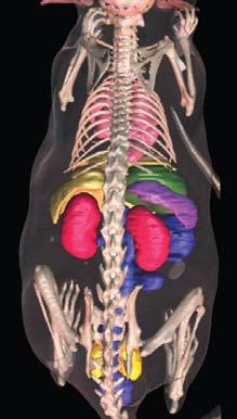
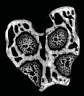
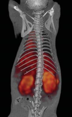
4 microcat Features Animal Bed Three styles of detachable animal beds are available: Mouse bed for up to 50 mm diameter animals Rat bed for up to 100 mm diameter animals Miniature bed for high resolution CT studies of small samples and for high resolution SPECT studies Each removable animal bed is interchangeable with Siemens micropet beds for easy transport of animals between instruments in dual modality studies. Positron emitting fiducial markers visible in the x-ray CT data set may be incorporated into the beds for easy image registration. Respiratory Gating The microcat is configured to accept respiratory gating signals from Biopac and BioTRIG physiological monitoring systems. Precision Motion Systems High-resolution images require high-precision moving components. Only the highest quality motion systems are employed in Siemens preclinical scanners. microspect All microcat systems can be configured with or upgraded to include a state-of-the-art SPECT imaging system for anatomic and functional data acquisition on a single platform. Applications Segmentation tools provide accurate quantification of anatomic data. Newly available preclinical microcat contrast agents provide previously unattainable soft tissue and vascular image contrast. Image courtesy of Oak Ridge National Laboratory. All microcat systems can be configured with optional high resolution SPECT detector heads for multi-modality studies. Image courtesy of the University of Tennessee Graduate School of Medicine, Knoxville, TN. With the optional variable focus x-ray source, images down to 15 micron resolution can be acquired in vivo. 4
5 Specifications Unit weight Unit height Unit width Unit depth (without front shield) Unit depth (with front shield) microcat Approximately 1800 lbs (820 kg) 70 in (1780 mm) 49 in (1245 mm) 33 in (840 mm) 55 in (1395 mm) Temperature and Humidity Operating room temperature F (7 24 C) Operating humidity: 30 70% non-condensing Note: The maximum power consumption of the microcat system is 2 kw (6800 BTU/hr). Electrical Requirements microcat and computer Note: Appropriate power connection will be provided for destination country. 110V/20A isolated outlet (20A outlet required) 240V/10A (Europe) Magnetic Field Magnetic field strength must be less than 10 Gauss. Radiation Safety The microcat is a cabinet x-ray system. The scanner is FDA registered and compliant with title 21 of the Code of Federal Regulations, Part (21 CFR ). 5
6 microcat +SPECT at a Glance Siemens microcat +SPECT generates high resolution, fully registered SPECT/CT data sets. With the largest commercially available pixilated detector heads, an extensive set of collimators, multiple energy windows, and independently adjustable detector head positions, the microcat +SPECT sets the standard for preclinical multimodality imaging. Key Features Largest commercially available pixilated detector heads 150 mm x 150 mm Large detector heads permit greater pinhole magnification, improving sensitivity while maintaining large Field of View Small pixels provide high resolution, even at low magnification and with parallel-hole collimators +SPECT data acquisition system is fully integrated into the microcat hardware and software microcat beds are compatible with micropet scanners for easy dual and triple modality studies microcat +SPECT data sets are fully compatible with the micropet ASIPro software package Detector Heads 150 mm x 150 mm active area Array of position sensitive photomultiplier tubes and pixilated NaI(Tl) 2.0 mm x 2.0 mm x 10 mm crystals 2.2 mm crystal spacing 4,600 pixels per detector head Collimators Parallel hole (1.2 mm and 2 mm aperture) Pinhole (0.5 mm, 1 mm, 2 mm, and 3 mm inserts) Multi-pinhole collimation will be introduced in early


7 Image courtesy of VA Hospital, Columbia, MO. 7
8 Amira is a registered trademark of Mercury Computer Systems. Biopac is a registered trademark of Biopac Systems, Inc. BioTRIG is a registered trademark of Spin Systems (QLD) Pty Ltd. Linux is a registered trademark of Sun Microsystems. microcat and micropet are registered trademark of Siemens reserves the right to modify the design and specifications contained herein without prior notice. Product performance depends on the choice of system configuration. Please contact your local Siemens sales representative for the most current information or contact one of the addresses listed below All rights reserved. All photographs 2005 Siemens Medical Solutions, USA. All rights reserved. Note: Original images always lose a certain amount of detail when reproduced. Molecular Imaging 2501 N. Barrington Road Hoffman Estates, IL USA Contact Addresses Molecular Imaging 2501 N. Barrington Road Hoffman Estates, IL USA Telephone: Headquarters 51 Valley Stream Parkway Malvern, PA USA Telephone: Molecular Imaging 810 Innovation Drive Knoxville, TN USA Telephone: , Siemens AG Order No. A91MI T-7600 Printed in USA 10/05 PA 1005/1.5
Inveon. No Limits on Discovery.
 Trademarks and service marks used in this material are property of Siemens Medical Solutions USA or Siemens AG. Inveon is a trademark of Siemens AG, its subsidiaries or affiliates. All other company, brand,
COMPUTED TOMOGRAPHY 1
 COMPUTED TOMOGRAPHY 1 Why CT? Conventional X ray picture of a chest 2 Introduction Why CT? In a normal X-ray picture, most soft tissue doesn't show up clearly. To focus in on organs, or to examine the
CHAPTER 2 COMMISSIONING OF KILO-VOLTAGE CONE BEAM COMPUTED TOMOGRAPHY FOR IMAGE-GUIDED RADIOTHERAPY
 14 CHAPTER 2 COMMISSIONING OF KILO-VOLTAGE CONE BEAM COMPUTED TOMOGRAPHY FOR IMAGE-GUIDED RADIOTHERAPY 2.1 INTRODUCTION kv-cbct integrated with linear accelerators as a tool for IGRT, was developed to
GE Healthcare. Senographe 2000D Full-field digital mammography system
 GE Healthcare Senographe 2000D Full-field digital mammography system Digital has arrived. The Senographe 2000D Full-Field Digital Mammography (FFDM) system gives you a unique competitive advantage. That
PET/CT Instrumentation Basics
 / Instrumentation Basics 1. Motivations for / imaging 2. What is a / Scanner 3. Typical Protocols 4. Attenuation Correction 5. Problems and Challenges with / 6. Examples Motivations for / Imaging Desire
New spectral benefi ts, proven low dose
 New spectral benefi ts, proven low dose Philips MicroDose mammography SI, technical data sheet Philips MicroDose SI with single-shot spectral imaging is a fullfi eld digital mammography solution that delivers
High-sensitivity. optical molecular imaging and high-resolution digital X-ray. In-Vivo Imaging Systems
 High-sensitivity optical molecular imaging and high-resolution digital X-ray In-Vivo Imaging Systems In vivo imaging solutions available in several packages Carestream Molecular Imaging offers a selection
2010 Philips BrightView XCT SPECT/CT
 2010 Philips BrightView XCT SPECT/CT Unit was purchased from Philips training center in 2015. Installed but never been used by the current facility. (Scroll for pictures) BrightView XCT Camera with PinPoint
NeuViz 16 Computed Tomography. Elevating routine imaging for exceptional results
 NeuViz 16 Computed Tomography Elevating routine imaging for exceptional results Essence NeuViz 16 Raising the bar on clinical utility in routine imaging. Get more. More clinical information for patients.
Research Article Feasibility and Initial Performance of Simultaneous SPECT-CT Imaging Using a Commercial Multi-Modality Preclinical Imaging System
 Hindawi Publishing Corporation International Journal of Molecular Imaging Volume 2015, Article ID 134768, 11 pages http://dx.doi.org/10.1155/2015/134768 Research Article Feasibility and Initial Performance
v tome x m microfocus CT
 GE Inspection Technologies v tome x m microfocus CT Uniting premium 3D metrology and inspection with quality and speed. gemeasurement.com/ct x plore precision CT line Inspect with precision, power, and
Research Support. Dual-Source CT: What is it and How Do I Test it? Cynthia H. McCollough, Ph.D.
 Dual-Source CT: What is it and How Do I Test it? Cynthia H. McCollough, Ph.D. CT Clinical Innovation Center Department of Radiology Mayo Clinic College of Medicine Rochester, MN Research Support National
Ergo TM Imaging System
 Ergo TM Imaging System Unparalleled Clinical Flexibility and Imaging Quality The Ergo Imaging System is Digirad s advanced solid-state large field-of-view (LFOV) general purpose nuclear medicine camera.
HISTORY. CT Physics with an Emphasis on Application in Thoracic and Cardiac Imaging SUNDAY. Shawn D. Teague, MD
 CT Physics with an Emphasis on Application in Thoracic and Cardiac Imaging Shawn D. Teague, MD DISCLOSURES 3DR- advisory committee CT PHYSICS WITH AN EMPHASIS ON APPLICATION IN THORACIC AND CARDIAC IMAGING
Multi-species Optical and X-ray Imaging System
 IVIS Lumina XRMS Series III P R O D U C T N O T E Pre-clinical in vivo imaging Key Features Optical and X-ray imaging Multi-species imaging including mice and rats High resolution, low dose digital X-ray
Philip Sperling. Sales Science and New Materials, YXLON International GmbH, Essener Bogen 15, Hamburg, Germany.
 A new generation of x-ray computed tomography devices for quality inspection and metrology inspection in the field of additive manufacturing and other sciences Philip Sperling Sales Science and New Materials,
Combined micropet /MR System: Performance Assessment of the Full PET Ring with Split Gradients 4.8
 Combined micropet /MR System: Performance Assessment of the Full PET Ring with Split Gradients 4.8 UNIVERSITY OF CAMBRIDGE 1.2 Rob C. Hawkes 1, Tim D. Fryer 1, Alun J. Lucas 1,2, Stefan B. Siegel 3, Richard
DELWORKS DR MEDICAL. take the next step
 DELWORKS DR MEDICAL take the next step DELWORKS MEDICAL DR If you are thinking of taking the next step to digital radiography, consider a DelWorks Medical DR Retrofit Package, the easy and affordable way
NM Module Section 2 6 th Edition Christian, Ch. 3
 NM 4303 Module Section 2 6 th Edition Christian, Ch. 3 Gas Filled Chamber Voltage Gas filled chamber uses Hand held detectors cutie pie Geiger counter Dose calibrators Cutie pie Chamber voltage in Ionization
ANALYTICAL MICRO X-RAY FLUORESCENCE SPECTROMETER
 Copyright(c)JCPDS-International Centre for Diffraction Data 2001,Advances in X-ray Analysis,Vol.44 325 ANALYTICAL MICRO X-RAY FLUORESCENCE SPECTROMETER ABSTRACT William Chang, Jonathan Kerner, and Edward
X-ray phase-contrast imaging
 ...early-stage tumors and associated vascularization can be visualized via this imaging scheme Introduction As the selection of high-sensitivity scientific detectors, custom phosphor screens, and advanced
Diffraction-enhanced X-ray Imaging (DEXI) Medical Solutions. More information using less radiation
 Medical Solutions. More information using less radiation") Diffraction-enhanced X-ray Imaging (DEXI) Medical Solutions More information using less radiation Medical Small Animal Security NDE/NDT Diffraction-Enhanced X-ray Imaging Medical Solutions Safe non-invasive
Diffraction-enhanced X-ray Imaging (DEXI) Medical Solutions More information using less radiation Medical Small Animal Security NDE/NDT Diffraction-Enhanced X-ray Imaging Medical Solutions Safe non-invasive
MAN Revision 002. InSight Mini C-arm Imaging System Technical Reference Manual
 MAN-00668 Revision 002 InSight Mini C-arm Imaging System Technical Reference Manual April 2007 The information contained in this Manual is confidential and proprietary to Hologic, Inc. This information
Dose Reduction and Image Preservation After the Introduction of a 0.1 mm Cu Filter into the LODOX Statscan unit above 110 kvp
 Dose Reduction and Image Preservation After the Introduction of a into the LODOX Statscan unit above 110 kvp Abstract: CJ Trauernicht 1, C Rall 1, T Perks 2, G Maree 1, E Hering 1, S Steiner 3 1) Division
First Results From the High-Resolution mousespect Annular Scintillation Camera
 First Results From the High-Resolution mousespect Annular Scintillation Camera Andrew L. Goertzen, Douglas W. Jones, Jurgen Seidel, King Li, and Michael V. Green Abstract High resolution SPECT imaging
160-slice CT SCANNER / New Standard for the Future
 TECHNOLOGY HISTORY For over 130 years, Toshiba has been a world leader in developing technology to improve the quality of life. Our 50,000 global patents demonstrate a long, rich history of leading innovation.
FMT18 FLOOR MOUNTED SYSTEM
 mas Time AEC 320 kvp 64 mas 320 ma 320 ma 320 DEN 0.0 mm Cu 17 in X 17 in 72.0 in FMT18 FLOOR MOUNTED SYSTEM with Synchronized Tracking System Overview Clinical Efficiency The FMT18 System was designed
X-RAD SmART (Small Animal RadioTherapy) IMAGE GUIDED BIOLOGICAL IRRADIATOR Technical Specifications (updated March 2015)
 IMAGE GUIDED BIOLOGICAL IRRADIATOR Technical Specifications (updated March 2015)") X-RAD SmART (Small Animal RadioTherapy) IMAGE GUIDED BIOLOGICAL IRRADIATOR Technical Specifications (updated March 2015) The X-RAD SmART Small Animal IGRT research system provides a high accuracy cone
X-RAD SmART (Small Animal RadioTherapy) IMAGE GUIDED BIOLOGICAL IRRADIATOR Technical Specifications (updated March 2015) The X-RAD SmART Small Animal IGRT research system provides a high accuracy cone
Radiology Physics Lectures: Digital Radiography. Digital Radiography. D. J. Hall, Ph.D. x20893
 Digital Radiography D. J. Hall, Ph.D. x20893 djhall@ucsd.edu Background Common Digital Modalities Digital Chest Radiograph - 4096 x 4096 x 12 bit CT - 512 x 512 x 12 bit SPECT - 128 x 128 x 8 bit MRI -
Veraviewepocs 2D High Speed Panoramic X-Ray Crystal Clear Images with Reduced Radiation
 Diagnostic and Imaging Equipment Treatment Units Handpieces and Instruments Endodontic Systems Laser Equipment Laboratory Devices Veraviewepocs 2D High Speed Panoramic X-Ray Crystal Clear Images with Reduced
First Applications of the YAPPET Small Animal Scanner
 First Applications of the YAPPET Small Animal Scanner Guido Zavattini Università di Ferrara CALOR2 Congress, Annecy - FRANCE YAP-PET scanner Scintillator: YAP:Ce Size: matrix of 2x2 match like crystals
Philips XPER FD10C R7.0.4
 Philips XPER FD10C R7.0.4 Reconditioned 2005 System- Upgraded to R7 in Oct 2010 The Allura Xper FD10 (Ceiling) single-plane cardiovascular system is comprised of a ceiling mounted C-arm stand and digital
In-Vivo IMAGING SYSTEMS. A complete line of high resolution optical & X-ray systems for pre-clinical imaging
 In-Vivo IMAGING SYSTEMS A complete line of high resolution optical & X-ray systems for pre-clinical imaging In-Vivo Imaging Systems Carestream is a strong, successful, multi-billion dollar, international
MC SIMULATION OF SCATTER INTENSITIES IN A CONE-BEAM CT SYSTEM EMPLOYING A 450 kv X-RAY TUBE
 MC SIMULATION OF SCATTER INTENSITIES IN A CONE-BEAM CT SYSTEM EMPLOYING A 450 kv X-RAY TUBE A. Miceli ab, R. Thierry a, A. Flisch a, U. Sennhauser a, F. Casali b a Empa - Swiss Federal Laboratories for
Introduction. Chapter 16 Diagnostic Radiology. Primary radiological image. Primary radiological image
 Introduction Chapter 16 Diagnostic Radiology Radiation Dosimetry I Text: H.E Johns and J.R. Cunningham, The physics of radiology, 4 th ed. http://www.utoledo.edu/med/depts/radther In diagnostic radiology
Acceptance Testing of a Digital Breast Tomosynthesis Unit
 Acceptance Testing of a Digital Breast Tomosynthesis Unit 2012 AAPM Spring Clinical Meeting Jessica Clements, M.S., DABR Objectives Review of technology and clinical advantages Acceptance Testing Procedures
MINIATURE X-RAY SOURCES AND THE EFFECTS OF SPOT SIZE ON SYSTEM PERFORMANCE
 228 MINIATURE X-RAY SOURCES AND THE EFFECTS OF SPOT SIZE ON SYSTEM PERFORMANCE D. CARUSO, M. DINSMORE TWX LLC, CONCORD, MA 01742 S. CORNABY MOXTEK, OREM, UT 84057 ABSTRACT Miniature x-ray sources present
Multimodal Co-registration Using the Quantum GX, G8 PET/CT and IVIS Spectrum Imaging Systems
 TECHNICAL NOTE Preclinical In Vivo Imaging Authors: Jen-Chieh Tseng, Ph.D. Jeffrey D. Peterson, Ph.D. PerkinElmer, Inc. Hopkinton, MA Multimodal Co-registration Using the Quantum GX, G8 PET/CT and IVIS
NDT Supply.com 7952 Nieman Road Lenexa, KS USA
 Durr NDT ScanX Computed Radiography System The Workhorse of Portable Digital Radiography in NDT There are 2 models available: ScanX Discover HR ScanX Discover HC ScanX Discover HR ScanX Discover HC The
Performance and care. all in one
 Performance and care all in one INNOVATION IS WHAT DRIVES US THINKING ABOUT THE FUTURE Preventive diagnostics remains an essential weapon in defeating breast cancer. Metaltronica s forward-thinking design
LSO PET/CT Pico Performance Improvements with Ultra Hi-Rez Option
 LSO PET/CT Pico Performance Improvements with Ultra Hi-Rez Option Y. Bercier, Member, IEEE, M. Casey, Member, IEEE, J. Young, Member, IEEE, T. Wheelock, Member, IEEE, T. Gremillion Abstract-- Factors which
Photomultiplier Tube
 Nuclear Medicine Uses a device known as a Gamma Camera. Also known as a Scintillation or Anger Camera. Detects the release of gamma rays from Radionuclide. The radionuclide can be injected, inhaled or
TECHNICAL DATA. GIOTTO IMAGE SDL/W is pre-arranged for Full Field Digital Biopsy examination with the patient in prone position.
 Ver. 01/06/07 TECHNICAL DATA GIOTTO IMAGE SDL/W LOW DOSE, FULL FIELD DIGITAL MAMMOGRAPHY UNIT USING AMORPHOUS SELENIUM (a-se) TECHNOLOGY DETECTOR (pre-arranged for stereotactic biopsy with the same digital
Get more from your images with Symphony Image Processing
 DIRECT RADIOGRAPHY The user-friendly DelWorks image acquisition and processing software provides a wide range of tools for a variety of image enhancements. Its user interface simplifies every step of the
Maximizing clinical outcomes
 Maximizing clinical outcomes Digital Tomosynthesis Dual Energy Subtraction Automated Long Length Imaging Improved image quality at a low dose Xray Xray Patented ISS capture technology promotes high sensitivity
Inside Biograph mct.
 Inside Biograph mct The technologies behind the world s first molecular CT. www.siemens.com/mi Large 78 cm bore helps reduce claustrophobia and provides more room for RTP positioning devices. 227 kg (500
Picasso-Trio 3-D Dental Imaging
 Picasso-Trio 3-D Dental Imaging Digital Panoramic & Cephalometric, CBCT X-ray Imaging System 3 in 1 System Product Data Model Picasso-Trio - NP Panoramic only [CT upgradeable] Picasso-Trio - NC Panoramic
PET Performance Evaluation of MADPET4: A Small Animal PET Insert for a 7-T MRI Scanner
 PET Performance Evaluation of MADPET4: A Small Animal PET Insert for a 7-T MRI Scanner September, 2017 Results submitted to Physics in Medicine & Biology Negar Omidvari 1, Jorge Cabello 1, Geoffrey Topping
Portable Digital X-ray
 Portable Digital X-ray Portable Digital X-ray Ideal choice for a complete range of radiographic procedure and eliminates the need for costly room modifications This is a complete outdoor X-ray system with
Maximum Performance, Minimum Space
 TECHNOLOGY HISTORY For over 130 years, Toshiba has been a world leader in developing technology to improve the quality of life. Our 50,000 global patents demonstrate a long, rich history of leading innovation.
Initial evaluation of the Indiana small animal PET scanner
 Initial evaluation of the Indiana small animal PET scanner Ned C. Rouze, Member, IEEE, Victor C. Soon, John W. Young, Member, IEEE, Stefan Siegel, Member, IEEE, and Gary D. Hutchins, Member, IEEE Abstract
XRC X-Ray Calibration System
 XRC X-Ray Calibration System Technical Description Contents 1. Equipment overview 2. X-ray Beam Specifications 3. The Control Console 4. Radiation Safety 5. Filters 6. X-ray Generator & Heat Exchange Equipment
Reach out for the power in mobile x-ray imaging MULTIMOBIL 10. Answers for life.
 Reach out for the power MULTIMOBIL 10 Answers for life. 1 MULTIMOBIL 10 Reach out for the power MULTIMOBIL 10 the ideal solution With an imaging power of 10 kw based on high frequency technology, MULTIMOBIL
LaBr 3 :Ce, the latest crystal for nuclear medicine
 10th Topical Seminar on Innovative Particle and Radiation Detectors 1-5 October 2006 Siena, Italy LaBr 3 :Ce, the latest crystal for nuclear medicine Roberto Pani On behalf of SCINTIRAD Collaboration INFN
Thermionic x-ray. Alternative technologies. Electron Field Emission. CNT Based Field Emission X-Ray Source
 Energy Level (ev) Multi-beam x-ray source array based on carbon nanotube field emission O. Zhou, JP Lu, X. Calderon-Colon, X. Qian, G. Yang, G. Cao, E. Gidcumb, A. Tucker, J. Shan University of North Carolina
Master of Science Thesis. SIMIND Based Pinhole Imaging
 Master of Science Thesis SIMIND Based Pinhole Imaging * Development and Validation Kurt Sundin Supervisor: Michael Ljungberg, PhD Medical Radiation Physics Clinical Sciences, Lund Lund University, 2006
Shad-o-Box HS Product Family
 Shad-o-Box HS Product Family DATASHEET Overview Key Features Large active area up to 10x15 cm Up to 10 lp/mm resolution Gigabit Ethernet interface (Camera Link optional) 14-bit digital video output Energy
Detector technology challenges for nuclear medicine and PET
 Nuclear Instruments and Methods in Physics Research A 513 (2003) 1 7 Detector technology challenges for nuclear medicine and PET Paul K. Marsden Guy s and St. Thomas Clinical PET Centre, King s College
MRI RF-Coils. Innovation with Integrity. Highest sensitivity for your preclinical MRI and MRS applications. Preclinical Imaging
 MRI RF-Coils Highest sensitivity for your preclinical MRI and MRS applications Innovation with Integrity Preclinical Imaging Molecular and Preclinical Imaging Preclinical magnetic resonance imaging of
I. PERFORMANCE OF X-RAY PRODUCTION COMPONENTS FLUOROSCOPIC ACCEPTANCE TESTING: TEST PROCEDURES & PERFORMANCE CRITERIA
 FLUOROSCOPIC ACCEPTANCE TESTING: TEST PROCEDURES & PERFORMANCE CRITERIA EDWARD L. NICKOLOFF DEPARTMENT OF RADIOLOGY COLUMBIA UNIVERSITY NEW YORK, NY ACCEPTANCE TESTING GOALS PRIOR TO 1st CLINICAL USAGE
Breast Tomosynthesis. Bob Liu, Ph.D. Department of Radiology Massachusetts General Hospital And Harvard Medical School
 Breast Tomosynthesis Bob Liu, Ph.D. Department of Radiology Massachusetts General Hospital And Harvard Medical School Outline Physics aspects of breast tomosynthesis Quality control of breast tomosynthesis
CR Basics and FAQ. Overview. Historical Perspective
 Page: 1 of 6 CR Basics and FAQ Overview Computed Radiography is a term used to describe a system that electronically records a radiographic image. Computed Radiographic systems use unique image receptors
Computed Tomography. The Fundamentals of... THE FUNDAMENTALS OF... Jason H. Launders, MSc. Current Technology
 The Fundamentals of... Computed Tomography Computed Tomography (CT) systems use x-rays to produce images of slices through a patient s anatomy. Despite having lower spatial resolution than other x-ray
ADVANCED MEDICAL SYSTEMS PTE LTD Singapore Malaysia India Australia
 Innovative design is combined with cutting-edge technology to yield a definitive diagnosis and never before seen ergonomics GIOTTO CLASS is the result of 25 years of experience in the research and development
R-AXIS RAPID. X-ray Single Crystal Structure Analysis System. Product Information
 The Rigaku Journal Vol. 15/ number 2/ 1998 Product Information X-ray Single Crystal Structure Analysis System R-AXIS RAPID 1. Introduction X-ray single crystal structure analysis is known as the easiest
MS260i 1/4 M IMAGING SPECTROGRAPHS
 MS260i 1/4 M IMAGING SPECTROGRAPHS ENTRANCE EXIT MS260i Spectrograph with 3 Track Fiber on input and InstaSpec IV CCD on output. Fig. 1 OPTICAL CONFIGURATION High resolution Up to three gratings, with
OTC18 OVERHEAD TUBE CRANE SYSTEM
 OTC18 OVERHEAD TUBE CRANE SYSTEM System Overview Clinical Performance Versatile and intuitive, the OTC18M System delivers enhanced patient comfort and optimized workflow. Precisely designed to withstand
China Resources Wandong Medical Equipment Co., Ltd. High Frequency 50kW Digital RF System - HF51-5
 China Resources Wandong Medical Equipment Co., Ltd. High Frequency 50kW Digital RF System - HF51-5 #3, No.9, Jiuxianqiaodong Road, Chaoyang District, Beijing 100015, P.R. China E-mail: international@wandong.com.cn
X-RAYS - NO UNAUTHORISED ENTRY
 Licencing of premises Premises Refer Guidelines A radiation warning sign and warning notice, X-RAYS - NO UNAUTHORISED ENTRY must be displayed at all entrances leading to the rooms where x-ray units are
Digital Panoramic X- Ray unit. Product Data
 Digital Panoramic X- Ray unit Product Data Standard examination programs Standard panoramic: adult/child panoramic exam TMJ open/close mouth: 4 slices are taken in the same image (left/right condyle, open/close
DOUBLE gated in vivo small animal cone beam micro
 Low Dose Phase Correlated Cone Beam Micro CT of Small Animals Stefan Sawall, Frank Bergner, Robert Lapp, Markus Mronz, Marek Karolczak, Andreas Hess, and Marc Kachelrieß, Member, IEEE I. INTRODUCTION DOUBLE
DALLA LUCE VISIBILE AI RAGGI X: NUOVI RIVELATORI DI IMMAGINI PER RAGGI X A DISCRIMINAZIONE IN ENERGIA ED APPLICAZIONI
 DALLA LUCE VISIBILE AI RAGGI X: NUOVI RIVELATORI DI IMMAGINI PER RAGGI X A DISCRIMINAZIONE IN ENERGIA ED APPLICAZIONI D. Pacella ENEA - Frascati LIMS, Frascati 14-15 ottobre 2015 Come per la fotografia:
Amorphous Selenium Direct Radiography for Industrial Imaging
 DGZfP Proceedings BB 67-CD Paper 22 Computerized Tomography for Industrial Applications and Image Processing in Radiology March 15-17, 1999, Berlin, Germany Amorphous Selenium Direct Radiography for Industrial
Distributed source x-ray tube technology for tomosynthesis imaging
 Distributed source x-ray tube technology for tomosynthesis imaging Authors: F. Sprenger a*, X. Calderon-Colon b, Y. Cheng a, K. Englestad a, J. Lu b, J. Maltz c, A. Paidi c, X. Qian b, D. Spronk a, S.
The Versatile and Powerful ACLxy. ACLxy
 The Versatile and Powerful ACLxy ACLxy Rolling into a Clinic, Imaging Center and Hospital Near You! COMPUTED RADIOGRAPHY (CR) IS RAPIDLY THE BEGINNING. THE OREX CR SOLUTION DRA- BECOMING A DRIVING FORCE
An Activity in Computed Tomography
 Pre-lab Discussion An Activity in Computed Tomography X-rays X-rays are high energy electromagnetic radiation with wavelengths smaller than those in the visible spectrum (0.01-10nm and 4000-800nm respectively).
THE increasing interest on pinhole collimation of gamma
 IEEE TRANSACTIONS ON NUCLEAR SCIENCE, VOL. 54, NO. 3, JUNE 2007 469 CsI(Tl) Micro-Pixel Scintillation Array for Ultra-high Resolution Gamma-ray Imaging M. N. Cinti, R. Scafè, R. Pellegrini, C. Trotta,
Software and Hardware in CCTA. Elly Castellano PhD
 Software and Hardware in CCTA Elly Castellano PhD Outline technical requirements for coronary CTA the modern cardiac CT scanner ECG-gating technology image reconstruction algorithms 2 Technical requirements
CHAPTER 8 GENERIC PERFORMANCE MEASURES
 GENERIC PERFORMANCE MEASURES M.E. DAUBE-WITHERSPOON Department of Radiology, University of Pennsylvania, Philadelphia, Pennsylvania, United States of America 8.1. INTRINSIC AND EXTRINSIC MEASURES 8.1.1.
1. Patient size AEC. Large Patient High ma. Small Patient Low ma
 Comparison of the function and performance of CT AEC systems CTUG meeting by Emily Field Trainee clinical scientist 14 th th Breakdown CT Automatic Exposure Control (AEC) Background Project Description
Pitfalls and Remedies of MDCT Scanners as Quantitative Instruments
 intensity m(e) m (/cm) 000 00 0 0. 0 50 0 50 Pitfalls and Remedies of MDCT Scanners as Jiang Hsieh, PhD GE Healthcare Technology University of Wisconsin-Madison Root-Causes of CT Number Inaccuracies Nature
Dedicated Veterinary Imaging Solutions Digital, CR and Analog Imaging Solutions for any size patient and any size budget.
 by Dedicated Veterinary Imaging Solutions Digital, CR and Analog Imaging Solutions for any size patient and any size budget. Serving the Veterinary Profession for Over 75 Years. ... We See Things Differently
2D Digital Radiography 3D Computed Tomography Inspection Services Laboratories. xrayinspectionservice.com
 2D Digital Radiography 3D Computed Tomography Inspection Services Laboratories 4nsi.com xrayinspectionservice.com Celebrating over 28 years of Innovation North Star Imaging was founded in 1986 by Ken Ness
Chiara Secco. PET Performance measurements of the new LSO-Based Whole Body PET/CT. Scanner biograph 16 HI-REZ using the NEMA NU Standard.
 Chiara Secco PET Performance measurements of the new LSO-Based Whole Body PET/CT Scanner biograph 16 HI-REZ using the NEMA NU 2-2001 Standard. INTRODUCTION Since its introduction, CT has become a fundamental
Oriel MS260i TM 1/4 m Imaging Spectrograph
 Oriel MS260i TM 1/4 m Imaging Spectrograph MS260i Spectrograph with 3 Track Fiber on input and InstaSpec CCD on output. The MS260i 1 4 m Imaging Spectrographs are economical, fully automated, multi-grating
How Gamma Camera s Head-Tilts Affect Image Quality of a Nuclear Scintigram?
 November 2014, Volume 1, Number 4 How Gamma Camera s Head-Tilts Affect Image Quality of a Nuclear Scintigram? Hojjat Mahani 1,2, Alireza Kamali-Asl 3, *, Mohammad Reza Ay 2, 4 1. Radiation Application
The image reconstruction influence in relative measurement in SPECT / CT animal
 BJRS BRAZILIAN JOURNAL OF RADIATION SCIENCES 0-01 (201) 01-09 The image reconstruction influence in relative measurement in SPECT / CT animal S.C.S. Soriano a ; S.A.L. Souza b ; T.Barboza b ; L.V. De Sá
inspexio SMX-225CT FPD HR
 Microfocus X-Ray CT System C251-E029A Advanced Operability and Excellent Image Quality That Overturns Conventional Assumptions Microfocus X-Ray CT System The is a high-performance microfocus X-ray CT system
Changing the Shape of Nuclear Medicine
 TRUTH IN IMAGING Changing the Shape of Nuclear Medicine Multi-Purpose SPECT Scanner Nothing Gets Closer Introducing 360 Body Contour Scanning With 360 degree detector coverage, and unique proximity sensors
BF-X2. In-line 3D automated X-ray inspection system for Semiconductor, Power module inspection
 In-line automated X-ray inspection system for Semiconductor, Power module inspection BF-X2 Visualize the inner structure with innovative automated inspection In-line automated X-ray inspection system for
Get more from your images with Symphony Image Processing
 DIRECT RADIOGRAPHY The user-friendly DelWorks image acquisition and processing software possesses a wide range of tools for a variety of image manipulations. Its user interface simplifies every step of
Investigation of Multiple Head Registration / Center of Rotation for SPECT Gamma Cameras
 Egyptian J. Nucl. Med., Vol 2, No. 2, Dec. 2009 82 PHYSICS, Original Artical Investigation of Multiple Head Registration / Center of Rotation for SPECT Gamma Cameras Abdelsattar, M.B. Ph.D.; BuHumaid,
SECTION I - CHAPTER 2 DIGITAL IMAGING PROCESSING CONCEPTS
 RADT 3463 - COMPUTERIZED IMAGING Section I: Chapter 2 RADT 3463 Computerized Imaging 1 SECTION I - CHAPTER 2 DIGITAL IMAGING PROCESSING CONCEPTS RADT 3463 COMPUTERIZED IMAGING Section I: Chapter 2 RADT
APD Quantum Efficiency
 APD Quantum Efficiency Development of a 64-channel APD Detector Module with Individual Pixel Readout for Submillimeter Spatial Resolution in PET Philippe Bérard a, Mélanie Bergeron a, Catherine M. Pepin
abc MHRA Philips Mx8000 IDT CT scanner technical evaluation September 2004 Best choice best practice nww.medical-devices.nhs.
 abc September 2004 MHRA 04099 Philips Mx8000 IDT CT scanner technical evaluation Best choice best practice www.mhra.gov.uk nww.medical-devices.nhs.uk About MHRA evaluation reports. What you can expect.
Industry Breakthrough
 Industry Breakthrough Dynamic SPECT Acquisition Quantifying Myocardial Blood Flow D-S P EC T Cardiac Imaging System Nuclear Cardiology in the 21st Century In the 21st century, most nuclear cameras are
Four-dimensional Computed Tomography (4D CT) Concepts and Preliminary Development
 Concepts and Preliminary Development") ORIGINAL ARTICLE ORIGINAL ARTICLE Radiation Medicine: Vol. 21 No. 1, 17 22 p.p., 2003 Four-dimensional Computed Tomography (4D CT) Concepts and Preliminary Development Masahiro Endo,* Takanori Tsunoo,*
ORIGINAL ARTICLE ORIGINAL ARTICLE Radiation Medicine: Vol. 21 No. 1, 17 22 p.p., 2003 Four-dimensional Computed Tomography (4D CT) Concepts and Preliminary Development Masahiro Endo,* Takanori Tsunoo,*
PET: New Technologies & Applications, Including Oncology
 PET: New Technologies & Applications, Including Oncology, PhD, FIEEE Imaging Research Laboratory Department of Radiology University of Washington, Seattle, WA Disclosures Research Contract, GE Healthcare
Technical data CAMARGUE CS-VH50/300. VARIABLE Height Bucky Table With Ceiling Suspension
 Technical data VARIABLE Height Bucky Table With Ceiling Suspension Model Variations CAMARGUE FH (Fixed Height) CAMARGUE FH Tomo CAMARGUE FH Ceiling suspension CAMARGUE VH (Variable Height) CAMARGUE VH
Discover new dimensions in flexibility
 YXLON FF85 CT High-power and high-resolution computed tomography (CT) inspection system for a wide sample spectrum Discover new dimensions in flexibility Technology with Passion Explore the art of detection