Inside Biograph mct.
|
|
|
- Darren Nash
- 6 years ago
- Views:
Transcription


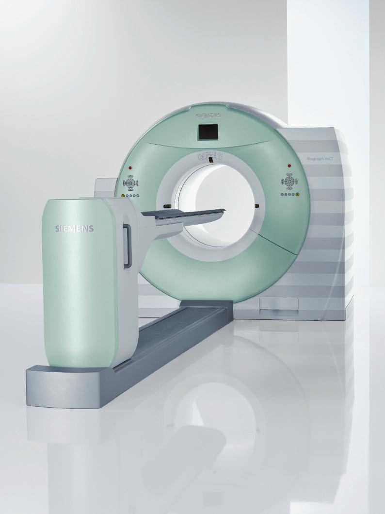
1 Inside Biograph mct The technologies behind the world s first molecular CT.


2 Large 78 cm bore helps reduce claustrophobia and provides more room for RTP positioning devices. 227 kg (500 lb.) bed limit with wide pallet helps accommodate bariatric patients. STRATON X-ray tube provides fast 0.3s CT rotation speed and high resolution. Adaptive Dose shield lowers CT dose by up to 20%. Adaptive 4D spiral allows whole-organ perfusion studies.


3 many-technologies-in-one-device CT. Welcome to the inner world of Biograph mct. Beyond the incredibly small dual-modality footprint, large 78 cm bore, and all the ergonomics is a universe of sophisticated technologies. Working precisely and in tandem, these technologies bring you magnificent images from the human body in both CT and PET to give you diagnostic capabilities like never before. LSO crystals and HI-REZ detectors offer industryleading PET performance. TrueV extends the PET field of view (FOV) by 33% and increases NEMA count rate by 70%. HD PET offers uniformity, high-resolution, and 2x better contrast over conventional PET. ultrahd PET provides uniformity, high-resolution, and 4x better contrast compared to conventional PET. Tubeguard monitors the condition of the STRATON tube and reduces unscheduled downtime.
4 CT TECHNOLOGY STRATON X-ray tube Our unparalleled 0 MHU STRATON X-ray tube has caused a paradigm shift in CT imaging. The tube s direct anode cooling eliminates the need for heat storage, permitting a compact design and fastest CT gantry rotation for all applications. Conventional tube technology In cardiac imaging, where the ability to freeze motion is critical, Biograph mct provides you with super-fast rotation time, making non-invasive cardiac diagnosis routinely available. To increase temporal resolution, the proprietary STRATON X-ray tube enables the market s fastest gantry rotation time of 0.30 s, resulting in 150 ms temporal resolution for motion- and artifact-free imaging of the heart. STRATON X-ray tube z-sharp technology Instead of decreasing the size of detector elements to improve spatial resolution, z-sharp Technology utilizes two overlapping X-ray beams, resulting in significantly increased resolution without a corresponding increase in dose. This provides you with the industry-leading isotropic resolution of 0.33 mm at any scan and rotation speed, and at any position within the scan field. In addition, with our proprietary z-uhr Technology, the system adapts for ultra-high resolution bone imaging for wrist, joint, or inner ear studies. We push the boundaries of spatial resolution even further providing unparalleled 0.24 mm isotropic resolution until now seen only with research flat panel and Micro CT technology. Over Sampling 0.6 mm Oversampling 0.6 mm 2


5 CT TECHNOLOGY CARE Dose4D: real-time dose modulation The CARE Dose4D feature utilizes an advanced computing technique that provides real-time dose modulation of the X-ray tube current according to the precise shape of the patient s body during both spiral and sequential scanning. It reduces the patient dose for low attenuation views, while the dose is kept at a nominally higher ma for high attenuation angles. From the initial topogram, a base ma setting is determined. During the scan, a detector element measures the attenuation through the patient and transfers that information to the output generator of the X-ray tube to keep the ma at a level which provides the accepted image quality. CARE Dose4D provides these economical and clinical benefits: Up to 66% dose reduction while maintaining the same image quality as compared to clinical protocols not employing CARE Dose4D Enhancement of the image quality and reduction of artifacts for scanning of asymmetric body regions, such as the shoulders or scans of the patient with arms alongside the body Lower power consumption and reduced heat load Longer spiral ranges and more flexibility for multiphase examinations, especially for obese patients Low dose examinations for pediatric patients Adaptive Dose Shield The Adaptive Dose Shield, which is unique to the CT industry, is part of the innovative new STRATON X-ray tube design. It moves collimators on the X-ray tube to block unnecessary radiation dose. The Adaptive Dose Shield dynamically opens at the beginning of a spiral range and then dynamically closes at the end. Now all clinically irrelevant dose is eliminated. Not only for dedicated applications, but for every single spiral acquisition. Giving you the ability to save an additional 20%* of dose in routine exams like abdominal CT. Conventional tube collimation *Results may vary. Data on file. Post-Spiral Dose Pre-Spiral Dose Image Area Conventional Technology Image Area STRATON with Adaptive Dose Shield No Pre-Spiral Dose No Post-Spiral Dose Image Area Unnecessary radiation Adaptive Dose Shield 3


6 CT TECHNOLOGY Adaptive ECG-Pulsing and Adaptive Cardio Sequence Adaptive ECG-Pulsing, our innovative heartbeatcontrolled dose modulation, applies the exact dose necessary to collect data projections of 180 degrees during the diastolic phase. Its real-time monitoring of the ECG enables a fully automated dose adjustment, instantly reacting to changes and abnormalities of the heartbeat. Instead of using multisegment reconstruction at higher heart rates, Biograph mct with its high temporal resolution, allows you to acquire cardiac images from a single heartbeat. The MinDose feature reduces the pulsing window to a minimum of 4%, giving you additional dose savings. Adaptive ECG-Pulsing in Spiral Mode 4% Minimum Dose Adaptive Cardio Sequence in Sequential Mode react scan move scan move scan react scan In addition to the Adaptive ECG-Pulsing, Biograph mct offers sequential cardiac acquisition mode. We call it Adaptive Cardio Sequence. It s an intelligently triggered sequence that shuts off radiation in the systolic phase and dynamically reacts to irregularities during the ECG trace. The real-time ECG monitoring reacts instantly to irregularities and stops the scan until the heart rate becomes stable again. This arrhythmia compensation method allows for high dose savings and an increased robustness of the scan. Ultra-fast ceramic CT detector Our proprietary Ultra-fast Ceramic (UFC) CT detector uses superior X-ray absorption, high light output and very short afterglow properties to produce stunning CT image quality. Compared to other detector materials, UFC has a much faster decay reaction, which enables the detector to measure more incoming photons per unit of time and contributes to improved image quality. This property, in combination with the short afterglow of UFC, allows very fast CT rotation times without an increase in image noise. 4
, mas, scan time, and slice width.")


7 CT TECHNOLOGY SureView: uncompromising image quality A patented solution for multislice CT scanning, SureView provides exceptional image quality at any pitch setting. To acquire a scan, you simply select the scan volume (range), mas, scan time, and slice width. All other parameters, such as pitch, are automatically calculated by the scanner, ensuring high quality imaging at any scanning speed. For spiral scanning, SureView yields a remarkably low image noise level. To produce the same noise level as sequential CT images, our spiral scan protocols are created with lower mas and therefore, can reduce doses by up to 20%. You specify the slice thickness according to your clinical needs, and SureView automatically provides the best image quality with reliable, excellent performance. SureView allows more patient coverage without loss of resolution. 49 mm/sec Siemens 40-slice CT with SureView 40 x 0.6 mm, 0.37 sec pitch 1.5 for z-resolution of 0.4 mm 55 mm/sec Competitive 64-slice without SureView 64 x mm, 0.4 sec limited to pitch 0.55 for z-resolution of 0.4 mm 87 mm/sec Siemens 64-slice CT with SureView 64 x 0.6 mm, 0.33 sec pitch 1.5 for z-resolution of 0.4 mm Adaptive 4D Spiral With today s conventional CT and its restricted coverage, whole organ perfusion studies i.e., long-range phase resolved CTA studies, are just not possible. The industry s initial attempts to overcome these limitations focused on extending the coverage, but were limited to twice the detector size. The result was an inability to cover the entire organ. Through our innovative Adaptive 4D Spiral, we apply a continuously repeated bi-directional table movement, moving the patient smoothly in and out of the gantry over the desired scan range. It s how we re able to overcome the coverage limitation of a static detector design. The Adaptive 4D Spiral enables you to adapt the coverage to exactly the range you need. For example, you can perform a phase resolved CTA study over a length of 270 mm, providing a clear separation of arterial and venous phase. Now for the first time in perfusion studies, you can cover virtually any organ in 4D, such as performing a complete stroke assessment of the entire brain. Conventional Perfusion Bi-Directional Table Adaptive 4D Spiral Gantry 5
8 PET TECHNOLOGY most-advanced-pet Biograph mct with HD PET and ultrahd PET gives you the ultimate in PET capabilities, offering uniform resolution throughout the field of view and up to 4x better contrast over conventional PET scanners. Resolution Uniformity Contrast Throughput Conventional PET imaging: Conventional PET imaging requires users to make compromises. An improvement in one key area comes at the cost of performance in other key areas, resulting in sub-optimal performance overall. Resolution Uniformity Contrast Throughput Biograph mct imaging: Biograph mct gives you optimum performance in all key areas, so you don t have to make compromises in daily operation. This results in unmatched clinical capabilities, providing high resolution, high contrast, uniformity, and high throughput. Standard High Definition LSO Crystals Pico-3D Electronics HI-REZ Detectors TrueC Scatter correction HD PET ultrahd PET Built on proven and patented PET technologies, Biograph mct offers true high-definition PET imaging. HI-REZ-FBP Without HD PET With HD PET and ultrahd PET HD PET and ultrahd PET Signal to Noise at 30cm 2x improvement in signal to noise 4x improvement in signal to noise PET HD PET ultrahd PET HD Uniformity: Images are distortion-free throughout the entire field of view, from center to edges, enabling more accurate visualization of fine detail no matter where you look. HD Resolution: HD PET and ultrahd PET offer 2 mm uniform resolution across the entire field of view for enhanced detectability and the highest level of detail. HD Contrast: With an unprecedented 2x and 4x improvement in signal-to-noise ratio, HD PET and ultrahd PET reveal sharper images, as well as greater distinctness within the image. 1 Measurements were taken with a line source suspended in air at radial positions from the center to 28 centimeters in 4 centimeter steps. The Biograph HI-REZ-FBP data were reconstructed with a standard filtered backprojection algorithm after FORE rebinning and the HD PET data were reconstructed with the TrueX algorithm using six iterations and 14 subsets. 6
 vs.")


9 Conventional PET ultrahd PET Conventional (top row) vs. HD PET (bottom row) With the industry-leading NEMA count rate, Biograph mct enables outstanding PET perfusion studies, as in this inferolateral myocardial infarction with total occlusion at mid RCA. Biograph mct offers excellent resolution and contrast in PET CT imaging, such as in this primary squamous cell carcinoma in the left lung with hilar lymph node metastases. 7
 will be calculated correctly because the photon will hit the crystal on an angle and continue traveling to")
10 PET TECHNOLOGY HD PET When a photon strikes a crystal, it travels a certain distance before its energy is converted into light. The further away from the center of the FOV, the less likely the line of response (LOR) will be calculated correctly because the photon will hit the crystal on an angle and continue traveling to another crystal before it lights up. A point spread function (PSF) describes the response of an imaging system to a point source or point object. A system that knows the response of a point source from everywhere in its FOV can use this information to recover the original shape and form of imaged objects. Conventional PET uses the same reconstruction principles across the entire FOV and does not take into account the detector geometry and mispositioning of the LORs. This results in fuzzy edges and increased distortion further from the center of the FOV. Conventional PET HD PET incorporates millions of accurately measured point spread functions in the reconstruction algorithms. Using measured PSFs, HD PET effectively positions the LORs in their actual geometric location, which dramatically reduces blurring and distortion in the final image. HD PET Data courtesy of the University of Tennesee. Benefits: HD uniformity HD resolution HD contrast 2x 8

11 PET TECHNOLOGY ultrahd PET ultrahd PET = HD PET + Siemens TOF Conventional PET Conventional PET detects coincidence photons and records individual LORs between the crystals. The actual location where the annihilation occurred along the LOR is not measured, which inherently generates blurring in the reconstructed image. ultrahd PET Siemens ultrahd PET with Time of Flight (TOF) measures the actual time difference between the detection of each coincidence photon. This additional timing information is used to better localize the event within a small range along each LOR. The better localization of each event using TOF reduces blurring in the reconstructed image. In this simplified example, four lines of response projections are summed together. The resulting image is a rough approximation of the real image with noticeable blurring. Conventional PET With ultrahd PET technology, the TOF projections are organized in time bins along each line of response. This results in a better estimate of the actual image with much less blurring. ultrahd PET Data courtesy of the University of Tennesee. Benefits: HD uniformity HD resolution HD contrast 4x 9


12 PET TECHNOLOGY TrueV: extended field of view TrueV widens the axial field of view (FOV) by 33%, which increases count rate performance by more than 70%, giving you the clinical flexibility to lower dose rates or scan times by 50%. To make the most of the additional FOV, the acceptance angle in the 3D PET acquisition is increased. In this way, more lines of response can be measured per a given unit of time. By increasing the lines of response and thereby, the count rate, scanning protocols can be more flexible. With TrueV you can improve image quality while shortening scan time or reducing the injected dose. Shortened scan time results in less patient motion, fewer motion artifacts, and more time for dedicated CT scans. TrueV widens the axial field of view by 33%. With TrueV Without TrueV Conventional Field of View Biograph with TrueV TrueV extended PET field of view from Siemens has raised all of our expectations as to what PET CT should be and what it can do to help our patients. Dr. David Townsend, University of Tennessee 33% wider field of view increases effective count rate by over 70% and reduces bed positions per scan by an average of 30%. TrueC: highly efficient scatter correction Scatter correction is a vital component of PET image quality, particularly in cardiology where scattered photons are often the result of the activity of structures near the heart, such as the liver and intestines. Without scatter correction, image degradation can result, making analysis difficult, if not impossible. To improve image analysis and thus, the assessment of patients, Biograph mct includes TrueC, a highly efficient, model-based Compton scatter correction system using Monte Carlo-based computational techniques. This single scatter simulation algorithm employs a unique, intuitive sampling technique organized as a summation over sample scattering points. TrueC is particularly efficient because it reuses the computed ray sums through the object to compute scatter contributions to multiple lines of response. Our single scatter based computational approach has been proven to offer the best balance of speed and accuracy for individual, patient-tailored scatter correction. 10

13 PET TECHNOLOGY LSO: state-of-the-art crystal technology With more than 10 years of experience in Lutetium Oxyorthosilicate (LSO) scintillator crystal technology, Siemens is a pioneer in innovative PET imaging techniques. The Biograph TruePoint PET CT employs patented LSO PET detectors that provide clear and fast images. A PET scanner s performance greatly depends on the scintillation properties of the detector crystal material. With a fast scintillation decay time of 40 ns and the highest density available, LSO crystals offer the best combination of properties of any PET scintillator known today. LSO offers a fast coincidence timing window of 4.5 ns for efficient rejection of random events and enough light output for high energy resolution discrimination to facilitate the efficient rejection of randoms all to provide high count rate statistics, which are essential to high speed PET scanning. HI-REZ: more than 250% improved volumetric resolution LSO is capable of tremendous light output, which enables very small individual detector crystals to be produced. The extremely small HI-REZ crystals result in exceptional isometric spatial resolution an improvement of 250% without any loss of sensitivity. Conventional PET HI-REZ PET Conventional PET detector block and annihilation LSO HI-REZ detector block and annihilation 11

14 PET TECHNOLOGY Siemens Remote Service Siemens Remote Service (SRS), a proactive remote-monitoring infrastructure for medical equipment, makes maintenance and repairs easy for your Biograph mct. With SRS, we can detect and fix most problems remotely and expedite on-site repair if new parts are required. But that s not all. SRS proactively monitors your system to detect and repair parameter deviations that could lead to future problems, making your Biograph mct extremely robust and reliable. And you can trust SRS to keep sensitive medical data secure. SRS employs sophisticated authentication and authorization procedures that are compliant with security and privacy standards recommended by US, European, and Japanese manufacturers. SRS advantages Fast and easy error detection Fast repair times Reduction in unscheduled downtime Improved patient planning and throughput Planned spare parts replacement Reduced on-site involvement with remote diagnosis Maximized system utilization and performance Tubeguard Biograph mct comes with the dependability of Tubeguard, a unique remote service that provides real-time condition reports of the scanner s STRATON tube. Tubeguard employs more than 10 proactive sensors to monitor the performance of the tube and assess its longevity. Based on real-time data from sensors and other complex algorithms, Tubeguard estimates how long the tube will last and when it is due for replacement. This helps schedule service and maintenance appointments and virtually eliminates the possibility of a sudden tube failure. 12
15
16 Trademarks and service marks used in this material are property of Siemens Medical Solutions USA or Siemens AG. All other company, brand, product and service names may be trademarks or registered trademarks of their respective holders. Siemens reserves the right to modify the design and specifications contained herein without prior notice. As is generally true for technical specifications, the data contained herein varies within defined tolerances. Some configurations are optional. Product performance depends on the choice of system configuration. Please contact your local Siemens Sales Representative for the most current information or contact one of the addresses listed below. Note: Original images always lose a certain amount of detail when reproduced. All photographs 2008 Siemens Medical Solutions USA, Inc. All rights reserved. Global Business Unit Address Siemens Medical Solutions USA, Inc. Molecular Imaging 2501 N. Barrington Road Hoffman Estates, IL USA Telephone: Global Siemens Headquarters Siemens AG Wittelsbacherplatz Munich Germany Global Siemens Headquarters Healthcare Headquarters Siemens AG Healthcare Sector Henkestrasse Erlangen Germany Telephone: Legal Manufacturer Siemens Medical Solutions USA, Inc. Molecular Imaging 810 Innovation Drive Knoxville, TN USA Telephone: Order No. A91MI C-7600 Printed in USA PA 0908/ , Siemens AG
Iterative Reconstruction in Image Space. Answers for life.
 Iterative Reconstruction in Image Space Answers for life. Iterative Reconstruction in Image Space * (IRIS) * Please note: IRIS is used as an abbreviation for Iterative Reconstruction in Image Space throughout
Software and Hardware in CCTA. Elly Castellano PhD
 Software and Hardware in CCTA Elly Castellano PhD Outline technical requirements for coronary CTA the modern cardiac CT scanner ECG-gating technology image reconstruction algorithms 2 Technical requirements
SAFIRE. Sinogram Affirmed Iterative Reconstruction. Answers for life.
 Neuro Thoracic Abdominal Abdominal Cardiovascular Pediatric SAFIRE Sinogram Affirmed Iterative Reconstruction Answers for life. SAFIRE * (Sinogram Affirmed Iterative Reconstruction) * The information
2D, 3D CT Intervention, and CT Fluoroscopy
 2D, 3D CT Intervention, and CT Fluoroscopy SOMATOM Definition, Definition AS, Definition Flash Answers for life. Siemens CT Vision Siemens CT Vision The justification for the existence of the entire medical
Research Support. Dual-Source CT: What is it and How Do I Test it? Cynthia H. McCollough, Ph.D.
 Dual-Source CT: What is it and How Do I Test it? Cynthia H. McCollough, Ph.D. CT Clinical Innovation Center Department of Radiology Mayo Clinic College of Medicine Rochester, MN Research Support National
160-slice CT SCANNER / New Standard for the Future
 TECHNOLOGY HISTORY For over 130 years, Toshiba has been a world leader in developing technology to improve the quality of life. Our 50,000 global patents demonstrate a long, rich history of leading innovation.
Data. microcat +SPECT
 Data microcat +SPECT microcat at a Glance Designed to meet the throughput, resolution and image quality requirements of academic and pharmaceutical research, the Siemens microcat sets the standard for
Designing an MR compatible Time of Flight PET Detector Floris Jansen, PhD, Chief Engineer GE Healthcare
 GE Healthcare Designing an MR compatible Time of Flight PET Detector Floris Jansen, PhD, Chief Engineer GE Healthcare There is excitement across the industry regarding the clinical potential of a hybrid
Multi-Access Biplane Lab
 Multi-Access Biplane Lab Advanced technolo gies deliver optimized biplane imaging Designed in concert with leading physicians, the Infinix VF-i/BP provides advanced, versatile patient access to meet the
PET/CT Instrumentation Basics
 / Instrumentation Basics 1. Motivations for / imaging 2. What is a / Scanner 3. Typical Protocols 4. Attenuation Correction 5. Problems and Challenges with / 6. Examples Motivations for / Imaging Desire
PET: New Technologies & Applications, Including Oncology
 PET: New Technologies & Applications, Including Oncology, PhD, FIEEE Imaging Research Laboratory Department of Radiology University of Washington, Seattle, WA Disclosures Research Contract, GE Healthcare
Pitfalls and Remedies of MDCT Scanners as Quantitative Instruments
 intensity m(e) m (/cm) 000 00 0 0. 0 50 0 50 Pitfalls and Remedies of MDCT Scanners as Jiang Hsieh, PhD GE Healthcare Technology University of Wisconsin-Madison Root-Causes of CT Number Inaccuracies Nature
Ysio Max. The most direct way to the image. Answers for life.
 Ysio Max The most direct way to the image www.siemens.com/ysio-max Answers for life. 2 It s more. It s MAX. MAX Multiple Advances in X-ray It s more than just single features or functions. MAX offers multiple
LSO PET/CT Pico Performance Improvements with Ultra Hi-Rez Option
 LSO PET/CT Pico Performance Improvements with Ultra Hi-Rez Option Y. Bercier, Member, IEEE, M. Casey, Member, IEEE, J. Young, Member, IEEE, T. Wheelock, Member, IEEE, T. Gremillion Abstract-- Factors which
Maximum Performance, Minimum Space
 TECHNOLOGY HISTORY For over 130 years, Toshiba has been a world leader in developing technology to improve the quality of life. Our 50,000 global patents demonstrate a long, rich history of leading innovation.
NeuViz 16 Computed Tomography. Elevating routine imaging for exceptional results
 NeuViz 16 Computed Tomography Elevating routine imaging for exceptional results Essence NeuViz 16 Raising the bar on clinical utility in routine imaging. Get more. More clinical information for patients.
Inveon. No Limits on Discovery.
 Trademarks and service marks used in this material are property of Siemens Medical Solutions USA or Siemens AG. Inveon is a trademark of Siemens AG, its subsidiaries or affiliates. All other company, brand,
Chiara Secco. PET Performance measurements of the new LSO-Based Whole Body PET/CT. Scanner biograph 16 HI-REZ using the NEMA NU Standard.
 Chiara Secco PET Performance measurements of the new LSO-Based Whole Body PET/CT Scanner biograph 16 HI-REZ using the NEMA NU 2-2001 Standard. INTRODUCTION Since its introduction, CT has become a fundamental
Brilliance in everything Philips CT products and services
 Brilliance in everything Philips CT products and services Ready for anything No one does more than Philips to help you gain the productivity you need with a comprehensive approach to CT that marries significant
Industry Breakthrough
 Industry Breakthrough Dynamic SPECT Acquisition Quantifying Myocardial Blood Flow Nuclear Cardiology in the 21st Century In the 21st century, most nuclear cameras are still relying on a technology invented
TOPICS: CT Protocol Optimization over the Range of Patient Age & Size and for Different CT Scanner Types: Recommendations & Misconceptions
 CT Protocol Optimization over the Range of Patient Age & Size and for Different CT Scanner Types: Recommendations & Misconceptions TOPICS: Computed Tomography Quick Overview CT Dosimetry Effects of CT
siemens.com/luminos-fusion Luminos Fusion The 2-in-1 system that fits your needs and fits your budget
 siemens.com/luminos-fusion Luminos Fusion The 2-in-1 system that fits your needs and fits your budget Luminos Fusion The 2-in-1 system that fits your needs and fits your budget Luminos Fusion provides
Liver imaging beyond expectations with Ingenia
 Publication for the Philips MRI Community Issue 47 2012/3 Liver imaging beyond expectations with Ingenia Contributed by John Penatzer, RT, MR clinical product specialist, Cleveland, OH, USA Publication
Image Quality and Dose. Image Quality and Dose. Image Quality and Dose Issues in MSCT. Scanner parameters affecting IQ and Dose
 Image Quality and Dose Issues in MSCT Image Quality and Dose Image quality Image noise Spatial resolution Contrast Artefacts Speckle and sharpness S. Edyvean St. George s Hospital London SW17 0QT Radiation
Detector technology in simultaneous spectral imaging
 Computed tomography Detector technology in simultaneous spectral imaging Philips IQon Spectral CT Z. Romman, I. Uman, Y. Yagil, D. Finzi, N. Wainer, D. Milstein; Philips Healthcare While CT has become
Radionuclide Imaging MII Single Photon Emission Computed Tomography (SPECT)
") Radionuclide Imaging MII 3073 Single Photon Emission Computed Tomography (SPECT) Single Photon Emission Computed Tomography (SPECT) The successful application of computer algorithms to x-ray imaging in
Radionuclide Imaging MII 3073 Single Photon Emission Computed Tomography (SPECT) Single Photon Emission Computed Tomography (SPECT) The successful application of computer algorithms to x-ray imaging in
12/21/2016. Siemens Medical Systems Research Agreement Philips Healthcare Research Agreement AAN and ASN Committees
 Joseph V. Fritz, PhD Nandor Pintor, MD Dent Neurologic Institute ASN 2017 Friday, January 20, 2017 Siemens Medical Systems Research Agreement Philips Healthcare Research Agreement AAN and ASN Committees
2010 Philips BrightView XCT SPECT/CT
 2010 Philips BrightView XCT SPECT/CT Unit was purchased from Philips training center in 2015. Installed but never been used by the current facility. (Scroll for pictures) BrightView XCT Camera with PinPoint
Industry Breakthrough
 Industry Breakthrough Dynamic SPECT Acquisition Quantifying Myocardial Blood Flow D-S P EC T Cardiac Imaging System Nuclear Cardiology in the 21st Century In the 21st century, most nuclear cameras are
Clinical Experience Using the Open Bore Multislice CT System Supria (16 slice CT) MEDIX VOL. 61 P.8 P.11
 MEDIX VOL. 61 P.8 P.11") Clinical Experience Using the Open Bore Multislice CT System Supria (16 slice CT) Hiroki Kadoya Yukiko Kitagawa MEDIX VOL. 61 P.8 P.11 Clinical Experience Using the Open Bore Multislice CT System Supria
Clinical Experience Using the Open Bore Multislice CT System Supria (16 slice CT) Hiroki Kadoya Yukiko Kitagawa MEDIX VOL. 61 P.8 P.11 Clinical Experience Using the Open Bore Multislice CT System Supria
Luminos RF Classic. Where value meets performance.
 Luminos RF Classic Where value meets performance www.siemens.com/healthcare What s good value in fluoroscopy? That s easy. Luminos RF Classic. 2 Whether for its handling convenience, outstanding image
The future of nuclear imaging is clear
 Cardius X-ACT The future of nuclear imaging is clear Increased regulations, growing competition, and concerns about radiation exposure are just a sampling of the current challenges facing the nuclear medicine
Image Quality. HTC Grid High Transmission Cellular Grid provides higher contrast images
 B R E A S T I M A G I N G S O L U T I O N S Setting the benchmark for mammography M-IV Series Innovations in breast imaging The Lorad M-IV Series exemplifies Hologic's commitment to developing advanced
Truly flexible to meet your clinical needs
 Truly flexible to meet your clinical needs 2 Adapting to meet your needs Flexible Fast and responsive Excellent image quality Designed with ergonomic efficiency Equipped with dose management tools 3 Three
P a n o r a m i c a n d C e p h a l o m e t r i c X - r a y s
 AN ALL AROUND PERFECT PICTURE. The perfect combination of image quality, efficiency and design. P a n o r a m i c a n d C e p h a l o m e t r i c X - r a y s Air Techniques experts in digital imaging ProVecta
Luminos drf Max. Taking 2-in-1 to the MAX in radiography and fluoroscopy. siemens.com/luminos-drf-max
 Luminos drf Max Taking 2-in-1 to the MAX in radiography and fluoroscopy siemens.com/luminos-drf-max 2 It s more. It s MAX. MAX Multiple Advances in X-ray It s more than just single features or functions.
1. Patient size AEC. Large Patient High ma. Small Patient Low ma
 Comparison of the function and performance of CT AEC systems CTUG meeting by Emily Field Trainee clinical scientist 14 th th Breakdown CT Automatic Exposure Control (AEC) Background Project Description
Introduction. Chapter 16 Diagnostic Radiology. Primary radiological image. Primary radiological image
 Introduction Chapter 16 Diagnostic Radiology Radiation Dosimetry I Text: H.E Johns and J.R. Cunningham, The physics of radiology, 4 th ed. http://www.utoledo.edu/med/depts/radther In diagnostic radiology
HISTORY. CT Physics with an Emphasis on Application in Thoracic and Cardiac Imaging SUNDAY. Shawn D. Teague, MD
 CT Physics with an Emphasis on Application in Thoracic and Cardiac Imaging Shawn D. Teague, MD DISCLOSURES 3DR- advisory committee CT PHYSICS WITH AN EMPHASIS ON APPLICATION IN THORACIC AND CARDIAC IMAGING
GE Healthcare. Senographe 2000D Full-field digital mammography system
 GE Healthcare Senographe 2000D Full-field digital mammography system Digital has arrived. The Senographe 2000D Full-Field Digital Mammography (FFDM) system gives you a unique competitive advantage. That
Image Quality. HTC Grid High Transmission Cellular Grid provides higher contrast images
 B R E A S T I M A G I N G S O L U T I O N S Setting the benchmark for mammography M-IV Series Innovations in breast imaging The Lorad M-IV Series exemplifies Hologic s commitment to developing advanced
The shortest distance to diagnosis Philips BrightView SPECT
 The shortest distance to diagnosis Philips Closer is better Simplicity is seeing something better right from the start. And is a completely new vision of what patient care can be, in a system as compact
Maximizing clinical outcomes
 Maximizing clinical outcomes Digital Tomosynthesis Dual Energy Subtraction Automated Long Length Imaging Improved image quality at a low dose Xray Xray Patented ISS capture technology promotes high sensitivity
Automated dose control in multi-slice CT. Nicholas Keat Formerly ImPACT, St George's Hospital, London
 Automated dose control in multi-slice CT Nicholas Keat Formerly ImPACT, St George's Hospital, London Introduction to presentation CT contributes ~50+ % of all medical radiation dose Ideally all patients
COMPUTED TOMOGRAPHY 1
 COMPUTED TOMOGRAPHY 1 Why CT? Conventional X ray picture of a chest 2 Introduction Why CT? In a normal X-ray picture, most soft tissue doesn't show up clearly. To focus in on organs, or to examine the
CHAPTER 8 GENERIC PERFORMANCE MEASURES
 GENERIC PERFORMANCE MEASURES M.E. DAUBE-WITHERSPOON Department of Radiology, University of Pennsylvania, Philadelphia, Pennsylvania, United States of America 8.1. INTRINSIC AND EXTRINSIC MEASURES 8.1.1.
PET Detectors. William W. Moses Lawrence Berkeley National Laboratory March 26, 2002
 PET Detectors William W. Moses Lawrence Berkeley National Laboratory March 26, 2002 Step 1: Inject Patient with Radioactive Drug Drug is labeled with positron (β + ) emitting radionuclide. Drug localizes
2/14/2019. Nuclear Medicine Artifacts. Symmetric energy windows
 Nuclear Medicine Artifacts SCPMG Medical Imaging Technology & Informatics Medical Physics Group Brian Helbig, MS, DABR 1 2 Symmetric energy windows 3 1 Dynamic clinical study Energy peak shift Electrical
Philips Astonish. Key advantages. including improved image quality and
 Philips Key advantages including improved image quality and with AC offers improved image quality, interpretative certainty, diagnostic Alternatively, simplify patient care by exposing patients to reduced
CT Basics: Image Quality Module 6
 Module 6 For educational and institutional use. This transcript is licensed for noncommercial, educational inhouse or online educational course use only in educational and corporate institutions. Any broadcast,
Celesteion Time-of-Flight Technology
 Celesteion Time-of-Flight Technology Bing Bai, PhD Clinical Sciences Manager, PET/CT Canon Medical Systems USA Introduction Improving the care for every patient while providing a high standard care to
3T Unlimited. ipat on MAGNETOM Allegra The Importance of ipat at 3T. medical
 3T Unlimited ipat on MAGNETOM Allegra The Importance of ipat at 3T s medical ipat on MAGNETOM Allegra The Importance of ipat at 3T The rise of 3T MR imaging Ultra High Field MR (3T) has flourished during
Invisible sophistication. Visible simplicity. CS Welcome to the simplicity of compact panoramic imaging
 Invisible sophistication. Visible simplicity. CS 8100 Welcome to the simplicity of compact panoramic imaging Introducing the CS 8100 The Carestream Dental Factor Humanized technology We keep our technology
Photomultiplier Tube
 Nuclear Medicine Uses a device known as a Gamma Camera. Also known as a Scintillation or Anger Camera. Detects the release of gamma rays from Radionuclide. The radionuclide can be injected, inhaled or
P a n o r a m i c a n d C e p h a l o m e t r i c X - r a y s
 AN ALL AROUND PERFECT PICTURE. The perfect combination of image quality, efficiency and design. P a n o r a m i c a n d C e p h a l o m e t r i c X - r a y s Air Techniques experts in digital imaging ProVecta
Reach out for the power in mobile x-ray imaging MULTIMOBIL 10. Answers for life.
 Reach out for the power MULTIMOBIL 10 Answers for life. 1 MULTIMOBIL 10 Reach out for the power MULTIMOBIL 10 the ideal solution With an imaging power of 10 kw based on high frequency technology, MULTIMOBIL
New Technology in Nuclear Medicine
 New Technology in Nuclear Medicine Reed G. Selwyn, PhD, DABR Vice Chair of Research & Imaging Sciences Associate Professor and Chief, Medical Physics Dept. of Radiology, University of New Mexico Objectives
Changing the Shape of Nuclear Medicine
 TRUTH IN IMAGING Changing the Shape of Nuclear Medicine Multi-Purpose SPECT Scanner Nothing Gets Closer Introducing 360 Body Contour Scanning With 360 degree detector coverage, and unique proximity sensors
Philips EasyDiagnost Eleva
 Philips EasyDiagnost Eleva The Philips EasyDiagnost Eleva Recognized for its ease of use and superb image quality, the EasyDiagnost Eleva has for many years been entrusted with a variety of R/F applications
SPECT Reconstruction & Filtering
 SPECT Reconstruction & Filtering Goals Understand the basics of SPECT Reconstruction Filtered Backprojection Iterative Reconstruction Make informed choices on filter selection and settings Pre vs. Post
R&F X-ray systsem. Savings With Every Exposure
 R&F X-ray systsem Savings With Every Exposure Savings with every exposure Versatility meets value Dependable performance User-friendly interface Expandable imaging capabilities Excellent image quality
diagnostic examination
 RADIOLOGICAL PHYSICS 2011 Raphex diagnostic examination Adel A. Mustafa, Ph.D., Editor PUBLISHED FOR: RAMPS (Radiological and Medical Physics Society of New York) preface The RAPHEX Diagnostic exam 2011
Fundamentals of Positron Emission Tomography (PET)
") Fundamentals of Positron Emission Tomography (PET) NPRE 435, Principles of Imaging with Ionizing Radiation, Fall 2017 Content Fundamentals of PET Camera & Detector Design Real World Considerations Performance
Fundamentals of Positron Emission Tomography (PET) NPRE 435, Principles of Imaging with Ionizing Radiation, Fall 2017 Content Fundamentals of PET Camera & Detector Design Real World Considerations Performance
Retrospective Transmit Beamformation. Whitepaper. ACUSON SC2000 Volume Imaging Ultrasound System. Answers for life.
 Whitepaper Retrospective Transmit Beamformation ACUSON SC2000 Volume Imaging Ultrasound System Chuck Bradley, Ph.D. Siemens Healthcare Sector Ultrasound Business Unit Mountain View, California USA Answers
Noise Characteristics of the FORE+OSEM(DB) Reconstruction Method for the MiCES PET Scanner
 Reconstruction Method for the MiCES PET Scanner") Noise Characteristics of the FORE+OSEM(DB) Reconstruction Method for the MiCES PET Scanner Kisung Lee, Member, IEEE, Paul E. Kinahan, Senior Member, Robert S. Miyaoka, Member, IEEE, Jeffrey A. Fessler,
Noise Characteristics of the FORE+OSEM(DB) Reconstruction Method for the MiCES PET Scanner Kisung Lee, Member, IEEE, Paul E. Kinahan, Senior Member, Robert S. Miyaoka, Member, IEEE, Jeffrey A. Fessler,
FMT18 FLOOR MOUNTED SYSTEM
 mas Time AEC 320 kvp 64 mas 320 ma 320 ma 320 DEN 0.0 mm Cu 17 in X 17 in 72.0 in FMT18 FLOOR MOUNTED SYSTEM with Synchronized Tracking System Overview Clinical Efficiency The FMT18 System was designed
SOMATOM Esprit A Bundle of Energy
 SOMATOM Esprit A Bundle of Energy DATA SOMATOM Esprit An economical CT scanner designed for...... Excellent spiral image quality... A wide range of clinical applications... Value performance and reliabilty
Computed Tomography. The Fundamentals of... THE FUNDAMENTALS OF... Jason H. Launders, MSc. Current Technology
 The Fundamentals of... Computed Tomography Computed Tomography (CT) systems use x-rays to produce images of slices through a patient s anatomy. Despite having lower spatial resolution than other x-ray
Medical Imaging. X-rays, CT/CAT scans, Ultrasound, Magnetic Resonance Imaging
 Medical Imaging X-rays, CT/CAT scans, Ultrasound, Magnetic Resonance Imaging From: Physics for the IB Diploma Coursebook 6th Edition by Tsokos, Hoeben and Headlee And Higher Level Physics 2 nd Edition
Initial Certification
 Initial Certification Nuclear Medical Physics (NMP) Study Guide Part 2 Content Guide and Sample Questions The content of all ABR exams is determined by a panel of experts who select the items based on
Lunar Technology Advantages
 Lunar Technology Advantages DXA stands for Dual-Energy X-ray Absorptiometry. It is a measurement method that uses the differences in the absorption of high energy and low energy X-ray photons by different
Wide-Detector CT for TAVR Planning:
 Wide-Detector CT for TAVR Planning: Impact on Iodine Dose, Radiation Dose, and Image Quality SCBTMR 2015 Annual Course Thursday, October 8 William P. Shuman MD FSCBTMR Department of Radiology University
TimTX TrueShape. The parallel transmit architecture of the future. Answers for life.
 www.siemens.com/trueshape TimTX TrueShape The parallel transmit architecture of the future. The product/feature (mentioned herein) is not commercially available. Due to regulatory reasons its future availability
PET Performance Evaluation of MADPET4: A Small Animal PET Insert for a 7-T MRI Scanner
 PET Performance Evaluation of MADPET4: A Small Animal PET Insert for a 7-T MRI Scanner September, 2017 Results submitted to Physics in Medicine & Biology Negar Omidvari 1, Jorge Cabello 1, Geoffrey Topping
ddr Compact Series Setting a new benchmark in digital radiography.
 ddr Compact Series Setting a new benchmark in digital radiography. ddrcompact When productivity and exceptional value come together. With the introduction of its newest DR system, the ddrcompact, Swissray
Performance characterization of a novel thin position-sensitive avalanche photodiode-based detector for high resolution PET
 2005 IEEE Nuclear Science Symposium Conference Record M11-126 Performance characterization of a novel thin position-sensitive avalanche photodiode-based detector for high resolution PET Jin Zhang, Member,
MUSICA Nerve Center. Artificial Intelligence. Intelligent tools for your Digital Radiography workflow. Fluoroscopy. Workflow Optimization
 Image Quality Bariatric Abdomen Pediatric Imaging Diagnostic Confidence Fluoroscopy Neonatal Imaging Scatter Suppression Dental Full Leg Full Spine Exposure Control Index Artificial Intelligence General
efficiency ddrcompact Series
 efficiency ddrcompact Series beyond conventional DR Swissray is the pioneer and worldwide leader in the design, manufacturing and marketing of state-of-the-art Digital Radiography systems. Swissray s
ADVANCED MEDICAL SYSTEMS PTE LTD Singapore Malaysia India Australia
 Innovative design is combined with cutting-edge technology to yield a definitive diagnosis and never before seen ergonomics GIOTTO CLASS is the result of 25 years of experience in the research and development
Data. Take the Lead in CT SOMATOM Sensation 64
 Data Take the Lead in CT SOMATOM Sensation 64 Imaging of this quality, sharpness, speed and gives us the opportunity to study the human anatomy at a level that has only been dreamt about. Werner A. Bautz,
CHAPTER 2 COMMISSIONING OF KILO-VOLTAGE CONE BEAM COMPUTED TOMOGRAPHY FOR IMAGE-GUIDED RADIOTHERAPY
 14 CHAPTER 2 COMMISSIONING OF KILO-VOLTAGE CONE BEAM COMPUTED TOMOGRAPHY FOR IMAGE-GUIDED RADIOTHERAPY 2.1 INTRODUCTION kv-cbct integrated with linear accelerators as a tool for IGRT, was developed to
CT Basics: Data Acquisition Module 3
 Module 3 Transcript For educational and institutional use. This transcript is licensed for noncommercial, educational inhouse or online educational course use only in educational and corporate institutions.
GE Healthcare. Performa. High-performance breast imaging
 GE Healthcare Performa High-performance breast imaging Moving mammography forward. And patients faster. GE Healthcare s unparalleled leadership across mammography begins with a deep understanding of breast
X-ray Imaging. PHYS Lecture. Carlos Vinhais. Departamento de Física Instituto Superior de Engenharia do Porto
 X-ray Imaging PHYS Lecture Carlos Vinhais Departamento de Física Instituto Superior de Engenharia do Porto cav@isep.ipp.pt Overview Projection Radiography Anode Angle Focal Spot Magnification Blurring
GE Healthcare. Essential for life. Senographe Essential Full-Field Digital Mammography system
 GE Healthcare Essential for life Senographe Essential Full-Field Digital Mammography system Excellence in FFDM is a process. An ongoing quest, fueled by our continuing breakthroughs in breast cancer detection
Get more from your images with Symphony Image Processing
 DIRECT RADIOGRAPHY The user-friendly DelWorks image acquisition and processing software provides a wide range of tools for a variety of image enhancements. Its user interface simplifies every step of the
make it easy, with Ray
 Lower dose - Quick scan times - Pulsed X-ray technology - Multiple scan modes 3 Dedicated detectors - Reliable performance - No damage - Long life span Easy upgrade - Ready to upgrade CBCT & Cephalometric
Revolutionizing 2D measurement. Maximizing longevity. Challenging expectations. R2100 Multi-Ray LED Scanner
 Revolutionizing 2D measurement. Maximizing longevity. Challenging expectations. R2100 Multi-Ray LED Scanner A Distance Ahead A Distance Ahead: Your Crucial Edge in the Market The new generation of distancebased
v tome x m microfocus CT
 GE Inspection Technologies v tome x m microfocus CT Uniting premium 3D metrology and inspection with quality and speed. gemeasurement.com/ct x plore precision CT line Inspect with precision, power, and
I. PERFORMANCE OF X-RAY PRODUCTION COMPONENTS FLUOROSCOPIC ACCEPTANCE TESTING: TEST PROCEDURES & PERFORMANCE CRITERIA
 FLUOROSCOPIC ACCEPTANCE TESTING: TEST PROCEDURES & PERFORMANCE CRITERIA EDWARD L. NICKOLOFF DEPARTMENT OF RADIOLOGY COLUMBIA UNIVERSITY NEW YORK, NY ACCEPTANCE TESTING GOALS PRIOR TO 1st CLINICAL USAGE
Performance Assessment of Pixelated LaBr 3 Detector Modules for TOF PET
 Performance Assessment of Pixelated LaBr 3 Detector Modules for TOF PET A. Kuhn, S. Surti, Member, IEEE, J. S. Karp, Senior Member, IEEE, G. Muehllehner, Fellow, IEEE, F.M. Newcomer, R. VanBerg Abstract--
QC by the MPE in Belgium
 Acceptance testing of state-of-the-art CT scanners using a new national protocol: first experience on a large number of scanners of different make and model the working group Radiology of the Belgian Hospital
QC Testing for Computed Tomography (CT) Scanner
 Scanner") QC Testing for Computed Tomography (CT) Scanner QA - Quality Assurance All planned and systematic actions needed to provide confidence on a structure, system or component. all-encompassing program, including
QC Testing for Computed Tomography (CT) Scanner QA - Quality Assurance All planned and systematic actions needed to provide confidence on a structure, system or component. all-encompassing program, including
LaBr 3 :Ce, the latest crystal for nuclear medicine
 10th Topical Seminar on Innovative Particle and Radiation Detectors 1-5 October 2006 Siena, Italy LaBr 3 :Ce, the latest crystal for nuclear medicine Roberto Pani On behalf of SCINTIRAD Collaboration INFN
productivity ddrformula Series
 productivity ddrformula Series beyond conventional DR Swissray is the pioneer and worldwide leader in the design, manufacturing and marketing of state-of-the-art Digital Radiography systems. Swissray
Think Digital. ddr Modulaire A quantum leap in radiography workflow and efficiency.
 Think Digital. ddr Modulaire A quantum leap in radiography workflow and efficiency. ddrmodulaire When performance is decisive. For the most demanding requirements. The ddrmodulaire is a space efficient,
Explain what is meant by a photon and state one of its main properties [2]
![Explain what is meant by a photon and state one of its main properties [2]](/thumbs/80/82516318.jpg "Explain what is meant by a photon and state one of its main properties [2]") 1 (a) A patient has an X-ray scan taken in hospital. The high-energy X-ray photons interact with the atoms inside the body of the patient. Explain what is meant by a photon and state one of its main properties....
1 (a) A patient has an X-ray scan taken in hospital. The high-energy X-ray photons interact with the atoms inside the body of the patient. Explain what is meant by a photon and state one of its main properties....
Redefining Ergonomics
 Samsung Electronics Co., Ltd. inspires the world and shapes the future with transformative ideas and technologies, redefining the worlds of TVs, smartphones, wearable devices, tablets, cameras, digital
Breast Tomosynthesis. Bob Liu, Ph.D. Department of Radiology Massachusetts General Hospital And Harvard Medical School
 Breast Tomosynthesis Bob Liu, Ph.D. Department of Radiology Massachusetts General Hospital And Harvard Medical School Outline Physics aspects of breast tomosynthesis Quality control of breast tomosynthesis
Simulation and evaluation of a cost-effective high-performance brain PET scanner.
 Research Article http://www.alliedacademies.org/biomedical-imaging-and-bioengineering/ Simulation and evaluation of a cost-effective high-performance brain PET scanner. Musa S Musa *, Dilber U Ozsahin,