TimTX TrueShape. The parallel transmit architecture of the future. Answers for life.
|
|
|
- Wilfred Carpenter
- 6 years ago
- Views:
Transcription
 is not commercially available.")
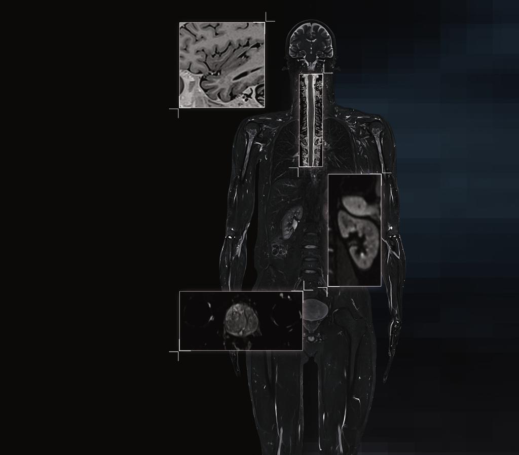
1 TimTX TrueShape The parallel transmit architecture of the future. The product/feature (mentioned herein) is not commercially available. Due to regulatory reasons its future availability cannot be guaranteed. Answers for life.

2 2
 applications.")
3 TimTX TrueShape Shape your pulse TimTX TrueShape is Siemens architecture for parallel transmit (ptx) technology. Based on an intelligent interaction of multiple, independent transmit channels, TimTX TrueShape breaks ground for a broad range of outstanding new magnetic resonance imaging (MRI) applications. Starting with syngo ZOOMit, a zoom function for MRI, TimTX TrueShape is the key to exciting future applications. Bringing TX array imaging into the clinical world with the first TrueShape application for clinical use, TimTX TrueShape also bears much potential for your research. 3


4 A whole new degree of freedom The sky is the limit Open up new horizons 4
5 The parallel transmit architecture of the future TimTX TrueShape provides multi-channel TX array imaging, excelling in higher image quality and shorter scan times. Shape the RF pulse according to your application and experience a whole new degree of freedom. The results are higher image quality and faster scan times. With two transmit channels fully integrated into the system architecture of the 3 Tesla scanner MAGNETOM Skyra, you are ready for the future. Designed for groundbreaking MRI applications The intelligent interaction of independent transmit channels in TimTX TrueShape, enables the selective excitation of a specific body area. That s the key to a broad range of outstanding new MRI applications like zooming with syngo ZOOMit. Only the sky is the limit. Parallel transmit for research and clinical use TimTX TrueShape transfers parallel transmit technology from research into the clinical world with syngo ZOOMit, the first TrueShape application for clinical use. Allow this powerful technology to boost your research activities and open up new horizons. 5
6 TimTX complements Tim ipat and ipat 2 applications syngo GRAPPA method techn msense parallel receive enables Tim tpat future applications 6
7 ology method TimTX TrueShape applications enables parallel transmit syngo ZOOMit As innovation leader in MRI, Siemens has set the trend in RF and coil technology. Around the world, Tim (Total imaging matrix) has changed the way of MR scanning. Our parallel receive technology is now in its 4th generation. Offering the highest channel configurations with up to 128 receive channels. With Tim accuracy, flexibility, and speed are the rule. Now, TimTX is completing Tim by adding transmit power to the parallel imaging intelligence of Tim. Tim and TimTX enable a broad range of existing standard and new MRI applications. future applications Tim and TimTX cover the full spectrum of MR imaging. 7
8 The way to outstanding new MRI applications Conventional MR imaging uses RF pulses at a single amplitude and phase. At higher field strengths (3T) this may lead to shading artifacts, so called B1 inhomogeneities. In 2007 Siemens was the first vendor to introduce TimTX TrueForm as standard B1 shimming solution for better image quality. TimTX TrueForm uses different RF pulse amplitudes and phases to reduce shading artifacts and increase image quality. Now, Siemens again sets the trend in parallel transmit MRI. With TimTX TrueShape you will be able to shape your RF pulse freely to achieve a new degree of freedom in MR imaging. This is opening up the way to outstanding new MRI applications. Starting with syngo ZOOMit, the first zoom function in MRI. This unique parallel transmit application allows zooming in MRI for better diagnostic confidence. In addition, it provides beneficial clinical features like focussed B1 Shimming, B1 Mitigation in zoom direction, B0 Compensation, and transmit SENSE. All paying into increased image quality and imaging speed. 8
9 Benefits of syngo ZOOMit at a glance: Due to Higher image quality Shorter echo trains Less distortions, less blurring Focussed B1 Shimming and B1 Mitigation on zoomed FoV Higher B1 homogeneity due to zooming B0 compensation Better fat saturation No signal from outside zoomed FoV Less motion and flow artifacts Only zoomed FoV must be encoded Increased spatial resolution in region of interest Higher image speed Only zoomed FoV must be encoded Shorter echo trains Fewer phase-encoding steps Shorter scan time First parallel transmit application now and many more in the future Future security 11


10 Conventional transmit Same amplitude Phase = 90 Circulary polarized Excitation of full FoV Shading effects can occur TimTX TrueForm Different amplitudes Phase 90 B1 shimming Excitation of full FoV Shading effects reduced TimTX TrueShape Totally free waveforms Fully parallel transmit Excitation of zoomed FoV Shading and distortions reduced, no infolding 9
11 Parallel transmit glossary B1 inhomogeneities If B1 is inhomogeneous, different locations in the image will have different signal and contrast. The image contrast cannot be made homogeneous by postprocessing. B1 inhomogeneity can arise at higher field strength (3T and above). The problem needs to be addressed at its roots, e.g. by B1 Shimming and B1 Mitigation. B1 Shimming Anatomy-specific B1 Shimming In conventional imaging, the ports of the RF body coil are fed with identical amplitudes and a fixed phase shift of 90. B1 Shimming means that the ports of the RF body coil can be fed with different amplitudes and a phase shift different from 90. It can be used to achieve a higher B1 homogeneity. There are two approaches to B1 Shimming, anatomy specific and patient-specific. Both are two different means of B1 Shimming to achieve the same goal, higher B1 homogeneity (see below). With anatomy-specific B1 Shimming, the different (amplitude/phase) settings for the ports of the RF body coil are taken from a look-up table consisting of anatomy specific pre-fixed values. TimTX TrueForm uses anatomy-specific B1 shimming. It achieves excellent B1 homogeneity without additional scan time or changing the workflow. Patient-specific B1 Shimming With patient-specific B1 Shimming, a B1 map needs to be measure. This changes the standard workflow and takes time. Based on the measured B1 map, the settings of the RF body coil are calculated. With TimTX TrueShape, you can also perform patient-specific B1 Shimming. But the benefit lies elsewhere: It enables fully parallel transmit applications like syngo ZOOMit. B1 Mitigation 1 Mitigation is a unique dynamic application to improve flip angle homogeneity beyond what can be achieved with B1 Shimming alone. This is achieved with sophisticated excitation pulses, typically multiple excitation "spokes" in the so-called excitation k-space. TimTX TrueShape and syngo ZOOMit features B1 Mitigation in the zoom direction, additionally to localized B1 Shimming of the excited FoV. B0 Compensation Transmit SENSE B0 is the main magnetic field. Conventionally, B0 homogeneity can be improved with B0 Shimming. Siemens unique B0 Compensation with dynamic parallel transmit pulses is an additional means to compensate for remnant B0 inhomogeneities. With sophisticated excitation pulses, the phase of the spins can locally be altered to counteract and compensate the phase shift due to B0 inhomogeneities. This will mitigate susceptibility artifacts and improve fat saturation. The principle of Transmit SENSE is the same as Parallel Imaging (msense or GRAPPA) on the receive side. Parallel imaging results in fewer phase-encoding steps for the same spatial resolution and, consequently, shorter scan times. Siemens unique Transmit SENSE results in a reduction of the length of the excitation pulse to achieve the same excitation quality, e.g. the steepness of the excitation profile, in less time. 10
12
 is not commercially available.")
13 syngo ZOOMit The first TimTX TrueShape application for clinical use. The product/feature (mentioned herein) is not commercially available. Due to regulatory reasons its future availability cannot be guaranteed. Answers for life.
14 syngo ZOOMit Shape your image syngo ZOOMit is the first zoom function for MR imaging ever. Based on TimTX TrueShape, Siemens parallel transmit technology, syngo ZOOMit creates a zoom effect during MR imaging. This unique application uses selective excitation to allow you to shape your image. Use syngo ZOOMit to highlight regions, organs, or even features of an organ. This speeds up the scan and improves the image quality in the selected zoom area. As innovation leader in MRI, Siemens introduced syngo ZOOMit as successful start of a broad range of TrueShape applications that will change the game in MRI. 2
15 3
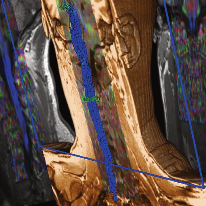
16 Create an MRI zoom through selective excitation Based on TimTX TrueShape, syngo ZOOMit creates a zoom effect during MR imaging. Thanks to multichannel transmit technology with its fully flexible wave forms, you can select a small stripe instead of a large excitation plane. This will allow you to perform a targeted excitation. Data volume is reduced from the very start, as well as time effort for phase-encoding steps. The scan will be faster and image quality in the selected zoom area is considerably improved. The first TrueShape application for clinical use With syngo ZOOMit, Siemens is the first company to provide a zoom function for MR imaging. Enabled by Siemens parallel transmit (ptx) technology TimTX TrueShape, it is the first of a broad range of TrueShape applications for clinical use. As innovation leader, Siemens creates the future of MRI impressively demonstrating how to get the most out of a promising technology like ptx. See more details and less distortions. In less time. Neurological Imaging In this example of a finger-tapping experiment activation patterns become visible in the moto-cortex. On the basis of zoomed data acquisition, you see more coherent better delineated activiation patterns. Be part of the future with TimTX TrueShape Collaboration is the key for innovative MRI technologies and applications. Based on Siemens large network of innovation partnerships, syngo ZOOMit is the result of fruitful collaboration backed by the ptx architecture TimTX TrueShape. And this is only the beginning of the capabilities of parallel transmit technology. Together, let s explore the limits. Due to susceptibility issues EPI in other body parts than the brain used to be difficult. Now, with zoomed EPI you can go down the CNS easily. In this case you see one of the first examples of tractography evaluation in the C-spine at pristine image quality! 4
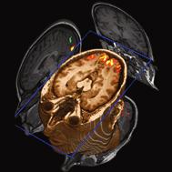
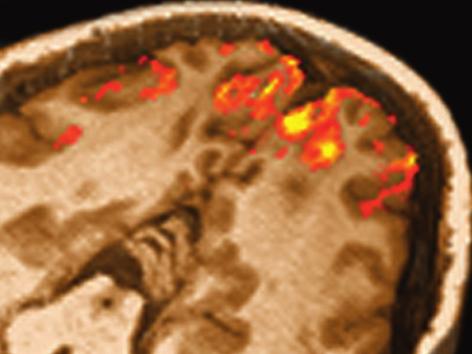
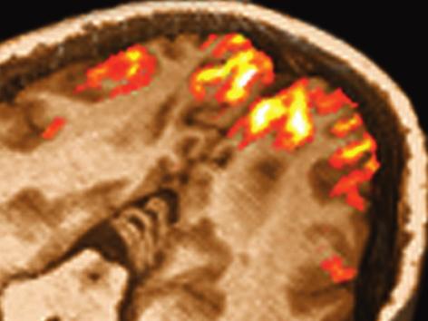
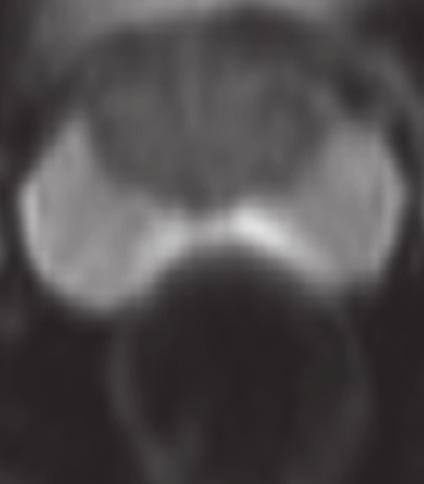
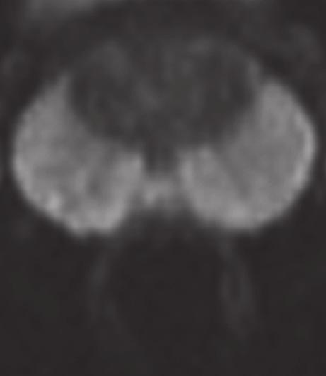
17 Functional Brain Mapping Conventional With syngo ZOOMit DTI of the spine 5
18 See more details and less distortions. In less time. Adding value to Neurology, Oncology, and MSK imaging syngo ZOOMit is especially beneficial for brain, prostate and pancreas, as well as spine applications, and in the visualization of the hip and shoulder joints. Based on EPI or SPACE sequences Higher resolution, less time, less artifacts Functional Prostate Imaging syngo ZOOMit leads to more accurate prostate imaging in multi-modal MRI. In this DWI case imaging time in the zoomed and conventional image are the same but spatial resolution increased along the phase encoding direction. With this the in-plane voxel size of the zoomed DWI is only 1.6 x 1.6 squared mm, at 3 mm slice thickness. Result: Close to distortion free DWI imaging agreeing very well with anatomical imaging. Muscoloskeletal Imaging You can acquire a high-resolution anatomical MRI of the lumbar spine up to 45% faster. 6


19 Anatomy Zoomed DWI Conventional DWI Conventional in 6:01 min (isotropic high-resolution SPACE) Zoomed in 3:20 min 7
20 On account of certain regional limitations of sales rights and service availability, we cannot guarantee that all products included in this brochure are available through the Siemens sales organization worldwide. Availability and packaging may vary by country and are subject to change without prior notice. All of the features and products described herein may not be available in the United States. All devices listed herein may not be licensed according to Canadian Medical Devices Regulations. The information in this document contains general technical descriptions of specifications and options as well as standard and optional features which do not always have to be present in individual cases. Siemens reserves the right to modify the design, packaging, specifications, and options described herein without prior notice. Please contact your local Siemens sales representative for the most current information. Note: Any technical data contained in this document may vary within defined tolerances. Original images always lose a certain amount of detail when reproduced. Please find fitting accessories: Local Contact Information In the USA Siemens Medical Solutions USA, Inc. 51 Valley Stream Parkway Malvern, PA Phone: Phone: Fax: In China Siemens Medical Park, Shanghai 278, Zhouzhu Road SIMZ, Nanhui District Shanghai, , P.R. China Phone: Fax: In Japan Siemens-Asahi Medical Technologies Ltd. Takanawa Park Tower 14F 20-14, Higashi-Gotanda 3-chome Shinagawa-ku, Tokyo Phone: In Asia Siemens Pte Ltd Healthcare Sector Regional Headquarters The Siemens Center 60 MacPherson Road, Singapore Phone: Fax: Global Business Unit Siemens AG Healthcare Sector Magnetic Resonance Henkestr Erlangen Germany Phone: Global Siemens Headquarters Siemens AG Wittelsbacherplatz Muenchen Germany Global Siemens Healthcare Headquarters Siemens AG Healthcare Sector Henkestrasse Erlangen Germany Phone: Legal Manufacturer Siemens AG Wittelsbacherplatz 2 DE Munich Germany Order No. A91MR-6-1C-7600 Printed in Germany CC MR WS , Siemens AG
3T Unlimited. ipat on MAGNETOM Allegra The Importance of ipat at 3T. medical
 3T Unlimited ipat on MAGNETOM Allegra The Importance of ipat at 3T s medical ipat on MAGNETOM Allegra The Importance of ipat at 3T The rise of 3T MR imaging Ultra High Field MR (3T) has flourished during
2D, 3D CT Intervention, and CT Fluoroscopy
 2D, 3D CT Intervention, and CT Fluoroscopy SOMATOM Definition, Definition AS, Definition Flash Answers for life. Siemens CT Vision Siemens CT Vision The justification for the existence of the entire medical
Luminos RF Classic. Where value meets performance.
 Luminos RF Classic Where value meets performance www.siemens.com/healthcare What s good value in fluoroscopy? That s easy. Luminos RF Classic. 2 Whether for its handling convenience, outstanding image
Advanced MSK MRI Protocols at 3.0T. Garry E. Gold, M.D. Associate Professor Department of Radiology Stanford University
 Advanced MSK MRI Protocols at 3.0T Garry E. Gold, M.D. Associate Professor Department of Radiology Stanford University Outline Why High Field for MSK? SNR and Relaxation Times Technical Issues Example
Retrospective Transmit Beamformation. Whitepaper. ACUSON SC2000 Volume Imaging Ultrasound System. Answers for life.
 Whitepaper Retrospective Transmit Beamformation ACUSON SC2000 Volume Imaging Ultrasound System Chuck Bradley, Ph.D. Siemens Healthcare Sector Ultrasound Business Unit Mountain View, California USA Answers
Ysio Max. The most direct way to the image. Answers for life.
 Ysio Max The most direct way to the image www.siemens.com/ysio-max Answers for life. 2 It s more. It s MAX. MAX Multiple Advances in X-ray It s more than just single features or functions. MAX offers multiple
Simultaneous Multi-Slice (Slice Accelerated) Diffusion EPI
 Diffusion EPI") Simultaneous Multi-Slice (Slice Accelerated) Diffusion EPI Val M. Runge, MD Institute for Diagnostic and Interventional Radiology Clinics for Neuroradiology and Nuclear Medicine University Hospital Zurich
Simultaneous Multi-Slice (Slice Accelerated) Diffusion EPI Val M. Runge, MD Institute for Diagnostic and Interventional Radiology Clinics for Neuroradiology and Nuclear Medicine University Hospital Zurich
The compact all-rounder for surgical imaging. SIREMOBIL Compact L. Answers for life.
 The compact all-rounder for surgical imaging SIREMOBIL Compact L Answers for life. A synonym for efficient performance Proven for its reliability and ease of use in intraoperative imaging, the multipurpose
Reach out for the power in mobile x-ray imaging MULTIMOBIL 10. Answers for life.
 Reach out for the power MULTIMOBIL 10 Answers for life. 1 MULTIMOBIL 10 Reach out for the power MULTIMOBIL 10 the ideal solution With an imaging power of 10 kw based on high frequency technology, MULTIMOBIL
[Maestro Class] MAGNETOM Harmony The New Degree of Perfection
![[Maestro Class] MAGNETOM Harmony The New Degree of Perfection](/thumbs/90/102664938.jpg "[Maestro Class] MAGNETOM Harmony The New Degree of Perfection") [Maestro Class] MAGNETOM Harmony The New Degree of Perfection MAGNETOM Family The Perfection of Care The aim of Siemens MR is the perfection of care. To create products and services that improve the quality
[Maestro Class] MAGNETOM Harmony The New Degree of Perfection MAGNETOM Family The Perfection of Care The aim of Siemens MR is the perfection of care. To create products and services that improve the quality
Iterative Reconstruction in Image Space. Answers for life.
 Iterative Reconstruction in Image Space Answers for life. Iterative Reconstruction in Image Space * (IRIS) * Please note: IRIS is used as an abbreviation for Iterative Reconstruction in Image Space throughout
See the big picture in urology. UROSKOP Omnia.
 See the big picture in urology UROSKOP Omnia www.siemens.com/omnia 2 How can I get real KUB in one shot? The answer is simple: with the new UROSKOP Omnia. Thanks to the 43 cm x 43 cm (17 inch x 17 inch)
SIEMENS MAGNETOM Skyra syngo MR D13
 Page 1 of 12 SIEMENS MAGNETOM Skyra syngo MR D13 \\USER\CIND\StudyProtocols\PTSA\*ep2d_M0Map_p2_TE15 TA:7.9 s PAT:2 Voxel size:2.5 2.5 3.0 mm Rel. SNR:1.00 :epfid Properties Routine Contrast Prio Recon
Liver imaging beyond expectations with Ingenia
 Publication for the Philips MRI Community Issue 47 2012/3 Liver imaging beyond expectations with Ingenia Contributed by John Penatzer, RT, MR clinical product specialist, Cleveland, OH, USA Publication
Compact yet Sophisticated
 Compact yet Sophisticated Hitachi has brought Open MRI one step further in its evolution, to better assist medical professionals who work at the forefront of healthcare. AIRIS Light MSK offers radiologists
1 Introduction. 2 The basic principles of NMR
 1 Introduction Since 1977 when the first clinical MRI scanner was patented nuclear magnetic resonance imaging is increasingly being used for medical diagnosis and in scientific research and application
ISSN X CODEN (USA): PCHHAX. The role of dual spin echo in increasing resolution in diffusion weighted imaging of brain
: PCHHAX. The role of dual spin echo in increasing resolution in diffusion weighted imaging of brain") Available online at www.derpharmachemica.com ISSN 0975-413X CODEN (USA): PCHHAX Der Pharma Chemica, 2016, 8(17):15-20 (http://derpharmachemica.com/archive.html) The role of in increasing resolution in
Available online at www.derpharmachemica.com ISSN 0975-413X CODEN (USA): PCHHAX Der Pharma Chemica, 2016, 8(17):15-20 (http://derpharmachemica.com/archive.html) The role of in increasing resolution in
MRI Metal Artifact Reduction
 MRI Metal Artifact Reduction PD Dr. med. Reto Sutter University Hospital Balgrist Zurich University of Zurich OUTLINE Is this Patient suitable for MR Imaging? Metal artifact reduction Is this Patient suitable
HETERONUCLEAR IMAGING. Topics to be Discussed:
 HETERONUCLEAR IMAGING BioE-594 Advanced MRI By:- Rajitha Mullapudi 04/06/2006 Topics to be Discussed: What is heteronuclear imaging. Comparing the hardware of MRI and heteronuclear imaging. Clinical applications
Siemens AG, Healthcare Sector. syngo MR D Operator Manual - Breast 0.0.
 Siemens AG, Healthcare Sector Cs2 syngo Breast Operator 2010-2012 MR-05019 630 02 English 06/2012 n.a. Informatik, Manual D13 Cape syngo MR D13 Operator Manual - Breast syngo MR D13 www.siemens.com/healthcare
MR Advance Techniques. Flow Phenomena. Class II
 MR Advance Techniques Flow Phenomena Class II Flow Phenomena In this class we will explore different phenomenona produced from nuclei that move during the acquisition of data. Flowing nuclei exhibit different
MRI Summer Course Lab 2: Gradient Echo T1 & T2* Curves
 MRI Summer Course Lab 2: Gradient Echo T1 & T2* Curves Experiment 1 Goal: Examine the effect caused by changing flip angle on image contrast in a simple gradient echo sequence and derive T1-curves. Image
SAFIRE. Sinogram Affirmed Iterative Reconstruction. Answers for life.
 Neuro Thoracic Abdominal Abdominal Cardiovascular Pediatric SAFIRE Sinogram Affirmed Iterative Reconstruction Answers for life. SAFIRE * (Sinogram Affirmed Iterative Reconstruction) * The information
High Field MRI: Technology, Applications, Safety, and Limitations
 High Field MRI: Technology, Applications, Safety, and Limitations R. Jason Stafford, Ph.D. The University of Texas M. D. Anderson Cancer Center, Houston, TX Introduction The amount of available signal
M R I Physics Course. Jerry Allison Ph.D., Chris Wright B.S., Tom Lavin B.S., Nathan Yanasak Ph.D. Department of Radiology Medical College of Georgia
 M R I Physics Course Jerry Allison Ph.D., Chris Wright B.S., Tom Lavin B.S., Nathan Yanasak Ph.D. Department of Radiology Medical College of Georgia M R I Physics Course Magnetic Resonance Imaging Spatial
Applications Guide. Spectral Editing with SVS. (Works-in-Progress) MAGNETOM TaTs and Verio Systems (3T)
 MAGNETOM TaTs and Verio Systems (3T)") Applications Guide Spectral Editing with SVS (Works-in-Progress) MAGNETOM TaTs and Verio Systems (3T) syngo MR Numaris 4 VB17A June 2009 Version 1.1 WIP #529 Important Note This document provides a description
Applications Guide Spectral Editing with SVS (Works-in-Progress) MAGNETOM TaTs and Verio Systems (3T) syngo MR Numaris 4 VB17A June 2009 Version 1.1 WIP #529 Important Note This document provides a description
Resona 6 Premium Ultrasound System
 The system Resona 6 is the newly developed, unique result of the mergence of leading companies Mindray Bio-medical Electronics Co. Ltd. and ZONARE Medical Systems, Inc.. By additions to the core competencies
Encoding of inductively measured k-space trajectories in MR raw data
 Downloaded from orbit.dtu.dk on: Apr 10, 2018 Encoding of inductively measured k-space trajectories in MR raw data Pedersen, Jan Ole; Hanson, Christian G.; Xue, Rong; Hanson, Lars G. Publication date:
CARING DESIGN. PRACTICAL TECHNOLOGY.
 GE Healthcare CARING DESIGN. PRACTICAL TECHNOLOGY. Brivo * MR355 1.5T Inspire ITS ELEGANT DESIGN AND WARM LIGHT ARE INTRIGUING. Brivo MR355 Inspire product manager Every piece of equipment you own represents
Pulse Sequence Design and Image Procedures
 Pulse Sequence Design and Image Procedures 1 Gregory L. Wheeler, BSRT(R)(MR) MRI Consultant 2 A pulse sequence is a timing diagram designed with a series of RF pulses, gradients switching, and signal readout
ACRIN 6686 / RTOG 0825
 ACRIN 6686 (RTOG 0825) Advanced MRI Imaging Manual ACRIN 6686 / RTOG 0825 A phase III double blind placebo controlled trial of conventional chemoradiation and adjuvant temozolomide plus bevacizumab vs
H 2 O and fat imaging
 H 2 O and fat imaging Xu Feng Outline Introduction benefit from the separation of water and fat imaging Chemical Shift definition of chemical shift origin of chemical shift equations of chemical shift
MARP. MR Accreditation Program Quality Control Beyond Just the Scans and Measurements July 2005
 ACR MRI accreditation program MR Accreditation Program Quality Control Beyond Just the Scans and Measurements July 2005 Carl R. Keener, Ph.D., DABMP, DABR keener@marpinc.com MARP Medical & Radiation Physics,
Diffusion and Functional MRI of the Spinal Cord Methods and Clinical Applications
 Diffusion and Functional MRI of the Spinal Cord Methods and Clinical Applications Susceptibility artifacts in DTI of the spinal cord J. Cohen-Adad Q-space imaging and axon diameter measurements Functional
Image Quality/Artifacts Frequency (MHz)
") The Larmor Relation 84 Image Quality/Artifacts (MHz) 42 ω = γ X B = 2πf 84 0.0 1.0 2.0 Magnetic Field (Tesla) 1 A 1D Image Magnetic Field Gradients Magnet Field Strength Field Strength / Gradient Coil
The Larmor Relation 84 Image Quality/Artifacts (MHz) 42 ω = γ X B = 2πf 84 0.0 1.0 2.0 Magnetic Field (Tesla) 1 A 1D Image Magnetic Field Gradients Magnet Field Strength Field Strength / Gradient Coil
MR in RTP. MR Data for Treatment Planning: Spatial Accuracy Issues, Protocol Optimization, and Applications (Preview of TG117 Report) Acknowledgements
 Acknowledgements") MR Data for Treatment Planning: Issues, Protocol Optimization, and s (Preview of TG117 Report) Debra H. Brinkmann Mayo Clinic, Rochester MN Acknowledgements TG-117 Use of MRI Data in Treatment Planning
MR Data for Treatment Planning: Issues, Protocol Optimization, and s (Preview of TG117 Report) Debra H. Brinkmann Mayo Clinic, Rochester MN Acknowledgements TG-117 Use of MRI Data in Treatment Planning
SIGNA Pioneer: Ultra High Efficiency Gradient System Advancing the gradient technology curve
 GE Healthcare SIGNA Pioneer: Ultra High Efficiency Gradient System Advancing the gradient technology curve NEW TECHNOLOGY 40W Watts spec is irrelevant. 4W LED bulb delivers same brightness as 40W incandescent
GE Healthcare. Discovery MR T. Simply powerful. Powerfully simple.
 GE Healthcare Discovery MR750 3.0T Simply powerful. Powerfully simple. Break free. The breast images you need in only two sequences. A complete liver study in a 15-minute time slot. Routine fmri with shorter
Application Guide & Release Notes
 Application Guide & Release Notes Inner-volume-imaging (IVI) EPI C2P Release 002a 1 September 2015 TMII Translational and Molecular Imaging Institute Conditions of Use This package is provided to support
siemens.com/luminos-fusion Luminos Fusion The 2-in-1 system that fits your needs and fits your budget
 siemens.com/luminos-fusion Luminos Fusion The 2-in-1 system that fits your needs and fits your budget Luminos Fusion The 2-in-1 system that fits your needs and fits your budget Luminos Fusion provides
Delivering Better Patient Care with SIGNA Architect
 Delivering etter Patient Care with SIGNA Architect As a regional leader in outpatient-based diagnostic imaging, the radiologists and staff at Inova Fairfax MRI Center are focused on one thing: the patient.
Field Simulation Software to Improve Magnetic Resonance Imaging
 Field Simulation Software to Improve Magnetic Resonance Imaging a joint project with the NRI in South Korea CST Usergroup Meeting 2010 Darmstadt Institute for Biometry and Medicine Informatics J. Mallow,
Module 2. Artefacts and Imaging Optimisation for single shot methods. Content: Introduction. Phase error. Phase bandwidth. Chemical shift review
 MRES 7005 - Fast Imaging Techniques Module 2 Artefacts and Imaging Optimisation for single shot methods Content: Introduction Phase error Phase bandwidth Chemical shift review Chemical shift in pixels
MR Basics: Module 8 Image Quality
 Module 8 Transcript For educational and institutional use. This transcript is licensed for noncommercial, educational inhouse or online educational course use only in educational and corporate institutions.
Spiral MRI on a 9.4T Vertical-bore Superconducting Magnet Using Unshielded and Self-shielded Gradient Coils
 Magn Reson Med Sci doi:10.2463/mrms.tn.2016-0049 Published Online: March 27, 2017 TECHNICAL NOTE Spiral MRI on a 9.4T Vertical-bore Superconducting Magnet Using Unshielded and Self-shielded Gradient Coils
BACKGROUND. ** 78% of all MRI scanners have Image Quality problems. *** *** 25% of all Multi-Channel RF coils have at least one bad channel.
 Range of Results from over 534 ACR-mandated Annual MRI Performance Evaluations on over 204 Magnets from 8 Vendors Spanning a 10-year Period Moriel NessAiver, Ph.D. - Simply Physics - Baltimore, MD moriel@simplyphysics.com
SECTION I - CHAPTER 2 DIGITAL IMAGING PROCESSING CONCEPTS
 RADT 3463 - COMPUTERIZED IMAGING Section I: Chapter 2 RADT 3463 Computerized Imaging 1 SECTION I - CHAPTER 2 DIGITAL IMAGING PROCESSING CONCEPTS RADT 3463 COMPUTERIZED IMAGING Section I: Chapter 2 RADT
Background (~EE369B)
") Background (~EE369B) Magnetic Resonance Imaging D. Nishimura Overview of NMR Hardware Image formation and k-space Excitation k-space Signals and contrast Signal-to-Noise Ratio (SNR) Pulse Sequences 13
Background (~EE369B) Magnetic Resonance Imaging D. Nishimura Overview of NMR Hardware Image formation and k-space Excitation k-space Signals and contrast Signal-to-Noise Ratio (SNR) Pulse Sequences 13
SIGNA Explorer Lift revives our MR
 Seiji Shiotani, MD, PhD Seirei Fuji Hospital in Fuji City, Shizuoka, Japan Masayoshi Sugimura Seirei Fuji Hospital in Fuji City, Shizuoka, Japan SIGN Explorer Lift revives our MR The clinical usefulness
Magnetic Resonance Imaging Principles, Methods, and Techniques
 Magnetic Resonance Imaging Principles, Methods, and Techniques Perry Sprawls Jr., Emory University Publisher: Medical Physics Publishing Corporation Publication Place: Madison, Wisconsin Publication Date:
Improving high-field MRI using parallel excitation
 review Improving high-field MRI using parallel excitation MRI at high magnetic field strengths promises to deliver clearer images of the body s structure and function. However, high-field MRI currently
Magnetic Resonance Imaging
 Magnetic Resonance Imaging Principles, Methods, and Techniques Perry Sprawls, Ph.D., FACR, FAAPM, FIOMP Distinguished Emeritus Professor Department of Radiology Emory University Atlanta, Georgia Medical
The SENSE Ghost: Field-of-View Restrictions for SENSE Imaging
 JOURNAL OF MAGNETIC RESONANCE IMAGING 20:1046 1051 (2004) Technical Note The SENSE Ghost: Field-of-View Restrictions for SENSE Imaging James W. Goldfarb, PhD* Purpose: To describe a known (but undocumented)
(N)MR Imaging. Lab Course Script. FMP PhD Autumn School. Location: C81, MRI Lab B0.03 (basement) Instructor: Leif Schröder. Date: November 3rd, 2010
MR Imaging. Lab Course Script. FMP PhD Autumn School. Location: C81, MRI Lab B0.03 (basement) Instructor: Leif Schröder. Date: November 3rd, 2010") (N)MR Imaging Lab Course Script FMP PhD Autumn School Location: C81, MRI Lab B0.03 (basement) Instructor: Leif Schröder Date: November 3rd, 2010 1 Purpose: Understanding the basic principles of MR imaging
(N)MR Imaging Lab Course Script FMP PhD Autumn School Location: C81, MRI Lab B0.03 (basement) Instructor: Leif Schröder Date: November 3rd, 2010 1 Purpose: Understanding the basic principles of MR imaging
MR in Tx Planning. Acknowledgements. Outline. Overview MR in RTP
 MR Data for Treatment Planning and Stereotactic Procedures: Sources of Distortion, Protocol Optimization, and Assessment (Preview of TG117 Report) Debra H. Brinkmann Mayo Clinic, Rochester MN Acknowledgements
MR Basics: Module 6 Pulse Sequences
 Module 6 Transcript For educational and institutional use. This transcript is licensed for noncommercial, educational inhouse or online educational course use only in educational and corporate institutions.
Alae Tracker: Tracking of the Nasal Walls in MR-Imaging
 Alae Tracker: Tracking of the Nasal Walls in MR-Imaging Katharina Breininger 1, Andreas K. Maier 1, Christoph Forman 1, Wilhelm Flatz 2, Catalina Meßmer 3, Maria Schuster 3 1 Pattern Recognition Lab, Friedrich-Alexander-Universität
MRI RF-Coils. Innovation with Integrity. Highest sensitivity for your preclinical MRI and MRS applications. Preclinical Imaging
 MRI RF-Coils Highest sensitivity for your preclinical MRI and MRS applications Innovation with Integrity Preclinical Imaging Molecular and Preclinical Imaging Preclinical magnetic resonance imaging of
Clear delineation of optic radiation and very small vessels using phase difference enhanced imaging (PADRE)
") Clear delineation of optic radiation and very small vessels using phase difference enhanced imaging (PADRE) Poster No.: C-2459 Congress: ECR 2010 Type: Scientific Exhibit Topic: Neuro Authors: T. Yoneda,
Clear delineation of optic radiation and very small vessels using phase difference enhanced imaging (PADRE) Poster No.: C-2459 Congress: ECR 2010 Type: Scientific Exhibit Topic: Neuro Authors: T. Yoneda,
Pulse Sequence Design Made Easier
 Pulse Sequence Design Made Easier Gregory L. Wheeler, BSRT(R)(MR) MRI Consultant gurumri@gmail.com 1 2 Pulse Sequences generally have the following characteristics: An RF line characterizing RF Pulse applications
Welcome at MR Achieva 3.0T TX. About the system. Index. 1.1 Introduction
 1 Welcome at MR Achieva 3.0T TX Welcome to the Introduction of and e-learning module for the Achieva 3 Tesla TX-system. This CBT is setup for those engineers who will soon receive a Achieva 3Tesla TX system.
MRI SYSTEM COMPONENTS Module One
 MRI SYSTEM COMPONENTS Module One 1 MAIN COMPONENTS Magnet Gradient Coils RF Coils Host Computer / Electronic Support System Operator Console and Display Systems 2 3 4 5 Magnet Components 6 The magnet The
2014 M.S. Cohen all rights reserved
 2014 M.S. Cohen all rights reserved mscohen@g.ucla.edu IMAGE QUALITY / ARTIFACTS SYRINGOMYELIA Source http://gait.aidi.udel.edu/res695/homepage/pd_ortho/educate/clincase/syrsco.htm Surgery is usually recommended
The digital 2-in-1 solution for fluoroscopy and radiography. AXIOM Luminos drf.
 The digital 2-in-1 solution for fluoroscopy and radiography AXIOM Luminos drf www.siemens.com/medical Double your benefits with a true 2-in-1 solution AXIOM Luminos drf tears down the wall between fluoroscopy
Annual Ceiling Amount: $
 High Field Magnetic Resonance Imaging Center Research MRI Suite, Basement Floor of Building 203 VAMC 4150 Clement Street, San Francisco, CA 94121 MR STUDY APPLICATION for Research Users Application date:
The Script of ZST + Presentation. MIS Upstream Marketing Team [ 日期 ]
![The Script of ZST + Presentation. MIS Upstream Marketing Team [ 日期 ]](/thumbs/94/119182132.jpg "The Script of ZST + Presentation. MIS Upstream Marketing Team [ 日期 ]") 1 The Script of ZST + Presentation MIS Upstream Marketing Team [ 日期 ] 1 The Script of ZST + Presentation Since Mindray was founded to develop ultrasound business, core technology has always been the engine
1 The Script of ZST + Presentation MIS Upstream Marketing Team [ 日期 ] 1 The Script of ZST + Presentation Since Mindray was founded to develop ultrasound business, core technology has always been the engine
Luminos drf Max. Taking 2-in-1 to the MAX in radiography and fluoroscopy. siemens.com/luminos-drf-max
 Luminos drf Max Taking 2-in-1 to the MAX in radiography and fluoroscopy siemens.com/luminos-drf-max 2 It s more. It s MAX. MAX Multiple Advances in X-ray It s more than just single features or functions.
Downloaded from by on 02/07/18 from IP address Copyright ARRS. For personal use only; all rights reserved
 Downloaded from www.ajronline.org by 46.3.192.5 on 02/07/18 from IP address 46.3.192.5. Copyright RRS. For personal use only; all rights reserved C oil sensitivity encoding (SENSE) is a new technique that
MR3T LASER SYSTEMS IN RT ESSENTIAL FOR PRECISE PATIENT ALIGNMENT
 MR3T LASER SYSTEMS IN RT ESSENTIAL FOR PRECISE PATIENT ALIGNMENT LAP IS YOUR IDEAL PARTNER FOR YOUR EXTERNAL LASER SYSTEMS OUR CUSTOMIZED MR3T LASER BRIDGE SYSTEMS ARE RECOMMENDED BY LEADING MRI SCANNER
MRI Systems and Coil Technology
 MRI for Technologists MRI Systems and Coil Technology PROGRAM INFORMATION MRI for Technologists is a training program designed to meet the needs of radiologic technologists entering or working in the field
TITLE: Prostate Cancer Detection Using High-Spatial Resolution MRI at 7.0 Tesla: Correlation with Histopathologic Findings at Radical Prostatectomy
 Award Number: W81XWH-11-1-0253 TITLE: Prostate Cancer Detection Using High-Spatial Resolution MRI at 7.0 Tesla: Correlation with Histopathologic Findings at Radical Prostatectomy PRINCIPAL INVESTIGATOR:
System/Imaging Imperfections
 System/Imaging Imperfections B0 variations: Shim, Susceptibility B1 variations: Transmit, Receive Gradient Imperfections: Non-linearities Delays and Eddy currents Concomitant terms 1 B0 Variations - Off-Resonance
2015 Spin echoes and projection imaging
 1. Spin Echoes 1.1 Find f0, transmit amplitudes, and shim settings In order to acquire spin echoes, we first need to find the appropriate scanner settings using the FID GUI. This was all done last week,
Cardiac MR. Dr John Ridgway. Leeds Teaching Hospitals NHS Trust, UK
 Cardiac MR Dr John Ridgway Leeds Teaching Hospitals NHS Trust, UK Cardiac MR Physics for clinicians: Part I Journal of Cardiovascular Magnetic Resonance 2010, 12:71 http://jcmr-online.com/content/12/1/71
DORADOnova MR3T MOVING LASER SYSTEM FOR PATIENT ALIGNMENT IN RT
 DORADOnova MRT MOVING LASER SYSTEM FOR PATIENT ALIGNMENT IN RT CERTIFIED The characteristic features of LAP laser systems are sophisticated technology, quality and design for more than 0 years. This level
12/21/2016. Siemens Medical Systems Research Agreement Philips Healthcare Research Agreement AAN and ASN Committees
 Joseph V. Fritz, PhD Nandor Pintor, MD Dent Neurologic Institute ASN 2017 Friday, January 20, 2017 Siemens Medical Systems Research Agreement Philips Healthcare Research Agreement AAN and ASN Committees
1.5T HIGH FIELD SMALL ANIMAL MRI
 1.5T HIGH FIELD SMALL ANIMAL MRI Designed Specifically for Veterinarians TECHNICAL GUIDE ADVANCING THE ART AND SCIENCE OF VETERINARY MRI The PetVet is the only high-field MRI system designed specifically
Designing Better Industrial Robots with Adams Multibody Simulation Software
 Designing Better Industrial Robots with Adams Multibody Simulation Software MSC Software: Designing Better Industrial Robots with Adams Multibody Simulation Software Introduction Industrial robots are
NEMA Standards Publication MS (R2014) Determination of Signal-to-Noise Ratio (SNR) in Diagnostic Magnetic Resonance Imaging
 Determination of Signal-to-Noise Ratio (SNR) in Diagnostic Magnetic Resonance Imaging") NEMA Standards Publication MS 1-2008 (R2014) Determination of Signal-to-Noise Ratio (SNR) in Diagnostic Magnetic Resonance Imaging Published by: National Electrical Manufacturers Association 1300 North
NEMA Standards Publication MS 1-2008 (R2014) Determination of Signal-to-Noise Ratio (SNR) in Diagnostic Magnetic Resonance Imaging Published by: National Electrical Manufacturers Association 1300 North
SmartExam helps to banish most repeat knee studies at Desert Medical Imaging
 I s s u e 3 2 - J u l y 2 0 0 7 F i e l d Strength Publication for the Philips MRI Community SmartExam helps to banish most repeat knee studies at Desert Medical Imaging Total scan automation boosts efficiency,
Cook Medical Peripheral Intervention MRI Information
 Page 1 of 13 Home My Account Help Careers Text size: A A A Home / Clinical / Peripheral Intervention / Products / I Information Browse by Product Family Disease State Alphabetical Cook Medical adheres
Data. microcat +SPECT
 Data microcat +SPECT microcat at a Glance Designed to meet the throughput, resolution and image quality requirements of academic and pharmaceutical research, the Siemens microcat sets the standard for
SIGNA Architect. Make the unimaginable the expected. Issue Spotlight. Issue Spotlight
 SIGNA Architect Make the unimaginable the expected The potential for MR is now even more astonishing. Introducing SIGNA Architect 3.0T, the most advanced and intuitive engineering in MR technology from
Imaging the brain at ultra-high resolution using 3D FatNavs
 Imaging the brain at ultra-high resolution using 3D FatNavs Daniel Gallichan Centre d Imagerie BioMédicale EPFL, Lausanne, Switzerland Overview Introduction How motion affects MRI scans Ways we can track
Lesson 06: Pulse-echo Imaging and Display Modes. These lessons contain 26 slides plus 15 multiple-choice questions.
 Lesson 06: Pulse-echo Imaging and Display Modes These lessons contain 26 slides plus 15 multiple-choice questions. These lesson were derived from pages 26 through 32 in the textbook: ULTRASOUND IMAGING
Maximum Performance, Minimum Space
 TECHNOLOGY HISTORY For over 130 years, Toshiba has been a world leader in developing technology to improve the quality of life. Our 50,000 global patents demonstrate a long, rich history of leading innovation.
Works-in-Progress package Version 1.0. For the SIEMENS Magnetom. Installation and User s Guide NUMARIS/4VA21B. January 22, 2003
 Works-in-Progress package Version 1.0 For the Installation and User s Guide NUMARIS/4VA21B January 22, 2003 Section of Medical Physics, University Hospital Freiburg, Germany Contact: Klaus Scheffler PhD,
Discover the difference in efficiency
 Y.CT Compact Fan-beam computed tomography (CT) inspection system for high-density medium and large-sized parts Discover the difference in efficiency Technology with Passion Explore the art of detection
Numerical Evaluation of an 8-element Phased Array Torso Coil for Magnetic Resonance Imaging
 Numerical Evaluation of an 8-element Phased Array Torso Coil for Magnetic Resonance Imaging Feng Liu, Joe Li, Ian Gregg, Nick Shuley and Stuart Crozier School of Information Technology and Electrical Engineering,
LG1627BXC Clocked Laser Driver
 Data Sheet LG1627BXC Clocked Laser Driver Features High data-rate clocked laser diode driver Clock disable mode for data feedthrough Adjustable high output current Operation up to 3 Gbits/s Applications
Fundamental and Clinical Studies for Effectiveness of Zero-filling Interpolation on k-space for Improvement of Sharpness in Magnetic Resonance Imaging
 Fundamental and Clinical Studies for Effectiveness of Zero-filling Interpolation on k-space for Improvement of Sharpness in Magnetic Resonance Imaging Poster No.: C-0709 Congress: ECR 2014 Type: Scientific
Noninvasive Blood Flow Mapping with Arterial Spin Labeling (ASL) Paul Kyu Han and Sung-Hong Park
 Paul Kyu Han and Sung-Hong Park") Noninvasive Blood Flow Mapping with Arterial Spin Labeling (ASL) Paul Kyu Han and Sung-Hong Park Department of Bio and Brain Engineering, Korea Advanced Institute of Science and Technology (KAIST), Daejeon,
Noninvasive Blood Flow Mapping with Arterial Spin Labeling (ASL) Paul Kyu Han and Sung-Hong Park Department of Bio and Brain Engineering, Korea Advanced Institute of Science and Technology (KAIST), Daejeon,
NeuViz 16 Computed Tomography. Elevating routine imaging for exceptional results
 NeuViz 16 Computed Tomography Elevating routine imaging for exceptional results Essence NeuViz 16 Raising the bar on clinical utility in routine imaging. Get more. More clinical information for patients.
MRI Anatomy and Positioning Series Module 12: Fat Suppression Techniques
 MRI Anatomy and Positioning Series Module 12: Fat Suppression Techniques 1 Introduction... 3 RF FatSat... 4 HOAST... 4 FatSat... 5 Segment FS... 8 PhaseCycle... 9 Water Excitation... 10 STIR... 12 FatSep...
Multiplexed Echo Planar Imaging for Sub-Second Whole Brain FMRI and Fast Diffusion Imaging
 Multiplexed Echo Planar Imaging for Sub-Second Whole Brain FMRI and Fast Diffusion Imaging David A. Feinberg 1,2,3 *, Steen Moeller 4, Stephen M. Smith 5, Edward Auerbach 4, Sudhir Ramanna 1,MattF. Glasser
Further Customization of Dot Engines: AutoCoverage
 Further Customization of Dot Engines: AutoCoverage Bart Schraa, MSc., Senior MR Application Specialist Siemens Canada Limited, Oakville, ON, Canada Introduction In 2009 Dot engines introduced a very powerful
RF PULSE DESIGN FOR PARALLEL TRANSMISSION IN ULTRA HIGH FIELD MAGNETIC RESONANCE IMAGING. Hai Zheng. B.S., Xi an JiaoTong University, 2005
 RF PULSE DESIGN FOR PARALLEL TRANSMISSION IN ULTRA HIGH FIELD MAGNETIC RESONANCE IMAGING by Hai Zheng B.S., Xi an JiaoTong University, 2005 Submitted to the Graduate Faculty of the Swanson School of Engineering
Multi-Access Biplane Lab
 Multi-Access Biplane Lab Advanced technolo gies deliver optimized biplane imaging Designed in concert with leading physicians, the Infinix VF-i/BP provides advanced, versatile patient access to meet the
Analysis of spatial dependence of acoustic noise transfer function in magnetic resonance imaging
 Analysis of spatial dependence of acoustic noise transfer function in magnetic resonance imaging Award: Magna Cum Laude Poster No.: C-1988 Congress: ECR 2014 Type: Scientific Exhibit Authors: T. Hamaguchi,
MOVING LASER SYSTEMS IN RT
 MOVING LASER SYSTEMS IN RT ESSENTIAL FOR PRECISE PATIENT ALIGNMENT NEXT GENERATION LAP IS YOUR IDEAL PARTNER FOR YOUR EXTERNAL LASER SYSTEMS OUR MOVING LASER SYSTEMS ARE RECOMMENDED BY LEADING CT SCANNER