Fundamentals of Positron Emission Tomography (PET)
|
|
|
- Sharleen Walker
- 5 years ago
- Views:
Transcription
1 Fundamentals of Positron Emission Tomography (PET) NPRE 435, Principles of Imaging with Ionizing Radiation, Fall 2017
2 Content Fundamentals of PET Camera & Detector Design Real World Considerations Performance Evaluation Clinical Uses NPRE 435, Principles of Imaging with Ionizing Radiation, Fall 2017
3 Positron Emission A positron is the anti-particle of electrons, which carries the same mass as an electron but is positively charged. Positrons are normally generated by those nuclides having a relatively low neutron-to-proton ratio. An typical example of positron emitter is 22 Na 22 Ne NPRE 435, Principles of Imaging with Ionizing Radiation, Fall 2017 Radiation Sources and Interactions
4 Annihilation Radiation following Positron Emission Beta - plus decay or positron decay : A Z X A Z 1 Y 10 NPRE 435, Principles of Imaging with Ionizing Radiation, Fall 2017 Radiation Sources and Interactions


5 Commonly Used PET Isotopes NPRE 435, Principles of Imaging with Ionizing Radiation, Fall 2017 Radiation Sources and Interactions
6 The Tracer Principle Again Drug is labeled with positron ( +, anti-particle of an electron) emitting radionuclide. Drug localizes in patient according to metabolic properties of that drug. Trace (pico-molar) quantities of drug are sufficient. Radiation dose fairly small (<1 rem).
7 Why PET Interesting Chemistry Easily incorporated into biologically active drugs. 1 Hour Half-Life Maximum study duration is 2 hours. Gives enough time to do the chemistry. Easily produced Short half life local production. 18 F 2 hour half-life 15 O, 11 C, 13 N 2 20 minute half-life
8 Ideal Tracer Isotope Tracers contain elements of life perfect for providing the functional information such as metabolism rate. Electronic collimation high sensitivity. Easier attenuation correction. 18 F 2 hour half-life 15 O, 11 C, 13 N 2 20 minute half-life
9 Attenuation of Internal Source P e (d1 d2) d1 d2 Event detection probability is product of individual photon detection probabilities.
10 Ring of Photon Detectors Detect Radioactive Decays Radionuclide emitting +. decays, + annihilates with e from tissue, forming back-to-back 511 kev photon pair. 511 kev photon pairs detected via time coincidence. Positron lies on line defined by detector pair (known as a chord or a line of response or a LOR). Detect Pair of Back-to Back 511 kev Photons
11 Multi-Layer PET Cameras Scintillator Tungsten Septum Lead Shield Can image several slices simultaneously. Can image cross-plane slices. Can remove septa to increase efficiency ( 3-D PET ) Planar Images Stacked to Form 3-D Image
12 Principle of Computed Tomography 2-Dimensional Object 1-Dimensional Vertical Projection 1-Dimensional Horizontal Projection By measuring all 1-dimensional projections of a 2-dimensional object, you can reconstruct the object
 LORs are organized into projections etc")
13 Organization of data PET data acquisition True counts in LORs are accumulated In some cases, groups of nearby LORs are grouped into one average LOR ( mashing ) LORs are organized into projections etc

14 PET data acquisition 2D and 3D acquisition modes 2D mode (= with septa) 3D mode (= no septa) septa In the 3D mode there are no septa: photons from a larger number of incident angles are accepted, increasing the sensitivity. Note that despite the name, the 2D mode provides three dimensional reconstructed images (a collection of transaxial, sagittal and transaxial slices), just like the 3D mode!
15 PET image reconstruction 2D Reconstruction 2D Reconstruction Each parallel slice is reconstructed independently (a 2D sinogram originates a 2D slice) Slices are stacked to form a 3D volume f(x,y,z) Plane 5 Plane 4 Plane 3 Plane 2 Plane 1 etc 2D reconstruction 2D reconstruction 2D reconstruction 2D reconstruction 2D reconstruction etc Slice 5 Slice 4 Slice 3 Slice 2 Slice 1
16 PET data acquisition 2D mode vs. 3D mode 2D mode (= with septa) 3D mode (= no septa) True True not detected (septa block photons) detected
17 Organization of data PET data acquisition In 3D, there are extra LORs relative to 2D D mode same planes as 2D + oblique planes


18 PET evolution: spatial resolution Human brain Monkey brain Animal PET ~1998 Image credits: Crump Institute, UCLA Image credits: CTI PET Systems
19 PET Camera & Detector Design Typical Parameters Detector Module Design


20 PET Cameras Patient port ~60 cm diameter. 24 to 48 layers, covering 15 cm axially. 4 5 mm fwhm spatial resolution. ~2% solid angle coverage. $1 $2 million dollars. Images courtesy of GE Medical Systems and Siemens / CTI PET Systems
21 What Do We Need for PET Detector? Efficient 511keV gamma rays are not easily stopped in detector. Excellent timing accuracy (typically a few ns) for coincidence measurements. Capability of a very high counting rate (e.g. 0.5MC/s per cm 2 ) High detector spatial resolution for high imaging resolution. Cost effective verylargedetectorvolumeisneeded for practical PET systems.
22 Early PET Detector Module Photomultiplier Tube (Converts Light to Electricity) Scintillator Crystal (Converts into Light) mm high (determines axial spatial resolution) 3 10 mm wide (determines in-plane spatial resolution) 30 mm deep (3 attenuation lengths) + BGO Scintillator (Bi 4 Ge 3 O 12 ). + Parallel Operation. Expensive. Difficult to Pack.
23 Block Design Using Anger Logic 50 mm 4 PMTs (25 mm square) Saw cuts direct light toward PMTs. Depth of cut determines light spread at PMTs. Crystal of interaction found with Anger logic (i.e. PMT light ratio). 50 mm 30 mm BGO Scintillator Crystal Block (sawed into 8x8 array, each crystal 6 mm square) Good Performance, Less Expensive, Easy to Pack
.")
24 Profile through Row 2 Crystal Identification with Anger Logic Y-Ratio Uniformly illuminate block. For each event, compute X-Ratio and Y-Ratio, then plot 2-D position. Individual crystals show up as dark regions. Profile shows overlap (i.e. identification not perfect). X-Ratio Can Decode Up To 64 Crystals with BGO
25 Event Rates Singles Events: ~3 ns timing accuracy 10 6 events / sec / module (25 cm 2 ) 200 modules 2x10 8 events / sec / camera Coincidence Events: Time window ~10 ns Lots of chords (~280,000,000 in 48 layer camera with septa removed). 5x10 6 coincidence events / sec Parallel Electronics is Necessary
26 Detector Requirements Detect 511 kev Photons With (in order of importance): >85% efficiency <5 mm spatial resolution low cost (<$100 / cm 2 ) low dead time (<1 μs cm 2 ) <5 ns fwhm timing resolution <100 kev energy resolution Based on Current PET Detector Modules
27 Variations (Present & Future) Quadrant Sharing Other Scintillators Partial Ring Animal PET Time of Flight PET / CT PET / SPECT
28 Quadrant Sharing Perspective View Front View Each PMT Services 4 Crystal Blocks (Not 1) (Number of PMTs = Number of Blocks) + Cost of PMTs Reduced 4x Dead Time Increased 9x
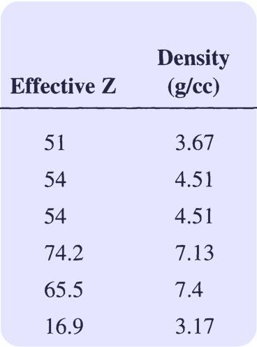
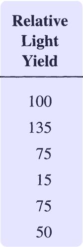
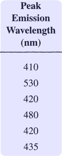
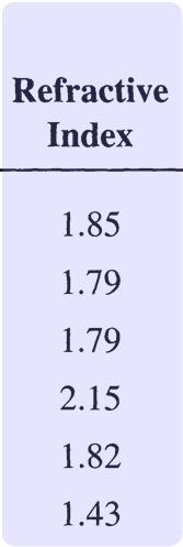
29 Scintillation Crystal Properties NPRE 435, Principles of Imaging with Ionizing Radiation, Fall 2017

30 Lutetium Orthosilicate (LSO) Scintillator Compared to BGO, LSO has: Same Attenuation Length: Good Spatial Resolution Higher Light Output: Decode More Crystals per Block Better SNR for Enhanced Readout (e.g. Depth of Interaction) Shorter Decay Time: Less Dead Time (Allows Larger Block Areas) Better Timing Resolution Reduce Cost OR Increase Performance

31 Image courtesy of Paul Lecoq, CERN Combine Best Properties of: LaBr 3 :30% Ce Timing resolution <100 ps Energy resolution <4% LuI 3 :Ce Light output >100,000 ph/mev PbWO 4 Density >8 g/cc High atomic number Inexpensive PET Performance Determined by Scintillator
 17 cm Detector Ring Diameter *Image courtesy of Simon Cherry, UC Davis Miniature Version of Standard PET")
32 Animal PET Camera Position Sensitive Photomultiplier Tube Fiber Optic Bundle LSO Scintillator Crystals (2x2x10 mm) 17 cm Detector Ring Diameter *Image courtesy of Simon Cherry, UC Davis Miniature Version of Standard PET Camera
33 Dual Modality: PET / SPECT Use SPECT Camera for PET) SPECT cameras optimized to image 140 kev (not 511 kev) photons. Detectors are thin (0.8 attenuation lengths) NaI:Tl. lower efficiency higher scatter fraction Large gaps in angular coverage rotate for complete sampling lower solid angle coverage. Detector area large dead time effects Less Expensive, But Not Optimized for PET
 PET CT Fused")

34 Dual Modality (PET / X-Ray CT) PET CT Fused PET + CT *Data courtesy of David Townsend, U. Tenn.
 ECAT ART Somatom AR.")

35 PET / X-Ray CT Artist s Conception PET detectors (BGO) Reality CT detectors (Xe) ECAT ART Somatom AR.SP PET & CT Scanners Must Be Separated Axially Cannot Image Same Slice Simultaneously! *Data courtesy of David Townsend, U. Tenn.
36 Standard Performance Evaluation NEMA Standards Publication NU : Performance Measurements of Positron Emission Tomographs Spatial Resolution Scatter Fraction Sensitivity Count Losses & Randoms Uniformity Correction (Scatter, Count Rate, Attenuation)
37 Real World Effects Limiting the Performance of PET Photon Attenuation Random Coincidences Scatter Radial Elongation
38 Photon Attenuation Attenuation length of 511 kev photons in water (i.e. tissue) is 10 cm. Brain is 20 cm diameter. up to e 2 = 86% of the events are lost. Loss fraction depends on position in patient. Need to correct for attenuation.


39 PET: Impaired Image Quality in Larger Patients Slim Patient Large Patient For an equivalent data signal to noise ratio, a 120 kg person would have to be scanned 2.3 times longer than a 60 kg person 1) 1) Optimizing Injected Dose in Clinical PET by Accurately Modeling the Counting Rate Response Functions Specific to Individual Patient Scans. Charles C. Watson, PhD et al Siemens Medical Solutions Molecular Imaging, Knoxville, Tennessee, JNM Vol. 46 No. 11, , 2005
 Possible Doubles Image Acquisition")
40 Attenuation Correction Quantitation Transverse Volume Rendered Corrected Uncorrected *Data courtesy of Duffy Cutler, Washington University + Accurate Quantitation (μci/cc) Possible Doubles Image Acquisition Time
41 Attenuation of Internal Source P e (d1 d2) d1 d2 Event detection probability is product of individual photon detection probabilities.

42 Transmission Scan Using an Isotopic Source Can reconstruct an image of the attenuation. Essentially a 511 kev x ray CT image. XBB
 PET CT: 70 kev Can use x-ray CT data to obtain attenuation data Attenuation coefficients are energy")
43 / 0.3 (cm/g) CT Attenuation Correction w/ X-Ray CT Bone Tissue Air Energy (kev) PET CT: 70 kev Can use x-ray CT data to obtain attenuation data Attenuation coefficients are energy dependent at 70 kev (x-ray CT energy) not equal to at 511 kev Scale data use CT to classify voxels as either air, tissue, or bone, then multiply by known ratio of 511 / 70 to do correction *Data courtesy of David Townsend, U. Tenn. Scaled: 511 kev
44 Random Coincidences Simultaneous decays can cause erroneous coincident events called Randoms. For 3-D PET, randoms can be as high as 50% of image. Random Rate is Rate 1 x Rate 2 x 2 t Randoms reduced by narrow coincidence window t. Time of flight across tomograph ring requires t > 4 ns. Random Rate (Activity Density) 2
45 What Is Actually Reconstructed? 3 Scans Taken: Hoop (external source with nothing in ring). Transmission (external source with patient in ring). Emission (patient after isotope injected). Recon. = (Emission Randoms) / Attenuation / Efficiency Attenuation = Transmission / Hoop Efficiency = Hoop / Hoop_Average
46 Scattered Events Compton scatter in patient produces erroneous coincidence events. ~15% of events are scattered in 2- D PET (i.e. if tungsten septa used). ~50% of events are scattered in 3-D Whole Body PET. ~30% of events are scattered in 3-D Brain PET.
47 Radial Elongation Penetration of 511 kev photons into crystal ring blurs measured position. Radial Projection Tangential Projection Blurring worsens as attenuation length increases. Effect variously known as Radial Elongation, Parallax Error, or Radial Astigmatism. Can be removed (in theory) by measuring depth of interaction.
48 Factor Spatial Resolution Shape FWHM Detector Crystal Width d d/2 180Þ± 0.25Þ Anger Logic Photon Noncollinearity 0 (individual coupling) 2.2 mm (Anger logic)* *empirically determined from published data 1.3 mm (head) 2.1 mm (heart) Positron Range Reconstruction Algorithm multiplicative factor 0.5 mm ( 18 F) 4.5 mm ( 82 Rb) 1.25 (in-plane) 1.0 (axial) Dominant Factor is Crystal Width Limit for 80 cm Ring w/ Block Detectors is 3.6 mm


49 Spatial Resolution Away From Center Point Source Images in 60 cm Ring Diameter Camera 1 cm Near Tomograph Center 14 cm from Tomograph Center Resolution Degrades Significantly...

50 Loss in Spatial Resolution Underlying Distribution Measured Distribution Gray / White Ratio = 4:1 Gray / White Ratio = 2.5:1

51 Controlled Charge Collection Apply electric field to drive the charge carriers Collection of visible photons in scintillator Collection of charge carriers in semiconductor In semiconductor, electron and holes are driven by electric field. Spatial spreading of the charge carriers can be better controlled, so that a better spatial resolution can be achieved. NPRE 435, Principles of Imaging with Ionizing Radiation, Fall 2017
 scintillation counter ~70keV ~81keV Pulse Amplitude (V) Measured energy spectrum from HgI 2 semiconductor, 1mm thick, 1x1mm 2 pixels NPRE 435,")
52 A Typical Measured Energy Spectrum Chn #2 Peak Position: ~5.26 V/581 kev Peak Position: 6.00 V/662 kev Peak Position: ~5.36 V/592 kev Counts Chn #3 E.R.: 0.9% ~5.96 kev Typical energy spectrum from a 3 inch NaI(Tl) scintillation counter ~70keV ~81keV Pulse Amplitude (V) Measured energy spectrum from HgI 2 semiconductor, 1mm thick, 1x1mm 2 pixels NPRE 435, Principles of Imaging with Ionizing Radiation, Fall 2017
53 Accurate Quantitation Large Regions Hot Spot Fraction = Activity Measured / True Activity Fraction Observed 100% 80% 60% 40% 20% Hot Spot Cold Spot Cold Spot Fraction = Activity Measured / Background Activity 0% Diameter / Camera FWHM Object Must Be 2x 4x Larger Than Scanner FWHM
54 Low Image Noise High Sensitivity Sensitivity Definition: Place 20 cm diameter phantom in camera. Measure True Event Rate. Sensitivity = True Event Rate / μci / cc. Sensitivity Measures Efficiency for Detecting Signal Increased Axial Extent Increases Sensitivity
55 Increase Sensitivity by Removing Septa Inter-Plane Septa No Septa 2-D (w/ Septa) + Septa Reduce Scatter Smaller Solid Angle for Trues 3-D (w/o Septa) No Scatter Suppression + Larger Solid Angle for Trues
56 Sensitivity Includes Noise from Background T = Trues S = Scatter R = Randoms Even when you do background subtraction, statistical noise from the background remains. Image Noise Not Determined by Sensitivity Alone!
57 Statistical Noise in PET If there are N counts in the image, SNR = Signals from Different Voxels are Coupled Statistical Noise Does Not Obey Counting Statistics
58 Noise Equivalent Count Rate (NECR) T 2 NECR = T + S + R T: true count rate, S: scattered count rate, R: random count rate NECR Properties: Like a Signal / Noise Ratio (Sensitivity only Includes Signal). Includes Noise from Backgrounds. Statistical Noise Variance NECR. Maximize NECR to Minimize Image Noise
59 NECR Depends On Activity Density 120 Count Rate D 3-D 20 cm Phantom Activity Concentration (µci/cc) At Small Activities, 3 D has Higher NECR Peak NECR in 2 D > Peak NECR in 3 D Very Sensitive to Scanner, Definitions, & Phantom Size!
60 Sensitivity Includes Noise from Background T = Trues S = Scatter R = Randoms Even when you do background subtraction, statistical noise from the background remains. Image Noise Not Determined by Sensitivity Alone!
61 Time of Flight Tomograph c = 1 foot/ns 500 ps timing resolution 8 cm localization D Can localize source along line of flight. Time of flight information reduces noise in images. Time of flight tomographs have been built with BaF 2 and CsF. Difficult to keep all detectors in accurate time coincidence. Variance Reduction Given by 2D/c t 500 ps Timing Resolution 5x Reduction in Variance!

62 Axial Position Determined Accurately w/ TOF ~15 cm PET Detector Ring ~80 cm 500 ps Time of Flight Localizes Source Position to ~7.5 cm fwhm Along Direction of Travel Axial Direction Because Chord is Nearly Vertical, Source Position Localization is 6x 200x Finer in Axial Direction Can Assign Chord to Correct Axial Plane Reduces Axial Blur in Reconstructed Image Turns 3 D Reconstruction into 2 D Much Faster!
 Conventional 1.")
63 Whole Body TOF Simulations 2x10 6 Trues, 1x10 6 Randoms, Attenuation Included OP OSEM w/ TOF Extensions, 2 Iterations, 14 Subsets Phantom (1:2:3 body:liver:tumor) Conventional 1.2 ns 700 ps 500 ps 300 ps Clear Improvement Visually *Data courtesy of Mike Casey, CPS Innovations


64 TruFlight : Enhanced Diagnostic Confidence Non TOF TOF Lymphoma within right iliopsoas muscle with central area of necrosis MIP 116 kg; BMI = mci; 2 hr post inj improved delineation of lymphoma activity Data courtesy of J. Karp, University of Pennsylvania
65 Clinical Uses Brain Dysfunction Tumor vs. Necrosis Alzheimer s Disease Epilepsy Heart Tissue Viability Cancer / Oncology

66 Tumor vs. Necrosis Brain tumor patient given radiation therapy. Symptoms recur. Too much or too little radiation? Check with PET. Too much radiation dead area. Too little radiation rapid metabolism. XBB

67 Alzheimer s Disease Decreased uptake in temporal and parietal regions. No known cure, but can tell if a curable disease is mis-diagnosed as Alzheimer s disease. XBB


68 Epilepsy XBB A NMR PET PET used to identify focal centers causing epilepsy. Focal centers surgically removed.

69 Heart Tissue Viability Damaged Area Patient has heart attack but lives. Heart always sustains some damage. How badly is the heart damaged? Badly Coronary bypass. Not Badly No surgery. PET measures degree of damage. Human Heart


70 Cancer / Oncology Brain Heart Metastases Shown with Arrows Normal Uptake in Other Organs Shown in Blue Bladder Many tumors have higher than normal uptake. Image the whole body to find metastases.
PET Detectors. William W. Moses Lawrence Berkeley National Laboratory March 26, 2002
 PET Detectors William W. Moses Lawrence Berkeley National Laboratory March 26, 2002 Step 1: Inject Patient with Radioactive Drug Drug is labeled with positron (β + ) emitting radionuclide. Drug localizes
CHAPTER 8 GENERIC PERFORMANCE MEASURES
 GENERIC PERFORMANCE MEASURES M.E. DAUBE-WITHERSPOON Department of Radiology, University of Pennsylvania, Philadelphia, Pennsylvania, United States of America 8.1. INTRINSIC AND EXTRINSIC MEASURES 8.1.1.
Chiara Secco. PET Performance measurements of the new LSO-Based Whole Body PET/CT. Scanner biograph 16 HI-REZ using the NEMA NU Standard.
 Chiara Secco PET Performance measurements of the new LSO-Based Whole Body PET/CT Scanner biograph 16 HI-REZ using the NEMA NU 2-2001 Standard. INTRODUCTION Since its introduction, CT has become a fundamental
LSO PET/CT Pico Performance Improvements with Ultra Hi-Rez Option
 LSO PET/CT Pico Performance Improvements with Ultra Hi-Rez Option Y. Bercier, Member, IEEE, M. Casey, Member, IEEE, J. Young, Member, IEEE, T. Wheelock, Member, IEEE, T. Gremillion Abstract-- Factors which
Photomultiplier Tube
 Nuclear Medicine Uses a device known as a Gamma Camera. Also known as a Scintillation or Anger Camera. Detects the release of gamma rays from Radionuclide. The radionuclide can be injected, inhaled or
First Applications of the YAPPET Small Animal Scanner
 First Applications of the YAPPET Small Animal Scanner Guido Zavattini Università di Ferrara CALOR2 Congress, Annecy - FRANCE YAP-PET scanner Scintillator: YAP:Ce Size: matrix of 2x2 match like crystals
Positron Emission Tomography - PET
 Positron Emission Tomography - PET Positron Emission Tomography Positron Emission Tomography (PET): Coincidence detection of annihilation radiation from positron-emitting isotopes followed by tomographic
New Technology in Nuclear Medicine
 New Technology in Nuclear Medicine Reed G. Selwyn, PhD, DABR Vice Chair of Research & Imaging Sciences Associate Professor and Chief, Medical Physics Dept. of Radiology, University of New Mexico Objectives
Performance Assessment of Pixelated LaBr 3 Detector Modules for TOF PET
 Performance Assessment of Pixelated LaBr 3 Detector Modules for TOF PET A. Kuhn, S. Surti, Member, IEEE, J. S. Karp, Senior Member, IEEE, G. Muehllehner, Fellow, IEEE, F.M. Newcomer, R. VanBerg Abstract--
PET: New Technologies & Applications, Including Oncology
 PET: New Technologies & Applications, Including Oncology, PhD, FIEEE Imaging Research Laboratory Department of Radiology University of Washington, Seattle, WA Disclosures Research Contract, GE Healthcare
LaBr 3 :Ce, the latest crystal for nuclear medicine
 10th Topical Seminar on Innovative Particle and Radiation Detectors 1-5 October 2006 Siena, Italy LaBr 3 :Ce, the latest crystal for nuclear medicine Roberto Pani On behalf of SCINTIRAD Collaboration INFN
Simulation and evaluation of a cost-effective high-performance brain PET scanner.
 Research Article http://www.alliedacademies.org/biomedical-imaging-and-bioengineering/ Simulation and evaluation of a cost-effective high-performance brain PET scanner. Musa S Musa *, Dilber U Ozsahin,
Noise Characteristics of the FORE+OSEM(DB) Reconstruction Method for the MiCES PET Scanner
 Reconstruction Method for the MiCES PET Scanner") Noise Characteristics of the FORE+OSEM(DB) Reconstruction Method for the MiCES PET Scanner Kisung Lee, Member, IEEE, Paul E. Kinahan, Senior Member, Robert S. Miyaoka, Member, IEEE, Jeffrey A. Fessler,
Noise Characteristics of the FORE+OSEM(DB) Reconstruction Method for the MiCES PET Scanner Kisung Lee, Member, IEEE, Paul E. Kinahan, Senior Member, Robert S. Miyaoka, Member, IEEE, Jeffrey A. Fessler,
Discovery ST. An Oncology System Designed For PET/CT. Revision: B Date: 30 Jan Page 1 of 47
 Discovery ST An Oncology System Designed For PET/CT Revision: B Date: 30 Jan 2003 Page 1 of 47 TABLE OF CONTENTS 1 Introduction...3 2 Design Requirements...4 2.1 The Design Objective...4 2.2 Design Philosophy...5
PET Performance Evaluation of MADPET4: A Small Animal PET Insert for a 7-T MRI Scanner
 PET Performance Evaluation of MADPET4: A Small Animal PET Insert for a 7-T MRI Scanner September, 2017 Results submitted to Physics in Medicine & Biology Negar Omidvari 1, Jorge Cabello 1, Geoffrey Topping
Future directions in Nuclear Medicine Instrumentation
 Future directions in Nuclear Medicine Instrumentation Where are we going - and why? First, the disclosure list My group at the University of Washington has research support from: NIH DOE General Electric
PET/CT Instrumentation Basics
 / Instrumentation Basics 1. Motivations for / imaging 2. What is a / Scanner 3. Typical Protocols 4. Attenuation Correction 5. Problems and Challenges with / 6. Examples Motivations for / Imaging Desire
Radionuclide Imaging MII Single Photon Emission Computed Tomography (SPECT)
") Radionuclide Imaging MII 3073 Single Photon Emission Computed Tomography (SPECT) Single Photon Emission Computed Tomography (SPECT) The successful application of computer algorithms to x-ray imaging in
Radionuclide Imaging MII 3073 Single Photon Emission Computed Tomography (SPECT) Single Photon Emission Computed Tomography (SPECT) The successful application of computer algorithms to x-ray imaging in
How Gamma Camera s Head-Tilts Affect Image Quality of a Nuclear Scintigram?
 November 2014, Volume 1, Number 4 How Gamma Camera s Head-Tilts Affect Image Quality of a Nuclear Scintigram? Hojjat Mahani 1,2, Alireza Kamali-Asl 3, *, Mohammad Reza Ay 2, 4 1. Radiation Application
Lawrence Berkeley National Laboratory Recent Work
 Lawrence Berkeley National Laboratory Recent Work Title Trends in PET imaging Permalink https://escholarship.org/uc/item/21m4690s Journal Nuclear Instruments and Methods in Physics Research A, 471(1/2/2008)
Designing an MR compatible Time of Flight PET Detector Floris Jansen, PhD, Chief Engineer GE Healthcare
 GE Healthcare Designing an MR compatible Time of Flight PET Detector Floris Jansen, PhD, Chief Engineer GE Healthcare There is excitement across the industry regarding the clinical potential of a hybrid
NM Module Section 2 6 th Edition Christian, Ch. 3
 NM 4303 Module Section 2 6 th Edition Christian, Ch. 3 Gas Filled Chamber Voltage Gas filled chamber uses Hand held detectors cutie pie Geiger counter Dose calibrators Cutie pie Chamber voltage in Ionization
Combined micropet /MR System: Performance Assessment of the Full PET Ring with Split Gradients 4.8
 Combined micropet /MR System: Performance Assessment of the Full PET Ring with Split Gradients 4.8 UNIVERSITY OF CAMBRIDGE 1.2 Rob C. Hawkes 1, Tim D. Fryer 1, Alun J. Lucas 1,2, Stefan B. Siegel 3, Richard
PET Performance Measurements for an LSO- Based Combined PET/CT Scanner Using the National Electrical Manufacturers Association NU Standard
 PET Performance Measurements for an LSO- Based Combined PET/CT Scanner Using the National Electrical Manufacturers Association NU 2-2001 Standard Yusuf E. Erdi, DSc 1 ; Sadek A. Nehmeh, PhD 1 ; Tim Mulnix,
Celesteion Time-of-Flight Technology
 Celesteion Time-of-Flight Technology Bing Bai, PhD Clinical Sciences Manager, PET/CT Canon Medical Systems USA Introduction Improving the care for every patient while providing a high standard care to
Radionuclide Imaging MII 3073 RADIONUCLIDE IMAGING SYSTEM
 Radionuclide Imaging MII 3073 RADIONUCLIDE IMAGING SYSTEM Preamplifiers and amplifiers The current from PMT must be further amplified before it can be processed and counted (the number of electrons yielded
Development of the LBNL Positron Emission Mammography Camera
 Development of the LBNL Positron Emission Mammography Camera J.S. Huber, Member, IEEE, W.S. Choong, Member, IEEE, J. Wang, Member, IEEE, J.S. Maltz, Member, IEEE, J. Qi, Member, IEEE, E. Mandelli, Member,
William Hallet - PRIMA IV 1
 Quantitative and application specific imaging PET: the measurement process,reconstruction, calibration, quantification Dr William Hallett Centre for Imaging Sciences Imperial College Hammersmith Hospital
Performance characterization of a novel thin position-sensitive avalanche photodiode-based detector for high resolution PET
 2005 IEEE Nuclear Science Symposium Conference Record M11-126 Performance characterization of a novel thin position-sensitive avalanche photodiode-based detector for high resolution PET Jin Zhang, Member,
Initial Certification
 Initial Certification Nuclear Medical Physics (NMP) Study Guide Part 2 Content Guide and Sample Questions The content of all ABR exams is determined by a panel of experts who select the items based on
Electronic Instrumentation for Radiation Detection Systems
 Electronic Instrumentation for Radiation Detection Systems January 23, 2018 Joshua W. Cates, Ph.D. and Craig S. Levin, Ph.D. Course Outline Lecture Overview Brief Review of Radiation Detectors Detector
Monte Carlo Simulation Study of a Dual-Plate PET Camera Dedicated to Breast Cancer Imaging
 IEEE Nuclear Science Symposium Conference Record M-9 Monte Carlo Simulation Study of a Dual-Plate PET Camera Dedicated to Breast Cancer Imaging Jin Zhang, Member, IEEE, Peter D. Olcott, Member, IEEE, Angela
Explain what is meant by a photon and state one of its main properties [2]
![Explain what is meant by a photon and state one of its main properties [2]](/thumbs/80/82516318.jpg "Explain what is meant by a photon and state one of its main properties [2]") 1 (a) A patient has an X-ray scan taken in hospital. The high-energy X-ray photons interact with the atoms inside the body of the patient. Explain what is meant by a photon and state one of its main properties....
1 (a) A patient has an X-ray scan taken in hospital. The high-energy X-ray photons interact with the atoms inside the body of the patient. Explain what is meant by a photon and state one of its main properties....
Primer on molecular imaging technology
 Primer on molecular imaging technology Craig S. Levin Division of Nuclear Medicine, Department of Radiology and Molecular Imaging Program at Stanford (MIPS), Stanford University School of Medicine, 300
Conceptual Study of Brain Dedicated PET Improving Sensitivity
 Original Article PROGRESS in MEDICAL PHYSICS 27(4), Dec. 2016 https://doi.org/10.14316/pmp.2016.27.4.236 pissn 2508-4445, eissn 2508-4453 Conceptual Study of Brain Dedicated PET Improving Sensitivity Han-Back
PD233: Design of Biomedical Devices and Systems
 PD233: Design of Biomedical Devices and Systems (Lecture-8 Medical Imaging Systems) (Imaging Systems Basics, X-ray and CT) Dr. Manish Arora CPDM, IISc Course Website: http://cpdm.iisc.ac.in/utsaah/courses/
Detector technology challenges for nuclear medicine and PET
 Nuclear Instruments and Methods in Physics Research A 513 (2003) 1 7 Detector technology challenges for nuclear medicine and PET Paul K. Marsden Guy s and St. Thomas Clinical PET Centre, King s College
Initial evaluation of the Indiana small animal PET scanner
 Initial evaluation of the Indiana small animal PET scanner Ned C. Rouze, Member, IEEE, Victor C. Soon, John W. Young, Member, IEEE, Stefan Siegel, Member, IEEE, and Gary D. Hutchins, Member, IEEE Abstract
A NOVEL CONCEPT FOR A POSITRON EMISSION TOMOGRAPHY SCANNER
 A NOVEL CONCEPT FOR A POSITRON EMISSION TOMOGRAPHY SCANNER An Undergraduate Research Scholars Thesis by BRIAN KELLY, MATTHEW LEE ELLIOT LEVIN and JEENA KHATRI Submitted to Honors and Undergraduate Research
Introduction. Chapter 16 Diagnostic Radiology. Primary radiological image. Primary radiological image
 Introduction Chapter 16 Diagnostic Radiology Radiation Dosimetry I Text: H.E Johns and J.R. Cunningham, The physics of radiology, 4 th ed. http://www.utoledo.edu/med/depts/radther In diagnostic radiology
CZT Technology: Fundamentals and Applications
 GE Healthcare CZT Technology: Fundamentals and Applications White Paper Abstract Nuclear Medicine traces its technology roots to the 1950 s, and while it has continued to evolve since the invention of
16 Instrumentation and Data Acquisition
 Instrumentation and Data Acquisition 275 16 Instrumentation and Data Acquisition Sibylle I. Ziegler and Magnus Dahlbom CONTENTS 16.1 Detectors and Imaging Systems 275 16.1.1 Principles of Scintillation
Pitfalls and Remedies of MDCT Scanners as Quantitative Instruments
 intensity m(e) m (/cm) 000 00 0 0. 0 50 0 50 Pitfalls and Remedies of MDCT Scanners as Jiang Hsieh, PhD GE Healthcare Technology University of Wisconsin-Madison Root-Causes of CT Number Inaccuracies Nature
Performance evaluation of a new highsensitivity time-of-flight clinical PET/CT system
 Huo et al. EJNMMI Physics (2018) 5:29 https://doi.org/10.1186/s40658-018-0229-4 EJNMMI Physics ORIGINAL RESEARCH Open Access Performance evaluation of a new highsensitivity time-of-flight clinical PET/CT
12/21/2016. Siemens Medical Systems Research Agreement Philips Healthcare Research Agreement AAN and ASN Committees
 Joseph V. Fritz, PhD Nandor Pintor, MD Dent Neurologic Institute ASN 2017 Friday, January 20, 2017 Siemens Medical Systems Research Agreement Philips Healthcare Research Agreement AAN and ASN Committees
Positron Emission Tomography
 Positron Emission Tomography UBC Physics & Astronomy / PHYS 409 1 Introduction Positron emission tomography (PET) is a non-invasive way to produce the functional 1 image of a patient. It works by injecting
COMPUTED TOMOGRAPHY 1
 COMPUTED TOMOGRAPHY 1 Why CT? Conventional X ray picture of a chest 2 Introduction Why CT? In a normal X-ray picture, most soft tissue doesn't show up clearly. To focus in on organs, or to examine the
Assessment of Image Quality of a PET/CT scanner for a Standarized Image situation Using a NEMA Body Phantom
 Assessment of Image Quality of a PET/CT scanner for a Standarized Image situation Using a NEMA Body Phantom The impact of Different Image Reconstruction Parameters on Image quality by QUAYE MICHAEL This
Medical Imaging. X-rays, CT/CAT scans, Ultrasound, Magnetic Resonance Imaging
 Medical Imaging X-rays, CT/CAT scans, Ultrasound, Magnetic Resonance Imaging From: Physics for the IB Diploma Coursebook 6th Edition by Tsokos, Hoeben and Headlee And Higher Level Physics 2 nd Edition
Quality control of Gamma Camera. By Dr/ Ibrahim Elsayed Saad 242 NMT
 Quality control of Gamma Camera By Dr/ Ibrahim Elsayed Saad 242 NMT WHAT IS QUALITY? The quality of a practice is to fulfill the expectations and demands from: Patient Clinicain Your self Quality assurance
Design Studies of A High-Performance Onboard Positron Emission Tomography For Integrated Small Animal PET/CT/RT Radiation Research Systems
 Proceedings of the International MultiConference of Engineers and Computer Scientists 2018 Vol II Design Studies of A High-Performance Onboard Positron Emission Tomography For Integrated Small Animal PET/CT/RT
arxiv: v1 [physics.med-ph] 29 Nov 2018
![arxiv: v1 [physics.med-ph] 29 Nov 2018](/thumbs/91/105290919.jpg "arxiv: v1 [physics.med-ph] 29 Nov 2018") Expected performance of the TT-PET scanner E. Ripiccini, a,b,1 D. Hayakawa, a,b G. Iacobucci, a M. Nessi, a,c E. Nowak, c L. Paolozzi, a O. Ratib, b P. Valerio a and D. Vitturini a a University of Geneva,
Expected performance of the TT-PET scanner E. Ripiccini, a,b,1 D. Hayakawa, a,b G. Iacobucci, a M. Nessi, a,c E. Nowak, c L. Paolozzi, a O. Ratib, b P. Valerio a and D. Vitturini a a University of Geneva,
Robert Pagnanelli BSRT(R)(N), CNMT, NCT, FASNC Chief Technologist, Nuclear Imaging Duke University Medical Center. Thursday September 8, 2011
(N), CNMT, NCT, FASNC Chief Technologist, Nuclear Imaging Duke University Medical Center. Thursday September 8, 2011") Robert Pagnanelli BSRT(R)(N), CNMT, NCT, FASNC Chief Technologist, Nuclear Imaging Duke University Medical Center Thursday September 8, 2011 Quality Control Quality control should be performed because:
Robert Pagnanelli BSRT(R)(N), CNMT, NCT, FASNC Chief Technologist, Nuclear Imaging Duke University Medical Center Thursday September 8, 2011 Quality Control Quality control should be performed because:
Lightburst Digital Detector
 GE Healthcare Lightburst Digital Detector INTRODUCTION In clinical practice, PET/CT imaging helps clinicians visualize disease at an early stage, before it metastasizes and involves other organs, tissues
MPPC and Liquid Xenon technologies from particle physics to medical imaging
 CANADA S NATIONAL LABORATORY FOR PARTICLE AND NUCLEAR PHYSICS Owned and operated as a joint venture by a consortium of Canadian universities via a contribution through the National Research Council Canada
Design of a Static Full-Ring Multi-Pinhole Collimator for Brain SPECT
 Design of a Static Full-Ring Multi-Pinhole Collimator for Brain SPECT Karen Van Audenhaege, Student Member, IEEE, Roel Van Holen, Member, IEEE, Karel Deprez, Joel S. Karp, Senior Member, IEEE, Scott Metzler,
Time-of-flight PET with SiPM sensors on monolithic scintillation crystals Vinke, Ruud
 University of Groningen Time-of-flight PET with SiPM sensors on monolithic scintillation crystals Vinke, Ruud IMPORTANT NOTE: You are advised to consult the publisher's version (publisher's PDF) if you
PERFORMANCE EVALUATION OF A SMALL FIELD-OF-VIEW, MOBILE PET/SPECT SYSTEM
 PERFORMANCE EVALUATION OF A SMALL FIELD-OF-VIEW, MOBILE PET/SPECT SYSTEM By MATTHEW THOMAS STUDENSKI A THESIS PRESENTED TO THE GRADUATE SCHOOL OF THE UNIVERSITY OF FLORIDA IN PARTIAL FULFILLMENT OF THE
CHAPTER 9 POSITION SENSITIVE PHOTOMULTIPLIER TUBES
 CHAPTER 9 POSITION SENSITIVE PHOTOMULTIPLIER TUBES The current multiplication mechanism offered by dynodes makes photomultiplier tubes ideal for low-light-level measurement. As explained earlier, there
Attenuation length in strip scintillators. Jonathan Button, William McGrew, Y.-W. Lui, D. H. Youngblood
 Attenuation length in strip scintillators Jonathan Button, William McGrew, Y.-W. Lui, D. H. Youngblood I. Introduction The ΔE-ΔE-E decay detector as described in [1] is composed of thin strip scintillators,
International Journal of Scientific & Engineering Research, Volume 4, Issue 9, September ISSN
 International Journal of Scientific & Engineering Research, Volume 4, Issue 9, September-013 06 Evaluating the effect of acquisition parameters on image quality and acquisition time with SPECT using collimator
... In vivo imaging in Nuclear Medicine. 1957: Anger camera (X;Y) X Y
 X Y") József Varga, PhD EMISSION IMAGING BASICS OF QUANTIFICATION Imaging devices Aims of image processing Reconstruction University of Debrecen Department of Nuclear Medicine. In vivo imaging in Nuclear Medicine
József Varga, PhD EMISSION IMAGING BASICS OF QUANTIFICATION Imaging devices Aims of image processing Reconstruction University of Debrecen Department of Nuclear Medicine. In vivo imaging in Nuclear Medicine
Instructions for gg Coincidence with 22 Na. Overview of the Experiment
 Overview of the Experiment Instructions for gg Coincidence with 22 Na 22 Na is a radioactive element that decays by converting a proton into a neutron: about 90% of the time through β + decay and about
Characterization of a 64 Channel PET Detector Using Photodiodes for Crystal Identification *
 Characterization of a 64 Channel PET Detector Using Photodiodes for Crystal Identification * J. S. Huber, Member, IEEE, W.W. Moses, Senior Member, IEEE, S.E. Derenzo, Senior Member, IEEE, M.H. Ho, M.S.
Focusing on high performance
 Advanced Molecular Imaging Vereos PET/CT Focusing on high performance Michael A. Miller, PhD, Philips, Advanced Molecular Imaging Physics This white paper presents a description of the Vereos digital PET/CT
SECTION I - CHAPTER 2 DIGITAL IMAGING PROCESSING CONCEPTS
 RADT 3463 - COMPUTERIZED IMAGING Section I: Chapter 2 RADT 3463 Computerized Imaging 1 SECTION I - CHAPTER 2 DIGITAL IMAGING PROCESSING CONCEPTS RADT 3463 COMPUTERIZED IMAGING Section I: Chapter 2 RADT
Data. microcat +SPECT
 Data microcat +SPECT microcat at a Glance Designed to meet the throughput, resolution and image quality requirements of academic and pharmaceutical research, the Siemens microcat sets the standard for
LaBr 3 :Ce scintillation gamma camera prototype for X and gamma ray imaging
 8th International Workshop on Radiation Imaging Detectors Pisa 2-6 July 2006 LaBr 3 :Ce scintillation gamma camera prototype for X and gamma ray imaging Roberto Pani On behalf of SCINTIRAD Collaboration
Introduction, Review of Signals & Systems, Image Quality Metrics
 Introduction, Review of Signals & Systems, Image Quality Metrics Yao Wang Polytechnic University, Brooklyn, NY 11201 Based on Prince and Links, Medical Imaging Signals and Systems and Lecture Notes by
Development of an innovative LSO-SiPM detector module for high-performance Positron Emission Tomography
 Development of an innovative LSO-SiPM detector module for high-performance Positron Emission Tomography Maria Leonor Trigo Franco Frazão leonorfrazao@ist.utl.pt Instituto Superior Técnico, Lisboa, Portugal
Industry Breakthrough
 Industry Breakthrough Dynamic SPECT Acquisition Quantifying Myocardial Blood Flow Nuclear Cardiology in the 21st Century In the 21st century, most nuclear cameras are still relying on a technology invented
Pinhole collimator design for nuclear survey system
 Annals of Nuclear Energy 29 (2002) 2029 2040 www.elsevier.com/locate/anucene Pinhole collimator design for nuclear survey system Wanno Lee*, Gyuseong Cho Department of Nuclear Engineering, Korea Advanced
HISTORY. CT Physics with an Emphasis on Application in Thoracic and Cardiac Imaging SUNDAY. Shawn D. Teague, MD
 CT Physics with an Emphasis on Application in Thoracic and Cardiac Imaging Shawn D. Teague, MD DISCLOSURES 3DR- advisory committee CT PHYSICS WITH AN EMPHASIS ON APPLICATION IN THORACIC AND CARDIAC IMAGING
FRCR Nuclear Medicine
 FRCR Nuclear Medicine FRCR LECTURES Lecture I 20/09/2016: Nuclear Medicine and Image Formation Lecture II 22/09/2016: Positron Emission Tomography & QA Lecture III 27/09/2016: Radiation Detectors - Radiation
An EM Reconstruction with Improved Signal-to. to-noise Ratio for Coded Aperture Imaging
 An EM Reconstruction with Improved Signal-to to-noise Ratio for Coded Aperture Imaging Cynthia Tozian, PhD UMass Lowell Bristol-Myers Squibb Medical Imaging Young Investigators Symposium April 12, 2006
QC Testing for Computed Tomography (CT) Scanner
 Scanner") QC Testing for Computed Tomography (CT) Scanner QA - Quality Assurance All planned and systematic actions needed to provide confidence on a structure, system or component. all-encompassing program, including
QC Testing for Computed Tomography (CT) Scanner QA - Quality Assurance All planned and systematic actions needed to provide confidence on a structure, system or component. all-encompassing program, including
Gamma Ray Spectroscopy with NaI(Tl) and HPGe Detectors
 and HPGe Detectors") Nuclear Physics #1 Gamma Ray Spectroscopy with NaI(Tl) and HPGe Detectors Introduction: In this experiment you will use both scintillation and semiconductor detectors to study γ- ray energy spectra. The
Nuclear Physics #1 Gamma Ray Spectroscopy with NaI(Tl) and HPGe Detectors Introduction: In this experiment you will use both scintillation and semiconductor detectors to study γ- ray energy spectra. The
Radiology Physics Lectures: Digital Radiography. Digital Radiography. D. J. Hall, Ph.D. x20893
 Digital Radiography D. J. Hall, Ph.D. x20893 djhall@ucsd.edu Background Common Digital Modalities Digital Chest Radiograph - 4096 x 4096 x 12 bit CT - 512 x 512 x 12 bit SPECT - 128 x 128 x 8 bit MRI -
Reconstruction Filtering in Industrial gamma-ray CT Application
 Reconstruction Filtering in Industrial gamma-ray CT Application Lakshminarayana Yenumula *, Rajesh V Acharya, Umesh Kumar, and Ashutosh Dash Industrial Tomography and Instrumentation Section, Isotope Production
Breast Tomosynthesis. Bob Liu, Ph.D. Department of Radiology Massachusetts General Hospital And Harvard Medical School
 Breast Tomosynthesis Bob Liu, Ph.D. Department of Radiology Massachusetts General Hospital And Harvard Medical School Outline Physics aspects of breast tomosynthesis Quality control of breast tomosynthesis
Design of a High-Resolution and High-Sensitivity Scintillation Crystal Array for PET With Nearly Complete Light Collection
 2236 IEEE TRANSACTIONS ON NUCLEAR SCIENCE, VOL. 49, NO. 5, OCTOBER 2002 Design of a High-Resolution and High-Sensitivity Scintillation Crystal Array for PET With Nearly Complete Light Collection Craig
A new operative gamma camera for Sentinel Lymph Node procedure
 A new operative gamma camera for Sentinel Lymph Node procedure A physicist device for physicians Samuel Salvador, Virgile Bekaert, Carole Mathelin and Jean-Louis Guyonnet 12/06/2007 e-mail: samuel.salvador@ires.in2p3.fr
Importance of Precise Timing for Medical Diagnostic Devices
 Importance of Precise Timing for Medical Diagnostic Devices M.C.S. Williams a,b, A. Zichichi a,b,c and the CERN-Bologna group. a CERN Geneva, Switzerland b INFN and Dipartimento di Fisica e Astronomia,
Acceptance Testing and Annual Physics Survey Recommendations for Gamma Camera, SPECT, and SPECT/CT Systems
 AAPM REPORT NO. 177 Acceptance Testing and Annual Physics Survey Recommendations for Gamma Camera, SPECT, and SPECT/CT Systems The Report of AAPM Task Group 177 February 2019 DISCLAIMER: This publication
Methods for High Resolution PET
 Methods for High Resolution PET Neal Clinthorne Radiology / Nuclear Medicine University of Michigan Ann Arbor PET Basics Ring of Photon Detectors Inject positron emitting radiotracer into patient Tracer
Highlights of Poster Session I: SiPMs
 Highlights of Poster Session I: SiPMs Yuri Musienko* FNAL(USA)/INR(Moscow) NDIP 2011, Lyon, 5.07.2011 Y. Musienko (Iouri.Musienko@cern.ch) 1 Poster Session I 21 contributions on SiPM characterization and
Simultaneous Reconstruction of the Activity Image and Registration of the CT image in TOF-PET. Ahmadreza Rezaei, Johan Nuyts
 Simultaneous Reconstruction of the Activity Image and Registration of the CT image in TOF-PET Ahmadreza Rezaei, Johan Nuyts Activity Reconstruction & Attenuation Registration Attenuation Correction, Background
Gas scintillation Glass GEM detector for high-resolution X-ray imaging and CT
 Gas scintillation Glass GEM detector for high-resolution X-ray imaging and CT Takeshi Fujiwara 1, Yuki Mitsuya 2, Hiroyuki Takahashi 2, and Hiroyuki Toyokawa 2 1 National Institute of Advanced Industrial
Detector technology in simultaneous spectral imaging
 Computed tomography Detector technology in simultaneous spectral imaging Philips IQon Spectral CT Z. Romman, I. Uman, Y. Yagil, D. Finzi, N. Wainer, D. Milstein; Philips Healthcare While CT has become
Multimodal Co-registration Using the Quantum GX, G8 PET/CT and IVIS Spectrum Imaging Systems
 TECHNICAL NOTE Preclinical In Vivo Imaging Authors: Jen-Chieh Tseng, Ph.D. Jeffrey D. Peterson, Ph.D. PerkinElmer, Inc. Hopkinton, MA Multimodal Co-registration Using the Quantum GX, G8 PET/CT and IVIS
2594 IEEE TRANSACTIONS ON NUCLEAR SCIENCE, VOL. 56, NO. 5, OCTOBER /$ IEEE
 2594 IEEE TRANSACTIONS ON NUCLEAR SCIENCE, VOL. 56, NO. 5, OCTOBER 2009 Investigation of Depth of Interaction Encoding for a Pixelated LSO Array With a Single Multi-Channel PMT Yongfeng Yang, Member, IEEE,
HF Upgrade Studies: Characterization of Photo-Multiplier Tubes
 HF Upgrade Studies: Characterization of Photo-Multiplier Tubes 1. Introduction Photomultiplier tubes (PMTs) are very sensitive light detectors which are commonly used in high energy physics experiments.
CURRENT AND FUTURE TRENDS IN PET INSTRUMENTATION
 ANALELE ŞTIINŢIFICE ALE UNIVERSITĂŢII AL. I. CUZA IAŞI Tomul III, s. Biofizică, Fizică medicală şi Fizica mediului 2007 CURRENT AND FUTURE TRENDS IN PET INSTRUMENTATION D. Mihăilescu 1, C. Borcia 1 KEYWORDS:
SYLLABUS. 1. Identification of Subject:
 SYLLABUS Date/ Revision : 30 January 2017/1 Faculty : Life Sciences Approval : Dean, Faculty of Life Sciences SUBJECT : Biophysics 1. Identification of Subject: Name of Subject : Biophysics Code of Subject
Inside Biograph mct.
 Inside Biograph mct The technologies behind the world s first molecular CT. www.siemens.com/mi Large 78 cm bore helps reduce claustrophobia and provides more room for RTP positioning devices. 227 kg (500
Radiation Detection Instrumentation
 Radiation Detection Instrumentation Principles of Detection and Gas-filled Ionization Chambers Neutron Sensitive Ionization Chambers Detection of radiation is a consequence of radiation interaction with
Medical Images Analysis and Processing
 Medical Images Analysis and Processing - 25642 Emad Course Introduction Course Information: Type: Graduated Credits: 3 Prerequisites: Digital Image Processing Course Introduction Reference(s): Insight
A modular VME or IBM PC based data acquisition system for multi-modality PET/CT scanners of different sizes and detector types
 A modular VME or IBM PC based data acquisition system for multi-modality PET/CT scanners of different sizes and detector types Abstract A modular, digital system, fully programmable and scalable for a
Development of the Pixelated Photon Detector. Using Silicon on Insulator Technology. for TOF-PET
 July 24, 2015 Development of the Pixelated Photon Detector Using Silicon on Insulator Technology for TOF-PET A.Koyama 1, K.Shimazoe 1, H.Takahashi 1, T. Orita 2, Y.Arai 3, I.Kurachi 3, T.Miyoshi 3, D.Nio
Solid-State Photomultiplier in CMOS Technology for Gamma-Ray Detection and Imaging Applications
 Solid-State Photomultiplier in CMOS Technology for Gamma-Ray Detection and Imaging Applications Christopher Stapels, Member, IEEE, William G. Lawrence, James Christian, Member, IEEE, Michael R. Squillante,