Positron Emission Tomography - PET
|
|
|
- Jody Goodman
- 6 years ago
- Views:
Transcription
1 Positron Emission Tomography - PET Positron Emission Tomography Positron Emission Tomography (PET): Coincidence detection of annihilation radiation from positron-emitting isotopes followed by tomographic reconstruction of 3-D activity distribution. Some unique features of PET: Use of electronic collimation instead of lead collimation. High detection efficiency Uniform resolution Accurate attenuation correction Absolute Quantification Use of short-lived biologically active radio-pharmaceuticals: 11 C-glucose 13 N-ammonia 15 O-water 18 FDG 18 FDOPA M. Dahlbom M284B Winter
2 Coincidence Detection Amp. Amp. PHA 511 kev? 511 kev? PHA Coinc. Counter M. Dahlbom M284B Winter


3 PET Gantry ECAT HR+ PET Gantry M. Dahlbom M284B Winter


4 Siemens Hi-Rez / Biograph 16 Hi-Rez Bernard Bendriem, Ph.D. Vice-President, R&D March 19, 2004 LSO 13x13 elements/block 4x4x20mm 2 detector elements Coincidence Detection M. Dahlbom M284B Winter
 2")


5 Coincidence Detection # of possible LORs: N = number of detectors N = LOR NN ( -1) 2 Spatial Resolution The spatial resolution in PET is primarily determined by: Detector size Physics of positron decay System geometry Detector material M. Dahlbom M284B Winter
6 Spatial Resolution For a source placed at the midpoint between two scintillation detectors with a width w d, the geometric line spread function has a triangular shape with a FWHM of w d /2. w d w d w FWHM= d 2 FWHM = w d /2 Spatial Resolution -Tangential For sources located between the midpoint and the detector surface the LSF will have a trapezoidal shape with width varying from w d /2 (at the center) and w d at the detector surface. M. Dahlbom M284B Winter
 Tangential 8 7 6 5 4 FWHM tang FWHM rad R=0 3 2 1 0 0 5 10 15 20 R")

 Recovery (%) 100 50 17 22 28 37 13")
7 Spatial Resolution - Radial Transaxial Resolution or ECAT EXACT HR Axial section Radial FWHM (mm) Tangential FWHM tang FWHM rad R= R (cm) Small Lesion Detection: A Phantom Study Bernard Bendriem, Ph.D. Vice-President, R&D March 19, 2004 All spheres contain the same activity concentration Profile (10 mm) Recovery (%) Standard 8 x 8 detector 0 10 Sphere diameter Recovery coefficients HI-REZ 13 x 13 detector M. Dahlbom M284B Winter
8 Spatial Resolution Although the most energetic positrons can travel several mm before annihilating, only a few of these are emitted. The average positron energy emitted is approximately 1/3-1/2 of the maximum energy. The total path length the positrons travel is not along a straight path. Through inelastic interactions with electrons in the positrons path is deflected. The distance from the mother nucleus is therefore much shorter. Positron Range From Levin & Hoffman PMB 44, 1999 M. Dahlbom M284B Winter

9 Positron Range ~65% ~50% 18 F 635 kev 124 I 1.53 & 2.14 MeV Non-colinearity ~0.3 FWHM 100 cm Ø ~ 2.5 mm FWHM 15 cm Ø ~ 0.3 mm FWHM M. Dahlbom M284B Winter
10 Spatial Resolution The measured resolution (intrinsic resolution) of the system is a convolution of the various resolution components. If the different resolution components are assumed to be Gaussian in shape and are described by a FWHM then the combined resolution is the squared sum of the individual resolution components: FWHM = FWHM + FWHM + FWHM total det ector positron angulation 3D Acquisition PET 2D Acquisition PET 3D Acquisition PET M. Dahlbom M284B Winter
 Dead-time problems when using slow detectors Image reconstruction is more complex More data M.")
11 3D vs. 2D PET The main advantage of the 3-D acquisition in PET is an improved sensitivity of ~5-7 times the 2-D sensitivity. The drawback is that the scatter fraction increases by a factor of 3. Non-uniform axial sensitivity Higher Randoms Rates Increased Noise (offsets sensitivity gain) Dead-time problems when using slow detectors Image reconstruction is more complex More data M. Dahlbom M284B Winter
12 Coincidence Detection Amp. Amp. PHA 511 kev? 511 kev? PHA Coinc. Counter Timing Resolution ns Counts FWHM 6 ns Channel Timing spectrum showing the PHA trigger time variation for a pair of BGO detectors in coincidence. The two peaks corresponds to two separate measurements where an additional delay of 64 ns of the stop pulse for channel-to-time calibration. M. Dahlbom M284B Winter
13 Coincidence Detection All coincidence detection systems have a finite time resolution BGO ~6 ns FWHM NaI ~4 ns FWHM GSO ~2 ns FWHM LSO ~0.5 ns FWHM Coincidence Detection Amp. Amp. PHA 511 kev? 511 kev? PHA Coinc. Counter M. Dahlbom M284B Winter
14 Random Coincidences Because of the finite width of the logic pulses that are fed into the coincidence circuit, there is a probability for random or accidental coincidences between unrelated events. True Single Coinc. Event Random Coinc. Detector 1 Detector 2 Time τ True Coinc. Single Event Random Coincidences If N 1 and N 2 are the individual average count rates of detector 1 and 2, respectively, then it can be shown that the random coincidence rate for the pair of detectors is: N R = 2τ N 1 N 2 Where 2τ is the coincidence window (or τ is the width of the singles pulses) M. Dahlbom M284B Winter

15 Event Types True Event Scattered Event Random Event Multiple Event Signal-to-Noise True Coincidences ~ Activity Good events! / ~ T S N T M. Dahlbom M284B Winter
16 Signal-to-Noise Random Coincidences ~ Activity 2 Can be accurately corrected for Correction increases image noise Detector material dependent S/ N ~ T T + 2R Signal-to-Noise Scattered Coincidences ~ Activity Reduces Image Contrast Requires correction Analytical estimation Correction increases image noise S/ N ~ T T + S+ 2R M. Dahlbom M284B Winter

17 Signal-to-Noise Multiple Coincidences: ~ Activity 3 Never saved Source of Dead time Improvements in PET Image Quality PET III 1975 NaI ECA T II 1976 NeuroECAT 1978 ECA T BGO ECA T EXACT HR CTI/Siemens M. Dahlbom M284B Winter


18 PET Detectors Most modern PET system use a different detector technology where a large number of scintillation crystals are coupled to a smaller number of PMTs. In the block detector, a matrix of cuts are made into a solid block of scintillator material to define the detector elements. The depth of the cuts are adjusted to direct the light to the PMTs. The light produced in each crystal, will produce a unique combination of signals in the PMTs, which will allow the detector to be identified. The Technology : HiRez Bernard Bendriem, Ph.D. Vice-President, R&D March 19, 2004 Standard Detector 6.4 mm x 6.4 mm 64 crystals/block 144 blocks/scanner 9216 crystals/scanner 3.4 mm slice width 47 slices HI-REZ Detector 4.0 mm x 4.0 mm 169 crystals/block 144 blocks/scanner crystals/scanner 2 mm slice width 81 slices M. Dahlbom M284B Winter
![Scintillator Materials NaI (Tl) BGO GSO LSO LYSO LaBr 3 Density [g/ml] 3.67 7.13 6.71 7.35 7.1 5.29 1/µ [cm] 2.88 1.](/docs-images/72/67357034/images/19-1.jpg "05 1.43 1.16 1.2 ~2 Index of Refraction 1.85 2.15 1.85 1.82 1.81 1.")
![9 Hygroscopic Yes No No No No Yes Rugged No Yes No Yes Yes Yes Peak Emission [nm] 410 480 430 420 420 380 Decay](/docs-images/72/67357034/images/19-2.jpg "Constant [ns] 230 300 60 40 41 25 Light Output 100 15 35 75 75 >100 Energy Resolution 7.8 20 8.9 <9 11 7.")
19 Scintillator Materials NaI (Tl) BGO GSO LSO LYSO LaBr 3 Density [g/ml] /µ [cm] ~2 Index of Refraction Hygroscopic Yes No No No No Yes Rugged No Yes No Yes Yes Yes Peak Emission [nm] Decay Constant [ns] Light Output >100 Energy Resolution < Presentation Title Goes In This Area M. Dahlbom M284B Winter


20 57 Presentation Title Goes In This Area Improvements in PET Image Quality LSO ECAT HRRT CTI/Siemens M. Dahlbom M284B Winter
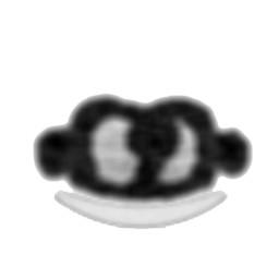
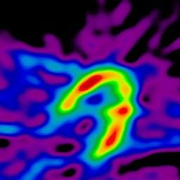
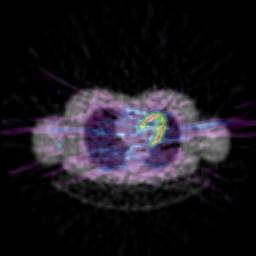
21 Corrections in PET In most nuclear medicine procedures, the goal is to produce an image in which the gray scale or count density is directly proportional to the regional isotope concentration. In order to achieve this in PET it is necessary to apply a number of corrections: Attenuation of photons in tissue Non-uniform response of detector elements Random coincidence events Detection of scattered events Loss of counts at high count rates - dead-time Isotope decay Absolute Calibration & cross calibration with other instruments How accurate these corrections are will have a direct impact on the quantitative measurement. Attenuation Correction M. Dahlbom M284B Winter


22 Attenuation Correction Attenuation Correction In PET imaging of the brain, the shape of the head can be approximated with an ellipse. The dimensions of the fitted ellipse can be estimated by first reconstructing the data without attenuation correction. Then an ellipse is drawn onto the image from which the attenuation correction can be derived. The attenuation correction is the applied to the data and the image is reconstructed again. This method can be fairly time consuming, especially on system producing a large number of transaxial slices. Atten. Corr.. = e µd M. Dahlbom M284B Winter
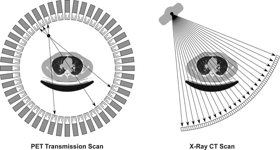

23 Attenuation Correction 68 Ge Source Blank Scan Transmission Scan Without Image Segmentation With Image Segmentation M. Dahlbom M284B Winter


 C TX after MC")
 M.")

24 A Early frame non-ac EM Original TX Fused TX-EM (Match) Early frame AC EM B Late frame non-ac EM Original TX Fused TX-EM (Mismatch) Late frame AC EM (before MC) C Late frame non-ac EM TX after MC Fused TX-EM (Match) Late frame AC EM (after MC) M. Dahlbom M284B Winter


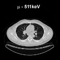
25 PET/CT GE Philips Siemens x H.U. µ 70 kev x M. Dahlbom M284B Winter
26 Time-of-flight PET R R Det 2 Det 1 x R - x R + x s = v t R + x = vt 1 R x = vt 2 2x = v(t 2 t 1 ) x = cδt 2 M. Dahlbom M284B Winter
27 Time-of-flight PET For ideal detectors, TOF would eliminate the need for image reconstruction, since the measurement would allow each event to be accurately positioned in space. All detectors have a finite time resolution, or uncertainty in timing. This translates to an uncertainty in positioning. BGO ~ 5 ns NaI ~ 1.5 ns CsF, LaBr 3 ~ 0.45 ns BaF 2, LSO, LYSO ~ 0.3 ns 75 cm 22.5 cm 6.7 cm 4.5 cm Time-of-flight PET Figure 1. Image elements contributing to a LOR, for conventional PET (left) and TOF PET (right). M. Dahlbom M284B Winter
28 Time-of-flight PET Even with a finite time resolution, using the TOF information an improvement in signal-to-noise ratio (S/N) can be achieved: 2 D D SNR SNR = SNR Δx cδt TOF conv. conv. Time-of-Flight vs. Conventional PET Better information sent to reconstruction Truth Conventional PET Image Formation Time-of-Flight Image Formation More precise localization of annihilation event improves image quality M. Dahlbom M284B Winter
29 Time-of-flight PET s Problems with TOF in the 80 s Poor detection efficiency of available scintillators TOF Gain did not offset the poor efficiency To improve the efficiency, large detector modules were used A more significant gain in S/N could be achieved by using high resolution detectors and conventional detection methods (Phelps, Hoffman, Huang, 1982). Time-of-flight PET Scintillators: CsF, BaF 2 LSO, LYSO - fast, high light, and dense Detectors/PMTs: 1:1 coupling 100:1 crystal encoding - spatial resolution Geometry: 2D (septa) 3D with large axial FOV - sensitivity Reconstruction: Analytic (FBP) iterative (list-mode) - system modeling Electronics: Accurate and stable M. Dahlbom M284B Winter

30 Can we see TOF improvement? non TOF TOF 5 min 3 min 1 min 6-to-1 contrast; 35-cm phantom J. Karp, U of Penn Noticeable improvement with TOF with large size phantom Gemini TF - patient study Rectal carcinoma, metastases in mesentery and bilateral iliac chains 114 kg; BMI = mci; 2 hr post-inj 3min/bed non-tof TOF J. Karp, U of Penn Lesion contrast (SUV) improves with TOF reconstruction M. Dahlbom M284B Winter


31 ME Phelps et. al. DH Silverman et. al. M. Dahlbom M284B Winter


32 DH Silverman et. al. DH Silverman et. al. M. Dahlbom M284B Winter


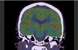
33 Low Grade Brain Tumor MRI FDG FDOPA FLT SUV 3.0 FDOPA Uptake Patterns Striatum Tumor Cerebellum minutes 80 Tumor reaches maximum before striatum M. Dahlbom M284B Winter


 Positrons in Magnetic Field M.")
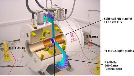
34 Integrated PET/MRI System Images courtesy Bernd Pichler Opportunities: direct and accurate registration of molecular PET signal with high resolution anatomy Anatomically guided analysis of PET data Improved quantification of PET data Good soft tissue contrast, no additional radiation dose time correlation of PET and MRI or MRS signal Interventional, therapeutic studies Dual-labeled agents ( 64 Cu, Gd) Positrons in Magnetic Field M. Dahlbom M284B Winter


35 MR Compatible PET System Animal MR System Concept PET Detectors Magnet Gradient Coils RF coil Simultaneous PET/MRI Imaging Shao Y, Cherry SR, Farahani K, et al. Phys Med Biol 42: ; mm ring diameter 72 2x2x25 mm LSO scintillators 200 g Rat - 18 F-FDG Brain Study M. Dahlbom M284B Winter

36 Challenges in Combining PET and MR imaging PET Detectors affected by: Static magnetic field Rapidly changing gradient field Radiofrequency signals MR affected by PET detectors and electronics B. Pichler et. al., 2008 M. Dahlbom M284B Winter


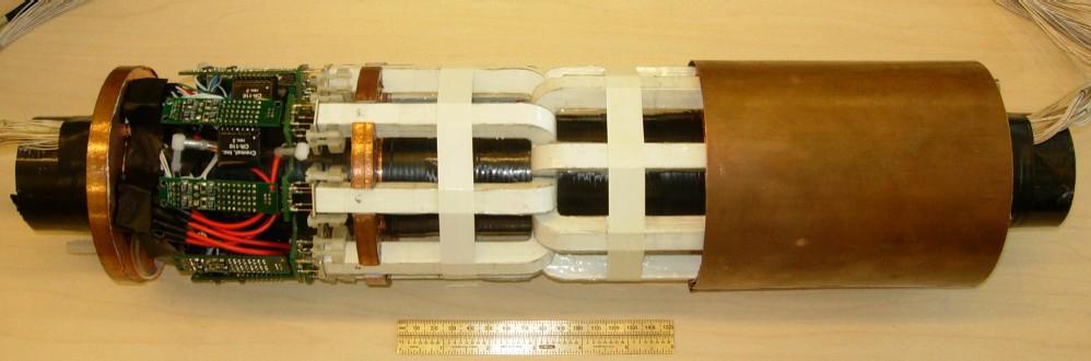
37 Solutions for combining PET-MR MR-Compatible PET Detector Module scintillator array optical fiber bundle PSAPD preamplifiers M. Dahlbom M284B Winter


38 PMT vs. APD/SiPM PET Insert preamplifiers PSAPDs optical fibers scintillator ring M. Dahlbom M284B Winter
 without PET insert (first row) and")
, 2006 Catana et.al., JNM 47 (12), 2006 M.")
39 MR phantom images: GE (left) and SE (right) sequences of a gadopentetate dimeglumine/h2o phantom (T1 = 250 ms) without PET insert (first row) and with PET insert unpowered (second row) and powered (third row). Catana et.al., JNM 47 (12), 2006 Catana et.al., JNM 47 (12), 2006 M. Dahlbom M284B Winter
 detector 6-detector")

40 MR/PET brain scanner prototype (conceptual design) detector 6-detector module (32 modules) gantry patient bed head coil bed rails M. Dahlbom M284B Winter


41 Test Setup n Concentric MR and PET n MR: n CP-TXRX-Head coil inner Diameter 27 cm n RF Shield id 36 cm n PET: n 512 LSO crystals in 2 modules n FOV 3.2 cm n Imaging by phantom rotation n MR/PET phantom n 1.0 mm mm diameter holes n Filled with water and 1.25 g NiSO 4 / litre and about 50 MBq FDG M. Dahlbom M284B Winter


42 SUPPLEMENTAL FIGURE 1 Diagram of the Biograph mmr, depicting how the PET detectors are located within the MR coils. M. Dahlbom M284B Winter


43 PET-MRI Attenuation Correction M. Dahlbom M284B Winter
 and Biograph PET/CT (B) fused views of whole-body 18F-fluoride scan of same patient.")
44 FIGURE 7. mmr PET/MR (A) and Biograph PET/CT (B) fused views of whole-body 18F-fluoride scan of same patient. mmr (C) and Verio (D) T2- weighted coronal views of healthy volunteer. M. Dahlbom M284B Winter
PET Detectors. William W. Moses Lawrence Berkeley National Laboratory March 26, 2002
 PET Detectors William W. Moses Lawrence Berkeley National Laboratory March 26, 2002 Step 1: Inject Patient with Radioactive Drug Drug is labeled with positron (β + ) emitting radionuclide. Drug localizes
Chiara Secco. PET Performance measurements of the new LSO-Based Whole Body PET/CT. Scanner biograph 16 HI-REZ using the NEMA NU Standard.
 Chiara Secco PET Performance measurements of the new LSO-Based Whole Body PET/CT Scanner biograph 16 HI-REZ using the NEMA NU 2-2001 Standard. INTRODUCTION Since its introduction, CT has become a fundamental
LSO PET/CT Pico Performance Improvements with Ultra Hi-Rez Option
 LSO PET/CT Pico Performance Improvements with Ultra Hi-Rez Option Y. Bercier, Member, IEEE, M. Casey, Member, IEEE, J. Young, Member, IEEE, T. Wheelock, Member, IEEE, T. Gremillion Abstract-- Factors which
First Applications of the YAPPET Small Animal Scanner
 First Applications of the YAPPET Small Animal Scanner Guido Zavattini Università di Ferrara CALOR2 Congress, Annecy - FRANCE YAP-PET scanner Scintillator: YAP:Ce Size: matrix of 2x2 match like crystals
PET: New Technologies & Applications, Including Oncology
 PET: New Technologies & Applications, Including Oncology, PhD, FIEEE Imaging Research Laboratory Department of Radiology University of Washington, Seattle, WA Disclosures Research Contract, GE Healthcare
PET Performance Evaluation of MADPET4: A Small Animal PET Insert for a 7-T MRI Scanner
 PET Performance Evaluation of MADPET4: A Small Animal PET Insert for a 7-T MRI Scanner September, 2017 Results submitted to Physics in Medicine & Biology Negar Omidvari 1, Jorge Cabello 1, Geoffrey Topping
Celesteion Time-of-Flight Technology
 Celesteion Time-of-Flight Technology Bing Bai, PhD Clinical Sciences Manager, PET/CT Canon Medical Systems USA Introduction Improving the care for every patient while providing a high standard care to
PET/CT Instrumentation Basics
 / Instrumentation Basics 1. Motivations for / imaging 2. What is a / Scanner 3. Typical Protocols 4. Attenuation Correction 5. Problems and Challenges with / 6. Examples Motivations for / Imaging Desire
Fundamentals of Positron Emission Tomography (PET)
") Fundamentals of Positron Emission Tomography (PET) NPRE 435, Principles of Imaging with Ionizing Radiation, Fall 2017 Content Fundamentals of PET Camera & Detector Design Real World Considerations Performance
Fundamentals of Positron Emission Tomography (PET) NPRE 435, Principles of Imaging with Ionizing Radiation, Fall 2017 Content Fundamentals of PET Camera & Detector Design Real World Considerations Performance
Combined micropet /MR System: Performance Assessment of the Full PET Ring with Split Gradients 4.8
 Combined micropet /MR System: Performance Assessment of the Full PET Ring with Split Gradients 4.8 UNIVERSITY OF CAMBRIDGE 1.2 Rob C. Hawkes 1, Tim D. Fryer 1, Alun J. Lucas 1,2, Stefan B. Siegel 3, Richard
An innovative detector concept for hybrid 4D-PET/MRI Imaging
 Piergiorgio Cerello (INFN - Torino) on behalf of the 4D-MPET* project *4 Dimensions Magnetic compatible module for Positron Emission Tomography INFN Perugia, Pisa, Torino; Polytechnic of Bari; University
New Technology in Nuclear Medicine
 New Technology in Nuclear Medicine Reed G. Selwyn, PhD, DABR Vice Chair of Research & Imaging Sciences Associate Professor and Chief, Medical Physics Dept. of Radiology, University of New Mexico Objectives
LaBr 3 :Ce, the latest crystal for nuclear medicine
 10th Topical Seminar on Innovative Particle and Radiation Detectors 1-5 October 2006 Siena, Italy LaBr 3 :Ce, the latest crystal for nuclear medicine Roberto Pani On behalf of SCINTIRAD Collaboration INFN
Simulation and evaluation of a cost-effective high-performance brain PET scanner.
 Research Article http://www.alliedacademies.org/biomedical-imaging-and-bioengineering/ Simulation and evaluation of a cost-effective high-performance brain PET scanner. Musa S Musa *, Dilber U Ozsahin,
APD Quantum Efficiency
 APD Quantum Efficiency Development of a 64-channel APD Detector Module with Individual Pixel Readout for Submillimeter Spatial Resolution in PET Philippe Bérard a, Mélanie Bergeron a, Catherine M. Pepin
CHAPTER 8 GENERIC PERFORMANCE MEASURES
 GENERIC PERFORMANCE MEASURES M.E. DAUBE-WITHERSPOON Department of Radiology, University of Pennsylvania, Philadelphia, Pennsylvania, United States of America 8.1. INTRINSIC AND EXTRINSIC MEASURES 8.1.1.
Development of PET using 4 4 Array of Large Size Geiger-mode Avalanche Photodiode
 2009 IEEE Nuclear Science Symposium Conference Record M09-8 Development of PET using 4 4 Array of Large Size Geiger-mode Avalanche Photodiode K. J. Hong, Y. Choi, J. H. Kang, W. Hu, J. H. Jung, B. J. Min,
Future directions in Nuclear Medicine Instrumentation
 Future directions in Nuclear Medicine Instrumentation Where are we going - and why? First, the disclosure list My group at the University of Washington has research support from: NIH DOE General Electric
PET Performance Measurements for an LSO- Based Combined PET/CT Scanner Using the National Electrical Manufacturers Association NU Standard
 PET Performance Measurements for an LSO- Based Combined PET/CT Scanner Using the National Electrical Manufacturers Association NU 2-2001 Standard Yusuf E. Erdi, DSc 1 ; Sadek A. Nehmeh, PhD 1 ; Tim Mulnix,
Performance evaluation of a new highsensitivity time-of-flight clinical PET/CT system
 Huo et al. EJNMMI Physics (2018) 5:29 https://doi.org/10.1186/s40658-018-0229-4 EJNMMI Physics ORIGINAL RESEARCH Open Access Performance evaluation of a new highsensitivity time-of-flight clinical PET/CT
Performance characterization of a novel thin position-sensitive avalanche photodiode-based detector for high resolution PET
 2005 IEEE Nuclear Science Symposium Conference Record M11-126 Performance characterization of a novel thin position-sensitive avalanche photodiode-based detector for high resolution PET Jin Zhang, Member,
William Hallet - PRIMA IV 1
 Quantitative and application specific imaging PET: the measurement process,reconstruction, calibration, quantification Dr William Hallett Centre for Imaging Sciences Imperial College Hammersmith Hospital
Primer on molecular imaging technology
 Primer on molecular imaging technology Craig S. Levin Division of Nuclear Medicine, Department of Radiology and Molecular Imaging Program at Stanford (MIPS), Stanford University School of Medicine, 300
Design Studies of A High-Performance Onboard Positron Emission Tomography For Integrated Small Animal PET/CT/RT Radiation Research Systems
 Proceedings of the International MultiConference of Engineers and Computer Scientists 2018 Vol II Design Studies of A High-Performance Onboard Positron Emission Tomography For Integrated Small Animal PET/CT/RT
Monte Carlo Simulation Study of a Dual-Plate PET Camera Dedicated to Breast Cancer Imaging
 IEEE Nuclear Science Symposium Conference Record M-9 Monte Carlo Simulation Study of a Dual-Plate PET Camera Dedicated to Breast Cancer Imaging Jin Zhang, Member, IEEE, Peter D. Olcott, Member, IEEE, Angela
Conceptual Study of Brain Dedicated PET Improving Sensitivity
 Original Article PROGRESS in MEDICAL PHYSICS 27(4), Dec. 2016 https://doi.org/10.14316/pmp.2016.27.4.236 pissn 2508-4445, eissn 2508-4453 Conceptual Study of Brain Dedicated PET Improving Sensitivity Han-Back
Detector technology challenges for nuclear medicine and PET
 Nuclear Instruments and Methods in Physics Research A 513 (2003) 1 7 Detector technology challenges for nuclear medicine and PET Paul K. Marsden Guy s and St. Thomas Clinical PET Centre, King s College
Performance Assessment of Pixelated LaBr 3 Detector Modules for TOF PET
 Performance Assessment of Pixelated LaBr 3 Detector Modules for TOF PET A. Kuhn, S. Surti, Member, IEEE, J. S. Karp, Senior Member, IEEE, G. Muehllehner, Fellow, IEEE, F.M. Newcomer, R. VanBerg Abstract--
MPPC and Liquid Xenon technologies from particle physics to medical imaging
 CANADA S NATIONAL LABORATORY FOR PARTICLE AND NUCLEAR PHYSICS Owned and operated as a joint venture by a consortium of Canadian universities via a contribution through the National Research Council Canada
NM Module Section 2 6 th Edition Christian, Ch. 3
 NM 4303 Module Section 2 6 th Edition Christian, Ch. 3 Gas Filled Chamber Voltage Gas filled chamber uses Hand held detectors cutie pie Geiger counter Dose calibrators Cutie pie Chamber voltage in Ionization
Designing an MR compatible Time of Flight PET Detector Floris Jansen, PhD, Chief Engineer GE Healthcare
 GE Healthcare Designing an MR compatible Time of Flight PET Detector Floris Jansen, PhD, Chief Engineer GE Healthcare There is excitement across the industry regarding the clinical potential of a hybrid
Initial evaluation of the Indiana small animal PET scanner
 Initial evaluation of the Indiana small animal PET scanner Ned C. Rouze, Member, IEEE, Victor C. Soon, John W. Young, Member, IEEE, Stefan Siegel, Member, IEEE, and Gary D. Hutchins, Member, IEEE Abstract
Study of a Prototype VP-PET Imaging System Based on highly. Pixelated CdZnTe Detectors
 Study of a Prototype VP-PET Imaging System Based on highly Pixelated CdZnTe Detectors Zheng-Qian Ye 1, Ying-Guo Li 1, Tian-Quan Wang 1, Ya-Ming Fan 1, Yong-Zhi Yin 1,*, Xi-Meng Chen 1 Affiliations: 1 School
2594 IEEE TRANSACTIONS ON NUCLEAR SCIENCE, VOL. 56, NO. 5, OCTOBER /$ IEEE
 2594 IEEE TRANSACTIONS ON NUCLEAR SCIENCE, VOL. 56, NO. 5, OCTOBER 2009 Investigation of Depth of Interaction Encoding for a Pixelated LSO Array With a Single Multi-Channel PMT Yongfeng Yang, Member, IEEE,
The Influence of Crystal Configuration and PMT on PET Time-of-Flight Resolution
 The Influence of Crystal Configuration and PMT on PET Time-of-Flight Resolution Christopher Thompson Montreal Neurological Institute and Scanwell Systems, Montreal, Canada Jason Hancock Cross Cancer Institute,
A NOVEL CONCEPT FOR A POSITRON EMISSION TOMOGRAPHY SCANNER
 A NOVEL CONCEPT FOR A POSITRON EMISSION TOMOGRAPHY SCANNER An Undergraduate Research Scholars Thesis by BRIAN KELLY, MATTHEW LEE ELLIOT LEVIN and JEENA KHATRI Submitted to Honors and Undergraduate Research
A new operative gamma camera for Sentinel Lymph Node procedure
 A new operative gamma camera for Sentinel Lymph Node procedure A physicist device for physicians Samuel Salvador, Virgile Bekaert, Carole Mathelin and Jean-Louis Guyonnet 12/06/2007 e-mail: samuel.salvador@ires.in2p3.fr
Attenuation Correction in Hybrid MR-BrainPET Imaging
 Mitglied der Helmholtz-Gemeinschaft Attenuation Correction in Hybrid MR-BrainPET Imaging Elena Rota Kops Institute of Neuroscience and Biophysics Medicine Brain Imaging Physics Interactions of 511 kev
Instructions for gg Coincidence with 22 Na. Overview of the Experiment
 Overview of the Experiment Instructions for gg Coincidence with 22 Na 22 Na is a radioactive element that decays by converting a proton into a neutron: about 90% of the time through β + decay and about
Journal of Radiology in press. Simultaneous PET/MR Images, acquired with a Compact MRI Compatible PET Detector in a 7 Tesla Magnet
 Journal of Radiology in press Simultaneous PET/MR Images, acquired with a Compact MRI Compatible PET Detector in a 7 Tesla Magnet Martin S. Judenhofer BS 1, Ciprian Catana 2, Brian, K. Swann 3, Stefan
Development of the LBNL Positron Emission Mammography Camera
 Development of the LBNL Positron Emission Mammography Camera J.S. Huber, Member, IEEE, W.S. Choong, Member, IEEE, J. Wang, Member, IEEE, J.S. Maltz, Member, IEEE, J. Qi, Member, IEEE, E. Mandelli, Member,
Design of a High Resolution and High Sensitivity Scintillation Crystal Array with Nearly Perfect Light Collection
 Design of a High Resolution and High Sensitivity Scintillation Crystal Array with Nearly Perfect Light Collection Craig S. Levin, Member, IEEE Abstract-- Spatial resolution improvements in Positron Emission
Performance measurements of a depth-encoding PET detector module based on positionsensitive
 Home Search Collections Journals About Contact us My IOPscience Performance measurements of a depth-encoding PET detector module based on positionsensitive avalanche photodiode read-out This article has
Focusing on high performance
 Advanced Molecular Imaging Vereos PET/CT Focusing on high performance Michael A. Miller, PhD, Philips, Advanced Molecular Imaging Physics This white paper presents a description of the Vereos digital PET/CT
Discovery ST. An Oncology System Designed For PET/CT. Revision: B Date: 30 Jan Page 1 of 47
 Discovery ST An Oncology System Designed For PET/CT Revision: B Date: 30 Jan 2003 Page 1 of 47 TABLE OF CONTENTS 1 Introduction...3 2 Design Requirements...4 2.1 The Design Objective...4 2.2 Design Philosophy...5
LaBr 3 :Ce scintillation gamma camera prototype for X and gamma ray imaging
 8th International Workshop on Radiation Imaging Detectors Pisa 2-6 July 2006 LaBr 3 :Ce scintillation gamma camera prototype for X and gamma ray imaging Roberto Pani On behalf of SCINTIRAD Collaboration
COMPUTED TOMOGRAPHY 1
 COMPUTED TOMOGRAPHY 1 Why CT? Conventional X ray picture of a chest 2 Introduction Why CT? In a normal X-ray picture, most soft tissue doesn't show up clearly. To focus in on organs, or to examine the
Initial Certification
 Initial Certification Nuclear Medical Physics (NMP) Study Guide Part 2 Content Guide and Sample Questions The content of all ABR exams is determined by a panel of experts who select the items based on
Currently, the spatial resolution of most dedicated smallanimal
 A Prototype High-Resolution Small-Animal PET Scanner Dedicated to Mouse Brain Imaging Yongfeng Yang 1,2, Julien Bec 1, Jian Zhou 1, Mengxi Zhang 1, Martin S. Judenhofer 1, Xiaowei Bai 1, Kun Di 1, Yibao
Radionuclide Imaging MII 3073 RADIONUCLIDE IMAGING SYSTEM
 Radionuclide Imaging MII 3073 RADIONUCLIDE IMAGING SYSTEM Preamplifiers and amplifiers The current from PMT must be further amplified before it can be processed and counted (the number of electrons yielded
Data. microcat +SPECT
 Data microcat +SPECT microcat at a Glance Designed to meet the throughput, resolution and image quality requirements of academic and pharmaceutical research, the Siemens microcat sets the standard for
Quality control of Gamma Camera. By Dr/ Ibrahim Elsayed Saad 242 NMT
 Quality control of Gamma Camera By Dr/ Ibrahim Elsayed Saad 242 NMT WHAT IS QUALITY? The quality of a practice is to fulfill the expectations and demands from: Patient Clinicain Your self Quality assurance
... In vivo imaging in Nuclear Medicine. 1957: Anger camera (X;Y) X Y
 X Y") József Varga, PhD EMISSION IMAGING BASICS OF QUANTIFICATION Imaging devices Aims of image processing Reconstruction University of Debrecen Department of Nuclear Medicine. In vivo imaging in Nuclear Medicine
József Varga, PhD EMISSION IMAGING BASICS OF QUANTIFICATION Imaging devices Aims of image processing Reconstruction University of Debrecen Department of Nuclear Medicine. In vivo imaging in Nuclear Medicine
A High-Resolution GSO-based Brain PET Camera
 A High-Resolution GSO-based Brain PET Camera J.S. Karp', Senior Member IEEE, L.E. Adam', R.Freifelder', Member IEEE, G. Muehllehner3 Senior Member IEEE, F. Liu"', Student Member IEEE, S. Surti"', Student
Investigation of low noise, low cost readout electronics for high sensitivity PET systems based on Avalanche Photodiode arrays
 Investigation of low noise, low cost readout electronics for high sensitivity PET systems based on Avalanche Photodiode arrays Frezghi Habte, Member, IEEE and Craig S.Levin, Member, IEEE Abstract A compact,
Time-of-flight PET with SiPM sensors on monolithic scintillation crystals Vinke, Ruud
 University of Groningen Time-of-flight PET with SiPM sensors on monolithic scintillation crystals Vinke, Ruud IMPORTANT NOTE: You are advised to consult the publisher's version (publisher's PDF) if you
2/14/2019. Nuclear Medicine Artifacts. Symmetric energy windows
 Nuclear Medicine Artifacts SCPMG Medical Imaging Technology & Informatics Medical Physics Group Brian Helbig, MS, DABR 1 2 Symmetric energy windows 3 1 Dynamic clinical study Energy peak shift Electrical
Pitfalls and Remedies of MDCT Scanners as Quantitative Instruments
 intensity m(e) m (/cm) 000 00 0 0. 0 50 0 50 Pitfalls and Remedies of MDCT Scanners as Jiang Hsieh, PhD GE Healthcare Technology University of Wisconsin-Madison Root-Causes of CT Number Inaccuracies Nature
Noise Characteristics of the FORE+OSEM(DB) Reconstruction Method for the MiCES PET Scanner
 Reconstruction Method for the MiCES PET Scanner") Noise Characteristics of the FORE+OSEM(DB) Reconstruction Method for the MiCES PET Scanner Kisung Lee, Member, IEEE, Paul E. Kinahan, Senior Member, Robert S. Miyaoka, Member, IEEE, Jeffrey A. Fessler,
Noise Characteristics of the FORE+OSEM(DB) Reconstruction Method for the MiCES PET Scanner Kisung Lee, Member, IEEE, Paul E. Kinahan, Senior Member, Robert S. Miyaoka, Member, IEEE, Jeffrey A. Fessler,
arxiv: v1 [physics.med-ph] 29 Nov 2018
![arxiv: v1 [physics.med-ph] 29 Nov 2018](/thumbs/91/105290919.jpg "arxiv: v1 [physics.med-ph] 29 Nov 2018") Expected performance of the TT-PET scanner E. Ripiccini, a,b,1 D. Hayakawa, a,b G. Iacobucci, a M. Nessi, a,c E. Nowak, c L. Paolozzi, a O. Ratib, b P. Valerio a and D. Vitturini a a University of Geneva,
Expected performance of the TT-PET scanner E. Ripiccini, a,b,1 D. Hayakawa, a,b G. Iacobucci, a M. Nessi, a,c E. Nowak, c L. Paolozzi, a O. Ratib, b P. Valerio a and D. Vitturini a a University of Geneva,
Performance Evaluation of SiPM Detectors for PET Imaging in the Presence of Magnetic Fields
 2008 IEEE Nuclear Science Symposium Conference Record M02-4 Performance Evaluation of SiPM Detectors for PET Imaging in the Presence of Magnetic Fields Samuel España, Student Member, IEEE, Gustavo Tapias,
PD233: Design of Biomedical Devices and Systems
 PD233: Design of Biomedical Devices and Systems (Lecture-8 Medical Imaging Systems) (Imaging Systems Basics, X-ray and CT) Dr. Manish Arora CPDM, IISc Course Website: http://cpdm.iisc.ac.in/utsaah/courses/
16 Instrumentation and Data Acquisition
 Instrumentation and Data Acquisition 275 16 Instrumentation and Data Acquisition Sibylle I. Ziegler and Magnus Dahlbom CONTENTS 16.1 Detectors and Imaging Systems 275 16.1.1 Principles of Scintillation
Recent Advances in Hybrid Molecular Imaging Systems
 103 Recent Advances in Hybrid Molecular Imaging Systems Jae Sung Lee, PhD 1 Joong Hyun Kim, PhD 2 1 Departments of Nuclear Medicine and Biomedical Sciences, Seoul National University College of Medicine,
Investigation of Multiple Head Registration / Center of Rotation for SPECT Gamma Cameras
 Egyptian J. Nucl. Med., Vol 2, No. 2, Dec. 2009 82 PHYSICS, Original Artical Investigation of Multiple Head Registration / Center of Rotation for SPECT Gamma Cameras Abdelsattar, M.B. Ph.D.; BuHumaid,
How Gamma Camera s Head-Tilts Affect Image Quality of a Nuclear Scintigram?
 November 2014, Volume 1, Number 4 How Gamma Camera s Head-Tilts Affect Image Quality of a Nuclear Scintigram? Hojjat Mahani 1,2, Alireza Kamali-Asl 3, *, Mohammad Reza Ay 2, 4 1. Radiation Application
Design of a High-Resolution and High-Sensitivity Scintillation Crystal Array for PET With Nearly Complete Light Collection
 2236 IEEE TRANSACTIONS ON NUCLEAR SCIENCE, VOL. 49, NO. 5, OCTOBER 2002 Design of a High-Resolution and High-Sensitivity Scintillation Crystal Array for PET With Nearly Complete Light Collection Craig
Recovery and normalization of triple coincidences in PET
 Universidad Carlos III de Madrid Repositorio institucional e-archivo Área de Imagen e Instrumentación (BiiG) http://e-archivo.uc3m.es DBIAB - BIIG - Journal Articles 2015-03 Recovery and normalization
PET has evolved from a research tool for studying
 Virtual-Pinhole PET Yuan-Chuan Tai 1,2, Heyu Wu 1, Debashish Pal 3, and Joseph A. O Sullivan 4 1 Department of Radiology, Washington University, St. Louis, Missouri; 2 Alvin J. Siteman Cancer Center, Washington
The PennPET Explorer Scanner for Total Body Applications
 The PennPET Explorer Scanner for Total Body Applications JS Karp, MJ Geagan, G Muehllehner, ME Werner, T McDermott, JP Schmall, V Viswanath, University of Pennsylvania, Philadelphia, PA AE Perkins, C-H
2010 Philips BrightView XCT SPECT/CT
 2010 Philips BrightView XCT SPECT/CT Unit was purchased from Philips training center in 2015. Installed but never been used by the current facility. (Scroll for pictures) BrightView XCT Camera with PinPoint
Introduction. Chapter 16 Diagnostic Radiology. Primary radiological image. Primary radiological image
 Introduction Chapter 16 Diagnostic Radiology Radiation Dosimetry I Text: H.E Johns and J.R. Cunningham, The physics of radiology, 4 th ed. http://www.utoledo.edu/med/depts/radther In diagnostic radiology
Photomultiplier Tube
 Nuclear Medicine Uses a device known as a Gamma Camera. Also known as a Scintillation or Anger Camera. Detects the release of gamma rays from Radionuclide. The radionuclide can be injected, inhaled or
UCLA UCLA Previously Published Works
 UCLA UCLA Previously Published Works Title Attenuation correction for small animal PET tomographs Permalink https://escholarship.org/uc/item/41n377p3 Journal Physics in Medicine and Biology, 5(8) ISSN
764 IEEE TRANSACTIONS ON NUCLEAR SCIENCE, VOL. 51, NO. 3, JUNE 2004
 764 IEEE TRANSACTIONS ON NUCLEAR SCIENCE, VOL. 51, NO. 3, JUNE 2004 Study of Low Noise Multichannel Readout Electronics for High Sensitivity PET Systems Based on Avalanche Photodiode Arrays Frezghi Habte,
Design, Calibration, and Evaluation of Depth-of- Interaction-Capable PET Detector Modules
 Texas Medical Center Library DigitalCommons@TMC UT GSBS Dissertations and Theses (Open Access) Graduate School of Biomedical Sciences 12-2012 Design, Calibration, and Evaluation of Depth-of- Interaction-Capable
Attenuation length in strip scintillators. Jonathan Button, William McGrew, Y.-W. Lui, D. H. Youngblood
 Attenuation length in strip scintillators Jonathan Button, William McGrew, Y.-W. Lui, D. H. Youngblood I. Introduction The ΔE-ΔE-E decay detector as described in [1] is composed of thin strip scintillators,
Image Quality Assessment of Pixellated Systems
 Image Quality Assessment of Pixellated Systems Andreas Goedicke, Herfried Wieczorek, Henrik Botterweck, Wolfgang Eckenbach, Ling Shao, Member, IEEE, Micheal Petrillo, Member, IEEE, Jinghan Ye, and John
Usefulness of noise adaptive non-linear Gaussian filter in FDG-PET study
 ORIGINAL ARTICLE Annals of Nuclear Medicine Vol. 19, No. 6, 469 477, 2005 Usefulness of noise adaptive non-linear Gaussian filter in FDG-PET study Makoto NAGAYOSHI,*, ** Kenya MURASE,* Kouichi FUJINO,**
Study of Silicon Photomultipliers for Positron Emission Tomography (PET) Application
 Application") Study of Silicon Photomultipliers for Positron Emission Tomography (PET) Application Eric Oberla 5 June 29 Abstract A relatively new photodetector, the silicon photomultiplier (SiPM), is well suited for
Study of Silicon Photomultipliers for Positron Emission Tomography (PET) Application Eric Oberla 5 June 29 Abstract A relatively new photodetector, the silicon photomultiplier (SiPM), is well suited for
Cross-Strip Multiplexed Electro-Optical Coupled Scintillation Detector for Integrated PET/MRI
 IEEE TRANSACTIONS ON NUCLEAR SCIENCE 1 Cross-Strip Multiplexed Electro-Optical Coupled Scintillation Detector for Integrated PET/MRI Peter D. Olcott, Member, IEEE, GaryGlover, Member, IEEE, and CraigS.Levin,
Positron Emission Tomography
 Positron Emission Tomography UBC Physics & Astronomy / PHYS 409 1 Introduction Positron emission tomography (PET) is a non-invasive way to produce the functional 1 image of a patient. It works by injecting
Time-of-flight PET with SiPM sensors on monolithic scintillation crystals Vinke, Ruud
 University of Groningen Time-of-flight PET with SiPM sensors on monolithic scintillation crystals Vinke, Ruud IMPORTANT NOTE: You are advised to consult the publisher's version (publisher's PDF) if you
Scintillation Counters
 PHY311/312 Detectors for Nuclear and Particle Physics Dr. C.N. Booth Scintillation Counters Unlike many other particle detectors, which exploit the ionisation produced by the passage of a charged particle,
Assessment of Image Quality of a PET/CT scanner for a Standarized Image situation Using a NEMA Body Phantom
 Assessment of Image Quality of a PET/CT scanner for a Standarized Image situation Using a NEMA Body Phantom The impact of Different Image Reconstruction Parameters on Image quality by QUAYE MICHAEL This
Lightburst Digital Detector
 GE Healthcare Lightburst Digital Detector INTRODUCTION In clinical practice, PET/CT imaging helps clinicians visualize disease at an early stage, before it metastasizes and involves other organs, tissues
VISTA-CT USER MANUAL. GE Healthcare 3000 N. Grandview Blvd Waukesha, WI USA
 VISTA-CT USER MANUAL GE Healthcare 3000 N. Grandview Blvd Waukesha, WI 53188 USA 2 VISTA-CT User s Manual Version 3.3.4 January, 2007 Copyright 2007 By Trident Imaging, Inc. All rights reserved Note: Information
Gamma Ray Spectroscopy with NaI(Tl) and HPGe Detectors
 and HPGe Detectors") Nuclear Physics #1 Gamma Ray Spectroscopy with NaI(Tl) and HPGe Detectors Introduction: In this experiment you will use both scintillation and semiconductor detectors to study γ- ray energy spectra. The
Nuclear Physics #1 Gamma Ray Spectroscopy with NaI(Tl) and HPGe Detectors Introduction: In this experiment you will use both scintillation and semiconductor detectors to study γ- ray energy spectra. The
PET is a noninvasive, diagnostic imaging technique that
 Performance Measurement of the micropet Focus 120 Scanner Jin Su Kim 1,2, Jae Sung Lee 1,2, Ki Chun Im 3, Su Jin Kim 1,2, Seog-Young Kim 3, Dong Soo Lee 1,2, and Dae Hyuk Moon 3 1 Department of Nuclear
4 Time walk correction for TOF-PET detectors based on a monolithic scintillation crystal coupled to a photosensor array
 4 Time walk correction for TOF-PET detectors based on a monolithic scintillation crystal coupled to a photosensor array This chapter has been published as: R. Vinke, H. Löhner, D. Schaart, H. van Dam,
GS Introduction to Medical Physics IV Laboratory 5 Gamma Camera Characteristics
 GS02 0193 Introduction to Medical Physics IV Laboratory 5 Gamma Camera Characteristics Purpose: To introduce some of the basic characteristics of a gamma camera. This lab will introduce gamma camera QC
Robert Pagnanelli BSRT(R)(N), CNMT, NCT, FASNC Chief Technologist, Nuclear Imaging Duke University Medical Center. Thursday September 8, 2011
(N), CNMT, NCT, FASNC Chief Technologist, Nuclear Imaging Duke University Medical Center. Thursday September 8, 2011") Robert Pagnanelli BSRT(R)(N), CNMT, NCT, FASNC Chief Technologist, Nuclear Imaging Duke University Medical Center Thursday September 8, 2011 Quality Control Quality control should be performed because:
Robert Pagnanelli BSRT(R)(N), CNMT, NCT, FASNC Chief Technologist, Nuclear Imaging Duke University Medical Center Thursday September 8, 2011 Quality Control Quality control should be performed because:
IEEE TRANSACTIONS ON NUCLEAR SCIENCE, VOL. 52, NO. 3, JUNE Investigation of the Block Effect on Spatial Resolution in PET Detectors
 IEEE TRANSACTIONS ON NUCLEAR SCIENCE, VOL. 52, NO. 3, JUNE 2005 599 Investigation of the Block Effect on Spatial Resolution in PET Detectors Nada Tomic, Student Member, IEEE, Christopher J. Thompson, Member,
12/21/2016. Siemens Medical Systems Research Agreement Philips Healthcare Research Agreement AAN and ASN Committees
 Joseph V. Fritz, PhD Nandor Pintor, MD Dent Neurologic Institute ASN 2017 Friday, January 20, 2017 Siemens Medical Systems Research Agreement Philips Healthcare Research Agreement AAN and ASN Committees
Design of a Static Full-Ring Multi-Pinhole Collimator for Brain SPECT
 Design of a Static Full-Ring Multi-Pinhole Collimator for Brain SPECT Karen Van Audenhaege, Student Member, IEEE, Roel Van Holen, Member, IEEE, Karel Deprez, Joel S. Karp, Senior Member, IEEE, Scott Metzler,
Radionuclide Imaging MII Single Photon Emission Computed Tomography (SPECT)
") Radionuclide Imaging MII 3073 Single Photon Emission Computed Tomography (SPECT) Single Photon Emission Computed Tomography (SPECT) The successful application of computer algorithms to x-ray imaging in
Radionuclide Imaging MII 3073 Single Photon Emission Computed Tomography (SPECT) Single Photon Emission Computed Tomography (SPECT) The successful application of computer algorithms to x-ray imaging in
SOLID state photodiode and avalanche photodiode scintillation
 2007 IEEE Nuclear Science Symposium Conference Record M14-1 Data acquisition system design for a 1 mm 3 resolution PSAPD-based PET system Peter D. Olcott,,Student Member, IEEE, Frances W. Y. Lau, Student
Multimodal Co-registration Using the Quantum GX, G8 PET/CT and IVIS Spectrum Imaging Systems
 TECHNICAL NOTE Preclinical In Vivo Imaging Authors: Jen-Chieh Tseng, Ph.D. Jeffrey D. Peterson, Ph.D. PerkinElmer, Inc. Hopkinton, MA Multimodal Co-registration Using the Quantum GX, G8 PET/CT and IVIS
Study of the performance of a novel 1 mm resolution dual-panel PET camera design dedicated to breast cancer imaging using Monte Carlo simulation
 Study of the performance of a novel 1 mm resolution dual-panel PET camera design dedicated to breast cancer imaging using Monte Carlo simulation Jin Zhang, a Peter D. Olcott, b Garry Chinn, c Angela M.
A PET detector module using FPGA-only MVT digitizers
 A PET detector module using FPGA-only MVT digitizers Daoming Xi, Student Member, IEEE, Chen Zeng, Wei Liu, Student Member, IEEE, Xiang Liu, Lu Wan, Student Member, IEEE, Heejong Kim, Member, IEEE, Luyao