Advanced Live Cell Imaging
|
|
|
- Collin Jenkins
- 6 years ago
- Views:
Transcription
 is the non-radiative transfer of photon energy from an")
.")
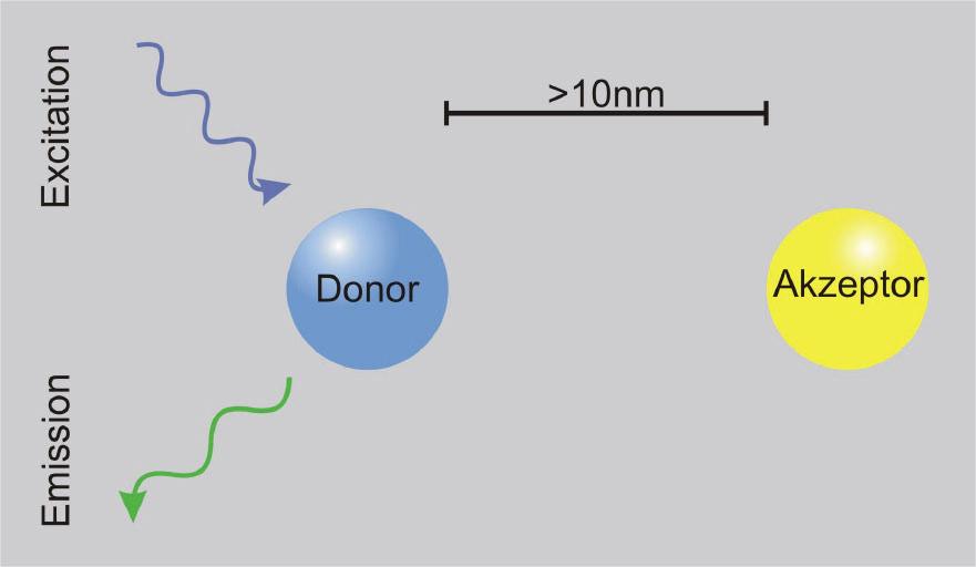
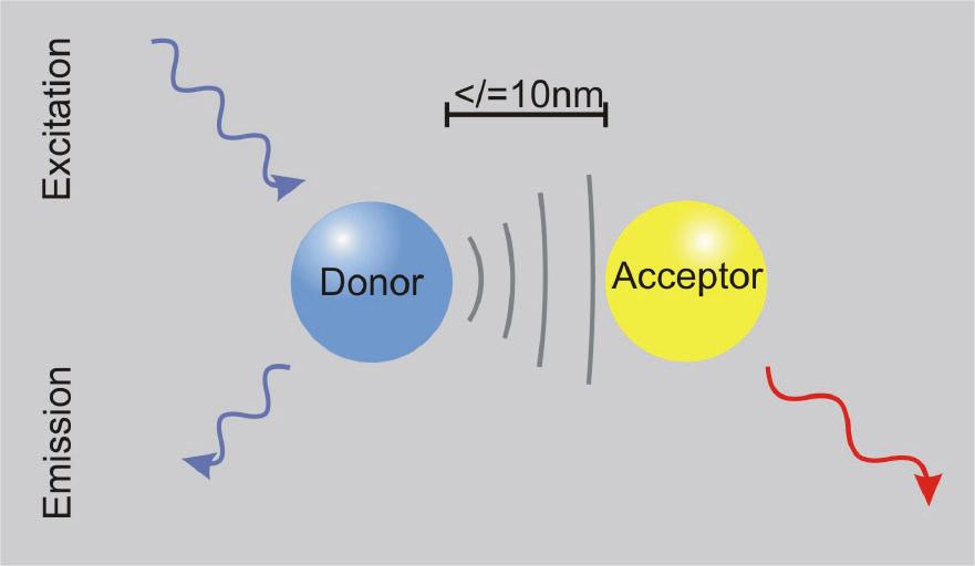
1 FRET Analysis in Laser Scanning Microscopy What is FRET? FRET (fluorescence resonance energy transfer) is the non-radiative transfer of photon energy from an excited fluorophore (the donor) to another fluorophore (the acceptor) when both are located within close proximity (1-10 nm). FRET applications Protein/protein interactions Detection of conformational changes Specialized FRET tools like yellow Chameleon for Ca++ Imaging


2 Preconditions for FRET Analysis Appropriate FRET pair with overlap between donor emission and acceptor excitation Specific staining of the molecule (protein) of interest Parallel orientation of the axis of interacting dye molecules EBFP & EGFP ECFP & EYFP EGFP & Rhodamine FITC & Rhodamine FITC & CY3 FRET CFP Em YFP Ex FRET


")

3 Quantitative Analysis using Filter FRET Track 1 Excitation of CFP Detection of CFP Excitation of YFP Detection of YFP Excitation of CFP Detection of YFP FRET signal (not corrected) CFP only YFP only CFP and YFP doing FRET

4 Sensitized Emission Calculation of Fc Fa capital letter : Filter Set small letter : Specimen Fc Fd Fa = Ff Df Af Dd Aa
5 Sensitized Emission Calculation of Fc Fd/Dd is a measure for the ratio of donor signal detected in the donor channel and emission crosstalk of donor signal detected in the FRET channel. Once determined for an experiment this value remains constant. Fa capital letter : Filter Set small letter : Specimen Fc Fd Fa = Ff Df Af Dd Aa
6 Sensitized Emission Calculation of Fc Fa/Aa is a measure for the ratio of acceptor signal detected in the acceptor channel and excitation crosstalk of acceptor signal detected in the FRET channel. Once determined for an experiment this value remains constant. Fa capital letter : Filter Set small letter : Specimen Fc Fd Fa = Ff Df Af Dd Aa
7 Sensitized Emission Calculation of Fc In a FRET experiment the values Df and Af correlate with donor and acceptor concentration respectively. Multiplied with the previously determined ratios Fd/Dd and Fa/Aa, the FRET value Ff can be corrected to get Fc. Fa capital letter : Filter Set small letter : Specimen Fc Fd Fa = Ff Df Af Dd Aa

8 Sensitized Emission The 3 Methods Method 1: Fc (FRETcorrected) D.C. Youvan et al Fc is corrected for donor and acceptor contribution to the signal measured with the FRET filter set. Fc is not normalized for the donor acceptor concentration. High Fc numbers occur were high concentration of donor and acceptor are present. Fc Fd Fa = Ff Df Af Dd Aa Fc = Ff [ Donor corr. ] [ Acc. corr. ]

9 Sensitized Emission The 3 Methods Method 2: Fn (FRET net) G.W. Gordon et al Fn is corrected for donor and acceptor contribution to the signal measured with the FRET filter set as Fc. Fn is given as Fc divided by the multiplied concentrations of donor and acceptor. This emphasize FRET occurring at low concentrations of donor and acceptor. Fn = Ff [ Donor corr. ] [ Acc. corr. ] G Df Af
10 Sensitized Emission The 3 Methods Method 3: NF (normalized FRET) X. Xia et al NF is corrected for donor and acceptor contribution to the signal measured with the FRET filter set as Fc. NF is given as Fc divided by the square root of the multiplied concentrations of donor and acceptor. This results in FRET values normalized for donor and acceptor concentration. NF = Ff [ Donorcorr. ] [ Acc. corr. ] G Df Af
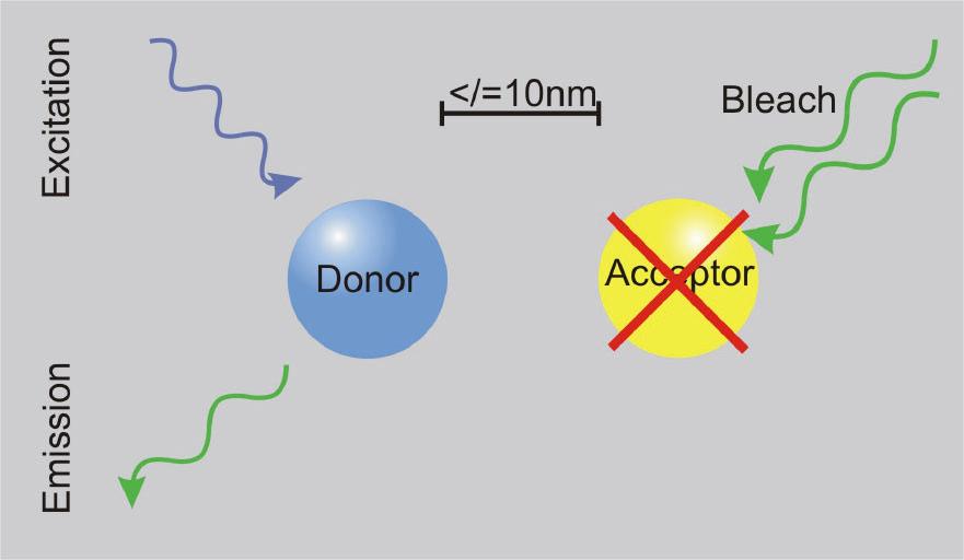
11 Quantitative FRET Analysis using Acceptor Bleach
 The donor signal increases (up to 30%) since no energy transfer to the acceptor is")
12 Quantitative FRET Analysis using Acceptor Bleach Principle Some donor (CFP) signal is transferred (FRET) to the acceptor (YFP) The acceptor is bleached (chemically destroyed) The donor signal increases (up to 30%) since no energy transfer to the acceptor is possible.

13 Quantitative FRET Analysis using Acceptor Bleach
14 Quantitative FRET Analysis using Acceptor Bleach

15 Quantitative FRET Analysis using Acceptor Bleach 100% YFP CFP in% 100% CFP X% YFP
16 Quantitative FRET Analysis using Acceptor Bleach CFPmax at total YFP bleaching Experimental Conditions Use non bleaching laser intensities of 458 and 514nm for CFP and YFP imaging Bleach YFP from 100 to 10% with 100% power of 514nm laser line E = F CFP max CFP min = F F CFP max 30% Apply linear regression analyses to yield values for CFP intensities without acceptor (F CFP-max at YFP = 0) Lit.: H.Amiri et al. in Cell Calcium (2003)
17 Methods and Systems for Quantitative FRET Analysis in LSM System Configuration LSM 5 PASCAL/510 plus AxioCam LSM 5 PASCAL LSM 510 LSM 510 META LSM 510 with FLIM Module FRET Analysis Method Sensitized Emission (Filter FERT) via FRET Macro Acceptor Photobleaching via FERT Macro Acceptor Photobleaching via manual calculation Fluorescence Lifetime FRET X/X X X X - (X)/X (X) X X - -/- - - X - -/ X - Calculation via FRET Macro requires Rel. 3.2 Add On - With the LSM 5 PASCAL no real regions of interest can be applied for bleaching - Calculation of Lifetime FRET requires external Hard and Software
Confocal Microscopy. Kristin Jensen
 Confocal Microscopy Kristin Jensen 17.11.05 References Cell Biological Applications of Confocal Microscopy, Brian Matsumoto, chapter 1 Studying protein dynamics in living cells,, Jennifer Lippincott-Schwartz
Shreyash Tandon M.S. III Year
 Shreyash Tandon M.S. III Year 20091015 Confocal microscopy is a powerful tool for generating high-resolution images and 3-D reconstructions of a specimen by using point illumination and a spatial pinhole
Multifluorescence The Crosstalk Problem and Its Solution
 Multifluorescence The Crosstalk Problem and Its Solution If a specimen is labeled with more than one fluorochrome, each image channel should only show the emission signal of one of them. If, in a specimen
TN378: Openlab Module - FRET. Topic. Discussion
 TN378: Openlab Module - FRET Topic This technical note describes the use of the Openlab FRET module in Openlab 3.1.4 and higher. Users of Openlab Server systems will require Openlab Server 3.0.1 or higher
Dynamic Confocal Imaging of Living Brain. Advantages and risks of multiphoton microscopy in physiology
 Dynamic Confocal Imaging of Living Brain Advantages and risks of multiphoton microscopy in physiology Confocal laser scanning microscopy In conventional optical microscopy focused and out-offocus light
Last updated: May 2014 Y.DeGraaf
 FLINDERS MICROSCOPY BIOMEDICAL SERVICES AVAILABLE MICROSCOPES AND SPECIFICATIONS & INFORMATION REGARDING TRAINING FOR NEW USERS Last updated: May 2014 Y.DeGraaf If you have new staff or students (Honours/Masters
LSM 510 META in Chang Gung University
 Content LSM 510 META in Chang ung University LSM 510 META 路 理 The features and applications of LSM 510 META 01-09 Introduction of the hardware 10-12 Fluorescence observation in conventional microscope
ADVANCED METHODS FOR CONFOCAL MICROSCOPY II. Jean-Yves Chatton Sept. 2006
 ADVANCED METHODS FOR CONFOCAL MICROSCOPY II Jean-Yves Chatton Sept. 2006 Workshop outline Confocal microscopy of living cells and tissues X-Z scanning Time series Bleach: FRAP, photoactivation Emission
April 2009 No.04 WIDEFIELD APPLICATION LETTER. resolution. FRET Sensitized Emission Wizard Widefield
 April 2009 No.04 WIDEFIELD APPLICATION LETTER resolution FRET Sensitized Emission Wizard Widefield FRET SE with the Leica Advanced Widefield Systems AF7000, AF6500 and AF6000 FRET Sensitized Emission (FRET
長庚大學共軛焦顯微鏡課程 長庚大學共軛焦顯微鏡課程. Spot light 長庚大學
 長庚大學共軛焦顯微鏡課程 Spot light 長庚大學共軛焦顯微鏡課程 20071030 長庚大學 Basic principle of Laser Scanning Confocal Microscopy The application of LSM 510 META detector Multiphoton microscopy basic principle and introduction
FLIM on a wide field fluorescence microscope
 FLIM on a wide field fluorescence microscope L.K. van Geest, K.W.J. Stoop Lambert Instruments, Turfweg 4, 933TH Leutingewolde, The Netherlands. Phone: +3 50 50846, Fax: +3 50 500034, e-mail: lkvgeest@lambert-instruments.com
Why and How? Daniel Gitler Dept. of Physiology Ben-Gurion University of the Negev. Microscopy course, Michmoret Dec 2005
 Why and How? Daniel Gitler Dept. of Physiology Ben-Gurion University of the Negev Why use confocal microscopy? Principles of the laser scanning confocal microscope. Image resolution. Manipulating the
Travel to New Dimensions- LSM 880. The Resolution of a Microscope is limited. The Resolution of a Microscope is limited. Image. Image. Object.
 Travel to New Dimensions- LSM 880 LSM 880: The Power of Sensitivity Our Latest Member of the LSM 880 with GaAsP Detectors Sensitivity, and Ease of Use Innovative High-End Laser Scanning Microscopes from
Confocal Laser Scanning Microscopy
 Name of the Core Facility: Confocal Laser Scanning Microscopy CORE Forschungszentrum Immunologie Mainz Welcome to the CSLM Core Facility: The CLSM Core Facility enables working groups to incorporate high
ZEISS LSM510META confocal manual
 ZEISS LSM510META confocal manual Switching on the system 1) Switch on the Remote Control button located on the table to the right of the microscope. This is the main switch for the whole system including
Zeiss 780 Training Notes
 Zeiss 780 Training Notes Turn on Main Switch, System PC and Components Switches 780 Start up sequence Do you need the argon laser (458, 488, 514 nm lines)? Yes Turn on the laser s main power switch and
Quick Start Guide. Leica SP5 X
 Quick Start Guide Leica SP5 X Please note: Some of the information in this guide was taken from Leica Microsystems Leica TCS SP5 LAS AF Guide for New Users. This work is licensed under the Creative Commons
MAKE SURE YOUR SLIDES ARE CLEAN (TOP & BOTTOM) BEFORE LOADING DO NOT LOAD SLIDES DURING SOFTWARE INITIALIZATION
 BEFORE LOADING DO NOT LOAD SLIDES DURING SOFTWARE INITIALIZATION") Olympus VS120-L100 Slide Scanner Standard Operating Procedure Startup 1) Red power bar switch (behind monitor) 2) Computer 3) Login: UserVS120 account (no password) 4) Double click: WAIT FOR INITIALIZATION
Olympus VS120-L100 Slide Scanner Standard Operating Procedure Startup 1) Red power bar switch (behind monitor) 2) Computer 3) Login: UserVS120 account (no password) 4) Double click: WAIT FOR INITIALIZATION
LSM 510 META Laser Scanning Microscope Fluorescence Signals Reliably Separated
 Microscopy from Carl Zeiss LSM 510 META Laser Scanning Microscope Fluorescence Signals Reliably Separated Highlights of Laser Scanning Microscopy 1982 The first Laser Scanning Microscope from Carl Zeiss.
TRAINING MANUAL. Multiphoton Microscopy LSM 510 META-NLO
 TRAINING MANUAL Multiphoton Microscopy LSM 510 META-NLO September 2010 Multiphoton Microscopy Training Manual Multiphoton microscopy is only available on the LSM 510 META-NLO system. This system is equipped
Introduction to light microscopy
 Center for Microscopy and Image Anaylsis Introduction to light Imaging with light / Overview of techniques Urs Ziegler ziegler@zmb.uzh.ch Light interacting with matter Absorbtion Refraction Diffraction
1 Co Localization and Working flow with the lsm700
 1 Co Localization and Working flow with the lsm700 Samples -1 slide = mousse intestine, Dapi / Ki 67 with Cy3/ BrDU with alexa 488. -1 slide = mousse intestine, Dapi / Ki 67 with Cy3/ no BrDU (but with
Microscopy Live Animal Imaging
 Microscopy Live Animal Imaging A collaborative environment that provides the knowledge, instruments, and expertise needed to visualize life at scales ranging from single molecules to entire animals. Project
Microscopy from Carl Zeiss LSM 710. The Power of Sensitivity. A New Dimension in Confocal Laser Scanning Microscopy
 Microscopy from Carl Zeiss LSM 710 The Power of Sensitivity A New Dimension in Confocal Laser Scanning Microscopy Sensitivity Is the Key Whether it is in live cell imaging, single molecule analysis or
presented at: EMBO Practical Course "Light Microscopy of Live Specimens" EMBL Heidelberg, May 2002 ABSTRACT 1. INTRODUCTION
 LIFA System for fluorescence lifetime imaging microscopy (FLIM) Karel W.J. Stoop *, Lambertus K. van Geest **, Cornelis J. R. van der Oord *** Lambert Instruments presented at: EMBO Practical Course "Light
The Next Level in Live Cell Imaging. Digital Imaging Solutions / Family
 The Next Level in Live Cell Imaging Digital Imaging Solutions / Family INTRODUCTION / CONTENTSINTRODUCTION / CONTENTS 2 / MODULAR IMAGING STATIONS LOOKING BEYOND THE IMAGE Dynamic processes and structural
Confocal, hyperspectral, spinning disk
 Confocal, hyperspectral, spinning disk Administrative HW 6 due on Fri Midterm on Wed Covers everything since previous midterm 8.5 x 11 sheet allowed, 1 side Guest lecture by Joe Dragavon on Mon 10/30 Last
Calculate Ratiometric FRET-Images with the FRET-Image-Script
 Tutorial Calculate Ratiometric FRET-Images with the FRET-Image-Script Summary This tutorial shows step-by-step, how the "FRET Image" script of SymPhoTime 64 can be used to calculate pixel-by-pixel the
SHORT INSTRUCTIONS FOR OPERATING LSM1/2 (Zeiss LSM510) AT CIAN Version 1.4, September 2014
 AT CIAN Version 1.4, September 2014") CIAN LSM1 or LSM2 short instructions, version 1.4, September 2014 page 1 of 6 SHORT INSTRUCTIONS FOR OPERATING LSM1/2 (Zeiss LSM510) AT CIAN Version 1.4, September 2014 Before starting To work with LSM1
CIAN LSM1 or LSM2 short instructions, version 1.4, September 2014 page 1 of 6 SHORT INSTRUCTIONS FOR OPERATING LSM1/2 (Zeiss LSM510) AT CIAN Version 1.4, September 2014 Before starting To work with LSM1
Bi Imaging. Multicolor Imaging: The Important Question of Co-Localization. Anna Smallcombe Bio-Rad Laboratories, Hemel Hempstead, UK
 Multicolor Imaging: The Important Question of Co-Localization Anna Smallcombe Bio-Rad Laboratories, Hemel Hempstead, UK The use of specific fluorescent probes, combined with confocal or multiphoton microscopy
Opterra II Multipoint Scanning Confocal Microscope. Innovation with Integrity
 Opterra II Multipoint Scanning Confocal Microscope Enabling 4D Live-Cell Fluorescence Imaging through Speed, Sensitivity, Viability and Simplicity Innovation with Integrity Fluorescence Microscopy The
Fundamentals of Light Microscopy II: Fluorescence, Deconvolution, Confocal, Multiphoton, Spectral microscopy. Integrated Microscopy Course
 Fundamentals of Light Microscopy II: Fluorescence, Deconvolution, Confocal, Multiphoton, Spectral microscopy Integrated Microscopy Course Review Lecture 1: Microscopy Basics Light train Kohler illumination*
Zeiss LSM880 Operating Instructions. UTMB Optical Microscopy Core Jan. 16, 2018
 Zeiss LSM880 Operating Instructions UTMB Optical Microscopy Core Jan. 16, 2018 1 1. Power up the microscope Sing the LOGBOOK Steps below will provide power to the computer and all of the microscope components.
Bio 407. Applied microscopy. Introduction into light microscopy. José María Mateos. Center for Microscopy and Image Analysis
 Center for Microscopy and Image Analysis Bio 407 Applied Introduction into light José María Mateos Fundamentals of light Compound microscope Microscope composed of an objective and an additional lens (eyepiece,
Multi-wavelength TCSPC lifetime imaging Wolfgang Becker a, Axel Bergmann a, Christoph Biskup b, Thomas Zimmer b, Nikolaj Klöcker c, Klaus Benndorf b
 Multi-wavelength TCSPC lifetime imaging Wolfgang Becker a, Axel Bergmann a, Christoph Biskup b, Thomas Zimmer b, Nikolaj Klöcker c, Klaus Benndorf b a Becker & Hickl GmbH, Nahmitzer Damm 30, D-12277 Berlin,
LSM 5 EXCITER Laser Scanning Microscope
 Microscopy from Carl Zeiss LSM 5 EXCITER Laser Scanning Microscope Tracking of Cellular Processes We make it visible. The LSM 5 EXCITER from Carl Zeiss is a confocal laser scanning microscope for fundamental
Sensitive imaging of spectrally overlapping fluorochromes using the LSM 510 META
 Invited Paper Sensitive imaging of spectrally overlapping fluorochromes using the LSM 510 META Mary E. Dickinson a*, Christopher W. Waters a, Gregory Bearman b, Ralf Wolleschensky c, Sebastian Tille d
Components of confocal and two-photon microscopes
 Components of confocal and two-photon microscopes Internal training 07/04/2016 A. GRICHINE Platform Optical microscopy Cell imaging, IAB, ISdV Plan Confocal laser scanning microscope o o o Principle Main
LSM 710 Confocal Microscope Standard Operation Protocol
 LSM 710 Confocal Microscope Standard Operation Protocol Basic Operation Turning on the system 1. Switch on Main power switch 2. Switch on System / PC power button 3. Switch on Components power button 4.
5/4/2015 INTRODUCTION TO LIGHT MICROSCOPY. Urs Ziegler MICROSCOPY WITH LIGHT. Image formation in a nutshell. Overview of techniques
 INTRODUCTION TO LIGHT MICROSCOPY Urs Ziegler ziegler@zmb.uzh.ch MICROSCOPY WITH LIGHT INTRODUCTION TO LIGHT MICROSCOPY Image formation in a nutshell Overview of techniques Widefield microscopy Resolution
Supplementary Figure S1: Schematic view of the confocal laser scanning STED microscope used for STED-RICS. For a detailed description of our
 Supplementary Figure S1: Schematic view of the confocal laser scanning STED microscope used for STED-RICS. For a detailed description of our home-built STED microscope used for the STED-RICS experiments,
6/3/15. The Anatomy of a Digital Image. Representative Intensities. Specimen: (molecular distribution)
") 2015 LMIC Imaging Workshop Sidney L. Shaw Technical Director An introduction of concepts for Super-Resolution Light Microscopy The Anatomy of a Digital Image Representative Intensities Specimen: (molecular
2015 LMIC Imaging Workshop Sidney L. Shaw Technical Director An introduction of concepts for Super-Resolution Light Microscopy The Anatomy of a Digital Image Representative Intensities Specimen: (molecular
Maria Smedh, Centre for Cellular Imaging. Maria Smedh, Centre for Cellular Imaging
 Nonlinear microscopy I: Two-photon fluorescence microscopy Multiphoton Microscopy What is multiphoton imaging? Applications Different imaging modes Advantages/disadvantages Scattering of light in thick
STORM/ PALM ANSWER KEY
 STORM/ PALM ANSWER KEY Phys598BP Spring 2016 University of Illinois at Urbana-Champaign Questions for Lab Report 1. How do you define a resolution in STORM imaging? If you are given a STORM setup, how
Precision-tracking of individual particles By Fluorescence Photo activation Localization Microscopy(FPALM) Presented by Aung K.
 Presented by Aung K.") Precision-tracking of individual particles By Fluorescence Photo activation Localization Microscopy(FPALM) Presented by Aung K. Soe This FPALM research was done by Assistant Professor Sam Hess, physics
Precision-tracking of individual particles By Fluorescence Photo activation Localization Microscopy(FPALM) Presented by Aung K. Soe This FPALM research was done by Assistant Professor Sam Hess, physics
Locating Molecules Using GSD Technology Project Folders: Organization of Experiment Files...1
 .....................................1 1 Project Folders: Organization of Experiment Files.................................1 2 Steps........................................................................2
Microscope Confocal LSM510 META
 Microscope Confocal LSM510 META Welcome to the Zeiss LSM 510 Meta Confocal tutorial. Before using the LSM 510 META, Log off any other computer that is open with your personal login. You will need to put
Operating Instructions for Zeiss LSM 510
 Operating Instructions for Zeiss LSM 510 Location: GNL 6.312q (BSL3) Questions? Contact: Maxim Ivannikov, maivanni@utmb.edu 1 Attend A Complementary Training Before Using The Microscope All future users
Zeiss 880 Training Notes Zen 2.3
 Zeiss 880 Training Notes Zen 2.3 1 Turn on the HXP 120V Lamp 2 Turn on Main Power Switch Turn on the Systems PC Switch Turn on the Components Switch. 3 4 5 Turn on the PC and log into your account. Start
BIO-RAD TECHNICAL NOTE 11. Written by: Anna Smallcombe and Duncan McMillan, Bio-Rad Microscopy Division, Hemel Hempstead, UK
 BIO-RAD TECHNICAL NOTE 11 Co-localisation: how is it determined, and how is it analysed with the Bio-Rad LaserPix image analysis software? Written by: Anna Smallcombe and Duncan McMillan, Bio-Rad Microscopy
Comparing FCS and FRAP as methodologies for calculating diffusion
 Bi/BE 227 Winter 2018 Assignment #4 Comparing FCS and FRAP as methodologies for calculating diffusion Schedule: Jan 29: Assignment Jan 29-Feb 14: Work on assignment Feb 14: Student PowerPoint presentations.
The DCS-120 Confocal Scanning FLIM System
 he DCS-120 Confocal Scanning FLIM System he bh DCS-120 confocal scanning FLIM system converts a conventional microscope into a high-performance fluorescence lifetime imaging system. he system is based
Nikon AZ100. Laser Scanning Macro Confocal Microscope. Jordan Briscoe Adam Fries Kyle Marchuk Kaitlin Corbin. May 2017.
 Nikon AZ100 Laser Scanning Macro Confocal Microscope Jordan Briscoe Adam Fries Kyle Marchuk Kaitlin Corbin May 2017 Contents 1 Introduction 2 2 Hardware - Startup 2 3 Software/Operation 4 3.1 Multidimensional
Rates of excitation, emission, ISC
 Bi177 Lecture 4 Fluorescence Microscopy Phenomenon of Fluorescence Energy Diagram Rates of excitation, emission, ISC Practical Issues Lighting, Filters More on diffraction Point Spread Functions Thus Far,
Welcome to: LMBR Imaging Workshop. Imaging Fundamentals Mike Meade, Photometrics
 Welcome to: LMBR Imaging Workshop Imaging Fundamentals Mike Meade, Photometrics Introduction CCD Fundamentals Typical Cooled CCD Camera Configuration Shutter Optic Sealed Window DC Voltage Serial Clock
Ratio Imaging. Dividing one image by another to detect changing conditions. Images collected at different times, wavelengths, polarities, etc
 Ratio Imaging Dividing one image by another to detect changing conditions Images collected at different times, wavelengths, polarities, etc Most common use of ratio imaging is to provide a quick spectral
3D light microscopy techniques
 3D light microscopy techniques The image of a point is a 3D feature In-focus image Out-of-focus image The image of a point is not a point Point Spread Function (PSF) 1D imaging 1 1 2! NA = 0.5! NA 2D imaging
An 8-Channel Parallel Multispectral TCSPC FLIM System
 An 8-Channel Parallel Multispectral TCSPC FLIM System Abstract. We describe a TCSPC FLIM system that uses 8 parallel TCSPC channels to record FLIM data at a peak count rate on the order of 50 10 6 s -1.
Alba v5 Laser Scanning Microscope
 D E S C R I P T I O N Alba v5 Laser Scanning Microscope The instrument for quantitative cell biology at single-molecule detection Alba is a laser scanning microscope that incorporates several measurement
DCS-120. Confocal Scanning FLIM Systems. Based on bh s Multidimensional Megapixel FLIM Technology
 Based on bh s Multidimensional Megapixel FLIM Technology Complete Laser Scanning FLIM Microscopes FLIM Upgrades for Existing Conventional Microscopes Multidimensional TCSPC technique High throughput dual-channel
LSM 780 Confocal Microscope Standard Operation Protocol
 LSM 780 Confocal Microscope Standard Operation Protocol Basic Operation Turning on the system 1. Sign on log sheet according to Actual start time 2. Check Compressed Air supply for the air table 3. Switch
Training Guide for Carl Zeiss LSM 510 META Confocal Microscope
 Training Guide for Carl Zeiss LSM 510 META Confocal Microscope AIM 4.2 Optical Imaging & Vital Microscopy Core Baylor College of Medicine (2017) Power ON Routine 1 2 Turn ON Components and System/PC switches
Boulevard du Temple Daguerrotype (Paris,1838) a busy street? Nyquist sampling for movement
 a busy street? Nyquist sampling for movement") Boulevard du Temple Daguerrotype (Paris,1838) a busy street? Nyquist sampling for movement CONFOCAL MICROSCOPY BioVis Uppsala, 2017 Jeremy Adler Matyas Molnar Dirk Pacholsky Widefield & Confocal Microscopy
Boulevard du Temple Daguerrotype (Paris,1838) a busy street? Nyquist sampling for movement CONFOCAL MICROSCOPY BioVis Uppsala, 2017 Jeremy Adler Matyas Molnar Dirk Pacholsky Widefield & Confocal Microscopy
Leica TCS SL Confocal Training. Neuroscience Imaging Core Staff. Core Director. Facility Manager
 Leica TCS SL Confocal Training Neuroscience Imaging Facility The Ohio State University Rightmire Hall 614-292-1367 Staff Core Director Anthony Brown, Ph. D. 060 Rightmire Hall 614-292-1205 brown.2302@osu.edu
DCS-120. Confocal Scanning FLIM Systems. Based on bh s Multidimensional Megapixel FLIM Technology
 DCS-120 Based on bh s Multidimensional Megapixel FLIM Technology Complete Laser Scanning FLIM Microscopes FLIM Upgrades for Existing Conventional Microscopes FLIM with up to 2048 x 2048 pixels Decay curves
LSM 710 NLO and LSM 780 NLO
 M i c r o s c o p y f r o m C a r l Z e i s s LSM 710 NLO and LSM 780 NLO Information in Depth Innovative Systems for Multiphoton Microscopy Providing Support for Progress and Innovation Biomedical sciences
Practical work no. 3: Confocal Live Cell Microscopy
 Practical work no. 3: Confocal Live Cell Microscopy Course Instructor: Mikko Liljeström (MIU) 1 Background Confocal microscopy: The main idea behind confocality is that it suppresses the signal outside
Sizing of nano structures below the diffraction limit using laser scanning microscopy
 Sizing of nano structures below the diffraction limit using laser scanning microscopy JAN BERGSTRAND Master s Thesis Supervisor: Stefan Wennmalm Examiner: Jerker Widengren trita? Abstract The resolution
Nature Methods: doi: /nmeth Supplementary Figure 1. Comparison of HySP and linear unmixing under different signal-to-noise ratios (SNRs).
.") Supplementary Figure 1 Comparison of HySP and linear unmixing under different signal-to-noise ratios (SNRs). (a) TrueColor images of 32 channel datasets of zebrafish labeled with H2B-Cerulean, kdrl:egfp,
Supplementary Figure 1 Comparison of HySP and linear unmixing under different signal-to-noise ratios (SNRs). (a) TrueColor images of 32 channel datasets of zebrafish labeled with H2B-Cerulean, kdrl:egfp,
Final Exam, 150 points PMB 185: Techniques in Light Microscopy
 Final Exam, 150 points Name PMB 185: Techniques in Light Microscopy Point value is in parentheses at the end of each question. Note: GFP = green fluorescent protein ; CFP = cyan fluorescent protein ; YFP
Microscope Confocal Sp2 Upright.
 Microscope Confocal Sp2 Upright. Welcome to the Leica Sp2 Confocal Upright tutorial. Before using the Sp2 Invert, You will need to put down your name on the reservation system = http://svintranet.epfl.ch/index.php?optio
Visa Smart Debit/Credit Certificate Authority Public Keys
 CHIP AND NEW TECHNOLOGIES Visa Smart Debit/Credit Certificate Authority Public Keys Overview The EMV standard calls for the use of Public Key technology for offline authentication, for aspects of online
Supplementary information, Figure S1A-S1H The thickness and the uniformity of the light sheet at different DOFs. By
 Supplementary information, Figure S1A-S1H The thickness and the uniformity of the light sheet at different DOFs. By imaging FITC-containing solution, the thickness of the light sheet generated by the P3A-DSLM
Operation Guide for the Leica SP2 Confocal Microscope Bio-Imaging Facility Hunter College October 2009
 Operation Guide for the Leica SP2 Confocal Microscope Bio-Imaging Facility Hunter College October 2009 Introduction of Fluoresence Confocal Microscopy The first confocal microscope was invented by Princeton
Shimadzu RF-5301 Fluorimeter operation guide for students
 Department of Chemistry Teaching Laboratories Shimadzu RF-5301 Fluorimeter operation guide for students General directions Detailed instructions for use of the fluorimeter may be given in the lab script,
WHITE PAPER FAST PROTEIN INTERACTION BINDING CURVES WITH INO S F-HS CONFOCAL MICROSCOPE
 WHITE PAPER FAST PROTEIN INTERACTION BINDING CURVES WITH INO S F-HS CONFOCAL MICROSCOPE Christian Tardif, Jean-Pierre Bouchard Pascal Gallant, Sebastien Roy, Ozzy Mermut September 2017 Introduction Protein-protein
Microscopy from Carl Zeiss LSM 700. Laser Scanning Microscope. High-End for All Uncompromised Quality and Operating Convenience
 Microscopy from Carl Zeiss LSM 700 Laser Scanning Microscope High-End for All Uncompromised Quality and Operating Convenience 2 3 The New LSM 700 from Carl Zeiss From a specialists system to the high-end
Multicolor 4D Fluorescence Microscopy using Ultrathin Bessel Light sheets
 SUPPLEMENTARY MATERIAL Multicolor 4D Fluorescence Microscopy using Ultrathin Bessel Light sheets Teng Zhao, Sze Cheung Lau, Ying Wang, Yumian Su, Hao Wang, Aifang Cheng, Karl Herrup, Nancy Y. Ip, Shengwang
User Guide to the IBIF Leica TCS SP8 MP Confocal Microscope
 User Guide to the IBIF Leica TCS SP8 MP Confocal Microscope This version: 7.24.14. Introduction The IBIF confocal microscope is made available on a fee-for-use-hour basis to all users who have been trained.
Becker & Hickl GmbH DCS-120. Confocal Scanning FLIM Systems. An Overview
 Becker & Hickl GmbH DCS-120 Confocal Scanning FLIM Systems An Overview 2015 The DCS-120 Confocal Scanning FLIM System An Overview Abstract: The DCS-120 system uses excitation by ps diode lasers or femtosecond
Introduction to light microscopy
 Center for Microscopy and Image Anaylsis Introduction to light Basic concepts of imaging with light Urs Ziegler ziegler@zmb.uzh.ch Microscopy with light 1 Light interacting with matter Absorbtion Refraction
Supplemental Method Information Zeiss LSM710
 Supplemental Method Information Zeiss LSM710 1 Under the Light Path window set up the confocal for imaging a green dye (Alexa488-EGFP). For example, set up the light path as shown here using the 488 nm
Supplementary Information. Stochastic Optical Reconstruction Microscopy Imaging of Microtubule Arrays in Intact Arabidopsis thaliana Seedling Roots
 Supplementary Information Stochastic Optical Reconstruction Microscopy Imaging of Microtubule Arrays in Intact Arabidopsis thaliana Seedling Roots Bin Dong 1,, Xiaochen Yang 2,, Shaobin Zhu 1, Diane C.
TCSPC at Wavelengths from 900 nm to 1700 nm
 TCSPC at Wavelengths from 900 nm to 1700 nm We describe picosecond time-resolved optical signal recording in the spectral range from 900 nm to 1700 nm. The system consists of an id Quantique id220 InGaAs
Fluorescent Indicators. Martin Thomas, Cairn Research Ltd
 Fluorescent Indicators Martin Thomas, Cairn Research Ltd ptical Measurements Are Sensitive! Electric current 1A = 6.25 x 10 18 electrons/sec Squid axon voltage clamp 1mA 10 16 charges/sec Microelectrode
Confocal Application Letter No. 13. Sequential Scan for Leica TCS NT/SP systems
 Confocal Application Letter No. 13 Sequential Scan for Leica TCS NT/SP systems Leica Microsystems Heidelberg GmbH Im Neuenheimer Feld 518 D-69120 Heidelberg Telephone +49 6221 4148 0 Fax +49 6221 414833
FLUORESCENCE MICROSCOPY. Matyas Molnar and Dirk Pacholsky
 FLUORESCENCE MICROSCOPY Matyas Molnar and Dirk Pacholsky 1 The human eye perceives app. 400-700 nm; best at around 500 nm (green) Has a general resolution down to150-300 μm (human hair: 40-250 μm) We need
Examination, TEN1, in courses SK2500/SK2501, Physics of Biomedical Microscopy,
 KTH Applied Physics Examination, TEN1, in courses SK2500/SK2501, Physics of Biomedical Microscopy, 2009-06-05, 8-13, FB51 Allowed aids: Compendium Imaging Physics (handed out) Compendium Light Microscopy
OPERATING INSTRUCTIONS
 Zeiss LSM 510 M eta Confocal M icroscope OPERATING INSTRUCTIONS Starting the System: 1. Turn the black knob on the laser box one-quarter turn from Off to On. You will hear the laser cooling mechanisms
Leica_Dye_Finder :53 Uhr Seite 6 Dye Finder LAS AF
 Dye Finder LAS AF Dye Finder Multicolor live cell fluorescence microscopy is limited by the availability of spectrally separable fluorescent dyes. Fluorescent dyes (or spectral GFP variants) with incongruent
Non-Descanned FLIM Detection in Multiphoton Microscopes
 Non-Descanned FLIM Detection in Multiphoton Microscopes Abstract. Multiphoton microscopes use a femtosecond NIR laser to excite fluorescence in the sample. Excitation is performed via a multi-photon absorption
In essence this means, that a certain proportion of a signal in one channel is actually derived from another dye spilling over into the channel.
 NNKQ táçéñáéäç=jìäíáåü~ååéä=råãáñáåö= _~ÅâÖêçìåÇ= Widefield Multichannel Unmixing is a new function for the removal of crosstalk between fluorescent dyes in multichannel images with up to eight fluorescence
Confocal 510 Tutorial
 Confocal 510 Tutorial You will have to log on in order to use the microscope. You will be charged according the time you are logged in, so please don t forget to log out after you are done. If you don
Nature Structural & Molecular Biology: doi: /nsmb Supplementary Figure 1
 Supplementary Figure 1 Supplemental correlative nanomanipulation-fluorescence traces probing nascent RNA and fluorescent Mfd during TCR initiation. Supplemental correlative nanomanipulation-fluorescence
Modes of light microscopy Choosing the appropriate system
 Modes of light microscopy Choosing the appropriate system Wide-field microscopy Confocal microscopy Multi-photon microscopy Wide-field, inverted fluorescence microscope Nikon MicroscopyU Endosome migration
Microscopy from Carl Zeiss LSM 710. The Power of Sensitivity. A New Dimension in Confocal Laser Scanning Microscopy
 Microscopy from Carl Zeiss LSM 710 The Power of Sensitivity A New Dimension in Confocal Laser Scanning Microscopy Providing Support for Progress and Innovation The biomedical sciences are considered some
Leica Sp5 II Confocal User Guide
 Leica Sp5 II Confocal User Guide Turning on the Confocal System (instructions are posted in the room) 1. Turn on Laser Power Button 2. Turn Key to On position 3. Turn on Scanner Power Button 4. Turn on
Spectral Imaging: Principles and Applications
 q 2006 International Society for Analytical Cytology Cytometry Part A 69A:735 747 (2006) Spectral Imaging: Principles and Applications Yuval Garini, 1,3 * Ian T. Young, 1 and George McNamara 2 1 Quantitative
Training Guide for Leica SP8 Confocal/Multiphoton Microscope
 Training Guide for Leica SP8 Confocal/Multiphoton Microscope LAS AF v3.3 Optical Imaging & Vital Microscopy Core Baylor College of Medicine (2017) Power ON Routine 1 2 Turn ON power switch for epifluorescence
DeltaMyc. Fluorescence Lifetime Mapping Microscope. Affordable Fluorescence Lifetime Imaging Microscopy (FLIM)
") DeltaMyc Fluorescence Lifetime Mapping Microscope Affordable Fluorescence Lifetime Imaging Microscopy (FLIM) DeltaMyc Affordable Fluorescence Imaging Lifetime Microscopy (FLIM) At last, an affordable yet
DeltaMyc Fluorescence Lifetime Mapping Microscope Affordable Fluorescence Lifetime Imaging Microscopy (FLIM) DeltaMyc Affordable Fluorescence Imaging Lifetime Microscopy (FLIM) At last, an affordable yet