Modes of light microscopy Choosing the appropriate system
|
|
|
- Christina Taylor
- 6 years ago
- Views:
Transcription
1 Modes of light microscopy Choosing the appropriate system Wide-field microscopy Confocal microscopy Multi-photon microscopy

2 Wide-field, inverted fluorescence microscope Nikon MicroscopyU

3 Endosome migration in living cells imaging a flat cell via wide field microscopy and a CCD

4 Some cells can be induced to be flat the cytoskeleton of a fibroblast grown on a solid substrate

5 But most cells are 3-dimensional 3D rendering of E-cadherin and nuclei in polarized epithelial cells
6 Cells are 3-dimensional the need for optical sections Conventional microscope Confocal microscope
7 Imaging 3-dimensional structures conventional epifluorescence Camera face
8 Imaging 3-dimensional structures Confocal microscopy


9 Serial confocal optical sections of the microtubule cytoskeleton of polarized epithelia Base Middle Apex 7.5µ 6.0µ

10 3-D rendering of the organization of microtubules in polarized epithelia Bob Bacallao
11 Resolution of confocal microscopy is only ~ 1.4 times better the big improvement lies in background rejection Conventional microscope Confocal microscope
12 Confocal microscopy builds an image by point scanning Need to acquire one point at a time. This limits acquisition to ~1 frame/sec. Limited by number of photons per pixel.
13 In a 1 second wide-field exposure all pixels are exposed (in parallel) for 1 second. For a 1 second acquisition using a confocal microscope, each pixel is collected for less than 4 µs, requiring 256,000 x higher incident intensity and 256,000 separate measurements. Point imaging is fundamentally different from wide-field imaging and generally requires lasers, sensitive detectors, and fast computers. These key components became available for common use in the 1980's.

14 Multi-color confocal microscopy - the apical recycling endosome is distinct from the trans-golgi network

15 Simultaneous analysis of multiple parameters - microtubules and chromosomes of a dividing epithelial cell Ruben Sandoval

16 Subcellular distributions of internalized transferrin and Rab25 in living MDCK cells

17 Multicolor confocal microscopy Image is built up through a raster scan requiring approximately 1 second figure through the unknowing courtesy of the Integrated Microscopy Resource, Madison, Wisconsin
18 BioRad MRC-1024 Lightpath M4 M2 PMT 2 Iris Aperture Double Dichroic (T1) Neutral Density Filter Wheel Laser Lines Emission Filter Wheel Excitor Filter Wheel PMT 3 560DCLP (T2A) 640DCSP M1 PMT 1 M3
19 BioRad 488 nm Excitation M4 M2 PMT 2 Iris Aperture Double Dichroic (T1) Neutral Density Filter Wheel Laser Lines Emission Filter Wheel Excitor Filter Wheel PMT 3 560DCLP (T2A) 640DCSP M1 PMT 1 M3
20 BioRad Fluorescein Emission M4 M2 PMT 2 Iris Aperture Double Dichroic (T1) Neutral Density Filter Wheel Laser Lines Emission Filter Wheel Excitor Filter Wheel PMT 3 560DCLP (T2A) 640DCSP M1 PMT 1 M3
21 BioRad Fluorescein Emission M4 M2 PMT 2 Iris Aperture Double Dichroic (T1) Neutral Density Filter Wheel Laser Lines Emission Filter Wheel Excitor Filter Wheel PMT 3 560DCLP (T2A) 640DCSP M1 PMT 1 M3
22 BioRad Fluorescein Emission M4 M2 PMT 2 Iris Aperture Double Dichroic (T1) Neutral Density Filter Wheel Laser Lines Emission Filter Wheel Excitor Filter Wheel PMT 3 560DCLP (T2A) 640DCSP M1 PMT 1 M3
23 BioRad Fluorescein Emission M4 M2 PMT 2 Iris Aperture Double Dichroic (T1) Neutral Density Filter Wheel Laser Lines Emission Filter Wheel Excitor Filter Wheel PMT 3 560DCLP (T2A) 640DCSP M1 PMT 1 M3
24 BioRad Fluorescein Emission M4 M2 PMT 2 Iris Aperture Double Dichroic (T1) Neutral Density Filter Wheel Laser Lines Emission Filter Wheel Excitor Filter Wheel PMT 3 560DCLP (T2A) 640DCSP M1 PMT 1 M3
25 BioRad 568 nm Excitation M4 M2 PMT 2 Iris Aperture Double Dichroic (T1) Neutral Density Filter Wheel Laser Lines Emission Filter Wheel Excitor Filter Wheel PMT 3 560DCLP (T2A) 640DCSP M1 PMT 1 M3
26 BioRad Rhodamine Emission M4 M2 PMT 2 Iris Aperture Double Dichroic (T1) Neutral Density Filter Wheel Laser Lines Emission Filter Wheel Excitor Filter Wheel PMT 3 560DCLP (T2A) 640DCSP M1 PMT 1 M3
27 BioRad Rhodamine Emission M4 M2 PMT 2 Iris Aperture Double Dichroic (T1) Neutral Density Filter Wheel Laser Lines Emission Filter Wheel Excitor Filter Wheel PMT 3 560DCLP (T2A) 640DCSP M1 PMT 1 M3
28 BioRad Rhodamine Emission M4 M2 PMT 2 Iris Aperture Double Dichroic (T1) Neutral Density Filter Wheel Laser Lines Emission Filter Wheel Excitor Filter Wheel PMT 3 560DCLP (T2A) 640DCSP M1 PMT 1 M3
29 BioRad Rhodamine Emission M4 M2 PMT 2 Iris Aperture Double Dichroic (T1) Neutral Density Filter Wheel Laser Lines Emission Filter Wheel Excitor Filter Wheel PMT 3 560DCLP (T2A) 640DCSP M1 PMT 1 M3
30 BioRad 647 nm Excitation M4 M2 PMT 2 Iris Aperture Double Dichroic (T1) Neutral Density Filter Wheel Laser Lines Emission Filter Wheel Excitor Filter Wheel PMT 3 560DCLP (T2A) 640DCSP M1 PMT 1 M3
31 BioRad Cy-5 Emission M4 M2 PMT 2 Iris Aperture Double Dichroic (T1) Neutral Density Filter Wheel Laser Lines Emission Filter Wheel Excitor Filter Wheel PMT 3 560DCLP (T2A) 640DCSP M1 PMT 1 M3
32 BioRad Cy-5 Emission M4 M2 PMT 2 Iris Aperture Double Dichroic (T1) Neutral Density Filter Wheel Laser Lines Emission Filter Wheel Excitor Filter Wheel PMT 3 560DCLP (T2A) 640DCSP M1 PMT 1 M3
33 BioRad Cy-5 Emission M4 M2 PMT 2 Iris Aperture Double Dichroic (T1) Neutral Density Filter Wheel Laser Lines Emission Filter Wheel Excitor Filter Wheel PMT 3 560DCLP (T2A) 640DCSP M1 PMT 1 M3
34 BioRad Cy-5 Emission M4 M2 PMT 2 Iris Aperture Double Dichroic (T1) Neutral Density Filter Wheel Laser Lines Emission Filter Wheel Excitor Filter Wheel PMT 3 560DCLP (T2A) 640DCSP M1 PMT 1 M3
35 BioRad Cy-5 Emission M4 M2 PMT 2 Iris Aperture Double Dichroic (T1) Neutral Density Filter Wheel Laser Lines Emission Filter Wheel Excitor Filter Wheel PMT 3 560DCLP (T2A) 640DCSP M1 PMT 1 M3
36 Advantages of scanning by Acousto-optical tuneable filters (AOTFs) Modulate individual lines Sequential multicolor Blank retrace Continuous modulation
37 Spectral confocal microscopy Zeiss LSM510-Meta detector system 32 channel Metadetector collects spectrum of each pixel deconvolution permits distinction of multiple, closely spaced fluorophores linear unmixing
38 Zeiss LSM510-Meta detector system

39 Zeiss LSM510-Meta system 20 channels of fluorescence

40
41 Zeiss 510 META linear unmixing of GFP and YFP Original image Image after linear unmixing
 + gut autofluorescence")

42 GFP versus autofluorescence VC4 and VC5 motor neurons (green) + gut autofluorescence (red) Removal of autofluorescence using spectral fitting and unmixing Imaged by David Miller
43 Practical confocal microscopy Image collection settings Objective choice
44 Practical confocal microscopy Image collection settings - prioritizing the pinhole, PMT and laser power

45 Effect of pinhole diameter on image quality Institut Jacques Monod
46 Effect of PMT Voltage on Signal, and Signal-to-Noise Ratio PMT1-mean PMT1-S/N 200 Biorad-PMT PMT1-mean intensity PMT1-signal-to-noise ratio PMT gain
47 Effect of Laser Power on Signal Relative fluorescence % NDF 3% NDF 10% NDF Number of scans 30% NDF
48 Practical confocal microscopy Image collection settings Objective choice
49 Practical confocal microscopy Image collection settings Objective choice Chromatic aberration
50 Axial chromatic aberration

51 Axial chromatic aberration

 5 0 0.")
52 Chromatic aberration - F-Cy5 ratios and the Plan fluor 40 Single section (solid line) percentage Projection (dashed line) R/F ratio
53 Practical confocal microscopy Image collection settings Objective choice Chromatic aberration Spherical aberration
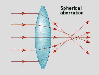
54 Spherical aberration

55 100x, Planapo, Oil immersion objective 0 microns depth 35 microns depth Spherical aberration 60x, Planapo, Water immersion objective 0 microns depth 66 microns depth

56 Collar adjustment of the 60x water immersion objective

57 Collar adjustment of the 60x water immersion objective Ruben Sandoval


58 Objective corrections require multiple lens elements Corrections: Color - Achromat, Apochromat Flat Field - Plan Immersion Media Cover Glass Polarization UV, IR transmission The more correction that a lens uses, the less transmission
59 100X, Oil immersion, NA X, Oil immersion, NA X, Oil immersion, NA X, Water immersion, NA X, Water immersion, NA.75 Light collection efficiency 10 4 x (NA 2 /mag) Corrections (# elements - transmission) Planapochromat 10 66% Plan-fluor 6 79% Planapochromat 10 66% Planapochromat 10 66% Plan-fluor 6 79% Ask yourself Is correction for chromatic aberration important to you?
60 100X, Oil immersion, NA X, Oil immersion, NA X, Oil immersion, NA X, Water immersion, NA X, Water immersion, NA.75 Light collection efficiency 10 4 x (NA 2 /mag) Corrections (# elements - transmission) Planapochromat 10 66% Plan-fluor 6 79% Planapochromat 10 66% Planapochromat 10 66% Plan-fluor 6 79% Ask yourself Is correction for chromatic aberration important to you?
61 Confocal microscopy - 2-dimensional imaging detectors
62 Confocal microscopy builds an image by point scanning and so is slow Need to acquire one point at a time. This limits acquisition to ~1 frame/sec. Limited by number of photons per pixel.

63 Spinning disk confocal microscope Spinning disk scans 1200 spots across a field at 30 Hz 360 Hz frame rate, we ve collected at up to 40 Hz Characterize rapid dynamics Signal to noise better than conventional confocal systems Reduce illumination for reduced phototoxicity and photobleaching Figure courtesy of Florida State Univ.

64 Transferrin labeled endosomes migrating around an MDCK cell Perkin-Elmer/Yokagawa spinning disk video-rate confocal microscope 1100 time points collected at 11 fps over 100 seconds

65 Real-time movement of Rab25 around an MDCK cell Perkin-Elmer/Yokagawa spinning disk confocal microscope 600 time points collected and displayed at 20 fps over 30 seconds

66 Living MDCK cells expressing GFP-tubulin and GFP-Rab7 Perkin-Elmer/Yokagawa spinning disk video-rate confocal microscope
67 Axial resolution of the Ultraview and the Zeiss 510 confocal microscopes 100x objectives 1 Vertical resolution 100x objective Relative intensity microns from focus
68 Axial resolution of the Ultraview and the Zeiss 510 confocal microscopes 60x objectives 1 Vertical resolution 60x objective Relative intensity microns from focus
69 Ultraview system shows large linear range characteristic of CCD detectors corrected mean s/n Perkin-Elmer Integration time test mean signal signal-to-noise ratio integration time (ms)


70 The Ultraview system for imaging living cells Rab25 in MDCK cells Zeiss 510 PerkinElmer Ultraview 200 images collected at 2 frames per second, 0.13 micron pixels, 12 bit pixel depth

71 Frame 1 Frame 200 PerkinElmer Ultraview Zeiss 510
72 1 Fraction of initial fluorescence frame number S/N - initial S/N - final Ultraview Comparison of imaging performance Zeiss 510 versus PerkinElmer Ultraview Images collected at 1.7 frames per second, micron pixels, 12 bit pixel depth Zeiss
73 Why is the Ultraview so good? 1/1200 the intensity less saturation Integrate 8 µs x 360 per second vs. 4 µs 2-fold higher quantum efficiency 360 breaks between illuminations Less digitizer and amplifier noise
74 Field imaging (Ultraview) versus point scanning confocal systems Field imaging detectors CCDs are very clean, with wide dynamic range Images can be collected rapidly Different colors must be collected sequentially, or on different detector arrays Point scanning detectors Multiple colors can be collected simultaneously PMTs are noisier than CCD systems Slow image acquisition and reasonable frame rates require very brief collection per pixel high illumination and few photons
75 Multi-photon microscopy

76 3D imaging of kidney tissue by confocal microscopy

77 3D imaging of kidney tissue by 2-photon microscopy
78 Multi-photon fluorescence excitation depends upon the simultaneous absorption of multiple photons
79 Multi-photon fluorescence excitation depends upon the simultaneous absorption of multiple photons What is simultaneous? Multiple photons must arrive within the duration of the intermediate virtual state of the electron ~ 1 attosecond (10-18 seconds) What is the relative frequency of such absorptions? Winfried Denk calculated that a molecule of rhodamine B exposed to direct sunlight will experience: A one-photon absorption around once per second. A two photon absorption once every 10,000 years. A three-photon absorption... well actually never in the history of the universe.
80 Multi-photon fluorescence excitation depends upon the simultaneous absorption of multiple photons What is simultaneous? Multiple photons must arrive within the duration of the intermediate virtual state of the electron ~ 1 attosecond (10-18 seconds) What is the relative frequency of such absorptions? Winfried Denk calculated that a molecule of rhodamine B exposed to direct sunlight will experience: A one-photon absorption around once per second. A two photon absorption once every 10,000 years. A three-photon absorption... well actually never in the history of the universe.
81 Multi-photon fluorescence excitation depends upon the simultaneous absorption of multiple photons What is simultaneous? Multiple photons must arrive within the duration of the intermediate virtual state of the electron ~ 1 attosecond (10-18 seconds) What is the relative frequency of such absorptions? Winfried Denk calculated that a molecule of rhodamine B exposed to direct sunlight will experience: A one-photon absorption around once per second. A two photon absorption once every 10,000 years. A three-photon absorption... well actually never in the history of the universe.
82 Multi-photon fluorescence excitation depends upon the simultaneous absorption of multiple photons What is simultaneous? Multiple photons must arrive within the duration of the intermediate virtual state of the electron ~ 1 attosecond (10-18 seconds) What is the relative frequency of such absorptions? Winfried Denk calculated that a molecule of rhodamine B exposed to direct sunlight will experience: A one-photon absorption around once per second. A two photon absorption once every 10,000 years. A three-photon absorption... well actually never in the history of the universe.
83 Multi-photon fluorescence excitation depends upon the simultaneous absorption of multiple photons What is simultaneous? Multiple photons must arrive within the duration of the intermediate virtual state of the electron ~ 1 attosecond (10-18 seconds) What is the relative frequency of such absorptions? Winfried Denk calculated that a molecule of rhodamine B exposed to direct sunlight will experience: A one-photon absorption around once per second. A two photon absorption once every 10,000 years. A three-photon absorption... well actually never in the history of the universe.
84 Multi-photon fluorescence excitation depends upon the simultaneous absorption of multiple photons What is simultaneous? Multiple photons must arrive within the duration of the intermediate virtual state of the electron ~ 1 attosecond (10-18 seconds) What is the relative frequency of such absorptions? Winfried Denk calculated that a molecule of rhodamine B exposed to direct sunlight will experience: A one-photon absorption around once per second. A two photon absorption once every 10,000 years. A three-photon absorption... well actually never in the history of the universe.
85 How to increase the probability of multi-photon absorption for multiphoton microscopy? You could increase illumination 600,000 fold.
86 How is the probability of multi-photon absorption increased in multiphoton microscopy? Photon crowding Dave Piston, Vanderbilt
87 How is the probability of multi-photon absorption increased in multiphoton microscopy? Photon crowding Photon crowding in space - the cross-sectional density of photons is highest at the focal point. Photon crowding in time - laser emissions are pulsed into brief (~100 femtosecond) packets
88 How is the probability of multi-photon absorption increased in multiphoton microscopy? Photon crowding Photon crowding in space - the cross-sectional density of photons is highest at the focal point. Photon crowding in time - laser emissions are pulsed into brief (~100 femtosecond) packets

89 Pulsed laser emissions provide for power sufficient for multiphoton absorption without photo-damage Pulsed laser provides low average power but peak power high enough for 2- photon absorption Sample illuminated for only 8 one million th of the pixel dwell time

90 One and two photon fluorescence excitation One photon absorption is proportional to illumination Fluorescence is stimulated throughout the lightpath Brad Amos - MRC Two-photon absorption is proportional to the squared power of illumination Photon density sufficient to excite fluorescence occurs only at the focal point
91 Fluorescence microscopy time pixel dwell time 4 microseconds 100X fluorescence lifetime 10 nanoseconds 100,000X 800 nm pulse length 100 femtoseconds 10,000X intermediate virtual state 10 attoseconds duration (seconds)
92 Benefits of Multi-Photon Microscopy no photobleaching in out-of-focus planes figure through the unknowing courtesy of the Integrated Microscopy Resource, Madison, Wisconsin

93 Figure courtesy of the National High Magnetic Field Laboratory Florida State University And Dave Piston, Vanderbilt Univ. Multiphoton photobleaching is limited to the focal point
94 Benefits of Multi-Photon Microscopy no photobleaching in out-of-focus planes no emission aperture - less loss to scattering figure through the unknowing courtesy of the Integrated Microscopy Resource, Madison, Wisconsin

95 Multi-photon microscopy is less sensitive to light scatter by tissues attenuation of signal Image courtesy of Istituto Nazionale per la Fisica della Materia - Genoa
96 Benefits of Multi-Photon Microscopy no photobleaching in out-of-focus planes no emission aperture - less loss to scattering IR light penetrates deeper, with less damage figure through the unknowing courtesy of the Integrated Microscopy Resource, Madison, Wisconsin

97 Multi-photon Excitation Allows Deeper Imaging in Intact Tissue Confocal Two-Photon Excitation Dave Piston, Vanderbilt


98 Imaging complex structures Kidney tubules of a newborn mouse kidney Carrie Phillips, Nephrology and Pathology

99 Imaging complex structures Kidney tubules of a newborn mouse kidney Carrie Phillips, Nephrology and Pathology

100 Single optical section of neural network

101 Imaging complex structures Neural network in mouse brain adapted to chronic alcohol Feng Zhou, Anatomy and Cell Biology

102 Imaging complex structures Segmentation of a single neuron Feng Zhou, Anatomy and Cell Biology

103 Imaging complex structures Neural network in mouse brain adapted to chronic alcohol Feng Zhou, Anatomy and Cell Biology
104 Multiphoton imaging of kidney

105 3D imaging of newborn mouse kidney by 2-photon microscopy Carrie Phillips

106 Imaging complex structures Glomerulus of a newborn mouse kidney Carrie Phillips, rendered by Anatomical Travel, Inc.

107 Imaging complex structures branching tubulogenesis in an embryonic mouse kidney Carrie Phillips, Nephrology and Pathology

108 140 micron thick volume of embryonic mouse kidney - branching tubulogenesis Carrie Phillips... and real time rendering on a PC using Voxx software Jeff Clendenon
109 Vital imaging by multiphoton microscopy The extended depth provided by multi-photon microscopy permits high-resolution imaging of the cells of living animals. The ability to image multiple fluorophores supports correlations of multiple proteins and physiological processes.

110 Experimental arrangement for intravital imaging of rat kidney Anesthesia is provided via 1% halothane and low-flow oxygen. Fluorescent probes are administered via tail-vein injection. Blood gases are monitored via femoral artery.
111 Two-photon image of kidney of living rat injected with Hoechst, 500 Kd fluorescein dextran and 3 Kd Texas-Red dextran Ruben Sandoval and Katrina Kelly
112 Multiple functions apparent in images of kidneys of animals injected with large and small dextrans proximal tubule endocytosis glomerular filtration tubular solute concentration capillary blood flow
113 Multiple functions apparent in images of kidneys of animals injected with large and small dextrans proximal tubule endocytosis glomerular filtration tubular solute concentration capillary blood flow

114 2-photon microscopy of glomerular filtration in a living rat Hoechst-labeled nuclei, 500 Kd fluorescein dextran in blood, 3 Kd Texas-Red dextran in lysosomes and tubule lumens Ruben Sandoval and Katrina Kelly
115 Multiple functions apparent in images of kidneys of animals injected with large and small dextrans proximal tubule endocytosis glomerular filtration tubular solute concentration capillary blood flow

116 2-photon microscopy of tubular solute concentration in a living rat Hoechst-labeled nuclei, rhodamine-albumin in blood, 3 Kd fluorescein dextran in lysosomes and tubule lumens Ruben Sandoval and Katrina Kelly

117 Multiple functions apparent in images of kidneys of animals injected with large and small dextrans proximal tubule endocytosis glomerular filtration tubular solute concentration capillary blood flow

118 Imaging capillary blood flow by 2-photon microscopy Rhodamine-albumin, 3K fluorescein dextran and Hoechst
 by 2-photon microscopy 500K fluorescein")
119 Imaging capillary blood flow (and proximal tubule autofluorescence) by 2-photon microscopy 500K fluorescein dextran and Hoechst
120 Practical multiphoton microscopy
121 Practical multiphoton microscopy Photobleaching Resolution Multiphoton fluorescence dyes Imaging multiple colors Laser choices Detector options

122 2 photon versus confocal microscopy POP go the endosomes One photon image at first, tenth, whatever scan Two photon image Two photon image at fourth scan
123 Practical multiphoton microscopy Photobleaching Resolution Multiphoton fluorescence dyes Imaging multiple colors Laser choices Detector options

124 Deep tissue imaging by multi-photon microscopy 2 photon image collected 120 microns into kidney section

125 Imaging complex structures Dendritic spines in mouse hippocampal neuron Feng Zhou, Anatomy and Cell Biology
126 Practical multiphoton microscopy Photobleaching Resolution Multiphoton fluorescence dyes Imaging multiple colors Laser choices Detector options
127 2-photon cross sections are not necessarily predicted by single photon excitation spectra BioRad

128 2-photon fluorescence excitation 2-photon cross sections Xu et al., BioImaging, 1996
129 Practical multiphoton microscopy Photobleaching Resolution Multiphoton fluorescence dyes Imaging multiple colors Laser choices Detector options

130 Imaging complex structures Comparing multiple probes in kidney tissue Ruben Sandoval, Nephrology
131 Practical multiphoton microscopy Photobleaching Resolution Multiphoton fluorescence dyes Imaging multiple colors Laser choices Detector options

132 2-photon image volume of kidney of a living rat (before any fluorescence labeling)


133 2-photon image autofluorescence and Texas-Red gentamicin A tuneable laser is a good idea 820 nm excitation 860 nm excitation
134 Practical multiphoton microscopy Photobleaching Resolution Multiphoton fluorescence dyes Imaging multiple colors Laser choices Detector options


135 Signal attenuation with depth into fixed tissues - Multiphoton microscopy Top view Side view


136 32% 80% Signal attenuation with depth in multiphoton microscopy 10 µm Live animal 34 µm 19 µm Fixed tissue 50 µm 81 µm 63 µm 2x

137 Multi-photon microscopy is less sensitive to light scatter by tissues attenuation of signal Mouse Imaging Centre The Hospital for Sick Children University of Toronto
138 Sources of signal attenuation at depth Fluorescence emissions scattering refraction spherical aberration Illumination scattering refraction spherical aberration

139 Red emissions Green emissions 12 µm Use Red fluors scattering decreases as the fourth power of wavelength 127 µm 860 nm excitation

140 Two color 2-photon imaging of 13 day old embryonic mouse kidney [ microns ] Red - peanut agglutinin Green - Len culinaris agglutinin


141 Non-descanned detectors collect scattered light more efficiently Descanned PMT Non-descanned PMT


142 Non-descanned detectors collect scattered light more efficiently Descanned PMT Non-descanned PMT
143 Sources of signal attenuation at depth Fluorescence emissions scattering refraction spherical aberration Illumination scattering refraction spherical aberration

144 17ocbleach Attenuation of illumination is reflected by lower photobleaching at depth After 1 scan After 40 scans surface 39 µm Attenuation of illumination photobleaching rates Live rat, 800 nm excitation, Hoechst
145 Illumination attenuation decreases at longer wavelengths light scatter decreases with wavelength nm 740 nm photobleaching decay rate depth
146 Signal attentuates with depth into kidneys, but can be regained with increased illumination

147 Signal attentuates with depth into kidneys, but can be regained with increased illumination And, unlike confocal microscopy, scatter has minimal effect on resolution
148 Practical multiphoton microscopy Photobleaching Resolution Multiphoton fluorescence dyes Imaging multiple colors Laser choices Detector options Objective choice
149 Objectives for Multiphoton Microscopy A new generation of microscope objectives is being developed that is optimized for infrared transmission, and designed for light collection rather than image formation.


150 The importance of high numerical aperture to 3d microscopy - XY planes of a kidney sample 20x, NA.75 60x, NA 1.2


151 The importance of high numerical aperture to 3d microscopy - XY planes of a kidney sample 20x, NA.75 60x, NA 1.2
152 Increase in the use of multiphoton microscopy since number of publications years since 1993
153 Increase in the use of multiphoton microscopy since 1993 total "kidney or renal, but not cultured cells" number of publications years since 1993
154 Summary Which system to choose? Do you need to image? Do you need optical sections? Confocal or image deconvolution? Confocal or multi-photon?
155 Summary Which system to choose? Do you need to image? Do you need optical sections? Confocal or image deconvolution? Confocal or multi-photon?
156 Summary Which system to choose? Do you need to image? Do you need optical sections? Confocal or image deconvolution? Confocal or multi-photon?
157 Summary Which system to choose? Do you need to image? Do you need optical sections? Confocal or image deconvolution? Confocal or multi-photon?
158 pixel dwell time 4 microseconds 100X fluorescence lifetime 10 nanoseconds 100,000X 800 nm pulse length 100 femtoseconds 10,000X intermediate virtual state 10 attoseconds duration (seconds)
Why and How? Daniel Gitler Dept. of Physiology Ben-Gurion University of the Negev. Microscopy course, Michmoret Dec 2005
 Why and How? Daniel Gitler Dept. of Physiology Ben-Gurion University of the Negev Why use confocal microscopy? Principles of the laser scanning confocal microscope. Image resolution. Manipulating the
Basics of confocal imaging (part I)
") Basics of confocal imaging (part I) Swiss Institute of Technology (EPFL) Faculty of Life Sciences Head of BIOIMAGING AND OPTICS BIOP arne.seitz@epfl.ch Lateral resolution BioImaging &Optics Platform Light
Basics of confocal imaging (part I) Swiss Institute of Technology (EPFL) Faculty of Life Sciences Head of BIOIMAGING AND OPTICS BIOP arne.seitz@epfl.ch Lateral resolution BioImaging &Optics Platform Light
Maria Smedh, Centre for Cellular Imaging. Maria Smedh, Centre for Cellular Imaging
 Nonlinear microscopy I: Two-photon fluorescence microscopy Multiphoton Microscopy What is multiphoton imaging? Applications Different imaging modes Advantages/disadvantages Scattering of light in thick
3D light microscopy techniques
 3D light microscopy techniques The image of a point is a 3D feature In-focus image Out-of-focus image The image of a point is not a point Point Spread Function (PSF) 1D imaging 1 1 2! NA = 0.5! NA 2D imaging
Boulevard du Temple Daguerrotype (Paris,1838) a busy street? Nyquist sampling for movement
 a busy street? Nyquist sampling for movement") Boulevard du Temple Daguerrotype (Paris,1838) a busy street? Nyquist sampling for movement CONFOCAL MICROSCOPY BioVis Uppsala, 2017 Jeremy Adler Matyas Molnar Dirk Pacholsky Widefield & Confocal Microscopy
Boulevard du Temple Daguerrotype (Paris,1838) a busy street? Nyquist sampling for movement CONFOCAL MICROSCOPY BioVis Uppsala, 2017 Jeremy Adler Matyas Molnar Dirk Pacholsky Widefield & Confocal Microscopy
INTRODUCTION TO MICROSCOPY. Urs Ziegler THE PROBLEM
 INTRODUCTION TO MICROSCOPY Urs Ziegler ziegler@zmb.uzh.ch THE PROBLEM 1 ORGANISMS ARE LARGE LIGHT AND ELECTRONS: ELECTROMAGNETIC WAVES v = Wavelength ( ) Speed (v) Frequency ( ) Amplitude (A) Propagation
TRAINING MANUAL. Multiphoton Microscopy LSM 510 META-NLO
 TRAINING MANUAL Multiphoton Microscopy LSM 510 META-NLO September 2010 Multiphoton Microscopy Training Manual Multiphoton microscopy is only available on the LSM 510 META-NLO system. This system is equipped
ADVANCED METHODS FOR CONFOCAL MICROSCOPY II. Jean-Yves Chatton Sept. 2006
 ADVANCED METHODS FOR CONFOCAL MICROSCOPY II Jean-Yves Chatton Sept. 2006 Workshop outline Confocal microscopy of living cells and tissues X-Z scanning Time series Bleach: FRAP, photoactivation Emission
Point Spread Function. Confocal Laser Scanning Microscopy. Confocal Aperture. Optical aberrations. Alternative Scanning Microscopy
 Bi177 Lecture 5 Adding the Third Dimension Wide-field Imaging Point Spread Function Deconvolution Confocal Laser Scanning Microscopy Confocal Aperture Optical aberrations Alternative Scanning Microscopy
MULTIPHOTON MICROSCOPY. Matyas Molnar Dirk Pacholsky
 MULTIPHOTON MICROSCOPY Matyas Molnar Dirk Pacholsky Information Information given here about 2 Photon microscopy were mainly taken from these sources: Background information on 2-Photon microscopy: http://micro.magnet.fsu.edu/primer/techniques/fluorescence/multiphoton/
Multifluorescence The Crosstalk Problem and Its Solution
 Multifluorescence The Crosstalk Problem and Its Solution If a specimen is labeled with more than one fluorochrome, each image channel should only show the emission signal of one of them. If, in a specimen
Invitation for a walk through microscopy. Sebastian Schuchmann Jörg Rösner
 Invitation for a walk through microscopy Sebastian Schuchmann Jörg Rösner joerg.roesner@charite.de Techniques in microscopy Conventional (light) microscopy bright & dark field, phase & interference contrast
LSM 510 META in Chang Gung University
 Content LSM 510 META in Chang ung University LSM 510 META 路 理 The features and applications of LSM 510 META 01-09 Introduction of the hardware 10-12 Fluorescence observation in conventional microscope
Shreyash Tandon M.S. III Year
 Shreyash Tandon M.S. III Year 20091015 Confocal microscopy is a powerful tool for generating high-resolution images and 3-D reconstructions of a specimen by using point illumination and a spatial pinhole
Confocal Microscopy. Kristin Jensen
 Confocal Microscopy Kristin Jensen 17.11.05 References Cell Biological Applications of Confocal Microscopy, Brian Matsumoto, chapter 1 Studying protein dynamics in living cells,, Jennifer Lippincott-Schwartz
Final Exam, 150 points PMB 185: Techniques in Light Microscopy
 Final Exam, 150 points Name PMB 185: Techniques in Light Microscopy Point value is in parentheses at the end of each question. Note: GFP = green fluorescent protein ; CFP = cyan fluorescent protein ; YFP
Bio 407. Applied microscopy. Introduction into light microscopy. José María Mateos. Center for Microscopy and Image Analysis
 Center for Microscopy and Image Analysis Bio 407 Applied Introduction into light José María Mateos Fundamentals of light Compound microscope Microscope composed of an objective and an additional lens (eyepiece,
3D light microscopy techniques
 3D light microscopy techniques The image of a point is a 3D feature In-focus image Out-of-focus image The image of a point is not a point Point Spread Function (PSF) 1D imaging 2D imaging 3D imaging Resolution
Nature Methods: doi: /nmeth Supplementary Figure 1. Schematic of 2P-ISIM AO optical setup.
 Supplementary Figure 1 Schematic of 2P-ISIM AO optical setup. Excitation from a femtosecond laser is passed through intensity control and shuttering optics (1/2 λ wave plate, polarizing beam splitting
BASICS OF CONFOCAL IMAGING (PART I)
") BASICS OF CONFOCAL IMAGING (PART I) INTERNAL COURSE 2012 LIGHT MICROSCOPY Lateral resolution Transmission Fluorescence d min 1.22 NA obj NA cond 0 0 rairy 0.61 NAobj Ernst Abbe Lord Rayleigh Depth of field
BASICS OF CONFOCAL IMAGING (PART I) INTERNAL COURSE 2012 LIGHT MICROSCOPY Lateral resolution Transmission Fluorescence d min 1.22 NA obj NA cond 0 0 rairy 0.61 NAobj Ernst Abbe Lord Rayleigh Depth of field
Opterra II Multipoint Scanning Confocal Microscope. Innovation with Integrity
 Opterra II Multipoint Scanning Confocal Microscope Enabling 4D Live-Cell Fluorescence Imaging through Speed, Sensitivity, Viability and Simplicity Innovation with Integrity Fluorescence Microscopy The
Bi/BE 227 Winter Assignment #3. Adding the third dimension: 3D Confocal Imaging
 Bi/BE 227 Winter 2016 Assignment #3 Adding the third dimension: 3D Confocal Imaging Schedule: Jan 20: Assignment Jan 20-Feb 8: Work on assignment Feb 10: Student PowerPoint presentations. Goals for this
Confocal Microscopy. (Increasing contrast and resolu6on using op6cal sec6oning) Lecture 7. November 2017
 Lecture 7. November 2017") Confocal Microscopy (Increasing contrast and resolu6on using op6cal sec6oning) Lecture 7 November 2017 3 Flavours of Microscope Confocal Laser Scanning Problem: Out of Focus Light Spinning disc 2-Photon
Confocal Microscopy (Increasing contrast and resolu6on using op6cal sec6oning) Lecture 7 November 2017 3 Flavours of Microscope Confocal Laser Scanning Problem: Out of Focus Light Spinning disc 2-Photon
Fundamentals of Light Microscopy II: Fluorescence, Deconvolution, Confocal, Multiphoton, Spectral microscopy. Integrated Microscopy Course
 Fundamentals of Light Microscopy II: Fluorescence, Deconvolution, Confocal, Multiphoton, Spectral microscopy Integrated Microscopy Course Review Lecture 1: Microscopy Basics Light train Kohler illumination*
Introduction to light microscopy
 Center for Microscopy and Image Anaylsis Introduction to light Imaging with light / Overview of techniques Urs Ziegler ziegler@zmb.uzh.ch Light interacting with matter Absorbtion Refraction Diffraction
Ratio Imaging. Dividing one image by another to detect changing conditions. Images collected at different times, wavelengths, polarities, etc
 Ratio Imaging Dividing one image by another to detect changing conditions Images collected at different times, wavelengths, polarities, etc Most common use of ratio imaging is to provide a quick spectral
Opterra. Multipoint Scanning Confocal Microscope. Innovation with Integrity. Cell-Friendly, High-Speed, High-Resolution Imaging
 Opterra Multipoint Scanning Confocal Microscope Cell-Friendly, High-Speed, High-Resolution Imaging Innovation with Integrity Fluorescence Microscopy Opterra Multipoint Scanning Confocal Microscope Superior
Supplementary Information. Stochastic Optical Reconstruction Microscopy Imaging of Microtubule Arrays in Intact Arabidopsis thaliana Seedling Roots
 Supplementary Information Stochastic Optical Reconstruction Microscopy Imaging of Microtubule Arrays in Intact Arabidopsis thaliana Seedling Roots Bin Dong 1,, Xiaochen Yang 2,, Shaobin Zhu 1, Diane C.
Confocal Microscopy and Related Techniques
 Confocal Microscopy and Related Techniques Chau-Hwang Lee Associate Research Fellow Research Center for Applied Sciences, Academia Sinica 128 Sec. 2, Academia Rd., Nankang, Taipei 11529, Taiwan E-mail:
Non-Descanned FLIM Detection in Multiphoton Microscopes
 Non-Descanned FLIM Detection in Multiphoton Microscopes Abstract. Multiphoton microscopes use a femtosecond NIR laser to excite fluorescence in the sample. Excitation is performed via a multi-photon absorption
Imaging Retreat - UMASS Customized real-time confocal and 2-photon imaging
 Imaging Retreat - UMASS 2012 Customized real-time confocal and 2-photon imaging Mike Sanderson Department of Microbiology and Physiological Systems University of Massachusetts Medical School Thanks for
1.The Problem LIGHT-LEVEL LEVEL IMAGING. light-level level Cameras. 3. Solutions. 2. Low-light LOW-LIGHT
 LOW-LIGHT LIGHT-LEVEL LEVEL IMAGING 1.The Problem 2. Low-light light-level level Cameras 3. Solutions How Much Light? I. Illumination system: 75 W Xenon Arc (~1mW/nm in visible) 490/10 nm exciter filter
MULTIPHOTON MICROSCOPY
 MULTIPHOTON MICROSCOPY Methods for Cell Analysis Course BioVis Uppsala, 2014 Matyas Molnar Dirk Pacholsky Information Information given here about 2 Photon microscopy were mainly taken from these sources:
Akinori Mitani and Geoff Weiner BGGN 266 Spring 2013 Non-linear optics final report. Introduction and Background
 Akinori Mitani and Geoff Weiner BGGN 266 Spring 2013 Non-linear optics final report Introduction and Background Two-photon microscopy is a type of fluorescence microscopy using two-photon excitation. It
Confocal, hyperspectral, spinning disk
 Confocal, hyperspectral, spinning disk Administrative HW 6 due on Fri Midterm on Wed Covers everything since previous midterm 8.5 x 11 sheet allowed, 1 side Guest lecture by Joe Dragavon on Mon 10/30 Last
Dynamic Confocal Imaging of Living Brain. Advantages and risks of multiphoton microscopy in physiology
 Dynamic Confocal Imaging of Living Brain Advantages and risks of multiphoton microscopy in physiology Confocal laser scanning microscopy In conventional optical microscopy focused and out-offocus light
Development of a High-speed Super-resolution Confocal Scanner
 Development of a High-speed Super-resolution Confocal Scanner Takuya Azuma *1 Takayuki Kei *1 Super-resolution microscopy techniques that overcome the spatial resolution limit of conventional light microscopy
The DCS-120 Confocal Scanning FLIM System
 he DCS-120 Confocal Scanning FLIM System he bh DCS-120 confocal scanning FLIM system converts a conventional microscope into a high-performance fluorescence lifetime imaging system. he system is based
Technology Note ZEISS LSM 880 with Airyscan
 Technology Note ZEISS LSM 880 with Airyscan Introducing the Fast Acquisition Mode ZEISS LSM 880 with Airyscan Introducing the Fast Acquisition Mode Author: Dr. Annette Bergter Carl Zeiss Microscopy GmbH,
Zeiss 780 Training Notes
 Zeiss 780 Training Notes Turn on Main Switch, System PC and Components Switches 780 Start up sequence Do you need the argon laser (458, 488, 514 nm lines)? Yes Turn on the laser s main power switch and
5/4/2015 INTRODUCTION TO LIGHT MICROSCOPY. Urs Ziegler MICROSCOPY WITH LIGHT. Image formation in a nutshell. Overview of techniques
 INTRODUCTION TO LIGHT MICROSCOPY Urs Ziegler ziegler@zmb.uzh.ch MICROSCOPY WITH LIGHT INTRODUCTION TO LIGHT MICROSCOPY Image formation in a nutshell Overview of techniques Widefield microscopy Resolution
Chemical Imaging. Whiskbroom Imaging. Staring Imaging. Pushbroom Imaging. Whiskbroom. Staring. Pushbroom
 Chemical Imaging Whiskbroom Chemical Imaging (CI) combines different technologies like optical microscopy, digital imaging and molecular spectroscopy in combination with multivariate data analysis methods.
ZEISS LSM510META confocal manual
 ZEISS LSM510META confocal manual Switching on the system 1) Switch on the Remote Control button located on the table to the right of the microscope. This is the main switch for the whole system including
Spectral Imaging with the Opterra Multipoint Scanning Confocal
 Spectral Imaging with the Opterra Multipoint Scanning Confocal Outline Opterra design overview Scan Modes Light Path Spectral Imaging with Opterra Drosophila larva heart. Opterra Design Overview Supravideo
FEMTOSMART. Benefits. Features
 FEMTOSMART Extremely large space under the objective For in vivo studies Field upgradability Patented imaging technologies Flexible scanning methods Maximal photon collection Elevated, column-based body
Zeiss 880 Training Notes Zen 2.3
 Zeiss 880 Training Notes Zen 2.3 1 Turn on the HXP 120V Lamp 2 Turn on Main Power Switch Turn on the Systems PC Switch Turn on the Components Switch. 3 4 5 Turn on the PC and log into your account. Start
You won t be able to measure the incident power precisely. The readout of the power would be lower than the real incident power.
 1. a) Given the transfer function of a detector (below), label and describe these terms: i. dynamic range ii. linear dynamic range iii. sensitivity iv. responsivity b) Imagine you are using an optical
1 Co Localization and Working flow with the lsm700
 1 Co Localization and Working flow with the lsm700 Samples -1 slide = mousse intestine, Dapi / Ki 67 with Cy3/ BrDU with alexa 488. -1 slide = mousse intestine, Dapi / Ki 67 with Cy3/ no BrDU (but with
EUV microscopy - a user s perspective Dimitri Scholz EUV,
 EUV microscopy - a user s perspective Dimitri Scholz EUV, 09.11.2011 Imaging technologies: available at UCD now and in the next future Begin ab ovo - Simple approaches direct to the goal - Standard methods
Training Guide for Carl Zeiss LSM 5 LIVE Confocal Microscope
 Training Guide for Carl Zeiss LSM 5 LIVE Confocal Microscope AIM 4.2 Optical Imaging & Vital Microscopy Core Baylor College of Medicine (2017) Power ON Routine 1 2 Verify that main power switches on the
TCSPC at Wavelengths from 900 nm to 1700 nm
 TCSPC at Wavelengths from 900 nm to 1700 nm We describe picosecond time-resolved optical signal recording in the spectral range from 900 nm to 1700 nm. The system consists of an id Quantique id220 InGaAs
Practical work no. 3: Confocal Live Cell Microscopy
 Practical work no. 3: Confocal Live Cell Microscopy Course Instructor: Mikko Liljeström (MIU) 1 Background Confocal microscopy: The main idea behind confocality is that it suppresses the signal outside
Bi Imaging. Multicolor Imaging: The Important Question of Co-Localization. Anna Smallcombe Bio-Rad Laboratories, Hemel Hempstead, UK
 Multicolor Imaging: The Important Question of Co-Localization Anna Smallcombe Bio-Rad Laboratories, Hemel Hempstead, UK The use of specific fluorescent probes, combined with confocal or multiphoton microscopy
Resolution. Diffraction from apertures limits resolution. Rayleigh criterion θ Rayleigh = 1.22 λ/d 1 peak at 2 nd minimum. θ f D
 Microscopy Outline 1. Resolution and Simple Optical Microscope 2. Contrast enhancement: Dark field, Fluorescence (Chelsea & Peter), Phase Contrast, DIC 3. Newer Methods: Scanning Tunneling microscopy (STM),
Nature Methods: doi: /nmeth Supplementary Figure 1
 . Supplementary Figure 1 Schematics and characterization of our AO two-photon fluorescence microscope. (a) Essential components of our AO two-photon fluorescence microscope: Ti:Sapphire laser; optional
Introduction to light microscopy
 Center for Microscopy and Image Anaylsis Introduction to light Basic concepts of imaging with light Urs Ziegler ziegler@zmb.uzh.ch Microscopy with light 1 Light interacting with matter Absorbtion Refraction
Prof. Enrico Gratton - Lecture 6 Fluorescence Microscopy
 Prof. Enrico Gratton - Lecture 6 Fluorescence Microscopy Instrumentation Light Sources: One-photon and Multi-photon Excitation Applications in Cells Lifetime Imaging Figures acknowledgements: E.D. Salmon
INTRODUCTION TO OPTICAL MICROSCOPY
 Experimental Biophysics TEK265, FYST23, TNF060, FAF010F Lab Exercise Supervisor: Karl Adolfsson Written by Peter Jönsson and Jason Beech Updated by Henrik Persson, Karl Adolfsson and Zhen Li karl.adolfsson@ftf.lth.se
Imaging Introduction. September 24, 2010
 Imaging Introduction September 24, 2010 What is a microscope? Merriam-Webster: an optical instrument consisting of a lens or combination of lenses for making enlarged images of minute objects; especially:
Components of confocal and two-photon microscopes
 Components of confocal and two-photon microscopes Internal training 07/04/2016 A. GRICHINE Platform Optical microscopy Cell imaging, IAB, ISdV Plan Confocal laser scanning microscope o o o Principle Main
Digital Camera Technologies for Scientific Bio-Imaging. Part 2: Sampling and Signal
 Digital Camera Technologies for Scientific Bio-Imaging. Part 2: Sampling and Signal Yashvinder Sabharwal, 1 James Joubert 2 and Deepak Sharma 2 1. Solexis Advisors LLC, Austin, TX, USA 2. Photometrics
長庚大學共軛焦顯微鏡課程 長庚大學共軛焦顯微鏡課程. Spot light 長庚大學
 長庚大學共軛焦顯微鏡課程 Spot light 長庚大學共軛焦顯微鏡課程 20071030 長庚大學 Basic principle of Laser Scanning Confocal Microscopy The application of LSM 510 META detector Multiphoton microscopy basic principle and introduction
Introduction to light microscopy
 Center for Microscopy and Image Anaylsis Introduction to light microscopy Basic concepts of imaging with light Urs Ziegler ziegler@zmb.uzh.ch Light interacting with matter Absorbtion Refraction Diffraction
Training Guide for Carl Zeiss LSM 510 META Confocal Microscope
 Training Guide for Carl Zeiss LSM 510 META Confocal Microscope AIM 4.2 Optical Imaging & Vital Microscopy Core Baylor College of Medicine (2017) Power ON Routine 1 2 Turn ON Components and System/PC switches
Rates of excitation, emission, ISC
 Bi177 Lecture 4 Fluorescence Microscopy Phenomenon of Fluorescence Energy Diagram Rates of excitation, emission, ISC Practical Issues Lighting, Filters More on diffraction Point Spread Functions Thus Far,
We attempted to separate the two dyes by acquiring images using a single excitation wavelength and just two emission wavelengths.
 TN437: Spectral Separation of monochrome images using Volocity 4.0 Introduction Spectral Separation is a technique that allows the user to separate images containing data from more than one fluorochrome
Nature Structural & Molecular Biology: doi: /nsmb Supplementary Figure 1
 Supplementary Figure 1 Supplemental correlative nanomanipulation-fluorescence traces probing nascent RNA and fluorescent Mfd during TCR initiation. Supplemental correlative nanomanipulation-fluorescence
Travel to New Dimensions- LSM 880. The Resolution of a Microscope is limited. The Resolution of a Microscope is limited. Image. Image. Object.
 Travel to New Dimensions- LSM 880 LSM 880: The Power of Sensitivity Our Latest Member of the LSM 880 with GaAsP Detectors Sensitivity, and Ease of Use Innovative High-End Laser Scanning Microscopes from
The Nature of Light. Light and Energy
 The Nature of Light Light and Energy - dependent on energy from the sun, directly and indirectly - solar energy intimately associated with existence of life -light absorption: dissipate as heat emitted
Fast, high-contrast imaging of animal development with scanned light sheet based structured-illumination microscopy
 nature methods Fast, high-contrast imaging of animal development with scanned light sheet based structured-illumination microscopy Philipp J Keller, Annette D Schmidt, Anthony Santella, Khaled Khairy,
Quick Start Guide. Leica SP5 X
 Quick Start Guide Leica SP5 X Please note: Some of the information in this guide was taken from Leica Microsystems Leica TCS SP5 LAS AF Guide for New Users. This work is licensed under the Creative Commons
Reflecting optical system to increase signal intensity. in confocal microscopy
 Reflecting optical system to increase signal intensity in confocal microscopy DongKyun Kang *, JungWoo Seo, DaeGab Gweon Nano Opto Mechatronics Laboratory, Dept. of Mechanical Engineering, Korea Advanced
Examination, TEN1, in courses SK2500/SK2501, Physics of Biomedical Microscopy,
 KTH Applied Physics Examination, TEN1, in courses SK2500/SK2501, Physics of Biomedical Microscopy, 2009-06-05, 8-13, FB51 Allowed aids: Compendium Imaging Physics (handed out) Compendium Light Microscopy
Operation Guide for the Leica SP2 Confocal Microscope Bio-Imaging Facility Hunter College October 2009
 Operation Guide for the Leica SP2 Confocal Microscope Bio-Imaging Facility Hunter College October 2009 Introduction of Fluoresence Confocal Microscopy The first confocal microscope was invented by Princeton
The Zeiss AiryScan System, Confocal Four.
 The Zeiss AiryScan System, Confocal Four. Overview. The Zeiss AiryScan module is a segmented, radially stacked GaASP detector and collector system designed to subsample the airy disk of a point emission
Confocal Laser Scanning Microscopy
 Name of the Core Facility: Confocal Laser Scanning Microscopy CORE Forschungszentrum Immunologie Mainz Welcome to the CSLM Core Facility: The CLSM Core Facility enables working groups to incorporate high
CMI STANDARD OPERATING PROCEDURE. Fluoview 300 laser scanning confocal microscope
 CMI STANDARD OPERATING PROCEDURE Fluoview 300 laser scanning confocal microscope CMI documentid:sop001 CONTACT INFORMATION: Peter Owens: 091 494036 (office) Peter.owens@nuigalway.ie Kerry Thompson: 091
3. are adherent cells (ie. cells in suspension are too far away from the coverslip)
") Before you begin, make sure your sample... 1. is seeded on #1.5 coverglass (thickness = 0.17) 2. is an aqueous solution (ie. fixed samples mounted on a slide will not work - not enough difference in refractive
Before you begin, make sure your sample... 1. is seeded on #1.5 coverglass (thickness = 0.17) 2. is an aqueous solution (ie. fixed samples mounted on a slide will not work - not enough difference in refractive
Microscopic Structures
 Microscopic Structures Image Analysis Metal, 3D Image (Red-Green) The microscopic methods range from dark field / bright field microscopy through polarisation- and inverse microscopy to techniques like
LSM 510 Meta Training Notes
 LSM 510 Meta Training Notes Turning on the system Turn on X-Cite power supply. This supplies light for epifluorescence for viewing your samples through the microscope. Turn on the remote control switch.
Training Guide for Leica SP8 Confocal/Multiphoton Microscope
 Training Guide for Leica SP8 Confocal/Multiphoton Microscope LAS AF v3.3 Optical Imaging & Vital Microscopy Core Baylor College of Medicine (2017) Power ON Routine 1 2 Turn ON power switch for epifluorescence
Inside the LSM 880 NLO + Airyscan
 Inside the LSM 880 NLO + Airyscan Overview of the Newest High-End Point Scanning Solution from Carl Zeiss Microscopy Matt Curtis 3D Imaging Specialist John Dirnberger Account Manager Washington University
Nikon C1si Spectral Laser Scanning Confocal Microscope. User Guide
 Nikon C1si Spectral Laser Scanning Confocal Microscope User Guide Contents: C1Si Turn-On/ShutDown Procedures... 2 Overview... 4 Setup for epi-illumination to view through the eyepieces:... 5 Setup for
Multiphoton confocal microscope. Multiphoton confocal microscope A1R MP
 Multiphoton confocal microscope Multiphoton confocal microscope A1R MP Nikon's provides deeper, faster and sharper imaging. The confocal microscope A1R, which has an excellent reputation for its high speed,
Multiphoton Microscopy
 Multiphoton Microscopy A. Neumann, Y. Kuznetsova Introduction Multi-Photon Fluorescence Microscopy is a relatively novel imaging technique in cell biology. It relies on the quasi-simultaneous absorption
NIH Public Access Author Manuscript Opt Lett. Author manuscript; available in PMC 2010 August 9.
 NIH Public Access Author Manuscript Published in final edited form as: Opt Lett. 2010 January 1; 35(1): 67 69. Autoconfocal transmission microscopy based on two-photon induced photocurrent of Si photodiodes
IC 2 S High Performance Objectives
 M i c r o s c o p y f r o m C a r l Z e i s s IC 2 S igh Performance Objectives for Biomedical Applications with Laser Based Imaging Systems LSM,, ConfoCor, TIRF and ELYRA Carl Zeiss offers a large range
MOM#3: LIGHT SHEET MICROSCOPY (LSM) Stanley Cohen, MD
 Stanley Cohen, MD") MOM#3: LIGHT SHEET MICROSCOPY (LSM) Stanley Cohen, MD Introduction. Although the technical details of light sheet imaging and its various permutations appear at first glance to be complex and require some
MOM#3: LIGHT SHEET MICROSCOPY (LSM) Stanley Cohen, MD Introduction. Although the technical details of light sheet imaging and its various permutations appear at first glance to be complex and require some
Megapixel FLIM with bh TCSPC Modules
 Megapixel FLIM with bh TCSPC Modules The New SPCM 64-bit Software Abstract: Becker & Hickl have recently introduced version 9.60 of their SPCM TCSPC data acquisition software. SPCM version 9.60 not only
Microscopy from Carl Zeiss
 Microscopy from Carl Zeiss Contents Page Contents... 1 Introduction... 1 Starting the System... 2 Introduction to ZEN Efficient Navigation... 5 Setting up the microscope... 10 Configuring the beam path
Single-photon excitation of morphology dependent resonance
 Single-photon excitation of morphology dependent resonance 3.1 Introduction The examination of morphology dependent resonance (MDR) has been of considerable importance to many fields in optical science.
Femtosecond laser microfabrication in. Prof. Dr. Cleber R. Mendonca
 Femtosecond laser microfabrication in polymers Prof. Dr. Cleber R. Mendonca laser microfabrication focus laser beam on material s surface laser microfabrication laser microfabrication laser microfabrication
a) How big will that physical image of the cells be your camera sensor?
 How big will that physical image of the cells be your camera sensor?") 1. Consider a regular wide-field microscope set up with a 60x, NA = 1.4 objective and a monochromatic digital camera with 8 um pixels, properly positioned in the primary image plane. This microscope is
1. Consider a regular wide-field microscope set up with a 60x, NA = 1.4 objective and a monochromatic digital camera with 8 um pixels, properly positioned in the primary image plane. This microscope is
Last updated: May 2014 Y.DeGraaf
 FLINDERS MICROSCOPY BIOMEDICAL SERVICES AVAILABLE MICROSCOPES AND SPECIFICATIONS & INFORMATION REGARDING TRAINING FOR NEW USERS Last updated: May 2014 Y.DeGraaf If you have new staff or students (Honours/Masters
Working Simultaneously. The Next Level of TIRF Microscopy. cell^tirf Illuminator Motorized Total Internal Reflection Fluorescence
 cell^tirf Illuminator Motorized Total Internal Reflection Fluorescence Four individually aligned illumination beams for simultaneous multi-color TIRF imaging Working Simultaneously The Next Level of TIRF
Leica SP8 TCS Users Manual
 Leica SP8 TCS Users Manual Follow the procedure for start up and log on as posted in the lab. Please log on with your account only and do not share your password with anyone. We track and confirm usage
FLUORESCENCE MICROSCOPY. Matyas Molnar and Dirk Pacholsky
 FLUORESCENCE MICROSCOPY Matyas Molnar and Dirk Pacholsky 1 The human eye perceives app. 400-700 nm; best at around 500 nm (green) Has a general resolution down to150-300 μm (human hair: 40-250 μm) We need
Multiphoton Detection Unit (MDU)
") Experts in Electrophysiology Microscope Equipment Multiphoton Detection Unit (MDU) For integration with the Scientifica SliceScope Pro motorised microscopy system 2 Multiphoton Detection Unit Two-photon
Experts in Electrophysiology Microscope Equipment Multiphoton Detection Unit (MDU) For integration with the Scientifica SliceScope Pro motorised microscopy system 2 Multiphoton Detection Unit Two-photon
Nature Methods: doi: /nmeth Supplementary Figure 1. Comparison of HySP and linear unmixing under different signal-to-noise ratios (SNRs).
.") Supplementary Figure 1 Comparison of HySP and linear unmixing under different signal-to-noise ratios (SNRs). (a) TrueColor images of 32 channel datasets of zebrafish labeled with H2B-Cerulean, kdrl:egfp,
Supplementary Figure 1 Comparison of HySP and linear unmixing under different signal-to-noise ratios (SNRs). (a) TrueColor images of 32 channel datasets of zebrafish labeled with H2B-Cerulean, kdrl:egfp,
Zeiss LSM 510 Confocor III Training Notes. Center for Cell Analysis & Modeling
 Zeiss LSM 510 Confocor III Training Notes Center for Cell Analysis & Modeling Confocor 3 Start Up Go to System Module Turn on Main Switch, System/ PC, and Components Switches Do you need the arc lamp?