Confocal Microscopy and Related Techniques
|
|
|
- Opal Fletcher
- 5 years ago
- Views:
Transcription
1 Confocal Microscopy and Related Techniques Chau-Hwang Lee Associate Research Fellow Research Center for Applied Sciences, Academia Sinica 128 Sec. 2, Academia Rd., Nankang, Taipei 11529, Taiwan 1
2 Imaging Microscopy 2

3 Light path in in an optical imaging microscope Images are from 3


4 Image formation = d d f o i Images are from 4

5 Specifications of of an objective Images are from 5

6 Achromatic Images are from 6
7 Types of of objectives Objective Spherical Chromatic Field Type Aberration Aberration Curvature Achromat 1 Color 2 Colors No Plan Achromat 1 Color 2 Colors Yes Fluorite 2-3 Colors 2-3 Colors No Plan Fluorite 3-4 Colors 2-4 Colors Yes Plan Apochromat 3-4 Colors 4-5 Colors Yes Images are from 7

8 Resolution Without resolution, magnified images cannot provide detailed information. Images are from 8


9 Numerical aperture Images are from 9

10 Numerical aperture and resolution Rayleigh criterion: resolution ~ 0.61λ /NA 0.61λ /NA For dry samples, NA < 1.0 clearly resolved resolution limit Ref: M. Born and E.Wolf, Principles of Optics, 6th ed. (Pergamon, Oxford, 1980), Chap

11 Depth of of field d = λn/(na) 2 Images are from 11
12 Contrast 12

 Images are from http://micro.")
13 Fluorescence microscopy False color images. Usually a monochrome camera is used to capture the images, and color is added in the digital image files. (emission) Images are from 13


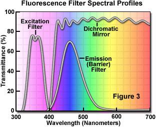
14 Fluorescence microscopy Images are from 14

15 Differential interference contrast (DIC) The contrast is from the gradient of the optical paths, not the optical paths. 15


16 Orientation in in DIC Ref: D. B. Murphy, Fundamentals of Light Microscopy and Electronic Imaging (Wiley-Liss, New York, 2001). 16

17 Comparison between phase contrast and DIC 17
")

18 Fluorescence resonance energy transfer (FRET) Microscopy Images are from 18
19 Confocal Microscopy 19


20 Confocal microscopy Conventional fluorescence microscopy Confocal microscopy Images are from Scientific American, August 1994, p


21 Confocal images Improved depth resolution Images are from 21
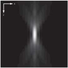
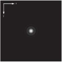
22 Three-dimensional point-spread function Ref: Carl Zeiss, Confocal Laser Scanning Microscopy 22

23 Effect of of the pinhole diameter and NA Ref: Carl Zeiss, Confocal Laser Scanning Microscopy 23

24 Scanning system Images are from 24
25 Nanometer depth resolution: differential confocal microscopy When signal light is from a single surface zero derivative signal linear region axial displacement (µm) 4 Typical slope in the linear region = 1/µm 10 nm displacement = 1% signal variation Ref: C.-H. Lee and J. Wang, Opt. Commun. 135, 233 (1997). 25
 aluminum (R = 80%) height (nm) 400 300 200 100 profiled by DCM profiled by AFM Ref: C.-W. Tsai, C.-H. Lee, and J. Wang, Opt. Lett. 24, 1732 (1999).")
26 Sample images of of DCM 70-nm deep H-trench on InGaAs glass slide human red blood cell cell surface The center recess is 570 nm. fused silica (R = 4%) aluminum (R = 80%) height (nm) profiled by DCM profiled by AFM Ref: C.-W. Tsai, C.-H. Lee, and J. Wang, Opt. Lett. 24, 1732 (1999) distance (µm) 80 26
27 Digital Images 27

28 A digitized image Images are from 28
")
29 Charge-coupled device (CCD) 29
 dark current ( 50 pa/cm 2 at 20 o C) dynamic range (> 85 db) digital or analog bit depth (10 bit; 12 bit; 14 bit...) 30")
30 Specifications of of CCD cameras pixel size (8 µm; 23 µm) pixel resolution (640 x 480; 1024 x 1024) spectral response (300 nm to 1000 nm) well depth (> 300,000 e - ) dark current ( 50 pa/cm 2 at 20 o C) dynamic range (> 85 db) digital or analog bit depth (10 bit; 12 bit; 14 bit...) 30

31 Signal digitization pixels 31

32 Sufficient sampling Sampling frequency 2 x signal bandwidth For CCD cameras, the pixel size on the image should be smaller than half the optical resolution. From Carl Zeiss, Confocal Laser Scanning Microscopy 32
33 Related Technologies 33


34 Multiphoton Microscopy IR light can penetrate deeper into the tissues. Femtosecond laser pulses are required to perform two-photon excitation. Images are from 34
.")
35 Widefield optically sectioning microscopy spatial phase shift: 2π/3 p Homodyne detection principle ( ) ( ) ( ) I = I I + I I + I I Axial response curve: focal plane sample Ref: M. A. A. Neil, R. Juskaitis, and T. Wilson, Optics Letters 22, 1905 (1997). Carl Zeiss, ApoTome

36 Sectioned fluorescence images without scanning Fluorescence Optically sectioned
 4 Typical slope in the linear region = 1/µm 10 nm displacement = 1% signal variation Ref: C.-H. Lee and J. Wang, Opt. Commun.")
37 The concept of of differential confocal microscopy When signal light is from a single surface zero derivative signal linear region axial displacement (µm) 4 Typical slope in the linear region = 1/µm 10 nm displacement = 1% signal variation Ref: C.-H. Lee and J. Wang, Opt. Commun. 135, 233 (1997). 37
 grid pattern 70 nm trench on InGaAs Water-immersion objective")
 250 200 150 100 50 0 5 AFM this technique 10 15 20 25 Distance")
. C.-H. Lee, H.-Y. Mong, and W.-C.")
38 The NIWOP technique stabilized lamp 14 bit CCD camera Band pass filter ( nm) grid pattern 70 nm trench on InGaAs Water-immersion objective All the components are added outside a bench-top microscope. Height (nm) AFM this technique Distance (µm) This technique is called non-interferometric widefield optical profilometry (NIWOP). C.-H. Lee, H.-Y. Mong, and W.-C. Lin, Optics Letters 27, 1773 (2002). 38


39 Observation of of membrane ripples of of a living cell 10 µ m Bright field image nm The ripples are moving away from the cell edge with an average speed about 1.3 µm/h. C.-C. Wang, J.-Y. Lin, and C.-H. Lee, Optics Express 13, (2005). 39


40 Highlighted in Virtual Journal for Biomedical Optics (January 2006) 40
 microscopy Ref: G.")

41 Stimulated emission depletion (STED) microscopy Ref: G. Donnert et al., Proc. Natl. Acad. Sci. USA 103, (2006). 41
.")
42 Confocal microscopy for single molecules Blinking Ref: W. E. Moerner and M. Orrit, Science 283, 1670 (1999). 42
. Ref: W.")
43 X-ray microscopy The resolution of a zone plate is almost equal to the smallest (outermost) zone width. With current e-beam lithography, the smallest zone width can be ~15 nm. Ref: C. Jacobsen, Trends Cell Biol. 9, 44 (1999). Ref: W. Chao et al, Nature 435, 1210 (2005). 43


44 Compact soft x-ray microscope Resolution ~ 100 nm Image of diatom R 3.37 nm Ref: M. Berglund et al., J. Microsc. 197, 268 (2000). 44


45 X-ray microtomography Commercial product available Vesicles inside a cell Capacitor Resolution ~ 250 nm Ref: Y.Wang et al., J. Microsc. 197, 80 (2000). Resolution < 10 µm 45
CCAM Microscope Objectives
 CCAM Microscope Objectives Things to consider when selecting an objective Magnification Numerical Aperture (NA) resolving power and light intensity of the objective Working Distance distance between the
CCAM s Selection of. Zeiss Microscope Objectives
 CCAM s Selection of Zeiss Microscope Objectives 1. Magnification Image scale 2. Resolution The minimum separation distance between two points that are clearly resolved. The resolution of an objective is
Introduction to light microscopy
 Center for Microscopy and Image Anaylsis Introduction to light microscopy Basic concepts of imaging with light Urs Ziegler ziegler@zmb.uzh.ch Light interacting with matter Absorbtion Refraction Diffraction
Bio 407. Applied microscopy. Introduction into light microscopy. José María Mateos. Center for Microscopy and Image Analysis
 Center for Microscopy and Image Analysis Bio 407 Applied Introduction into light José María Mateos Fundamentals of light Compound microscope Microscope composed of an objective and an additional lens (eyepiece,
3D light microscopy techniques
 3D light microscopy techniques The image of a point is a 3D feature In-focus image Out-of-focus image The image of a point is not a point Point Spread Function (PSF) 1D imaging 1 1 2! NA = 0.5! NA 2D imaging
Introduction to light microscopy
 Center for Microscopy and Image Anaylsis Introduction to light Basic concepts of imaging with light Urs Ziegler ziegler@zmb.uzh.ch Microscopy with light 1 Light interacting with matter Absorbtion Refraction
Imaging Introduction. September 24, 2010
 Imaging Introduction September 24, 2010 What is a microscope? Merriam-Webster: an optical instrument consisting of a lens or combination of lenses for making enlarged images of minute objects; especially:
Microscopy: Fundamental Principles and Practical Approaches
 Microscopy: Fundamental Principles and Practical Approaches Simon Atkinson Online Resource: http://micro.magnet.fsu.edu/primer/index.html Book: Murphy, D.B. Fundamentals of Light Microscopy and Electronic
Resolution. Diffraction from apertures limits resolution. Rayleigh criterion θ Rayleigh = 1.22 λ/d 1 peak at 2 nd minimum. θ f D
 Microscopy Outline 1. Resolution and Simple Optical Microscope 2. Contrast enhancement: Dark field, Fluorescence (Chelsea & Peter), Phase Contrast, DIC 3. Newer Methods: Scanning Tunneling microscopy (STM),
Examination, TEN1, in courses SK2500/SK2501, Physics of Biomedical Microscopy,
 KTH Applied Physics Examination, TEN1, in courses SK2500/SK2501, Physics of Biomedical Microscopy, 2009-06-05, 8-13, FB51 Allowed aids: Compendium Imaging Physics (handed out) Compendium Light Microscopy
5/4/2015 INTRODUCTION TO LIGHT MICROSCOPY. Urs Ziegler MICROSCOPY WITH LIGHT. Image formation in a nutshell. Overview of techniques
 INTRODUCTION TO LIGHT MICROSCOPY Urs Ziegler ziegler@zmb.uzh.ch MICROSCOPY WITH LIGHT INTRODUCTION TO LIGHT MICROSCOPY Image formation in a nutshell Overview of techniques Widefield microscopy Resolution
3D light microscopy techniques
 3D light microscopy techniques The image of a point is a 3D feature In-focus image Out-of-focus image The image of a point is not a point Point Spread Function (PSF) 1D imaging 2D imaging 3D imaging Resolution
Microscopic Structures
 Microscopic Structures Image Analysis Metal, 3D Image (Red-Green) The microscopic methods range from dark field / bright field microscopy through polarisation- and inverse microscopy to techniques like
Point Spread Function. Confocal Laser Scanning Microscopy. Confocal Aperture. Optical aberrations. Alternative Scanning Microscopy
 Bi177 Lecture 5 Adding the Third Dimension Wide-field Imaging Point Spread Function Deconvolution Confocal Laser Scanning Microscopy Confocal Aperture Optical aberrations Alternative Scanning Microscopy
Development of a new multi-wavelength confocal surface profilometer for in-situ automatic optical inspection (AOI)
") Development of a new multi-wavelength confocal surface profilometer for in-situ automatic optical inspection (AOI) Liang-Chia Chen 1#, Chao-Nan Chen 1 and Yi-Wei Chang 1 1. Institute of Automation Technology,
Development of a new multi-wavelength confocal surface profilometer for in-situ automatic optical inspection (AOI) Liang-Chia Chen 1#, Chao-Nan Chen 1 and Yi-Wei Chang 1 1. Institute of Automation Technology,
Invitation for a walk through microscopy. Sebastian Schuchmann Jörg Rösner
 Invitation for a walk through microscopy Sebastian Schuchmann Jörg Rösner joerg.roesner@charite.de Techniques in microscopy Conventional (light) microscopy bright & dark field, phase & interference contrast
Lecture 23 MNS 102: Techniques for Materials and Nano Sciences
 Lecture 23 MNS 102: Techniques for Materials and Nano Sciences Reference: #1 C. R. Brundle, C. A. Evans, S. Wilson, "Encyclopedia of Materials Characterization", Butterworth-Heinemann, Toronto (1992),
k λ NA Resolution of optical systems depends on the wavelength visible light λ = 500 nm Extreme ultra-violet and soft x-ray light λ = 1-50 nm
 Resolution of optical systems depends on the wavelength visible light λ = 500 nm Spatial Resolution = k λ NA EUV and SXR microscopy can potentially resolve full-field images with 10-100x smaller features
INTRODUCTION TO MICROSCOPY. Urs Ziegler THE PROBLEM
 INTRODUCTION TO MICROSCOPY Urs Ziegler ziegler@zmb.uzh.ch THE PROBLEM 1 ORGANISMS ARE LARGE LIGHT AND ELECTRONS: ELECTROMAGNETIC WAVES v = Wavelength ( ) Speed (v) Frequency ( ) Amplitude (A) Propagation
IC 2 S High Performance Objectives
 M i c r o s c o p y f r o m C a r l Z e i s s IC 2 S igh Performance Objectives for Biomedical Applications with Laser Based Imaging Systems LSM,, ConfoCor, TIRF and ELYRA Carl Zeiss offers a large range
Microscopy Techniques that make it easy to see things this small.
 Microscopy Techniques that make it easy to see things this small. What is a Microscope? An instrument for viewing objects that are too small to be seen easily by the naked eye. Dutch spectacle-makers Hans
Introduction to Light Microscopy. (Image: T. Wittman, Scripps)
") Introduction to Light Microscopy (Image: T. Wittman, Scripps) The Light Microscope Four centuries of history Vibrant current development One of the most widely used research tools A. Khodjakov et al. Major
Introduction to Light Microscopy (Image: T. Wittman, Scripps) The Light Microscope Four centuries of history Vibrant current development One of the most widely used research tools A. Khodjakov et al. Major
Heisenberg) relation applied to space and transverse wavevector
 relation applied to space and transverse wavevector") 2. Optical Microscopy 2.1 Principles A microscope is in principle nothing else than a simple lens system for magnifying small objects. The first lens, called the objective, has a short focal length (a
2. Optical Microscopy 2.1 Principles A microscope is in principle nothing else than a simple lens system for magnifying small objects. The first lens, called the objective, has a short focal length (a
Why and How? Daniel Gitler Dept. of Physiology Ben-Gurion University of the Negev. Microscopy course, Michmoret Dec 2005
 Why and How? Daniel Gitler Dept. of Physiology Ben-Gurion University of the Negev Why use confocal microscopy? Principles of the laser scanning confocal microscope. Image resolution. Manipulating the
Measuring chromatic aberrations in imaging systems using plasmonic nano particles
 Measuring chromatic aberrations in imaging systems using plasmonic nano particles Sylvain D. Gennaro, Tyler R. Roschuk, Stefan A. Maier, and Rupert F. Oulton* Department of Physics, The Blackett Laboratory,
Introduction to light microscopy
 Center for Microscopy and Image Anaylsis Introduction to light Imaging with light / Overview of techniques Urs Ziegler ziegler@zmb.uzh.ch Light interacting with matter Absorbtion Refraction Diffraction
Microscope anatomy, image formation and resolution
 Microscope anatomy, image formation and resolution Ian Dobbie Buy this book for your lab: D.B. Murphy, "Fundamentals of light microscopy and electronic imaging", ISBN 0-471-25391-X Visit these websites:
Supplementary Figure 1. Effect of the spacer thickness on the resonance properties of the gold and silver metasurface layers.
 Supplementary Figure 1. Effect of the spacer thickness on the resonance properties of the gold and silver metasurface layers. Finite-difference time-domain calculations of the optical transmittance through
Development of a High-speed Super-resolution Confocal Scanner
 Development of a High-speed Super-resolution Confocal Scanner Takuya Azuma *1 Takayuki Kei *1 Super-resolution microscopy techniques that overcome the spatial resolution limit of conventional light microscopy
Optical Components for Laser Applications. Günter Toesko - Laserseminar BLZ im Dezember
 Günter Toesko - Laserseminar BLZ im Dezember 2009 1 Aberrations An optical aberration is a distortion in the image formed by an optical system compared to the original. It can arise for a number of reasons
Sensitive measurement of partial coherence using a pinhole array
 1.3 Sensitive measurement of partial coherence using a pinhole array Paul Petruck 1, Rainer Riesenberg 1, Richard Kowarschik 2 1 Institute of Photonic Technology, Albert-Einstein-Strasse 9, 07747 Jena,
Basics of confocal imaging (part I)
") Basics of confocal imaging (part I) Swiss Institute of Technology (EPFL) Faculty of Life Sciences Head of BIOIMAGING AND OPTICS BIOP arne.seitz@epfl.ch Lateral resolution BioImaging &Optics Platform Light
Basics of confocal imaging (part I) Swiss Institute of Technology (EPFL) Faculty of Life Sciences Head of BIOIMAGING AND OPTICS BIOP arne.seitz@epfl.ch Lateral resolution BioImaging &Optics Platform Light
Femtosecond laser microfabrication in. Prof. Dr. Cleber R. Mendonca
 Femtosecond laser microfabrication in polymers Prof. Dr. Cleber R. Mendonca laser microfabrication focus laser beam on material s surface laser microfabrication laser microfabrication laser microfabrication
Confocal Imaging Through Scattering Media with a Volume Holographic Filter
 Confocal Imaging Through Scattering Media with a Volume Holographic Filter Michal Balberg +, George Barbastathis*, Sergio Fantini % and David J. Brady University of Illinois at Urbana-Champaign, Urbana,
Objectives from Carl Zeiss Exceeding Your Expectations
 Microscopy from Carl Zeiss bjectives from Carl Zeiss Exceeding Your Expectations Brilliant Imaging for Research and Routine Work in Life Sciences When Your Research Pushes the Boundaries of What Is Visible,
Applications of Optics
 Nicholas J. Giordano www.cengage.com/physics/giordano Chapter 26 Applications of Optics Marilyn Akins, PhD Broome Community College Applications of Optics Many devices are based on the principles of optics
CFIM MICROSCOPY COURSE PROGRAMME PRINCIPLES OF MICROSCOPY CONFOCAL AND FLUORESCENCE MICROSCOPY
 CFIM MICROSCOPY COURSE PROGRAMME PRINCIPLES OF MICROSCOPY 11.01.16-15.01.2016 CONFOCAL AND FLUORESCENCE MICROSCOPY 25.01.16-29.01.2016 PhD Course - University of Copenhagen Department of Biomedical Sciences
EE-527: MicroFabrication
 EE-57: MicroFabrication Exposure and Imaging Photons white light Hg arc lamp filtered Hg arc lamp excimer laser x-rays from synchrotron Electrons Ions Exposure Sources focused electron beam direct write
Prof. Enrico Gratton - Lecture 6 Fluorescence Microscopy
 Prof. Enrico Gratton - Lecture 6 Fluorescence Microscopy Instrumentation Light Sources: One-photon and Multi-photon Excitation Applications in Cells Lifetime Imaging Figures acknowledgements: E.D. Salmon
Katarina Logg, Kristofer Bodvard, Mikael Käll. Dept. of Applied Physics. 12 September Optical Microscopy. Supervisor s signature:...
 Katarina Logg, Kristofer Bodvard, Mikael Käll Dept. of Applied Physics 12 September 2007 O1 Optical Microscopy Name:.. Date:... Supervisor s signature:... Introduction Over the past decades, the number
TRAINING MANUAL. Multiphoton Microscopy LSM 510 META-NLO
 TRAINING MANUAL Multiphoton Microscopy LSM 510 META-NLO September 2010 Multiphoton Microscopy Training Manual Multiphoton microscopy is only available on the LSM 510 META-NLO system. This system is equipped
Light microscopy BMB 173, Lecture 14, Feb. 21, 2018
 Light microscopy The Structural Biology Continuum Next two lectures: Light microscopy Many slides taken from Scott Fraser, Murphy s Fundamentals of light microscopy, Alberts Molecular Biology of the Cell,
Characteristics of point-focus Simultaneous Spatial and temporal Focusing (SSTF) as a two-photon excited fluorescence microscopy
 as a two-photon excited fluorescence microscopy") Characteristics of point-focus Simultaneous Spatial and temporal Focusing (SSTF) as a two-photon excited fluorescence microscopy Qiyuan Song (M2) and Aoi Nakamura (B4) Abstracts: We theoretically and experimentally
Characteristics of point-focus Simultaneous Spatial and temporal Focusing (SSTF) as a two-photon excited fluorescence microscopy Qiyuan Song (M2) and Aoi Nakamura (B4) Abstracts: We theoretically and experimentally
Maria Smedh, Centre for Cellular Imaging. Maria Smedh, Centre for Cellular Imaging
 Nonlinear microscopy I: Two-photon fluorescence microscopy Multiphoton Microscopy What is multiphoton imaging? Applications Different imaging modes Advantages/disadvantages Scattering of light in thick
Boulevard du Temple Daguerrotype (Paris,1838) a busy street? Nyquist sampling for movement
 a busy street? Nyquist sampling for movement") Boulevard du Temple Daguerrotype (Paris,1838) a busy street? Nyquist sampling for movement CONFOCAL MICROSCOPY BioVis Uppsala, 2017 Jeremy Adler Matyas Molnar Dirk Pacholsky Widefield & Confocal Microscopy
Boulevard du Temple Daguerrotype (Paris,1838) a busy street? Nyquist sampling for movement CONFOCAL MICROSCOPY BioVis Uppsala, 2017 Jeremy Adler Matyas Molnar Dirk Pacholsky Widefield & Confocal Microscopy
SETTING UP OF A TOTAL INTERNAL REFLECTION FLUORESCENT MICROSCOPE (TIRFM) SYSTEM: A DETAILED OVERVIEW
 SYSTEM: A DETAILED OVERVIEW") PK ISSN 0022-2941; CODEN JNSMAC Vol. 51, (2011) PP 31-45 SETTING UP OF A TOTAL INTERNAL REFLECTION FLUORESCENT MICROSCOPE (TIRFM) SYSTEM: A DETAILED OVERVIEW A. R. KHAN 1 *, S. AKHLAQ 1, M. N. B. ABID
PK ISSN 0022-2941; CODEN JNSMAC Vol. 51, (2011) PP 31-45 SETTING UP OF A TOTAL INTERNAL REFLECTION FLUORESCENT MICROSCOPE (TIRFM) SYSTEM: A DETAILED OVERVIEW A. R. KHAN 1 *, S. AKHLAQ 1, M. N. B. ABID
Observing Microorganisms through a Microscope LIGHT MICROSCOPY: This type of microscope uses visible light to observe specimens. Compound Light Micros
 PHARMACEUTICAL MICROBIOLOGY JIGAR SHAH INSTITUTE OF PHARMACY NIRMA UNIVERSITY Observing Microorganisms through a Microscope LIGHT MICROSCOPY: This type of microscope uses visible light to observe specimens.
INTRODUCTION TO OPTICAL MICROSCOPY
 Experimental Biophysics TEK265, FYST23, TNF060, FAF010F Lab Exercise Supervisor: Karl Adolfsson Written by Peter Jönsson and Jason Beech Updated by Henrik Persson, Karl Adolfsson and Zhen Li karl.adolfsson@ftf.lth.se
Supplementary Figure S1: Schematic view of the confocal laser scanning STED microscope used for STED-RICS. For a detailed description of our
 Supplementary Figure S1: Schematic view of the confocal laser scanning STED microscope used for STED-RICS. For a detailed description of our home-built STED microscope used for the STED-RICS experiments,
Immersed transparent microsphere magnifying sub-diffraction-limited objects
 Immersed transparent microsphere magnifying sub-diffraction-limited objects Seoungjun Lee, 1, * Lin Li, 1 Zengbo Wang, 1 Wei Guo, 1 Yinzhou Yan, 1 and Tao Wang 2 1 School of Mechanical, Aerospace and Civil
NIH Public Access Author Manuscript Opt Lett. Author manuscript; available in PMC 2010 August 9.
 NIH Public Access Author Manuscript Published in final edited form as: Opt Lett. 2010 January 1; 35(1): 67 69. Autoconfocal transmission microscopy based on two-photon induced photocurrent of Si photodiodes
Aberrations and adaptive optics for biomedical microscopes
 Aberrations and adaptive optics for biomedical microscopes Martin Booth Department of Engineering Science And Centre for Neural Circuits and Behaviour University of Oxford Outline Rays, wave fronts and
Non-Descanned FLIM Detection in Multiphoton Microscopes
 Non-Descanned FLIM Detection in Multiphoton Microscopes Abstract. Multiphoton microscopes use a femtosecond NIR laser to excite fluorescence in the sample. Excitation is performed via a multi-photon absorption
High resolution extended depth of field microscopy using wavefront coding
 High resolution extended depth of field microscopy using wavefront coding Matthew R. Arnison *, Peter Török #, Colin J. R. Sheppard *, W. T. Cathey +, Edward R. Dowski, Jr. +, Carol J. Cogswell *+ * Physical
OPELCO OPtical ELements COrporation LB Objective Series for Biological Use
 LB Objective Series for Biological Use 105 Executive Drive Suite 100 Dulles, VA 20166-9558 Tel: (703) 471-0080 S PLAN APOCHROMAT OBJECTIVES These objectives compensate for three wavelength of chromatic
Introduction to light microscopy
 Center for Microscopy and Image Anaylsis Introduction to light microscopy (an overview) Microscopy with light Components of a light microscope 1. Light source 2. Objective 3. Sample or specimen holder
FLUORESCENCE MICROSCOPY. Matyas Molnar and Dirk Pacholsky
 FLUORESCENCE MICROSCOPY Matyas Molnar and Dirk Pacholsky 1 The human eye perceives app. 400-700 nm; best at around 500 nm (green) Has a general resolution down to150-300 μm (human hair: 40-250 μm) We need
BASICS IN BIOIMAGING AND OPTICS PLATFORM EPFL SV PTBIOP LIGHT MICROSCOPY
 BASICS IN LIGHT MICROSCOPY OVERVIEW 1. Motivation 2. Basic in optics 3. How microscope works 4. Illumination and resolution 5. Microscope optics 6. Contrasting methods -2- MOTIVATION Why do we need microscopy?
Final Exam, 150 points PMB 185: Techniques in Light Microscopy
 Final Exam, 150 points Name PMB 185: Techniques in Light Microscopy Point value is in parentheses at the end of each question. Note: GFP = green fluorescent protein ; CFP = cyan fluorescent protein ; YFP
X-ray generation by femtosecond laser pulses and its application to soft X-ray imaging microscope
 X-ray generation by femtosecond laser pulses and its application to soft X-ray imaging microscope Kenichi Ikeda 1, Hideyuki Kotaki 1 ' 2 and Kazuhisa Nakajima 1 ' 2 ' 3 1 Graduate University for Advanced
A 3D Profile Parallel Detecting System Based on Differential Confocal Microscopy. Y.H. Wang, X.F. Yu and Y.T. Fei
 Key Engineering Materials Online: 005-10-15 ISSN: 166-9795, Vols. 95-96, pp 501-506 doi:10.408/www.scientific.net/kem.95-96.501 005 Trans Tech Publications, Switzerland A 3D Profile Parallel Detecting
OPTICAL PRINCIPLES OF MICROSCOPY. Interuniversity Course 28 December 2003 Aryeh M. Weiss Bar Ilan University
 OPTICAL PRINCIPLES OF MICROSCOPY Interuniversity Course 28 December 2003 Aryeh M. Weiss Bar Ilan University FOREWORD This slide set was originally presented at the ISM Workshop on Theoretical and Experimental
Education in Microscopy and Digital Imaging
 Contact Us Carl Zeiss Education in Microscopy and Digital Imaging ZEISS Home Products Solutions Support Online Shop ZEISS International ZEISS Campus Home Interactive Tutorials Basic Microscopy Spectral
Microscopy. Matti Hotokka Department of Physical Chemistry Åbo Akademi University
 Microscopy Matti Hotokka Department of Physical Chemistry Åbo Akademi University What s coming Anatomy of a microscope Modes of illumination Practicalities Special applications Basic microscope Ocular
Very short introduction to light microscopy and digital imaging
 Very short introduction to light microscopy and digital imaging Hernan G. Garcia August 1, 2005 1 Light Microscopy Basics In this section we will briefly describe the basic principles of operation and
Enhancement of the lateral resolution and the image quality in a line-scanning tomographic optical microscope
 Summary of the PhD thesis Enhancement of the lateral resolution and the image quality in a line-scanning tomographic optical microscope Author: Dudás, László Supervisors: Prof. Dr. Szabó, Gábor and Dr.
Specimen-induced aberrations and adaptive optics for microscopy
 Specimen-induced aberrations and adaptive optics for microscopy Martin J. Booth, Michael Schwertner and Tony Wilson Department of Engineering Science, University of Oxford, U.K. ABSTRACT The imaging properties
MULTIPHOTON MICROSCOPY. Matyas Molnar Dirk Pacholsky
 MULTIPHOTON MICROSCOPY Matyas Molnar Dirk Pacholsky Information Information given here about 2 Photon microscopy were mainly taken from these sources: Background information on 2-Photon microscopy: http://micro.magnet.fsu.edu/primer/techniques/fluorescence/multiphoton/
Confocal and 2-photon Imaging. October 15, 2010
 Confocal and 2-photon Imaging October 15, 2010 Review Optical Elements Adapted from Sluder & Nordberg 2007 Review Optical Elements Collector Lens Adapted from Sluder & Nordberg 2007 Review Optical Elements
Water-Window Microscope Based on Nitrogen Plasma Capillary Discharge Source
 2015 International Workshop on EUV and Soft X-Ray Sources Water-Window Microscope Based on Nitrogen Plasma Capillary Discharge Source T. Parkman 1, M. F. Nawaz 2, M. Nevrkla 2, M. Vrbova 1, A. Jancarek
Coherent anti-stokes Raman scattering microscopy with dynamic speckle illumination
 Coherent anti-stokes Raman scattering microscopy with dynamic speckle illumination To cite this article: Christoph Heinrich et al 2008 New J. Phys. 10 023029 View the article online for updates and enhancements.
BASICS OF CONFOCAL IMAGING (PART I)
") BASICS OF CONFOCAL IMAGING (PART I) INTERNAL COURSE 2012 LIGHT MICROSCOPY Lateral resolution Transmission Fluorescence d min 1.22 NA obj NA cond 0 0 rairy 0.61 NAobj Ernst Abbe Lord Rayleigh Depth of field
BASICS OF CONFOCAL IMAGING (PART I) INTERNAL COURSE 2012 LIGHT MICROSCOPY Lateral resolution Transmission Fluorescence d min 1.22 NA obj NA cond 0 0 rairy 0.61 NAobj Ernst Abbe Lord Rayleigh Depth of field
Supporting Information
 Copyright WILEY-VCH Verlag GmbH & Co. KGaA, 69469 Weinheim, Germany, 2012. Supporting Information for Adv. Mater., DOI: 10.1002/adma.201203033 Solid Immersion Facilitates Fluorescence Microscopy with Nanometer
Bandpass Edge Dichroic Notch & More
 Edmund Optics BROCHURE Filters COPYRIGHT 217 EDMUND OPTICS, INC. ALL RIGHTS RESERVED 1/17 Bandpass Edge Dichroic Notch & More Contact us for a Stock or Custom Quote Today! USA: +1-856-547-3488 EUROPE:
Confocal Microscopy. Kristin Jensen
 Confocal Microscopy Kristin Jensen 17.11.05 References Cell Biological Applications of Confocal Microscopy, Brian Matsumoto, chapter 1 Studying protein dynamics in living cells,, Jennifer Lippincott-Schwartz
Opterra II Multipoint Scanning Confocal Microscope. Innovation with Integrity
 Opterra II Multipoint Scanning Confocal Microscope Enabling 4D Live-Cell Fluorescence Imaging through Speed, Sensitivity, Viability and Simplicity Innovation with Integrity Fluorescence Microscopy The
ELECTRON MICROSCOPY. 13:10 16:00, Oct. 6, 2008 Institute of Physics, Academia Sinica. Tung Hsu
 ELECTRON MICROSCOPY 13:10 16:00, Oct. 6, 2008 Institute of Physics, Academia Sinica Tung Hsu Department of Materials Science and Engineering National Tsing Hua University Hsinchu 300, TAIWAN Tel. 03-5742564
Light Microscopy. Upon completion of this lecture, the student should be able to:
 Light Light microscopy is based on the interaction of light and tissue components and can be used to study tissue features. Upon completion of this lecture, the student should be able to: 1- Explain the
Adaptive optics two-photon fluorescence microscopy
 Adaptive optics two-photon fluorescence microscopy Yaopeng Zhou 1, Thomas Bifano 1 and Charles Lin 2 1. Manufacturing Engineering Department, Boston University 15 Saint Mary's Street, Brookline MA, 02446
More fancy SPIM, Even fancier SPIM
 More fancy SPIM, Even fancier SPIM Last class Light sheet microscopy Fancy SPIM (ispim, dspim, etc ) This class Multi camera SPIM SIM SPIM Bessels d x,y = λ em 2 NA d z = 2 NA λ ex + n(1 cosθ λ em 1 IsoView
Microscopy. Lecture 2: Optical System of the Microscopy II Herbert Gross. Winter term
 Microscopy Lecture 2: Optical System of the Microscopy II 212-1-22 Herbert Gross Winter term 212 www.iap.uni-jena.de Preliminary time schedule 2 No Date Main subject Detailed topics Lecturer 1 15.1. Optical
Development of Laser Confocal Microscopy for Internal Defect Measurement
 Development of Laser Confocal Microscopy for Internal Defect Measurement Chia-Liang Yeh*, Fu-Cheng Yang, Wei-Hsiung Tsai, and Keng-Li Lin Center for Measurement Standards, Industrial Technology Research
Reflecting optical system to increase signal intensity. in confocal microscopy
 Reflecting optical system to increase signal intensity in confocal microscopy DongKyun Kang *, JungWoo Seo, DaeGab Gweon Nano Opto Mechatronics Laboratory, Dept. of Mechanical Engineering, Korea Advanced
Rates of excitation, emission, ISC
 Bi177 Lecture 4 Fluorescence Microscopy Phenomenon of Fluorescence Energy Diagram Rates of excitation, emission, ISC Practical Issues Lighting, Filters More on diffraction Point Spread Functions Thus Far,
a) How big will that physical image of the cells be your camera sensor?
 How big will that physical image of the cells be your camera sensor?") 1. Consider a regular wide-field microscope set up with a 60x, NA = 1.4 objective and a monochromatic digital camera with 8 um pixels, properly positioned in the primary image plane. This microscope is
1. Consider a regular wide-field microscope set up with a 60x, NA = 1.4 objective and a monochromatic digital camera with 8 um pixels, properly positioned in the primary image plane. This microscope is
The Compound Microscope. Brightfield: Köhler Illumination
 Outline History of Microscopy The Magnifying Glass The Compound Microscope Brightfield: Köhler Illumination Microscopy µικροσ (mikros): small σκοπειν (skopein): to observe History of Microscopy Well :
Nature Methods: doi: /nmeth Supplementary Figure 1. Schematic of 2P-ISIM AO optical setup.
 Supplementary Figure 1 Schematic of 2P-ISIM AO optical setup. Excitation from a femtosecond laser is passed through intensity control and shuttering optics (1/2 λ wave plate, polarizing beam splitting
Multiphoton Microscopy
 Multiphoton Microscopy A. Neumann, Y. Kuznetsova Introduction Multi-Photon Fluorescence Microscopy is a relatively novel imaging technique in cell biology. It relies on the quasi-simultaneous absorption
EE119 Introduction to Optical Engineering Spring 2002 Final Exam. Name:
 EE119 Introduction to Optical Engineering Spring 2002 Final Exam Name: SID: CLOSED BOOK. FOUR 8 1/2 X 11 SHEETS OF NOTES, AND SCIENTIFIC POCKET CALCULATOR PERMITTED. TIME ALLOTTED: 180 MINUTES Fundamental
Εισαγωγική στην Οπτική Απεικόνιση
 Εισαγωγική στην Οπτική Απεικόνιση Δημήτριος Τζεράνης, Ph.D. Εμβιομηχανική και Βιοϊατρική Τεχνολογία Τμήμα Μηχανολόγων Μηχανικών Ε.Μ.Π. Χειμερινό Εξάμηνο 2015 Light: A type of EM Radiation EM radiation:
Practical Flatness Tech Note
 Practical Flatness Tech Note Understanding Laser Dichroic Performance BrightLine laser dichroic beamsplitters set a new standard for super-resolution microscopy with λ/10 flatness per inch, P-V. We ll
Biomedical Imaging 生物醫學影像學
 Biomedical Imaging 生物醫學影像學 楊自森助理教授 牙體技術學系 2013/02/24 tsyang@tmu.edu.tw 1 Course Outline 1. Course Introduction 2. Basic Optics and Light Microscopes 3. Fluorescence/Confocal/TIRF Microscopes 4. FRET Techniques
Flatness of Dichroic Beamsplitters Affects Focus and Image Quality
 Flatness of Dichroic Beamsplitters Affects Focus and Image Quality Flatness of Dichroic Beamsplitters Affects Focus and Image Quality 1. Introduction Even though fluorescence microscopy has become a routine
Fundamentals of Light Microscopy II: Fluorescence, Deconvolution, Confocal, Multiphoton, Spectral microscopy. Integrated Microscopy Course
 Fundamentals of Light Microscopy II: Fluorescence, Deconvolution, Confocal, Multiphoton, Spectral microscopy Integrated Microscopy Course Review Lecture 1: Microscopy Basics Light train Kohler illumination*
Three-dimensional quantitative phase measurement by Commonpath Digital Holographic Microscopy
 Available online at www.sciencedirect.com Physics Procedia 19 (2011) 291 295 International Conference on Optics in Precision Engineering and Nanotechnology Three-dimensional quantitative phase measurement
Microscopy Live Animal Imaging
 Microscopy Live Animal Imaging A collaborative environment that provides the knowledge, instruments, and expertise needed to visualize life at scales ranging from single molecules to entire animals. Project
Measuring incidence angle for throughthe-objective
 Measuring incidence angle for throughthe-objective total internal reflection fluorescence microscopy Thomas P. Burghardt Journal of Biomedical Optics 17(12), 126007 (December 2012) Measuring incidence
Shaping light in microscopy:
 Shaping light in microscopy: Adaptive optical methods and nonconventional beam shapes for enhanced imaging Martí Duocastella planet detector detector sample sample Aberrated wavefront Beamsplitter Adaptive
Advanced 3D Optical Profiler using Grasshopper3 USB3 Vision camera
 Advanced 3D Optical Profiler using Grasshopper3 USB3 Vision camera Figure 1. The Zeta-20 uses the Grasshopper3 and produces true color 3D optical images with multi mode optics technology 3D optical profiling
Specimen-induced distortions in light microscopy
 Journal of Microscopy, Vol. 228, Pt 1 27, pp. 97 12 Received 29 June 26; accepted 11 April 27 Specimen-induced distortions in light microscopy M. S C H W E RT N E R, M. J. B O O T H & T. W I L S O N Department