INTRODUCTION TO MICROSCOPY. Urs Ziegler THE PROBLEM
|
|
|
- Melvyn Wilkins
- 5 years ago
- Views:
Transcription

1 INTRODUCTION TO MICROSCOPY Urs Ziegler THE PROBLEM 1

 Frequency ( ) Amplitude (A)")


2 ORGANISMS ARE LARGE LIGHT AND ELECTRONS: ELECTROMAGNETIC WAVES v = Wavelength ( ) Speed (v) Frequency ( ) Amplitude (A) Propagation direction Vibration direction Light and electrons as a probe of matter Electromagnetic wave Murphy and Davidson,

 t Typically Phase shift of livingcells is /4, not visible for human eyes courtesy Heiko Gäthje, Olympus")
3 INTERACTION OFELECTROMAGNETIC WAVES WITHMATTER Murphy and Davidson, 2013 LIGHT (ELECTROMAGNETIC WAVES) INTERACTS WITH MATTER Stained Specimen Medium Amplitude object Light is absorbed by parts of the specimen and so changed in brightness and colour Phase object Phase shift depends on the refractive index and thickness of the object. 2 ( n 2 n1 ) t Typically Phase shift of livingcells is /4, not visible for human eyes courtesy Heiko Gäthje, Olympus Europe SE & Co. KG 3

 or generate sections Sample preparation to allow processing of")
 Choice of imaging method depending on sample and")

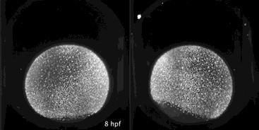
4 ORGANISMS ARE LARGE SAMPLE PREPARATION CHOOSING THE RIGHT TOOL FOR IMAGING Typical Organisms in Life Science Solutions to allow imaging Thin samples (e.g. cells) or generate sections Sample preparation to allow processing of tissue without deterioration (e.g. fixation, freezing) Choice of imaging method depending on sample and resolution to be achieved Confocal laser scanning microscopy In vivo microscopy Selective plane illumination microscopy Superresolution techniques Transmission electron microscopy Scanning electron mciroscopy MICROSCOPY WITH LIGHT 4


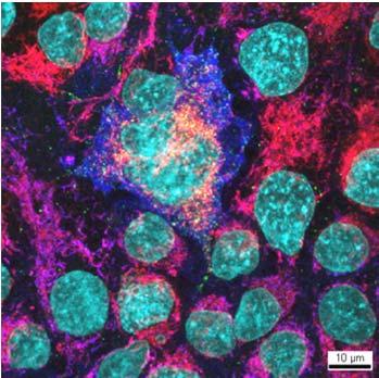
5 FLUORESCENCE IN MICROSCOPY DNA Bax Mitochondria Cytochrome C DNA Bax Mitochondria Cytochrome C DNA Bax Mitochondria Cytochrome C WIDEFIELD MICROSCOPY 5

6 ESSENTIAL PARTS OF A MICROSCOPE Compound microscope Main parts of a microscope Illumination light source Focusing of light collector lenses and condensor Sample holder Objective Eyepiece Focus WIDEFIELD MICROSCOPY Problem Classical example of widefield imaging various points of an object are viewed simultaneously points of planes, other than the object plane, produce background illumination lowering the contrast stunningdepth field photographs/ Principle of widefield imaging Example from microscopy: histology 6


7 WIDEFIELD MICROSCOPY Problem Classical example of widefield imaging various points of an object are viewed simultaneously points of planes, other than the object plane, produce background illumination lowering the contrast stunningdepth field photographs/ Principle of widefield imaging Example from microscopy: histology FUNDAMENTALSETUP OF LIGHT MICROSCOPES 7

8 CONFOCAL LASERSCANNING MICROSCOPY CONFOCAL LASERSCANNING MICROSCOPY: CLSM Problem in widefield microscopy various points of an object are viewed simultaneously points of planes, other than the object plane, produce background illumination lowering the contrast Principle of widefield confocal imaging Solution 1. Illuminate a point in the object (using a focused laser which is scanned over the object hence the name laserscanning) 2. Introduce a pinhole in the image plane 3. The image plane is confocal to the focused object plane hence the name: confocal Motoneuronal endplate: widefield CLSM data data 8
")


9 TEMPORAL RESOLUTION NIPKOW DISK (SPINNING DISK TANDEM) SCANNING MICROSCOPY Problem Solution Speed in (single) point scanning confocal is limited! Scanning with multiple focused laser spots 1 to a few frames per seconds Schematics Illumination Detection campus.magnet.fsu.edu/tutorials MULTIPHOTON (LASERSCANNING) MICROSCOPY 9


")
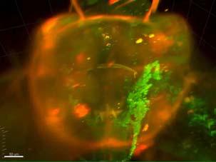
10 MULTIPHOTON MICROSCOPY 3D sectioning without pinhole Schematics Excitation with one photon linearly depends on the amount of photons from the light source. Multiphoton excitation is proportional to the square of the intensity of light. Exponential drop in excitation out of focus in multiphoton excitation. No pinhole needed because no emitted light from out of focus. microsystems.com/science lab MULTIPHOTON MICROSCOPY Imaging in scattering tissue and deep into tissue Pulsed infrared laser ( nm) excites fluorochromes by multiphoton absorbtion Excitation in a small volume defined by the probability (densitiy of photons high) of a simultaneous multiphoton absorbtion All fluorescent photons provide useful signals. Helmchen and Denk, Nature Methods


11 MULTIPHOTON MICROSCOPY Kidney Brain Living mouse: kidney (Hoechst, 10kD dextran FITC, 150kD dextran Texas Red Helmchen, F., and W. Denk Deep tissue two photon microscopy. Nature methods. 2: LIGHTSHEET (SELECTIVE PLANE ILLUMINATION) MICROSCOPY 11

 Light sheet imaging technique Better signal to")

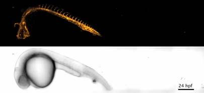
12 SELECTIVE PLANE ILLUMINATION MICROSCOPY LIGHTSHEET MICROSCOPY 3D Imaging with low phototoxicity and high speed Excitation of focal plane only Detection of whole plane (CCD parallel) Light sheet imaging technique Better signal to noise ratio Low phototoxicity 4D imaging Huisken J, Stainier D Y R Development 2009;136: SELECTIVE PLANE ILLUMINATION MICROSCOPY LIGHTSHEET MICROSCOPY Excitation of focal plane only Detection of whole plane (CCD parallel) Light sheet imaging technique Better signal to noise ratio Low phototoxicity 4D imaging Huisken J, Stainier D Y R Development 2009;136:


13 SUPERRESOLUTION MICROSCOPY SUPERRESOLUTION IMAGING Why superresolution imaging? Is there a limit in resolution that cannot be overcome? Why do we want to overcome the limit in resolution? 13
/ _ = ( λ)/ ^2 These formula")


14 RESOLUTION LIMITS _ = (0.61 λ)/ _ = ( λ)/ ^2 These formula are used for the calculation of resolution in widefield microscopy. In other techniques like confocal laser scanning, multiphoton microscopy, etc slightly other formulas are used. SUPERRESOLUTION MICROSCOPY: STATISTICAL MICROSCOPY LIKE PALM, STORM, GSD PALM: PhotoActivated LightMicroscopy STORM: Stochastic Optical Reconstruction Microscopy GSD: Ground State Depletion microscopy stochastic photoswitching of fluorescent proteins where most of the molecules remain dark 14


15 STIMULATED EMISSION DEPLETION MICROSCOPY : STED In STED, an initial excitation pulse is focused on a spot. The spot is narrowed by a second, donut shaped pulse that prompts all excited fluorophores in the body of the donut to emit (this is the emission depletion part of STED). This leaves only the hole of the donut in an excited state, and only this narrow hole is detected as an emitted fluorescence. ELECTRON MICROSCOPY 15

")
 The")

")

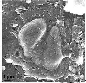
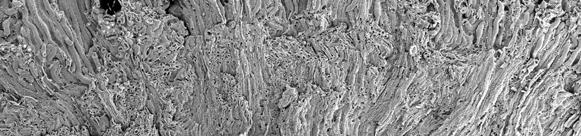
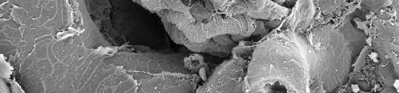
16 THE TYPES OF ELECTRON MICROSCOPES Transmission electron microscope (TEM) Scanning electron microscope (SEM) The types of electron microscopes Transmission electron microscope (TEM) Electron beam Scanning electron microscope (SEM) Electron beam Specimen ~100 nm Projection Specimen Surface Hela Cells 16


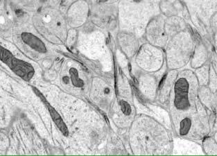
17 Transmission electron microscope vs. Widefield light microscope Transmission electron microscope Widefield light microscope Illumination Condenser lens Specimen Objective lens Projector lens Final image Examples TEM Mouse cerebellum Microtubule Dendrite Synapse Ribosomes Golgi Mitochondrium Nucleus 500 nm Specimen courtesy of B. Sobottka, Institute of Experimental Immunology, University of Zurich 17


18 Examples TEM Mouse cerebellum Mitochondrium Lipid bilayer Golgi Nucleus 100 nm Scanning electron microscope vs. Confocal laser scanning microscope Scanning electron microscope Confocal laser scanning microscope Illumination Detector Lens system Beam scanner Lens system Specimen 18


19 Examples SEM Mouse kidney 500 µm Examples SEM Mouse kidney (glomerulus) 10 µm 19
20 LITERATURE AND ACKNOWLEDGMENTS Literature Fundamentals of light microscopy and electronic imaging, Douglas B. Murphy; Wiley Liss, 2001 ISBN X Light Microscopy in Biology A practical approach, A. J. Lacey; Oxford University Press, 2004 Light and Electron Microscopy, E. M. Slayter, H. S. Slayter; Cambridge University Press, Acknowledgments 20
5/4/2015 INTRODUCTION TO LIGHT MICROSCOPY. Urs Ziegler MICROSCOPY WITH LIGHT. Image formation in a nutshell. Overview of techniques
 INTRODUCTION TO LIGHT MICROSCOPY Urs Ziegler ziegler@zmb.uzh.ch MICROSCOPY WITH LIGHT INTRODUCTION TO LIGHT MICROSCOPY Image formation in a nutshell Overview of techniques Widefield microscopy Resolution
Introduction to light microscopy
 Center for Microscopy and Image Anaylsis Introduction to light microscopy (an overview) Microscopy with light Components of a light microscope 1. Light source 2. Objective 3. Sample or specimen holder
Introduction to light microscopy
 Center for Microscopy and Image Anaylsis Introduction to light Imaging with light / Overview of techniques Urs Ziegler ziegler@zmb.uzh.ch Light interacting with matter Absorbtion Refraction Diffraction
Introduction to light microscopy
 Center for Microscopy and Image Anaylsis Introduction to light microscopy Basic concepts of imaging with light Urs Ziegler ziegler@zmb.uzh.ch Light interacting with matter Absorbtion Refraction Diffraction
Introduction to light microscopy
 Center for Microscopy and Image Anaylsis Introduction to light Basic concepts of imaging with light Urs Ziegler ziegler@zmb.uzh.ch Microscopy with light 1 Light interacting with matter Absorbtion Refraction
Bio 407. Applied microscopy. Introduction into light microscopy. José María Mateos. Center for Microscopy and Image Analysis
 Center for Microscopy and Image Analysis Bio 407 Applied Introduction into light José María Mateos Fundamentals of light Compound microscope Microscope composed of an objective and an additional lens (eyepiece,
Light Microscopy. Upon completion of this lecture, the student should be able to:
 Light Light microscopy is based on the interaction of light and tissue components and can be used to study tissue features. Upon completion of this lecture, the student should be able to: 1- Explain the
3D light microscopy techniques
 3D light microscopy techniques The image of a point is a 3D feature In-focus image Out-of-focus image The image of a point is not a point Point Spread Function (PSF) 1D imaging 1 1 2! NA = 0.5! NA 2D imaging
Development of a High-speed Super-resolution Confocal Scanner
 Development of a High-speed Super-resolution Confocal Scanner Takuya Azuma *1 Takayuki Kei *1 Super-resolution microscopy techniques that overcome the spatial resolution limit of conventional light microscopy
Invitation for a walk through microscopy. Sebastian Schuchmann Jörg Rösner
 Invitation for a walk through microscopy Sebastian Schuchmann Jörg Rösner joerg.roesner@charite.de Techniques in microscopy Conventional (light) microscopy bright & dark field, phase & interference contrast
Maria Smedh, Centre for Cellular Imaging. Maria Smedh, Centre for Cellular Imaging
 Nonlinear microscopy I: Two-photon fluorescence microscopy Multiphoton Microscopy What is multiphoton imaging? Applications Different imaging modes Advantages/disadvantages Scattering of light in thick
Point Spread Function. Confocal Laser Scanning Microscopy. Confocal Aperture. Optical aberrations. Alternative Scanning Microscopy
 Bi177 Lecture 5 Adding the Third Dimension Wide-field Imaging Point Spread Function Deconvolution Confocal Laser Scanning Microscopy Confocal Aperture Optical aberrations Alternative Scanning Microscopy
EUV microscopy - a user s perspective Dimitri Scholz EUV,
 EUV microscopy - a user s perspective Dimitri Scholz EUV, 09.11.2011 Imaging technologies: available at UCD now and in the next future Begin ab ovo - Simple approaches direct to the goal - Standard methods
MOM#3: LIGHT SHEET MICROSCOPY (LSM) Stanley Cohen, MD
 Stanley Cohen, MD") MOM#3: LIGHT SHEET MICROSCOPY (LSM) Stanley Cohen, MD Introduction. Although the technical details of light sheet imaging and its various permutations appear at first glance to be complex and require some
MOM#3: LIGHT SHEET MICROSCOPY (LSM) Stanley Cohen, MD Introduction. Although the technical details of light sheet imaging and its various permutations appear at first glance to be complex and require some
Basics of confocal imaging (part I)
") Basics of confocal imaging (part I) Swiss Institute of Technology (EPFL) Faculty of Life Sciences Head of BIOIMAGING AND OPTICS BIOP arne.seitz@epfl.ch Lateral resolution BioImaging &Optics Platform Light
Basics of confocal imaging (part I) Swiss Institute of Technology (EPFL) Faculty of Life Sciences Head of BIOIMAGING AND OPTICS BIOP arne.seitz@epfl.ch Lateral resolution BioImaging &Optics Platform Light
Resolution. Diffraction from apertures limits resolution. Rayleigh criterion θ Rayleigh = 1.22 λ/d 1 peak at 2 nd minimum. θ f D
 Microscopy Outline 1. Resolution and Simple Optical Microscope 2. Contrast enhancement: Dark field, Fluorescence (Chelsea & Peter), Phase Contrast, DIC 3. Newer Methods: Scanning Tunneling microscopy (STM),
3D light microscopy techniques
 3D light microscopy techniques The image of a point is a 3D feature In-focus image Out-of-focus image The image of a point is not a point Point Spread Function (PSF) 1D imaging 2D imaging 3D imaging Resolution
MULTIPHOTON MICROSCOPY. Matyas Molnar Dirk Pacholsky
 MULTIPHOTON MICROSCOPY Matyas Molnar Dirk Pacholsky Information Information given here about 2 Photon microscopy were mainly taken from these sources: Background information on 2-Photon microscopy: http://micro.magnet.fsu.edu/primer/techniques/fluorescence/multiphoton/
TRAINING MANUAL. Multiphoton Microscopy LSM 510 META-NLO
 TRAINING MANUAL Multiphoton Microscopy LSM 510 META-NLO September 2010 Multiphoton Microscopy Training Manual Multiphoton microscopy is only available on the LSM 510 META-NLO system. This system is equipped
Confocal and 2-photon Imaging. October 15, 2010
 Confocal and 2-photon Imaging October 15, 2010 Review Optical Elements Adapted from Sluder & Nordberg 2007 Review Optical Elements Collector Lens Adapted from Sluder & Nordberg 2007 Review Optical Elements
Observing Microorganisms through a Microscope LIGHT MICROSCOPY: This type of microscope uses visible light to observe specimens. Compound Light Micros
 PHARMACEUTICAL MICROBIOLOGY JIGAR SHAH INSTITUTE OF PHARMACY NIRMA UNIVERSITY Observing Microorganisms through a Microscope LIGHT MICROSCOPY: This type of microscope uses visible light to observe specimens.
Chapter 2 The Study of Microbial Structure: Microscopy and Specimen Preparation
 Chapter 2 The Study of Microbial Structure: Microscopy and Specimen Preparation 1 Lenses and the Bending of Light light is refracted (bent) when passing from one medium to another refractive index a measure
Observing Microorganisms through a Microscope
 2016/2/19 PowerPoint Lecture Presentations prepared by Bradley W. Christian, McLennan Community College CHAPTER 3 Observing Microorganisms through a Microscope 1 Figure 3.2 Microscopes and Magnification.
Microscopy. CS/CME/BioE/Biophys/BMI 279 Nov. 2, 2017 Ron Dror
 Microscopy CS/CME/BioE/Biophys/BMI 279 Nov. 2, 2017 Ron Dror 1 Outline Microscopy: the basics Fluorescence microscopy Resolution limits The diffraction limit Beating the diffraction limit 2 Microscopy:
Confocal Microscopy. Kristin Jensen
 Confocal Microscopy Kristin Jensen 17.11.05 References Cell Biological Applications of Confocal Microscopy, Brian Matsumoto, chapter 1 Studying protein dynamics in living cells,, Jennifer Lippincott-Schwartz
BASICS OF CONFOCAL IMAGING (PART I)
") BASICS OF CONFOCAL IMAGING (PART I) INTERNAL COURSE 2012 LIGHT MICROSCOPY Lateral resolution Transmission Fluorescence d min 1.22 NA obj NA cond 0 0 rairy 0.61 NAobj Ernst Abbe Lord Rayleigh Depth of field
BASICS OF CONFOCAL IMAGING (PART I) INTERNAL COURSE 2012 LIGHT MICROSCOPY Lateral resolution Transmission Fluorescence d min 1.22 NA obj NA cond 0 0 rairy 0.61 NAobj Ernst Abbe Lord Rayleigh Depth of field
Script Bio 407 Applied Microscopy Light microscopy
 Center for Microscopy and Image Analysis Dr. José María Mateos Center for Microscopy and Image Analysis Winterthurerstrasse 190 CH-8057 Zurich Phone 044 635 98 20 mateos@zmb.uzh.ch www.zmb.uzh.ch Script
Tissue Preparation ORGANISM IMAGE TISSUE PREPARATION. 1) Fixation: halts cell metabolism, preserves cell/tissue structure
 Fixation: halts cell metabolism, preserves cell/tissue structure") Lab starts this week! ANNOUNCEMENTS - Tuesday or Wednesday 1:25 ISB 264 - Read Lab 1: Microscopy and Imaging (see Web Page) - Getting started on Lab Group project - Organ for investigation - Lab project
Lab starts this week! ANNOUNCEMENTS - Tuesday or Wednesday 1:25 ISB 264 - Read Lab 1: Microscopy and Imaging (see Web Page) - Getting started on Lab Group project - Organ for investigation - Lab project
Confocal Microscopy and Related Techniques
 Confocal Microscopy and Related Techniques Chau-Hwang Lee Associate Research Fellow Research Center for Applied Sciences, Academia Sinica 128 Sec. 2, Academia Rd., Nankang, Taipei 11529, Taiwan E-mail:
Microscopy Techniques that make it easy to see things this small.
 Microscopy Techniques that make it easy to see things this small. What is a Microscope? An instrument for viewing objects that are too small to be seen easily by the naked eye. Dutch spectacle-makers Hans
Light microscopy BMB 173, Lecture 14, Feb. 21, 2018
 Light microscopy The Structural Biology Continuum Next two lectures: Light microscopy Many slides taken from Scott Fraser, Murphy s Fundamentals of light microscopy, Alberts Molecular Biology of the Cell,
Shreyash Tandon M.S. III Year
 Shreyash Tandon M.S. III Year 20091015 Confocal microscopy is a powerful tool for generating high-resolution images and 3-D reconstructions of a specimen by using point illumination and a spatial pinhole
Lecture 4 to 5 MICROSCOPY-PRINCIPLES AND TYPES
 Lecture 4 to 5 MICROSCOPY-PRINCIPLES AND TYPES Microorganisms are too small to be seen by our unaided eyes and the microscopes are of crucial importance as they help to view the microbes. A microscope
Practical Flatness Tech Note
 Practical Flatness Tech Note Understanding Laser Dichroic Performance BrightLine laser dichroic beamsplitters set a new standard for super-resolution microscopy with λ/10 flatness per inch, P-V. We ll
Advanced Optical Microscopy
 Nanosystems I - Seminar TU München 8th December 2008 1 Introduction to Classical Optical Microscopy Denitions in Optical Microscopy Contrast and Contrast Enhancement 1 Introduction to Classical Optical
Final Exam, 150 points PMB 185: Techniques in Light Microscopy
 Final Exam, 150 points Name PMB 185: Techniques in Light Microscopy Point value is in parentheses at the end of each question. Note: GFP = green fluorescent protein ; CFP = cyan fluorescent protein ; YFP
Dynamic Confocal Imaging of Living Brain. Advantages and risks of multiphoton microscopy in physiology
 Dynamic Confocal Imaging of Living Brain Advantages and risks of multiphoton microscopy in physiology Confocal laser scanning microscopy In conventional optical microscopy focused and out-offocus light
Boulevard du Temple Daguerrotype (Paris,1838) a busy street? Nyquist sampling for movement
 a busy street? Nyquist sampling for movement") Boulevard du Temple Daguerrotype (Paris,1838) a busy street? Nyquist sampling for movement CONFOCAL MICROSCOPY BioVis Uppsala, 2017 Jeremy Adler Matyas Molnar Dirk Pacholsky Widefield & Confocal Microscopy
Boulevard du Temple Daguerrotype (Paris,1838) a busy street? Nyquist sampling for movement CONFOCAL MICROSCOPY BioVis Uppsala, 2017 Jeremy Adler Matyas Molnar Dirk Pacholsky Widefield & Confocal Microscopy
S200 Course LECTURE 1 TEM
 S200 Course LECTURE 1 TEM Development of Electron Microscopy 1897 Discovery of the electron (J.J. Thompson) 1924 Particle and wave theory (L. de Broglie) 1926 Electromagnetic Lens (H. Busch) 1932 Construction
Confocal Imaging Through Scattering Media with a Volume Holographic Filter
 Confocal Imaging Through Scattering Media with a Volume Holographic Filter Michal Balberg +, George Barbastathis*, Sergio Fantini % and David J. Brady University of Illinois at Urbana-Champaign, Urbana,
Modes of light microscopy Choosing the appropriate system
 Modes of light microscopy Choosing the appropriate system Wide-field microscopy Confocal microscopy Multi-photon microscopy Wide-field, inverted fluorescence microscope Nikon MicroscopyU Endosome migration
Shaping light in microscopy:
 Shaping light in microscopy: Adaptive optical methods and nonconventional beam shapes for enhanced imaging Martí Duocastella planet detector detector sample sample Aberrated wavefront Beamsplitter Adaptive
Multiphoton Microscopy
 Multiphoton Microscopy A. Neumann, Y. Kuznetsova Introduction Multi-Photon Fluorescence Microscopy is a relatively novel imaging technique in cell biology. It relies on the quasi-simultaneous absorption
Opterra II Multipoint Scanning Confocal Microscope. Innovation with Integrity
 Opterra II Multipoint Scanning Confocal Microscope Enabling 4D Live-Cell Fluorescence Imaging through Speed, Sensitivity, Viability and Simplicity Innovation with Integrity Fluorescence Microscopy The
Confocal Microscopy. (Increasing contrast and resolu6on using op6cal sec6oning) Lecture 7. November 2017
 Lecture 7. November 2017") Confocal Microscopy (Increasing contrast and resolu6on using op6cal sec6oning) Lecture 7 November 2017 3 Flavours of Microscope Confocal Laser Scanning Problem: Out of Focus Light Spinning disc 2-Photon
Confocal Microscopy (Increasing contrast and resolu6on using op6cal sec6oning) Lecture 7 November 2017 3 Flavours of Microscope Confocal Laser Scanning Problem: Out of Focus Light Spinning disc 2-Photon
Low Voltage Electron Microscope
 LVEM5 Low Voltage Electron Microscope Nanoscale from your benchtop LVEM5 Delong America DELONG INSTRUMENTS COMPACT BUT POWERFUL The LVEM5 is designed to excel across a broad range of applications in material
Fundamentals of Light Microscopy II: Fluorescence, Deconvolution, Confocal, Multiphoton, Spectral microscopy. Integrated Microscopy Course
 Fundamentals of Light Microscopy II: Fluorescence, Deconvolution, Confocal, Multiphoton, Spectral microscopy Integrated Microscopy Course Review Lecture 1: Microscopy Basics Light train Kohler illumination*
Technology Note ZEISS LSM 880 with Airyscan
 Technology Note ZEISS LSM 880 with Airyscan Introducing the Fast Acquisition Mode ZEISS LSM 880 with Airyscan Introducing the Fast Acquisition Mode Author: Dr. Annette Bergter Carl Zeiss Microscopy GmbH,
Supplementary Information. Stochastic Optical Reconstruction Microscopy Imaging of Microtubule Arrays in Intact Arabidopsis thaliana Seedling Roots
 Supplementary Information Stochastic Optical Reconstruction Microscopy Imaging of Microtubule Arrays in Intact Arabidopsis thaliana Seedling Roots Bin Dong 1,, Xiaochen Yang 2,, Shaobin Zhu 1, Diane C.
Akinori Mitani and Geoff Weiner BGGN 266 Spring 2013 Non-linear optics final report. Introduction and Background
 Akinori Mitani and Geoff Weiner BGGN 266 Spring 2013 Non-linear optics final report Introduction and Background Two-photon microscopy is a type of fluorescence microscopy using two-photon excitation. It
Compound Light Microscopy. Microscopy. Things to remember... 1/22/2017. This is what we use in the laboratory
 Compound Light Microscopy This is what we use in the laboratory Microscopy Chapter 3 BIO 440 A series of finely ground lenses is used to form a magnified image Specimen is illuminated with visible light
Center for Microscopy and Image Analysis. A short introduction to light and electron microscopy
 A short introduction to light and electron microscopy 2 1. General Introduction Microscopy enables a direct imaging of organisms, tissues, cells, organelles, molecular assemblies and even individual proteins.
Operation Guide for the Leica SP2 Confocal Microscope Bio-Imaging Facility Hunter College October 2009
 Operation Guide for the Leica SP2 Confocal Microscope Bio-Imaging Facility Hunter College October 2009 Introduction of Fluoresence Confocal Microscopy The first confocal microscope was invented by Princeton
Imaging Retreat - UMASS Customized real-time confocal and 2-photon imaging
 Imaging Retreat - UMASS 2012 Customized real-time confocal and 2-photon imaging Mike Sanderson Department of Microbiology and Physiological Systems University of Massachusetts Medical School Thanks for
ADVANCED METHODS FOR CONFOCAL MICROSCOPY II. Jean-Yves Chatton Sept. 2006
 ADVANCED METHODS FOR CONFOCAL MICROSCOPY II Jean-Yves Chatton Sept. 2006 Workshop outline Confocal microscopy of living cells and tissues X-Z scanning Time series Bleach: FRAP, photoactivation Emission
More fancy SPIM, Even fancier SPIM
 More fancy SPIM, Even fancier SPIM Last class Light sheet microscopy Fancy SPIM (ispim, dspim, etc ) This class Multi camera SPIM SIM SPIM Bessels d x,y = λ em 2 NA d z = 2 NA λ ex + n(1 cosθ λ em 1 IsoView
VISUAL PHYSICS ONLINE DEPTH STUDY: ELECTRON MICROSCOPES
 VISUAL PHYSICS ONLINE DEPTH STUDY: ELECTRON MICROSCOPES Shortly after the experimental confirmation of the wave properties of the electron, it was suggested that the electron could be used to examine objects
Biology The Microscope. May 20 1:19 PM. Using a Microscope to Explore the Cell
 Biology 2201 1.2 The Microscope Using a Microscope to Explore the Cell Resolution or Resolving power The ability of the eye, or other instrument, to distinguish between two objects that are close together
Application Note. The New 2D Superresolution Mode for ZEISS Airyscan 120 nm Lateral Resolution without Acquiring a Z-stack
 The New 2D Superresolution Mode for ZEISS Airyscan 120 nm Lateral Resolution without Acquiring a Z-stack The New 2D Superresolution Mode for ZEISS Airyscan 120 nm Lateral Resolution without Acquiring a
Confocal Laser Scanning Microscopy
 Name of the Core Facility: Confocal Laser Scanning Microscopy CORE Forschungszentrum Immunologie Mainz Welcome to the CSLM Core Facility: The CLSM Core Facility enables working groups to incorporate high
Imaging Introduction. September 24, 2010
 Imaging Introduction September 24, 2010 What is a microscope? Merriam-Webster: an optical instrument consisting of a lens or combination of lenses for making enlarged images of minute objects; especially:
Reflecting optical system to increase signal intensity. in confocal microscopy
 Reflecting optical system to increase signal intensity in confocal microscopy DongKyun Kang *, JungWoo Seo, DaeGab Gweon Nano Opto Mechatronics Laboratory, Dept. of Mechanical Engineering, Korea Advanced
長庚大學共軛焦顯微鏡課程 長庚大學共軛焦顯微鏡課程. Spot light 長庚大學
 長庚大學共軛焦顯微鏡課程 Spot light 長庚大學共軛焦顯微鏡課程 20071030 長庚大學 Basic principle of Laser Scanning Confocal Microscopy The application of LSM 510 META detector Multiphoton microscopy basic principle and introduction
ANSWER KEY Lab 2 (IGB): Bright Field and Fluorescence Optical Microscopy and Sectioning
: Bright Field and Fluorescence Optical Microscopy and Sectioning") Phys598BP Spring 2016 University of Illinois at Urbana-Champaign ANSWER KEY Lab 2 (IGB): Bright Field and Fluorescence Optical Microscopy and Sectioning Location: IGB Core Microscopy Facility Microscope:
Phys598BP Spring 2016 University of Illinois at Urbana-Champaign ANSWER KEY Lab 2 (IGB): Bright Field and Fluorescence Optical Microscopy and Sectioning Location: IGB Core Microscopy Facility Microscope:
Multifluorescence The Crosstalk Problem and Its Solution
 Multifluorescence The Crosstalk Problem and Its Solution If a specimen is labeled with more than one fluorochrome, each image channel should only show the emission signal of one of them. If, in a specimen
Burton's Microbiology for the Health Sciences
 Burton's Microbiology for the Health Sciences Chapter 2. Viewing the Microbial World Chapter 2 Outline Introduction Using the metric system to express the sizes of microbes Microscopes Simple microscopes
Ex 1: Introduction to the microscope
 Ex 1: Introduction to the microscope So what exactly is a microorganism? Microorganisms = any living thing that is too small to be seen with the unaided eye fungus protist bacteria virus Parasitic worm
Travel to New Dimensions- LSM 880. The Resolution of a Microscope is limited. The Resolution of a Microscope is limited. Image. Image. Object.
 Travel to New Dimensions- LSM 880 LSM 880: The Power of Sensitivity Our Latest Member of the LSM 880 with GaAsP Detectors Sensitivity, and Ease of Use Innovative High-End Laser Scanning Microscopes from
3. are adherent cells (ie. cells in suspension are too far away from the coverslip)
") Before you begin, make sure your sample... 1. is seeded on #1.5 coverglass (thickness = 0.17) 2. is an aqueous solution (ie. fixed samples mounted on a slide will not work - not enough difference in refractive
Before you begin, make sure your sample... 1. is seeded on #1.5 coverglass (thickness = 0.17) 2. is an aqueous solution (ie. fixed samples mounted on a slide will not work - not enough difference in refractive
6/3/15. The Anatomy of a Digital Image. Representative Intensities. Specimen: (molecular distribution)
") 2015 LMIC Imaging Workshop Sidney L. Shaw Technical Director An introduction of concepts for Super-Resolution Light Microscopy The Anatomy of a Digital Image Representative Intensities Specimen: (molecular
2015 LMIC Imaging Workshop Sidney L. Shaw Technical Director An introduction of concepts for Super-Resolution Light Microscopy The Anatomy of a Digital Image Representative Intensities Specimen: (molecular
Advanced Optical Microscopy lecture. 03. December 2012 Kai Wicker
 Advanced Optical Microscopy lecture 03. December 2012 Kai Wicker Today: Optical transfer functions (OTF) and point spread functions (PSF) in incoherent imaging. 1. Quick revision: the incoherent wide-field
MICROSCOPE LAB. Resolving Power How well specimen detail is preserved during the magnifying process.
 AP BIOLOGY Cells ACTIVITY #2 MICROSCOPE LAB OBJECTIVES 1. Demonstrate proper care and use of a compound microscope. 2. Identify the parts of the microscope and describe the function of each part. 3. Compare
Examination, TEN1, in courses SK2500/SK2501, Physics of Biomedical Microscopy,
 KTH Applied Physics Examination, TEN1, in courses SK2500/SK2501, Physics of Biomedical Microscopy, 2009-06-05, 8-13, FB51 Allowed aids: Compendium Imaging Physics (handed out) Compendium Light Microscopy
Aberrations and adaptive optics for biomedical microscopes
 Aberrations and adaptive optics for biomedical microscopes Martin Booth Department of Engineering Science And Centre for Neural Circuits and Behaviour University of Oxford Outline Rays, wave fronts and
Confocal Microscopy Confocal Microscopy Acousto-optic products
 Confocal Microscopy Confocal Microscopy Acousto-optic products AA OPTO-ELECTRONIC QUANTA TECH Introduction Confocal microscopy is an imaging technique used to increase micrograph contrast and/or to reconstruct
Digital Camera Technologies for Scientific Bio-Imaging. Part 2: Sampling and Signal
 Digital Camera Technologies for Scientific Bio-Imaging. Part 2: Sampling and Signal Yashvinder Sabharwal, 1 James Joubert 2 and Deepak Sharma 2 1. Solexis Advisors LLC, Austin, TX, USA 2. Photometrics
Transmission electron Microscopy
 Transmission electron Microscopy Image formation of a concave lens in geometrical optics Some basic features of the transmission electron microscope (TEM) can be understood from by analogy with the operation
OPTICAL PRINCIPLES OF MICROSCOPY. Interuniversity Course 28 December 2003 Aryeh M. Weiss Bar Ilan University
 OPTICAL PRINCIPLES OF MICROSCOPY Interuniversity Course 28 December 2003 Aryeh M. Weiss Bar Ilan University FOREWORD This slide set was originally presented at the ISM Workshop on Theoretical and Experimental
Education in Microscopy and Digital Imaging
 Contact Us Carl Zeiss Education in Microscopy and Digital Imaging ZEISS Home Products Solutions Support Online Shop ZEISS International ZEISS Campus Home Interactive Tutorials Basic Microscopy Spectral
Last updated: May 2014 Y.DeGraaf
 FLINDERS MICROSCOPY BIOMEDICAL SERVICES AVAILABLE MICROSCOPES AND SPECIFICATIONS & INFORMATION REGARDING TRAINING FOR NEW USERS Last updated: May 2014 Y.DeGraaf If you have new staff or students (Honours/Masters
INTRODUCTION TO OPTICAL MICROSCOPY
 Experimental Biophysics TEK265, FYST23, TNF060, FAF010F Lab Exercise Supervisor: Karl Adolfsson Written by Peter Jönsson and Jason Beech Updated by Henrik Persson, Karl Adolfsson and Zhen Li karl.adolfsson@ftf.lth.se
FLUORESCENCE MICROSCOPY. Matyas Molnar and Dirk Pacholsky
 FLUORESCENCE MICROSCOPY Matyas Molnar and Dirk Pacholsky 1 The human eye perceives app. 400-700 nm; best at around 500 nm (green) Has a general resolution down to150-300 μm (human hair: 40-250 μm) We need
Practical work no. 3: Confocal Live Cell Microscopy
 Practical work no. 3: Confocal Live Cell Microscopy Course Instructor: Mikko Liljeström (MIU) 1 Background Confocal microscopy: The main idea behind confocality is that it suppresses the signal outside
attocfm I for Surface Quality Inspection NANOSCOPY APPLICATION NOTE M01 RELATED PRODUCTS G
 APPLICATION NOTE M01 attocfm I for Surface Quality Inspection Confocal microscopes work by scanning a tiny light spot on a sample and by measuring the scattered light in the illuminated volume. First,
LSM 510 META in Chang Gung University
 Content LSM 510 META in Chang ung University LSM 510 META 路 理 The features and applications of LSM 510 META 01-09 Introduction of the hardware 10-12 Fluorescence observation in conventional microscope
Chapter 3. Observing Microorganisms Through a Microscope
 Chapter 3 Observing Microorganisms Through a Microscope Microbial Size Macroscopic organisms can be measured in the range from meters (m) to centimeters (cm) Microscopic organisms fall into the range
Optical Microscopy and Imaging ( Part 2 )
") 1 Optical Microscopy and Imaging ( Part 2 ) Chapter 7.1 : Semiconductor Science by Tudor E. Jenkins Saroj Kumar Patra, Department of Electronics and Telecommunication, Norwegian University of Science and
1 Optical Microscopy and Imaging ( Part 2 ) Chapter 7.1 : Semiconductor Science by Tudor E. Jenkins Saroj Kumar Patra, Department of Electronics and Telecommunication, Norwegian University of Science and
:... resolution is about 1.4 μm, assumed an excitation wavelength of 633 nm and a numerical aperture of 0.65 at 633 nm.
 PAGE 30 & 2008 2007 PRODUCT CATALOG Confocal Microscopy - CFM fundamentals :... Over the years, confocal microscopy has become the method of choice for obtaining clear, three-dimensional optical images
The microscope is useful in making observations and collecting data in scientific experiments. Microscopy involves three basic concepts:
 AP BIOLOGY Chapter 6 NAME DATE Block MICROSCOPE LAB PART I: COMPOUND MICROSCOPE OBJECTIVES: After completing this exercise you should be able to: Demonstrate proper care and use of a compound microscope.
Lecture 23 MNS 102: Techniques for Materials and Nano Sciences
 Lecture 23 MNS 102: Techniques for Materials and Nano Sciences Reference: #1 C. R. Brundle, C. A. Evans, S. Wilson, "Encyclopedia of Materials Characterization", Butterworth-Heinemann, Toronto (1992),
BIOIMAGING AND OPTICS PLATFORM EPFL SV PTBIOP LASER SCANNING CONFOCAL MICROSCOPY PRACTICAL CONSIDERATIONS
 LASER SCANNING CONFOCAL MICROSCOPY PRACTICAL CONSIDERATIONS IMPORTANT PARAMETERS Pixel dwell time Zoom and pixel number PIXEL DWELL TIME How much time signal is collected at every pixel Very small values,
Enhancement of the lateral resolution and the image quality in a line-scanning tomographic optical microscope
 Summary of the PhD thesis Enhancement of the lateral resolution and the image quality in a line-scanning tomographic optical microscope Author: Dudás, László Supervisors: Prof. Dr. Szabó, Gábor and Dr.
Adaptive optics two-photon fluorescence microscopy
 Adaptive optics two-photon fluorescence microscopy Yaopeng Zhou 1, Thomas Bifano 1 and Charles Lin 2 1. Manufacturing Engineering Department, Boston University 15 Saint Mary's Street, Brookline MA, 02446
MULTIPHOTON MICROSCOPY
 MULTIPHOTON MICROSCOPY Methods for Cell Analysis Course BioVis Uppsala, 2014 Matyas Molnar Dirk Pacholsky Information Information given here about 2 Photon microscopy were mainly taken from these sources:
Administrative details:
 Administrative details: Anything from your side? www.photonics.ethz.ch 1 What are we actually doing here? Optical imaging: Focusing by a lens Angular spectrum Paraxial approximation Gaussian beams Method
The light microscope
 What is a microscope? The microscope is an essential tool in modern biology. It allows us to view structural details of organs, tissue, and cells not visible to the naked eye. The microscope should always
Supplementary Figure 1. Effect of the spacer thickness on the resonance properties of the gold and silver metasurface layers.
 Supplementary Figure 1. Effect of the spacer thickness on the resonance properties of the gold and silver metasurface layers. Finite-difference time-domain calculations of the optical transmittance through
Low Voltage Electron Microscope. Nanoscale from your benchtop LVEM5. Delong America
 LVEM5 Low Voltage Electron Microscope Nanoscale from your benchtop LVEM5 Delong America DELONG INSTRUMENTS COMPACT BUT POWERFUL The LVEM5 is designed to excel across a broad range of applications in material
Microscopy Training & Overview
 Microscopy Training & Overview Product Marketing October 2011 Stephan Briggs - PLE OVERVIEW AND PRESENTATION FLOW Glossary and Important Terms Introduction Timeline Innovation and Advancement Primary Components
Single-photon excitation of morphology dependent resonance
 Single-photon excitation of morphology dependent resonance 3.1 Introduction The examination of morphology dependent resonance (MDR) has been of considerable importance to many fields in optical science.