Travel to New Dimensions- LSM 880. The Resolution of a Microscope is limited. The Resolution of a Microscope is limited. Image. Image. Object.
|
|
|
- Reynard Dickerson
- 6 years ago
- Views:
Transcription


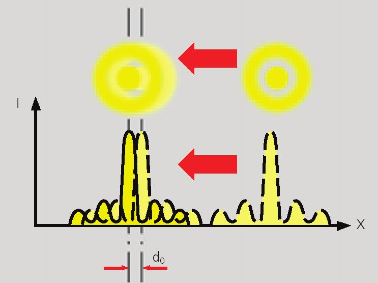
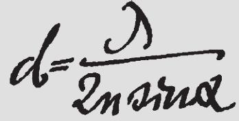
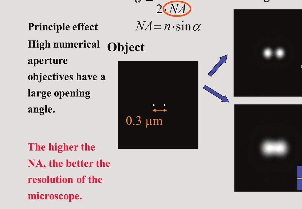
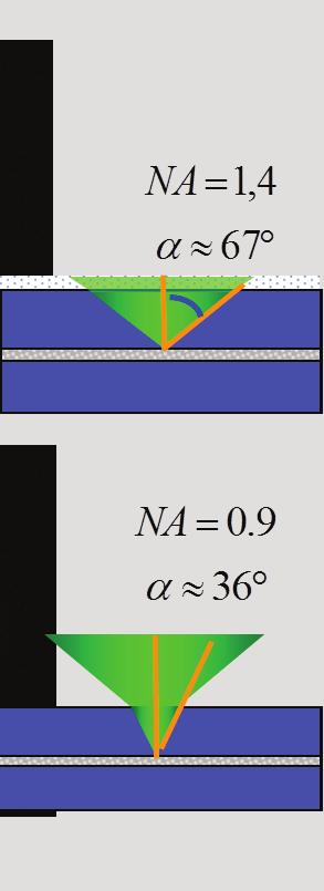
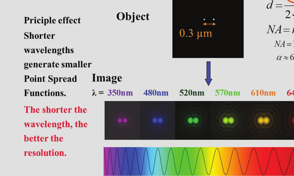
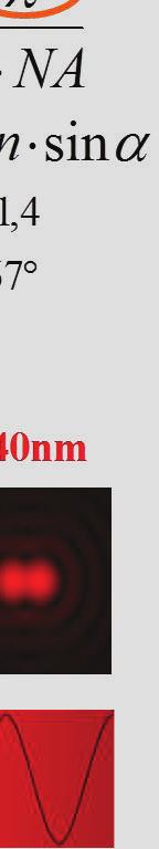
1 Travel to New Dimensions- LSM 880 LSM 880: The Power of Sensitivity Our Latest Member of the LSM 880 with GaAsP Detectors Sensitivity, and Ease of Use Innovative High-End Laser Scanning Microscopes from Carl Zeiss Page Page 2 The Resolution of a Microscope is limited The Resolution of a Microscope is limited Object Image Object Image What does that mean? The image of a point-like structure is not a point, but a diffraction pattern with a finite extension. This 2-dimensional pattern in the image plane is also called the Airy-disc. In general, the image of a pointlike structure is called the Point Spread Function (PSF). d=1.0 µm Definition The resolution limit is reached, when two point-like objects can not be imaged as two distinct structures anymore. The distance between the objects is called the resolution limit. d=0.4 µm d=0.3 µm Page 3 Page 4


2 The Point-Spread-Function is a 3-dimensional function The Resolution of a Microscope is limited. The axial shape of the PSF is completely different from the lateral one. Object Image The axial extension is larger than the lateral. y x z àa microscope has a lateral and an axial resolution. Prof. Ernst Abbe ( ) (1876) Page 5 Page 6 Page 8 Page 10
, von Willebrand Factor (Oregon-green), cell nucleus (DAPI).")

3 Different Beam Path of Image Formation Fluorescence -- Wavelength of visible light Confocal Laser Scanning Microscopy Optical sectioning: elimination of out-of-focus light Excitation Emission Human endothial cells with 3 fluorescence markers: Actin (Phalloidin/TRITC), von Willebrand Factor (Oregon-green), cell nucleus (DAPI). Page 12 The Comparison Between the LSM and the Conventional Light Microscope Excitation Emission Mercury or Xenon Lamp Laser Illuminated Field Wide Field Spot Image Acquisition Parallel, Frame at Once Confocal Page 13 The Point-Spread-Function is a 3-dimensional function Object Image The axial shape of the PSF is completely different from the lateral one. The axial extension is larger than the lateral. y x z Sequential, Pixel wise Signal Separation Dichroic Beam Splitter, Emission Filter Beam Splitter Cascade, Emission Filter Detector Eye or CCD Camera Diffraction limited by pinhole àphotomultiplier (PMT) Confocal Laser Scanning Microscope Light Source Wide Field. Wide Field Microscope Wide Field àa microscope has a lateral and an axial resolution. Page 14 Page 15

.")
4 Conventional/Widefield Fluorescence Why do we need Optical Sections? Background emission from deeper image planes The Fundamental Problem conventional image Conventional Images Conventional images from 3dimensional objects consists of light from structures, which are in focus and the light from structures which are not in focus. out-of-focus structures in-focus structures + Structures which are out-of-focus become visible in conventional widefield-fluorescence. Because of the focal depth inherent in all objectives, they are visible as an image blur (haze, image fog). Page 16 Excitation PMT Page 17 Emission Excitation Emission Wide Field Wide Field Confocal Confocal Pinhole Laser The pinhole diameter directly controls the thickness of the optical section. Confocal Laser Scanning Microscopy Optical sectioning: elimination of out-of-focus light The confocal principle A minute diaphragm, situated in a conjugated focal plane, prevents out of focus light to be detected. x Sample Page 20 Page 21


 Data recording at shorter pixel times Need for")
 Sample: Drosophila larva developing brain and eye.")
5 Confocal: Point Scanning From Spot to Image LSM 710/780/880 Innovative Beam Path Technology To get a 2 dimensional image from the specimen, the excitation spot has to be moved over the specimen The scanning mirrors move the excitation beam in a line wise fashion In VIS Laser Ports VIS XY scanning Point scanning confocal systems Page 22 Page 23 3 Channel Spectral with one GaAsP Detector Unmatched sensitivity Get More Results With GaAsP Detectors Applications Benefit from Improved Sensitivity in Many Ways GaAsP (Gallium Arsenide Phosphide) is a semiconductor material with ideal characteristics for converting photons into electrical signals. Better image quality Higher sensitivity equals better signalto-noise ratio (detection of faint signals) Faster scanning Benefits of GaAsP detectors: Almost two times better SNR than PMTs (resulting in higher sensitivity, better image quality and higher acquisition speed). GaAsP detectors can be operated in integration mode as well as in photon counting mode. Typical sensitivity of detectors GaAsP detector (schematic illustration) Data recording at shorter pixel times Need for averaging strategies largely reduced 13 fps Acquisition of more data Data recording at lower laser power (reduced bleaching and photo-toxic effects in live cell imaging) Sample: Drosophila larva developing brain and eye. Labeled with three FP s. Fluorescence boosted by Alexa conjugated antibodies against the FP s. Page 24 Page 25


 images Page 30")
6 LSM 880 The power of sensitivity LSM 880 The power of sensitivity Sensitivity - the enabling factor Improved S/N ratio: Black background Image longer Scan faster Look deeper Improved signal recording: Crisp details, clear image data Page 28 Page 29 LSM 880 Laser line ZEN 2 - Efficient Navigation Powerful software for powerful LSM systems Laser line Fluorochrome 405 nm DAPI, Hoechst, Alexa 405, BFP 458 nm ECFP 488 nm Alexa 488, Fluo-4, FITC, egfp 514 nm EYFP 561 nm Rhodamine, Alexa 546, 555, 568, Cy3, TRITC, DsRed, Texas Red, MitoTracker Red, mcherry Ease of Use & Low Maintenance 633 nm Alexa 633, Cy5 Detectors: QUASAR Detection (3) for fluorescence images 1 transmitted PMT detector for Bright Field (PH/DIC) images Page 30 Page 31


 Any geometry: 1x4.")
 Occurrence of two")


7 ZEN 2 Load configuration ZEN 2 Reuse function: recur all parameters and setting Page 32 Page 33 Major tasks of a LSM Laser and scanning mirror control Two independent scanning mirrors Major tasks of a LSM Colocalization in Confocal Microscopy Acquisition of Crosstalk free images required Free scan field rotation (0-360 o ) Free online zooming (0.6~40x (zoom=66.7x) Any geometry: 1x *6144 Faster rectangular acquisition (e.g. video rate) Occurrence of two fluorescent emission signals inside the same detection volume Identical size of detection volumes for different color channels required Intensities and position of the signals inside the detection volume may vary Page 34 Page 35


8 Major tasks of a LSM Optimal optical sectioning in thick tissue Z stack Select all 1 AU pihhole This plane represents an optical section X/Y/Z Stack Z-Drive 3 D information is acquired by moving the excitation focus not only in XY direction but also in Z direction The result is a 3 D data stack consisting of number of XY images representing different optical sections from the specimen 36 Page Page 37 Major tasks of a LSM Optimal optical sectioning in thick tissue Z stack Number of sections Major tasks of a LSM Optimal optical sectioning in thick tissue Z stack Optimal Number of sections : no missing information at minimal number of sections Missing Information Optical thickness depends on: wavelenght l objective lens, N.A. refractive index n pinhole diameter P d ~ P n l / (N.A) 2 Sample bleached and much data, Nyquist- or Sampling- Theorem slices overlap by the 50% of their thickness LSM software: One click for best resolution Page 38 Page 39

 of these single images results in an image")


9 Major tasks of a LSM Optimal optical sectioning in thick tissue Z stack Major tasks of a LSM Optimal optical sectioning in thick tissue 0 µm 2 µm 4 µm 6 µm 8 µm 10 µm 12 µm 14 µm 16 µm 18 µm An overlay (maximum projection) of these single images results in an image with an enhanced depth of focus This image contains all information from the specimen 20 µm 22 µm 24 µm 26 µm 28 µm A series of of confocal images from different optical planes contains the image information from the whole specimen Every detail is in focus! Page 40 Page 41 Tile scanning with motorized scanning stage Versatile LSM 880 Get More Results! Innovative High-End LSMs from Carl Zeiss Sensitiviy Extremely light-efficient instrument design New super-sensitive GaAsP detectors for LSM 880 QUASAR detection unit allows for maximum flexibility in signal recording Modularity: Configuration of sophisticated imaging platforms through integration of LSM with additional detection modules 40X objective, 10X9 Ease of Use ZEN 2: Powerful software for sophisticated LSM applications User-friendly graphical interface Page 42 Page 43


 is acceptable if the pinhole is set to 1 AU. Fixed registration of excitation spot (blue) and detection unit Airyscan pinhole diameters) and additional spatial information about the signal!")
10 LSM 880: The Power of Sensitivity Our Latest Member of the LSM 880 with GaAsP Detectors Airyscan introduces a revolutionary new concept designed to overcome a classical limitation of LSMs Emission filters Zoom optics Array of detection elements Array of highly sensitive GaAsP-based detector elements Page 44 Page 45 In practice, confocal imaging is mostly a compromise that tries to balance resolving power and SNR Airyscan overcomes a classical limitation of LSMs with its arrayed detector elements all utilized in parallel Point-like l emitter r The Problem: The resolving power of LSMs Point-like l emitter r Solution: Array of detection elements Scanning direction stays far below its potential maximum when setting the confocal pinhole to 1 AU. Scanning direction Benefits: Improved SNR (utilizes light otherwise rejected at small Fixed registration of excitation spot (blue) and detection unit Conventional LSM Note: The signal-to-noise ratio (SNR) is acceptable if the pinhole is set to 1 AU. Fixed registration of excitation spot (blue) and detection unit Airyscan pinhole diameters) and additional spatial information about the signal! Note: Each detector element compares to a confocal Pinhole: 1.0 AU pinhole set to 0.2 AU ( sub- Airy sampling )

.")

 11.09.")


 and J. Fitzpatrick Ph.D. (Director, Waitt Advanced Biophotonics Core), The Salk Institute, La Jolla, USA.")
11 In brief: Airyscan takes advantage of spatial information not recorded with conventional LSMs LSM 880 Airyscan detects intensity distribution Narrower PSF means improved resolution Airy pattern of a point-like emitter The offset of individual detectors to the optical axis provides additional spatial information in Airyscan (detectors of a conventional LSM just integrate all light passing through its pinhole). Linear deconvolution assigns all signals (and Array detector of Airyscan frequencies) recorded by individual detector elements to their appropriate locations. Result: Isotropic 1.7-fold increase in resolving power! Consider intensity Consider distribution Scan Both objects that were indistinguishable now become resolved in space. (Further reading: White paper on Airyscan) LSM 880 Airyscan enhances resolution, boosts SNR...thereby allowing for a much more accurate quantification (Karlseder and Fitzpatrick, The Salk Institute, La Jolla, CA, USA) Airyscan reveals more details in your samples by increasing the resolution of LSM up to 1.7-fold Confocal Airyscan 2 µm 2 µm Telomere replication without RTEL1: Stalled forks and telomere breakage visualized as doubled dots using Airyscan. Resolution is meaningless without good SNR. Courtesy: J. Karlseder Ph.D. (Molecular and Cell Biology Laboratory) and J. Fitzpatrick Ph.D. (Director, Waitt Advanced Biophotonics Core), The Salk Institute, La Jolla, USA. Cultivated mitotic cells stained for tubulin Peter O Toole, Ian Morrison (York, UK) 2 µm

 Airyscan delivers exceptional data of live samples using")


 Sample courtesy of:")

 10 µm 11.09.")
12 With its drastically improved SNR, Airyscan delivers quality images previously impossible with LSMs Confocal (excitation: 1.8%) Airyscan delivers exceptional data of live samples using the same laser power than in confocal imaging Airyscan (excitation: %) Mitosis in HeLa-Kyoto cell line during mitosis. Imaged with LSM 880 / Airyscan. Video showing Histone 2B (H2B, red, mcherry) and microtubule end-binding protein 3 (EB3, blue, EGFP) Sample courtesy of: Jan Ellenberg, EMBL, Heidelberg. 10 µm Arabidopsis root cells expressing GFP-MBD (GFP fused to microtubule binding domain) Olga Samajova (Olomouc, Czech Republic) 10 µm Airyscan performs multi-color imaging of samples stained with up to four fluorescent labels LSM Airyscan: Software Integration Airyscan 32x ü ü ü 16bit ü ü 2 µm Page 61 - LSM 880 Sales Training Page
 using the Airyscan detector to produce effectively small pinholes.")
 pinhole.")


13 LSM 880 with Airyscan: Easy of use 3 different modes of Airyscan detector LSM 880 with Airyscan: Easy of use SR Mode Detector View SR: Superresolution (up to 1,7 fold) using the Airyscan detector to produce effectively small pinholes. Oversampling and deconvolution are used to generate images with up to 140 nm resolution in xy and 400 nm in z. VP: In virtual pinhole mode images are collected with an open (> 3 A.u.) pinhole. Using the distribution on the array pinhole can be adjusted as needed in a post acquisition step. CO: Confocal mode just uses the sum total signal from the array, using it as a single extra channel. Page Page LSM 880 with Airyscan: Easy of use VP Mode Select VP mode BEFORE acquisition LSM 510 Meta LSM 880 / Airyscan 8, 12 bit 8, 12, 16 bit 18 mm 20 mm 0.7x~40x 0.6x~40x 5 fps (512x512) 13 fps (512x512) ~10 nm ~3 nm In the Airyscan processing tab, instead of SR strength, the software display VP parameters (1-4 AU) E D C A B T-PMT AIM ZEN Ar laser (458, 477, 488, 514nm); HeNe laser 543nm; HeNe laser 633nm; Diode laser 405nm T-PMT Diode laser 405nm; Ar laser 458, 488, 514nm; DPSS-laser 561nm; HeNe laser 633nm; confocal GaAsP (SR, VP, CO mode) Page Page 66
14 Thank you for your attention!! Page 67
Technology Note ZEISS LSM 880 with Airyscan
 Technology Note ZEISS LSM 880 with Airyscan Introducing the Fast Acquisition Mode ZEISS LSM 880 with Airyscan Introducing the Fast Acquisition Mode Author: Dr. Annette Bergter Carl Zeiss Microscopy GmbH,
LSM 510 META in Chang Gung University
 Content LSM 510 META in Chang ung University LSM 510 META 路 理 The features and applications of LSM 510 META 01-09 Introduction of the hardware 10-12 Fluorescence observation in conventional microscope
長庚大學共軛焦顯微鏡課程 長庚大學共軛焦顯微鏡課程. Spot light 長庚大學
 長庚大學共軛焦顯微鏡課程 Spot light 長庚大學共軛焦顯微鏡課程 20071030 長庚大學 Basic principle of Laser Scanning Confocal Microscopy The application of LSM 510 META detector Multiphoton microscopy basic principle and introduction
Why and How? Daniel Gitler Dept. of Physiology Ben-Gurion University of the Negev. Microscopy course, Michmoret Dec 2005
 Why and How? Daniel Gitler Dept. of Physiology Ben-Gurion University of the Negev Why use confocal microscopy? Principles of the laser scanning confocal microscope. Image resolution. Manipulating the
Zeiss 880 Training Notes Zen 2.3
 Zeiss 880 Training Notes Zen 2.3 1 Turn on the HXP 120V Lamp 2 Turn on Main Power Switch Turn on the Systems PC Switch Turn on the Components Switch. 3 4 5 Turn on the PC and log into your account. Start
Practical work no. 3: Confocal Live Cell Microscopy
 Practical work no. 3: Confocal Live Cell Microscopy Course Instructor: Mikko Liljeström (MIU) 1 Background Confocal microscopy: The main idea behind confocality is that it suppresses the signal outside
Point Spread Function. Confocal Laser Scanning Microscopy. Confocal Aperture. Optical aberrations. Alternative Scanning Microscopy
 Bi177 Lecture 5 Adding the Third Dimension Wide-field Imaging Point Spread Function Deconvolution Confocal Laser Scanning Microscopy Confocal Aperture Optical aberrations Alternative Scanning Microscopy
Operation Guide for the Leica SP2 Confocal Microscope Bio-Imaging Facility Hunter College October 2009
 Operation Guide for the Leica SP2 Confocal Microscope Bio-Imaging Facility Hunter College October 2009 Introduction of Fluoresence Confocal Microscopy The first confocal microscope was invented by Princeton
Basics of confocal imaging (part I)
") Basics of confocal imaging (part I) Swiss Institute of Technology (EPFL) Faculty of Life Sciences Head of BIOIMAGING AND OPTICS BIOP arne.seitz@epfl.ch Lateral resolution BioImaging &Optics Platform Light
Basics of confocal imaging (part I) Swiss Institute of Technology (EPFL) Faculty of Life Sciences Head of BIOIMAGING AND OPTICS BIOP arne.seitz@epfl.ch Lateral resolution BioImaging &Optics Platform Light
Operating Instructions for Zeiss LSM 510
 Operating Instructions for Zeiss LSM 510 Location: GNL 6.312q (BSL3) Questions? Contact: Maxim Ivannikov, maivanni@utmb.edu 1 Attend A Complementary Training Before Using The Microscope All future users
Zeiss 780 Training Notes
 Zeiss 780 Training Notes Turn on Main Switch, System PC and Components Switches 780 Start up sequence Do you need the argon laser (458, 488, 514 nm lines)? Yes Turn on the laser s main power switch and
Microscopy from Carl Zeiss
 Microscopy from Carl Zeiss Contents Page Contents... 1 Introduction... 1 Starting the System... 2 Introduction to ZEN Efficient Navigation... 5 Setting up the microscope... 10 Configuring the beam path
Quick Guide. LSM 5 MP, LSM 510 and LSM 510 META. Laser Scanning Microscopes. We make it visible. M i c r o s c o p y f r o m C a r l Z e i s s
 LSM 5 MP, LSM 510 and LSM 510 META M i c r o s c o p y f r o m C a r l Z e i s s Quick Guide Laser Scanning Microscopes LSM Software ZEN 2007 August 2007 We make it visible. Contents Page Contents... 1
Confocal Microscopy. (Increasing contrast and resolu6on using op6cal sec6oning) Lecture 7. November 2017
 Lecture 7. November 2017") Confocal Microscopy (Increasing contrast and resolu6on using op6cal sec6oning) Lecture 7 November 2017 3 Flavours of Microscope Confocal Laser Scanning Problem: Out of Focus Light Spinning disc 2-Photon
Confocal Microscopy (Increasing contrast and resolu6on using op6cal sec6oning) Lecture 7 November 2017 3 Flavours of Microscope Confocal Laser Scanning Problem: Out of Focus Light Spinning disc 2-Photon
BASICS OF CONFOCAL IMAGING (PART I)
") BASICS OF CONFOCAL IMAGING (PART I) INTERNAL COURSE 2012 LIGHT MICROSCOPY Lateral resolution Transmission Fluorescence d min 1.22 NA obj NA cond 0 0 rairy 0.61 NAobj Ernst Abbe Lord Rayleigh Depth of field
BASICS OF CONFOCAL IMAGING (PART I) INTERNAL COURSE 2012 LIGHT MICROSCOPY Lateral resolution Transmission Fluorescence d min 1.22 NA obj NA cond 0 0 rairy 0.61 NAobj Ernst Abbe Lord Rayleigh Depth of field
ZEISS LSM510META confocal manual
 ZEISS LSM510META confocal manual Switching on the system 1) Switch on the Remote Control button located on the table to the right of the microscope. This is the main switch for the whole system including
Inside the LSM 880 NLO + Airyscan
 Inside the LSM 880 NLO + Airyscan Overview of the Newest High-End Point Scanning Solution from Carl Zeiss Microscopy Matt Curtis 3D Imaging Specialist John Dirnberger Account Manager Washington University
Training Guide for Carl Zeiss LSM 510 META Confocal Microscope
 Training Guide for Carl Zeiss LSM 510 META Confocal Microscope AIM 4.2 Optical Imaging & Vital Microscopy Core Baylor College of Medicine (2017) Power ON Routine 1 2 Turn ON Components and System/PC switches
1 Co Localization and Working flow with the lsm700
 1 Co Localization and Working flow with the lsm700 Samples -1 slide = mousse intestine, Dapi / Ki 67 with Cy3/ BrDU with alexa 488. -1 slide = mousse intestine, Dapi / Ki 67 with Cy3/ no BrDU (but with
Zeiss LSM880 Operating Instructions. UTMB Optical Microscopy Core Jan. 16, 2018
 Zeiss LSM880 Operating Instructions UTMB Optical Microscopy Core Jan. 16, 2018 1 1. Power up the microscope Sing the LOGBOOK Steps below will provide power to the computer and all of the microscope components.
LSM 710 Confocal Microscope Standard Operation Protocol
 LSM 710 Confocal Microscope Standard Operation Protocol Basic Operation Turning on the system 1. Switch on Main power switch 2. Switch on System / PC power button 3. Switch on Components power button 4.
Boulevard du Temple Daguerrotype (Paris,1838) a busy street? Nyquist sampling for movement
 a busy street? Nyquist sampling for movement") Boulevard du Temple Daguerrotype (Paris,1838) a busy street? Nyquist sampling for movement CONFOCAL MICROSCOPY BioVis Uppsala, 2017 Jeremy Adler Matyas Molnar Dirk Pacholsky Widefield & Confocal Microscopy
Boulevard du Temple Daguerrotype (Paris,1838) a busy street? Nyquist sampling for movement CONFOCAL MICROSCOPY BioVis Uppsala, 2017 Jeremy Adler Matyas Molnar Dirk Pacholsky Widefield & Confocal Microscopy
Training Guide for Carl Zeiss LSM 5 LIVE Confocal Microscope
 Training Guide for Carl Zeiss LSM 5 LIVE Confocal Microscope AIM 4.2 Optical Imaging & Vital Microscopy Core Baylor College of Medicine (2017) Power ON Routine 1 2 Verify that main power switches on the
Microscope Confocal LSM510 META
 Microscope Confocal LSM510 META Welcome to the Zeiss LSM 510 Meta Confocal tutorial. Before using the LSM 510 META, Log off any other computer that is open with your personal login. You will need to put
Application Note. The New 2D Superresolution Mode for ZEISS Airyscan 120 nm Lateral Resolution without Acquiring a Z-stack
 The New 2D Superresolution Mode for ZEISS Airyscan 120 nm Lateral Resolution without Acquiring a Z-stack The New 2D Superresolution Mode for ZEISS Airyscan 120 nm Lateral Resolution without Acquiring a
Leica TCS SP8 Quick Start Guide
 Leica TCS SP8 Quick Start Guide Leica TCS SP8 System Overview Start-Up Procedure 1. Turn on the CTR Control Box, Fluorescent Light for the microscope stand. 2. Turn on the Scanner Power (1) on the front
Spectral Imaging with the Opterra Multipoint Scanning Confocal
 Spectral Imaging with the Opterra Multipoint Scanning Confocal Outline Opterra design overview Scan Modes Light Path Spectral Imaging with Opterra Drosophila larva heart. Opterra Design Overview Supravideo
Multifluorescence The Crosstalk Problem and Its Solution
 Multifluorescence The Crosstalk Problem and Its Solution If a specimen is labeled with more than one fluorochrome, each image channel should only show the emission signal of one of them. If, in a specimen
Cell Biology and Bioimaging Core
 Cell Biology and Bioimaging Core Leica TCS SP5 Operating Instructions Starting up the instrument 1. First, log in the log book located on the confocal desk. Include your name, your lab s PI, an account
3D light microscopy techniques
 3D light microscopy techniques The image of a point is a 3D feature In-focus image Out-of-focus image The image of a point is not a point Point Spread Function (PSF) 1D imaging 1 1 2! NA = 0.5! NA 2D imaging
Supplemental Method Information Zeiss LSM710
 Supplemental Method Information Zeiss LSM710 1 Under the Light Path window set up the confocal for imaging a green dye (Alexa488-EGFP). For example, set up the light path as shown here using the 488 nm
Supplementary Information. Stochastic Optical Reconstruction Microscopy Imaging of Microtubule Arrays in Intact Arabidopsis thaliana Seedling Roots
 Supplementary Information Stochastic Optical Reconstruction Microscopy Imaging of Microtubule Arrays in Intact Arabidopsis thaliana Seedling Roots Bin Dong 1,, Xiaochen Yang 2,, Shaobin Zhu 1, Diane C.
The Zeiss AiryScan System, Confocal Four.
 The Zeiss AiryScan System, Confocal Four. Overview. The Zeiss AiryScan module is a segmented, radially stacked GaASP detector and collector system designed to subsample the airy disk of a point emission
Training Guide for Leica SP8 Confocal/Multiphoton Microscope
 Training Guide for Leica SP8 Confocal/Multiphoton Microscope LAS AF v3.3 Optical Imaging & Vital Microscopy Core Baylor College of Medicine (2017) Power ON Routine 1 2 Turn ON power switch for epifluorescence
Components of confocal and two-photon microscopes
 Components of confocal and two-photon microscopes Internal training 07/04/2016 A. GRICHINE Platform Optical microscopy Cell imaging, IAB, ISdV Plan Confocal laser scanning microscope o o o Principle Main
Confocal, Airyscan and Structured Illumination Superresolution microscopy Mayandi Sivaguru
 Confocal, Airyscan and Structured Illumination Superresolution microscopy Mayandi Sivaguru Theory, Light Path, Resolution Comparison Performance comparison Why and When Choose Airyscan OR SR-SIM Superresolution
Leica TCS SP8 Quick Start Guide
 Leica TCS SP8 Quick Start Guide Leica TCS SP8 System Overview Start-Up Procedure 1. Turn on the CTR Control Box, EL6000 fluorescent light source for the microscope stand. 2. Turn on the Scanner Power
TRAINING MANUAL. Multiphoton Microscopy LSM 510 META-NLO
 TRAINING MANUAL Multiphoton Microscopy LSM 510 META-NLO September 2010 Multiphoton Microscopy Training Manual Multiphoton microscopy is only available on the LSM 510 META-NLO system. This system is equipped
Quick Start Guide. Leica SP5 X
 Quick Start Guide Leica SP5 X Please note: Some of the information in this guide was taken from Leica Microsystems Leica TCS SP5 LAS AF Guide for New Users. This work is licensed under the Creative Commons
ADVANCED METHODS FOR CONFOCAL MICROSCOPY II. Jean-Yves Chatton Sept. 2006
 ADVANCED METHODS FOR CONFOCAL MICROSCOPY II Jean-Yves Chatton Sept. 2006 Workshop outline Confocal microscopy of living cells and tissues X-Z scanning Time series Bleach: FRAP, photoactivation Emission
contents TABLE OF The SECOM platform Applications - sections Applications - whole cells Features Integrated workflow Automated overlay
 S E C O M TABLE OF contents The SECOM platform 4 Applications - sections 5 Applications - whole cells 8 Features 9 Integrated workflow 12 Automated overlay ODEMIS - integrated software Specifications 13
Bi/BE 227 Winter Assignment #3. Adding the third dimension: 3D Confocal Imaging
 Bi/BE 227 Winter 2016 Assignment #3 Adding the third dimension: 3D Confocal Imaging Schedule: Jan 20: Assignment Jan 20-Feb 8: Work on assignment Feb 10: Student PowerPoint presentations. Goals for this
Training Guide for Carl Zeiss LSM 880 with AiryScan FAST
 Training Guide for Carl Zeiss LSM 880 with AiryScan FAST ZEN 2.3 Optical Imaging & Vital Microscopy Core Baylor College of Medicine (2018) Power ON Routine 1 2 Turn ON Main Switch from the remote control
Bio 407. Applied microscopy. Introduction into light microscopy. José María Mateos. Center for Microscopy and Image Analysis
 Center for Microscopy and Image Analysis Bio 407 Applied Introduction into light José María Mateos Fundamentals of light Compound microscope Microscope composed of an objective and an additional lens (eyepiece,
CONFOCAL MICROSCOPE (Zeiss LSM 510 META v4.2)
") Wellcome Trust Centre for Human Genetics Molecular Cytogenetics and Microscopy Core CONFOCAL MICROSCOPE (Zeiss LSM 510 META v4.2) 1) STARTING THE SYSTEM Abridged INSTRUCTIONS Switch on the mercury bulb
Wellcome Trust Centre for Human Genetics Molecular Cytogenetics and Microscopy Core CONFOCAL MICROSCOPE (Zeiss LSM 510 META v4.2) 1) STARTING THE SYSTEM Abridged INSTRUCTIONS Switch on the mercury bulb
EUV microscopy - a user s perspective Dimitri Scholz EUV,
 EUV microscopy - a user s perspective Dimitri Scholz EUV, 09.11.2011 Imaging technologies: available at UCD now and in the next future Begin ab ovo - Simple approaches direct to the goal - Standard methods
Opterra. Multipoint Scanning Confocal Microscope. Innovation with Integrity. Cell-Friendly, High-Speed, High-Resolution Imaging
 Opterra Multipoint Scanning Confocal Microscope Cell-Friendly, High-Speed, High-Resolution Imaging Innovation with Integrity Fluorescence Microscopy Opterra Multipoint Scanning Confocal Microscope Superior
Things to check before start-up.
 Byeong Cha Page 1 11/24/2009 Manual for Leica SP2 Confocal Microscope Enter you name, the date, the time, and the account number in the user log book. Things to check before start-up. Make sure that your
Instant super-resolution imaging in live cells and embryos via analog image processing
 Nature Methods Instant super-resolution imaging in live cells and embryos via analog image processing Andrew G. York, Panagiotis Chandris, Damian Dalle Nogare, Jeffrey Head, Peter Wawrzusin, Robert S.
Confocal imaging on the Leica TCS SP8. 1) Turn the system on. 2) Use TCS user account. 3) Start LAS X software:
 Turn the system on. 2) Use TCS user account. 3) Start LAS X software:") Confocal imaging on the Leica TCS SP8 1) Turn the system on. 2) Use TCS user account. 3) Start LAS X software: 4) Do not touch the microscope while the software is initializing. Choose your options: Turn
Confocal imaging on the Leica TCS SP8 1) Turn the system on. 2) Use TCS user account. 3) Start LAS X software: 4) Do not touch the microscope while the software is initializing. Choose your options: Turn
Development of a High-speed Super-resolution Confocal Scanner
 Development of a High-speed Super-resolution Confocal Scanner Takuya Azuma *1 Takayuki Kei *1 Super-resolution microscopy techniques that overcome the spatial resolution limit of conventional light microscopy
Contents. Introduction
 Contents Page Contents... 1 Introduction... 1 Starting the System... 2 Introduction to ZEN Efficient Navigation... 5 Setting up the microscope... 10 Configuring the beam path and lasers... 12 Scanning
Zeiss LSM 510 Confocor III Training Notes. Center for Cell Analysis & Modeling
 Zeiss LSM 510 Confocor III Training Notes Center for Cell Analysis & Modeling Confocor 3 Start Up Go to System Module Turn on Main Switch, System/ PC, and Components Switches Do you need the arc lamp?
LSM 800 Confocal Microscope Standard Operation Protocol
 LSM 800 Confocal Microscope Standard Operation Protocol Turning on the system 1. Switch on the Main switch (labeled 1 and 2 ) mounted on the wall. 2. Turn the Laser Key (labeled 3 ) 90 clockwise for power
LSM 510 Meta Training Notes
 LSM 510 Meta Training Notes Turning on the system Turn on X-Cite power supply. This supplies light for epifluorescence for viewing your samples through the microscope. Turn on the remote control switch.
OPERATING INSTRUCTIONS
 Zeiss LSM 510 M eta Confocal M icroscope OPERATING INSTRUCTIONS Starting the System: 1. Turn the black knob on the laser box one-quarter turn from Off to On. You will hear the laser cooling mechanisms
Confocal Microscopy. Kristin Jensen
 Confocal Microscopy Kristin Jensen 17.11.05 References Cell Biological Applications of Confocal Microscopy, Brian Matsumoto, chapter 1 Studying protein dynamics in living cells,, Jennifer Lippincott-Schwartz
High resolution extended depth of field microscopy using wavefront coding
 High resolution extended depth of field microscopy using wavefront coding Matthew R. Arnison *, Peter Török #, Colin J. R. Sheppard *, W. T. Cathey +, Edward R. Dowski, Jr. +, Carol J. Cogswell *+ * Physical
LEICA TCS SP5 AOBS TANDEM USER MANUAL
 LEICA TCS SP5 AOBS TANDEM USER MANUAL STARTING THE SYSTEM...2 THE LAS AF SOFTWARE...3 THE «ACQUIRE» MENU...5 CHOOSE AND CREATE A SETTING...6 THE CONTROL PANEL...8 THE DMI6000B MICROSCOPE...10 ACQUIRE ONE
Leica SPEII confocal microscope. Short Manual
 Leica SPEII confocal microscope Short Manual Switching ON sequence: 1. Turn on the Workstation under the bench (top, far right). 2. Turn on the Supply Unit - Laser box (big green switch first and then
Confocal Laser Scanning Microscopy
 Name of the Core Facility: Confocal Laser Scanning Microscopy CORE Forschungszentrum Immunologie Mainz Welcome to the CSLM Core Facility: The CLSM Core Facility enables working groups to incorporate high
INTRODUCTION TO MICROSCOPY. Urs Ziegler THE PROBLEM
 INTRODUCTION TO MICROSCOPY Urs Ziegler ziegler@zmb.uzh.ch THE PROBLEM 1 ORGANISMS ARE LARGE LIGHT AND ELECTRONS: ELECTROMAGNETIC WAVES v = Wavelength ( ) Speed (v) Frequency ( ) Amplitude (A) Propagation
Quick Guide for Zeiss 710 Laser Scanning Confocal MGH Cancer Center
 Quick Guide for Zeiss 710 Laser Scanning Confocal MGH Cancer Center For any questions or concerns, please contact: Linda Nieman lnieman@mgh.harvard.edu Office: (617) 643-9684 Cell: (512) 565-8076 Chenyue
ANSWER KEY Lab 2 (IGB): Bright Field and Fluorescence Optical Microscopy and Sectioning
: Bright Field and Fluorescence Optical Microscopy and Sectioning") Phys598BP Spring 2016 University of Illinois at Urbana-Champaign ANSWER KEY Lab 2 (IGB): Bright Field and Fluorescence Optical Microscopy and Sectioning Location: IGB Core Microscopy Facility Microscope:
Phys598BP Spring 2016 University of Illinois at Urbana-Champaign ANSWER KEY Lab 2 (IGB): Bright Field and Fluorescence Optical Microscopy and Sectioning Location: IGB Core Microscopy Facility Microscope:
Leica SP8 TCS Users Manual
 Leica SP8 TCS Users Manual Follow the procedure for start up and log on as posted in the lab. Please log on with your account only and do not share your password with anyone. We track and confirm usage
Leica_Dye_Finder :53 Uhr Seite 6 Dye Finder LAS AF
 Dye Finder LAS AF Dye Finder Multicolor live cell fluorescence microscopy is limited by the availability of spectrally separable fluorescent dyes. Fluorescent dyes (or spectral GFP variants) with incongruent
LSM 510 Training Notes
 LSM 510 Training Notes Turning on the system Turn on the arc lamp, found on the bench top left of the microscope. This supplies light for epifluorescence for viewing your samples through the microscope.
Megapixel FLIM with bh TCSPC Modules
 Megapixel FLIM with bh TCSPC Modules The New SPCM 64-bit Software Abstract: Becker & Hickl have recently introduced version 9.60 of their SPCM TCSPC data acquisition software. SPCM version 9.60 not only
LSM 780 Confocal Microscope Standard Operation Protocol
 LSM 780 Confocal Microscope Standard Operation Protocol Basic Operation Turning on the system 1. Sign on log sheet according to Actual start time 2. Check Compressed Air supply for the air table 3. Switch
Microscope Confocal Sp2 Upright.
 Microscope Confocal Sp2 Upright. Welcome to the Leica Sp2 Confocal Upright tutorial. Before using the Sp2 Invert, You will need to put down your name on the reservation system = http://svintranet.epfl.ch/index.php?optio
Opterra II Multipoint Scanning Confocal Microscope. Innovation with Integrity
 Opterra II Multipoint Scanning Confocal Microscope Enabling 4D Live-Cell Fluorescence Imaging through Speed, Sensitivity, Viability and Simplicity Innovation with Integrity Fluorescence Microscopy The
Rapid three-dimensional isotropic imaging of living cells using Bessel beam plane illumination
 Nature Methods Rapid three-dimensional isotropic imaging of living cells using beam plane illumination Thomas A Planchon, Liang Gao, Daniel E Milkie, Michael W Davidson, James A Galbraith, Catherine G
Topics. - How to calibrate the LSM scanner. - How to clean the microscope. - How to adjust the pinhole alignment. - How to adjust the Collimator
 Topics - How to calibrate the LSM scanner - How to measure the PSF - How to clean the microscope - How to adjust the pinhole alignment - How to adjust the Collimator How to calibrate the LSM scanner The
Nature Methods: doi: /nmeth Supplementary Figure 1. Schematic of 2P-ISIM AO optical setup.
 Supplementary Figure 1 Schematic of 2P-ISIM AO optical setup. Excitation from a femtosecond laser is passed through intensity control and shuttering optics (1/2 λ wave plate, polarizing beam splitting
Invitation for a walk through microscopy. Sebastian Schuchmann Jörg Rösner
 Invitation for a walk through microscopy Sebastian Schuchmann Jörg Rösner joerg.roesner@charite.de Techniques in microscopy Conventional (light) microscopy bright & dark field, phase & interference contrast
Supplemental Figure 1: Histogram of 63x Objective Lens z axis Calculated Resolutions. Results from the MetroloJ z axis fits for 5 beads from each
 Supplemental Figure 1: Histogram of 63x Objective Lens z axis Calculated Resolutions. Results from the MetroloJ z axis fits for 5 beads from each lens with a 1 Airy unit pinhole setting. Many water lenses
3D light microscopy techniques
 3D light microscopy techniques The image of a point is a 3D feature In-focus image Out-of-focus image The image of a point is not a point Point Spread Function (PSF) 1D imaging 2D imaging 3D imaging Resolution
Confocal Application Letter No. 13. Sequential Scan for Leica TCS NT/SP systems
 Confocal Application Letter No. 13 Sequential Scan for Leica TCS NT/SP systems Leica Microsystems Heidelberg GmbH Im Neuenheimer Feld 518 D-69120 Heidelberg Telephone +49 6221 4148 0 Fax +49 6221 414833
Fundamentals of Light Microscopy II: Fluorescence, Deconvolution, Confocal, Multiphoton, Spectral microscopy. Integrated Microscopy Course
 Fundamentals of Light Microscopy II: Fluorescence, Deconvolution, Confocal, Multiphoton, Spectral microscopy Integrated Microscopy Course Review Lecture 1: Microscopy Basics Light train Kohler illumination*
Leica Sp5 II Confocal User Guide
 Leica Sp5 II Confocal User Guide Turning on the Confocal System (instructions are posted in the room) 1. Turn on Laser Power Button 2. Turn Key to On position 3. Turn on Scanner Power Button 4. Turn on
Shreyash Tandon M.S. III Year
 Shreyash Tandon M.S. III Year 20091015 Confocal microscopy is a powerful tool for generating high-resolution images and 3-D reconstructions of a specimen by using point illumination and a spatial pinhole
Rates of excitation, emission, ISC
 Bi177 Lecture 4 Fluorescence Microscopy Phenomenon of Fluorescence Energy Diagram Rates of excitation, emission, ISC Practical Issues Lighting, Filters More on diffraction Point Spread Functions Thus Far,
MULTIPHOTON MICROSCOPY. Matyas Molnar Dirk Pacholsky
 MULTIPHOTON MICROSCOPY Matyas Molnar Dirk Pacholsky Information Information given here about 2 Photon microscopy were mainly taken from these sources: Background information on 2-Photon microscopy: http://micro.magnet.fsu.edu/primer/techniques/fluorescence/multiphoton/
Advanced Optical Microscopy lecture. 03. December 2012 Kai Wicker
 Advanced Optical Microscopy lecture 03. December 2012 Kai Wicker Today: Optical transfer functions (OTF) and point spread functions (PSF) in incoherent imaging. 1. Quick revision: the incoherent wide-field
Quick Guide for Zeiss 710 Laser Scanning Confocal MGH Cancer Center
 Quick Guide for Zeiss 710 Laser Scanning Confocal MGH Cancer Center For any questions or concerns, please contact: Linda Nieman lnieman@mgh.harvard.edu Office: (617) 643-9684 Cell: (512) 565-8076 Chenyue
ZEISS LSM 710 CONFOCAL MICROSCOPE USER MANUAL
 ZEISS LSM 710 CONFOCAL MICROSCOPE USER MANUAL START THE SYSTEM... 2 START ZEN SOFTWARE... 3 SET THE TEMPERATURE AND THE CO2 CONTROLLERS... OBSERVATION AT OCULARS... 5 STATIF PRESENTATION... 6 ACQUIRE ONE
3 Choose the Channels button and set the Channel Settings. Set the Pinhole to 1 Airy unit.
 1 Set up the confocal light path for imaging a green dye (e.g. Alexa488-EGFP). For example, under the Configuration Control window the light path could be set up as shown here using the 488 nm LASER (found
The DCS-120 Confocal Scanning FLIM System
 he DCS-120 Confocal Scanning FLIM System he bh DCS-120 confocal scanning FLIM system converts a conventional microscope into a high-performance fluorescence lifetime imaging system. he system is based
Leica SP8 TCS Users Manual
 Version : 07/08/0 Leica SP8 TCS Users Manual Start up:. Turn the PC Microscope, Scanner Power, Laser Power, and the Laser Emission key to on (bottom right of desk).. Turn on the fluorescent lamp (top left
TRAINING MANUAL. Olympus FV1000
 TRAINING MANUAL Olympus FV1000 September 2014 TABLE OF CONTENTS A. Start-Up Procedure... 1 B. Visual Observation under the Microscope... 1 C. Image Acquisition... 4 A brief Overview of the Settings...
MIF ZEISS VIOLET CONFOCAL ZEN 2009 PROTOCOL
 MIF ZEISS VIOLET CONFOCAL ZEN 2009 PROTOCOL START-UP On the Switchbox, turn both black switches to the ON position. Wait for the microscope to boot up completely (watch the screen on the side of the microscope).
Nikon. King s College London. Imaging Centre. N-SIM guide NIKON IMAGING KING S COLLEGE LONDON
 N-SIM guide NIKON IMAGING CENTRE @ KING S COLLEGE LONDON Starting-up / Shut-down The NSIM hardware is calibrated after system warm-up occurs. It is recommended that you turn-on the system for at least
Pixel shift in fluorescence microscopy
 Pixel shift in fluorescence microscopy 1. Introduction Multicolor imaging in fluorescence microscopy is typically performed by sequentially acquiring images of different colors. An overlay of these images
Introduction to light microscopy
 Center for Microscopy and Image Anaylsis Introduction to light Basic concepts of imaging with light Urs Ziegler ziegler@zmb.uzh.ch Microscopy with light 1 Light interacting with matter Absorbtion Refraction
Training Guide for Carl Zeiss LSM 7 MP Multiphoton Microscope
 Training Guide for Carl Zeiss LSM 7 MP Multiphoton Microscope ZEN 2009 Optical Imaging & Vital Microscopy Core Baylor College of Medicine (2017) Power ON Routine 1 2 Turn Chameleon TiS laser key from Standby
Lecture 16. OMX - Structured Illumination Microscopy Ian Dobbie x Microscopy Course Lecture 16 1
 Lecture 16 OMX - Structured Illumination Microscopy Ian Dobbie x13323 Microscopy Course 2014 - Lecture 16 1 Super-resolution fluorescence microscopy Specificity Sensitivity Non-invasive (in situ & in vivo)
Introduction to light microscopy
 Center for Microscopy and Image Anaylsis Introduction to light microscopy Basic concepts of imaging with light Urs Ziegler ziegler@zmb.uzh.ch Light interacting with matter Absorbtion Refraction Diffraction
Dynamic Confocal Imaging of Living Brain. Advantages and risks of multiphoton microscopy in physiology
 Dynamic Confocal Imaging of Living Brain Advantages and risks of multiphoton microscopy in physiology Confocal laser scanning microscopy In conventional optical microscopy focused and out-offocus light
Light Microscopy. Upon completion of this lecture, the student should be able to:
 Light Light microscopy is based on the interaction of light and tissue components and can be used to study tissue features. Upon completion of this lecture, the student should be able to: 1- Explain the
1 Set up the confocal light path for imaging a green dye (Alexa488-EGFP). For example, the
. For example, the") 1 Set up the confocal light path for imaging a green dye (Alexa488-EGFP). For example, the light path as shown here using the 488 nm LASER (Laser Unit 1) reflecting off of the 405/488 nm Dichroic mirror
1 Set up the confocal light path for imaging a green dye (Alexa488-EGFP). For example, the light path as shown here using the 488 nm LASER (Laser Unit 1) reflecting off of the 405/488 nm Dichroic mirror