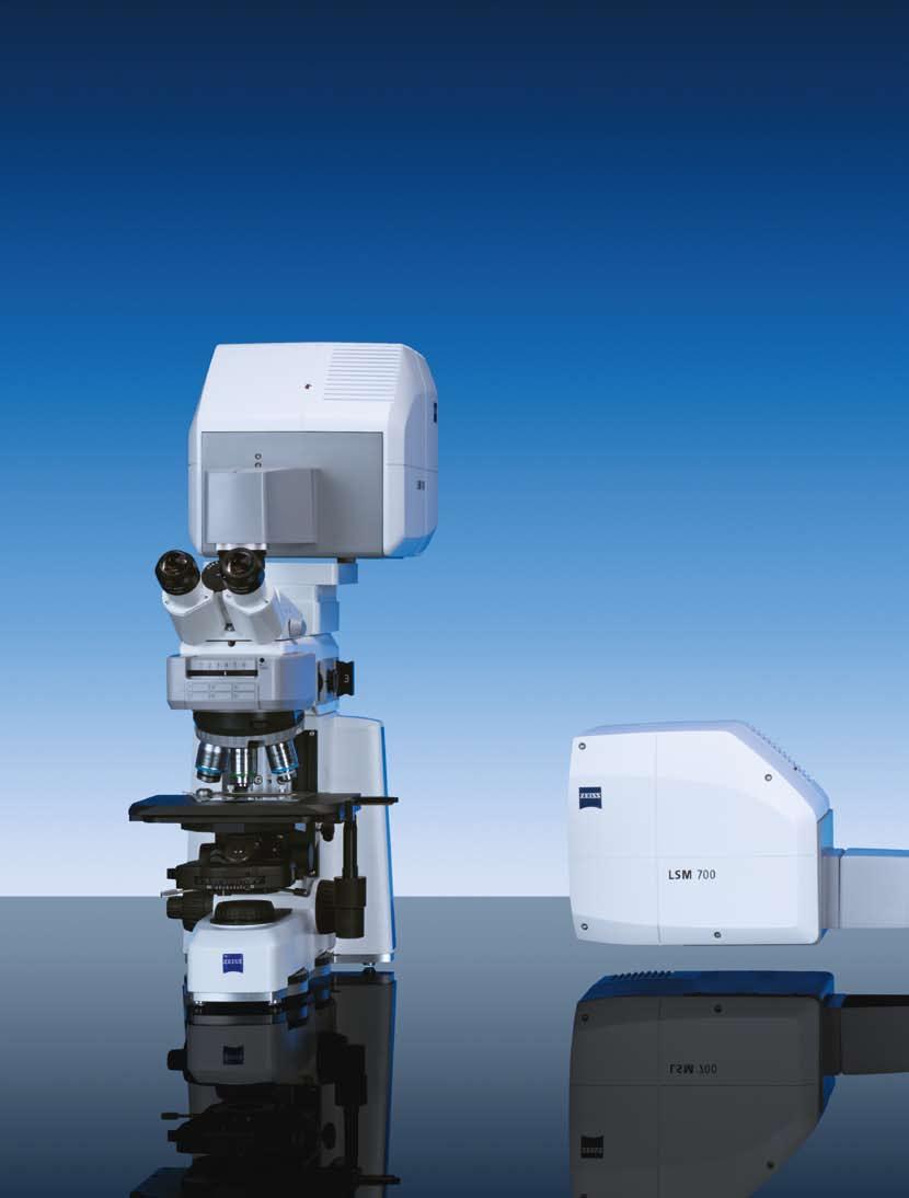
Microscopy from Carl Zeiss LSM 700. Laser Scanning Microscope. High-End for All Uncompromised Quality and Operating Convenience
|
|
|
- Gregory Allen
- 6 years ago
- Views:
Transcription


1 Microscopy from Carl Zeiss LSM 700 Laser Scanning Microscope High-End for All Uncompromised Quality and Operating Convenience
2 2
3 3
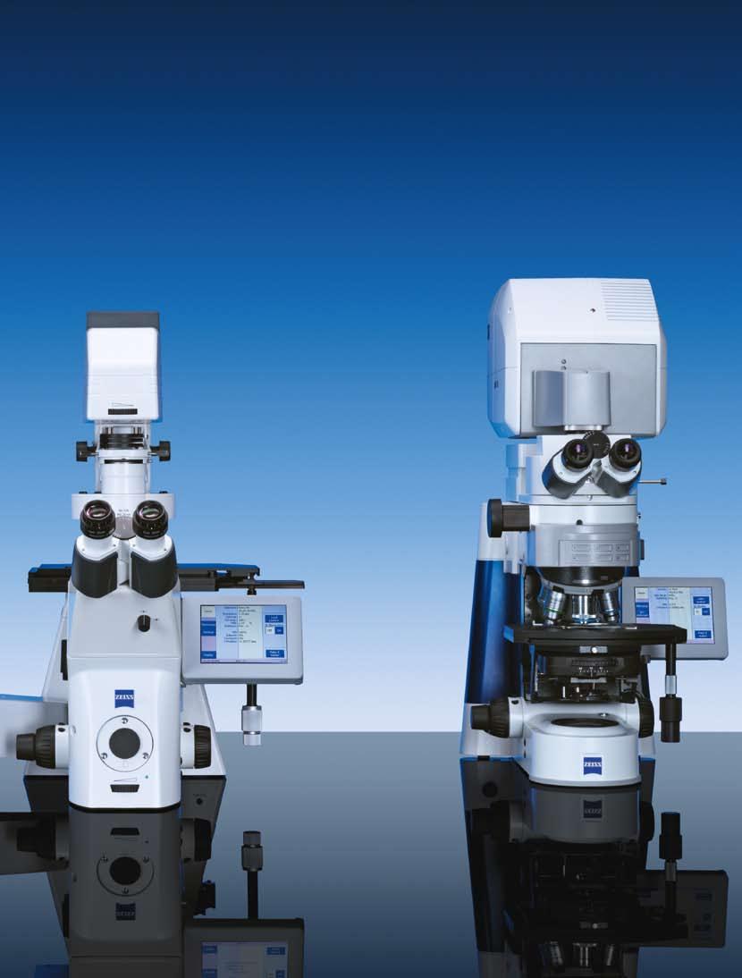
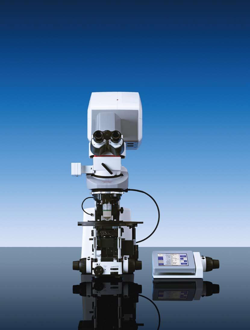
4 The New LSM 700 from Carl Zeiss From a specialists system to the high-end microscope for all the LSM 700 represents the next big step in the evolution of confocal microscopy. The LSM 700 is a member of the seventh generation of confocal microscopes from Carl Zeiss a product family that is characterized throughout by a wealth of genuinely innovative ideas and technologies. Top-grade system components ensure superior performance. The LSM 700 concept combines Carl Zeiss quality, exceptional ease of operation and an attractive price level, resulting in an excellent price/performance ratio, which is evident in many details. Tried-and-tested ZEISS quality for basic applications and complex requirements Designed for complex tasks while being easy to operate, the LSM 700 meets every challenge, whether in a single- or multiple-user environment. It fits on many different microscope stands to suit a wide range of personal or application requirements. One image acquisition system for simple and demanding tasks The LSM 700 is ideal for multi-user facilities, where it can complement larger systems and relieve their workload. For many users in biomedical research it will provide an entry to high-end applications previously limited to just the highest end systems. With frame rates of up to five fps (512 x 512 pixels), a freely 360 rotatable scanning field and freely definable regions of interest (ROIs), the LSM 700 provides the experimental freedom required for many applications. The system features outstanding sensitivity, thanks to its mature, highly corrected optics and the efficiency of its detectors and electronic components. Together with the user-friendly ZEN software the system makes up a package that is suitable for classical confocal microscopy and special applications alike, such as live cell imaging, spectral imaging and raster image correlation spectroscopy (RICS). 4
5 5
6 6
7 7
8 8

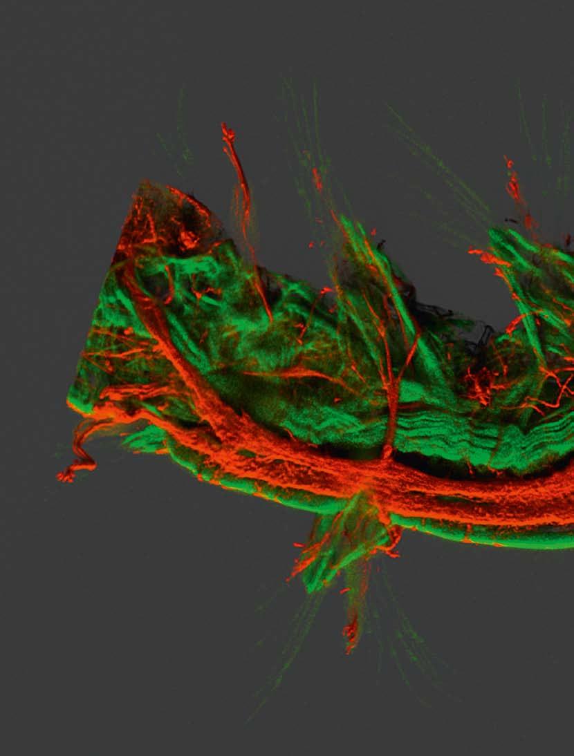
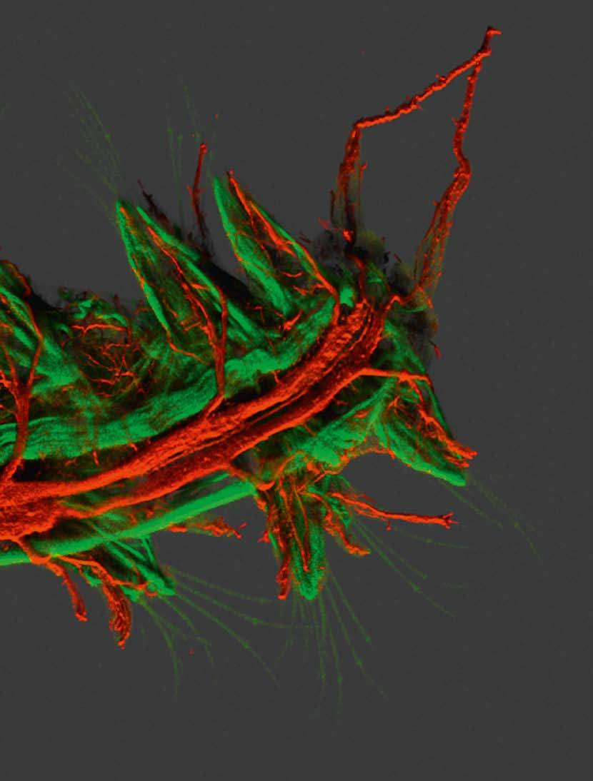
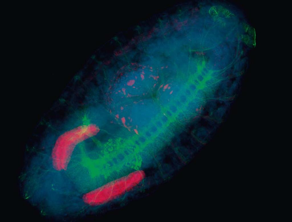
9 Fresh Impetus to Your Research The LSM 700 fits many applications that are extraordinary for its price bracket. Spectral Imaging and Linear Unmixing are but two examples of techniques that demand a system of top quality. 3D imaging 3D imaging is the standard application of a laser scanning microscope. Brilliant 3D images require excellent optical quality and precisely controlled image acquisition. The LSM 700 assists you in configuring the acquisition parameters, from choosing the pixel resolution, via setting the diameter of the confocal pinhole to the Z spacing of the optical sections. Subsequently image acquisition is performed automatically and fully motorized. The ZEN software reconstructs your highly resolved 3D images and meaningfully presents them, e.g., in the form of projection or animations. Human lymphocytes transmitting the HIV virus from cell to cell. Red: HIV (Gag), Alexa 546. Green: Actin-phalloidin-Alexa 488. Blue: Cytosol marker. Domenika Rudnika Nathalie Sol-Foulon and Olivier Schwartz, Institut Pasteur, Virus and Immunity Unit, Paris, France Drosophila Melanogaster Embryo, Blue: DAPI. Green: Alexa 488. Red: Cy3 9

10 Section of a mouse stomach. Blue: Plasma membrane, stained with Alexa Fluor 350 WGA (wheat germ agglutinin). Red: Actin, stained with Alexa 488. Green: Nuclei, stained with Sytox Green Multiple fluorescence and colocalization analyses In multicolor fluorescence imaging, the use of several fluorophores permits the observation of spatial relations between several cell constituents. 2 fluorescence detectors in the LSM 700 detect up to four color signals in a (quasi-)simultaneous mode, at frame rates of up to 5 fps for 512 x 512 pixels. Efficient separation of the fluorescence signals by selective laser excitation, and efficient splitting by means of the VSD (Variable Secondary Dichroic) beamsplitter prevent crosstalk and ensure unambiguous results, especially in colocalization analyses. Emission Fingerprinting Spectral imaging and subsequent linear unmixing precisely separate fluorescent signals even of greatly overlapping color signals whether you use, for example, GFP and YFP simultaneously or whether broad-band autofluorescence is present. The integration of the VSD beamsplitter into the Emission Fingerprinting concept of the LSM 700 provides an innovative, highly efficient method of spectral image acquisition. Unlike conventional sequential methods, all parts of the spectrum emitted by the specimen are utilized for determining each spectral data point. 10


11 DAPI-stained nuclei and mitosis visualized in live cultured cells Live Cell Imaging High light intensities and long irradiation lead to phototoxic reactions in living cells and tissues. The high sensitivity of the LSM 700, combined with pixel-precise control of illumination, preserves your specimens and permits you to observe fast biological processes over long periods of time. Ion imaging The well thought-out functions of the ZEN software support not only image acquisition but also image analysis in the observation of ion activities in live specimens. Online creation of ratiometric images allows the results of your experiments to be displayed in real time during the acquisition. 11

12 FRAP, FLIP, photoactivation and photoconversion Transport processes in live cells and organisms can be observed by means of targeted localized photobleaching, or by means of photoactivation or color conversion of fluorophores such as PA-GFP or Kaede. Thanks to precise real-time control of the excitation laser light and scanning mirror movements in the LSM 700, pixelprecise local illumination in up to 99 regions of interest is possible, as is the change between manipulation and imaging modes within milliseconds. Photoconversion of the fluorescent protein Kikume in a transgenic mouse embryo. Specimen: Dr. Heather Young, Anatomy Department, University of Melbourne, Australia 12


13 Stage Stage A software joystick supports the control of the motorized XY scanning stage. Z-Stack Use this module to configure the acquisition of image Z stacks. The software controls the Z movement of the microscope at the correct intervals and synchronizes its movements with image acquisition. Step sizes can be computed automatically or determined interactively. If you want to check the settings made before starting your experiment (e.g., for Z stacks versus time), a graph will provide an informative overview. Z Stack Light Path With this function you can select the position of the VSD beamsplitter, and thus, the desired detection range. Do this interactively, or automatically with the Smart Setup tool. Light Path 17


 a microscope system with a laser")
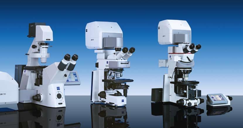
14 Confocal Microscopy 3D Imaging of Objects Confocal Laser Scanning Microscopy: Sharp three-dimensional images even of thick specimens. In the mid-17th century, biologists and doctors enthusiastically welcomed the first microscopes, which led to an enormous leap in their research. Three centuries later, scientists were amazed again: The arrival of confocal microscopy in 1957 opened up a new dimension. Instead of 2D images affected by out-of-focus light, the invention made possible the imaging of extended three-dimensional specimens with excellent depth discrimination. In 1982, Carl Zeiss launched the first commercially available laser scanning microscope (LSM) a microscope system with a laser beam scanning the specimen, and electronic image processing. Beam path in the confocal microscope 18
15 Every Photon Counts the LSM 700 Beam Path In the scanning module, light rays are guided from the sample to the detectors with the absolute minimum loss. This gives the LSM 700 its high sensitivity. High sensitivity of the LSM 700 is guaranteed by the sophisticated, innovative optical design by Carl Zeiss, which conducts the light emitted by the specimen onto the detectors with next to no photon loss. This is the path of light rays from the source to the detectors in the LSM 700 scanning module: Excitation light from up to four lasers is coupled into the scanning module via optical fibers (1). It falls on the beamcombining mirror cascade (2), where it is centered and aligned with the optical axis. Two scanning galvanometer mirrors (3) direct the light onto the specimen, which is scanned by the light beam in a point-by-point mode. allows fluorescent light originating from the objective s focal plane to pass. This reaches another beamsplitter (6), where it is split up and directed onto the two detectors PMT1 (7) and PMT2 (8). From the signals they detect, the computer assembles an electronic 3D image. Filters (9) may optionally be positioned in the beam path between the VSD beamsplitter and the detectors. The scanning module can be combined with a wide range of Carl Zeiss microscopes. The fluorescent light emitted by the specimen is contaminated by a small amount of reflected laser light; this is efficiently blocked by the FixGate main beamsplitter (4). The remaining emission light is directed through the fully automatic, high-precision pinhole (5), which exclusively 20

16 Beam path of the LSM 700 scanning module 21

17 FixGate main beamsplitter Thanks to its special geometric arrangement, the FixGate main beamsplitter separates the fluorescent signal returned by the specimen from the excitation radiation with great efficiency. The resultant superb laser blocking in the LSM 700 permits imaging of even the faintest fluorescence signals in critical specimens. PTC lasers and mirror cascade The LSM 700 operates with up to four stable solid-state lasers (405/444, 488, 555 and 639 nm). Each of them is connected to the scanning module by a separate optical fiber and a precision connector (PTC pigtail concept). This replaces large laser modules and substantially reduces the space occupied by the LSM 700. The mirror cascade in the scanning module directs all excitation wavelengths onto the system s optical axis precisely and color-corrected. These components are responsible for the excellent optical quality of the LSM 700. As the precision connectors need no adjustment, lasers can be retrofitted to the LSM 700 in the lab within a few minutes. But this is not the only feature to make the system so easy to use: The laser module is very small compared to others, hence the LSM 700 has a small footprint and needs no special, bulky antivibration table. 24


18 The LSM 700 Facilitates Your Research Sophisticated and yet robust technologies, an easy-to-operate, intuitive software and simple configuration for experiments make the LSM 700 the genuine workhorse of a research team whether as a basic confocal system or as a supplement to existing LSM systems. The LSM 700 is especially suitable for... service providers who want to make one LSM system available to several users: Calibration objective and System Maintenance Tool permit quick and easy calibration by every system manager. The intuitive ZEN software not only shortens the training period for novices but also saves the workstation settings of each user and thus makes experiments reproducible. The Laser Life Extender helps save costs. Thanks to its small size, the LSM 700 can easily be accommodated even in a cramped lab environment. With its compact design, small footprint, and high quality optics the LSM 700 is ideal for imaging facilities where laboratory space is at premium. Further, the automated maintenance protocols ensure that imaging performance can be verified by users. The training is largely facilitated with the new ZEN environment where all functions are in a single window. With Smart Setup acquisition protocols are designed by selecting fluorophore spectra from a database. Users can then choose the best compromise between speed and good spectral detection, simply by telling the ZEN software what overall performance criteria they wish to achieve and Smart Setup does the rest. With this feature training new users could not have been made any simpler. The optics design of the LSM 700 enables a wide variety of confocal applications. Sophisticated tasks, like spectral acquisition, are achieved very easily; all with excellent image quality and rapid scan speed. Molecular parameters such as diffusion can be acquired using Imaging Correlation Spectroscopy. The LSM 700 provides much of the functionality and performance usually restricted to higher priced systems. Dr. Spencer Shorte Imaging Facility Manager Dr. Emanuelle Perret Scientific Staff Dr. Pascal Roux Scientific Staff 26


19 users who plan ambitious experiments and need a highly sensitive, absolutely precise LSM system: The fully automatic pinhole ensures precision in multiple fluorescence work. Software options facilitate techniques such as FRAP, FRET, FLIP or RICS The system s sensitivity permits fast, specimen-preserving scanning. users who want fast results without in-depth study of laser scanning microscopy: The intuitive ZEN software almost explains itself and the system capabilities. The Smart Setup concept allows straightforward configuration. The first images are obtained quickly. The impressive results are ideal for publications. Images of excellent quality thanks to high-grade optics and electronics a matter of course with a Carl Zeiss product. Dr. Spencer Shorte Dr. Emanuelle Perret Dr. Pascal Roux Plate Forme d Imagerie Dynamique Dr. Peter O Toole Head of Imaging and Cytometry Technological Facility Dr. Dave Spiller School of Biological Sciences Center for Cell Imaging This is the most compact Laser Scanning Microscope system I have ever seen, the image quality has not been compromised. Moreover the footprint of the system is so small it could fit into any lab. The LSM 700 surpassed my expectations of a basic LSM. I found it extremely competent and able to perform most applications with ease. The real bonus is that the system is extremely sensitive and capable of imaging almost any probe. From a multiuser perspective, with the ZEN software my users would not need to be trained on the basic operations and can easily move between systems. Dr. Dave Spiller Principal Experimental Officer Dr. Peter O Toole Head of Imaging Department 27
20 Specification LSM 700 Microscopes Upright stands Inverted stands Z drive, smallest increment Accessories Axio Imager.Z1, Axio Imager.M1, Axio Examiner, Axio Scope mot for LSM Axio Observer.Z1 (SP) Axiovert 200M (SP) Axio Imager.Z1, Axio Imager.M1, Axio Observer.Z1: < 25 nm; Axio Examiner: < 30 nm; fast piezo objective or stage focus accessory; Definite Focus for inverted microscopes; XY stage, option: motorized XY scanning stage, with Mark & Find function (XYZ) and tile (mosaic) scan AxioCam digital microscope camera; integration of incubation chambers Scanning module Scanning module Scanners Scan resolution Scan speed Line scan mode 1 or 2 reflection/fluorescence (R/FL) detection channels, each with highly sensitive PMT detectors, prepared for lasers of wavelengths 405, 445, 488, 555 and 639 nm; option: 1 external transmitted-light channel (DIC-capable) Two independent galvanometer mirrors with ultrashort line and frame flyback 4 1 up to 2048 x 2048 pixels, also for two channels, continuously variable Up to 5 fps of 512 x 512 pixels (and, e.g., 27 fps with 512 x 96 pixels, or 154 fps with 512 x 32 pixels) in two channels, selection of 26 speed levels Scaleable from 4 to 2600 lines/s with 512x1 pixels Scan zoom 0.5x to 40x, digitally variable by increments of 0.1 Scan rotation Scan field Pinhole Beam conduction Spectral detection Data depth Free 360 rotation, variable by increments of 1, free XY offset 18 mm field diagonal (max.) in the intermediate image plane, with full pupil illumination Motorized master pinhole, continuously variable diameter Main color beamsplitter, outstanding laser line suppression Simultaneously in two confocal reflection channels, with high-sensitivity, low-noise PMTs, adjustable (increment 1 nm) Selectable between 8, 12 and 16 bit Laser modules Laser modules (VIS, V) Laser lines Pigtail-coupled solid-state lasers with polarization-preserving single-mode fibers; up to 4 V/VIS lasers directly connectable to the scanning module 405 nm 5 mw or 445 nm 5 mw; 488 nm 10 mw; 555 nm 10 mw; 639 nm 5 mw (each at the fiber output end). Fast (pixel-precise) individually variable intensity setting of all laser lines (direct modulation). Automatic power down of lasers not in use 28
21 Electronics module Real-time electronics integrated in PC; communication with PC via PCI Express Control of microscope, lasers, scanning module and accessory components; monitoring of data acquisition and synchronization Oversampling read-out logics for best sensitivity and twice the SNR; online data extraction possible already during image acquisition User PC generously equipped with main memory and hard-disk capacity; ergonomic, high-resolution 16:10 TFT flat-panel display Many accessories; Windows VISTA operating system, multi-user capability Ethernet connection to local area network Standard software ZEN Configuration of all motorized functions of microscope, scanning module and lasers Configurable and savable workspace (user interface) Saving and restitution of application-specific configurations (ReUse) System self-test: Calibrating and testing tool for automatic system checking and adjustment Smart Setup; Automatic setting of the system according to a selection of dyes Acquisition modes: Spot, Line/Spline, Frame, Z stack, Lambda Stack, Time Series and all combinations (XYZ t) Online computation and presentation of ratio images; averaging and summation (linewise, framewise, configurable), Step Scan (for higher frame rates) Crop function: Convenient selection of scan areas (zoom, offset, rotation simultaneously); RealROI Scan, Spline Scan, scan of up to 99 ROIs of any shape, pixel-precise laser blanking; scan along a freely defined line ROI Bleach: Localized bleaching in up to 99 bleaching-rois for applications such as FRAP or Uncaging; use of different speeds for bleaching and image acquisition, use of different laser lines for different ROIs. Multitracking: Fast change of excitation lines when acquiring multiple fluorescences, for minimizing signal crosstalk Lambda Scan: Sequential acquisition of image stacks with spectral information for every pixel Linear Unmixing: Generation of crosstalk-free multifluorescence images with simultaneous excitation; online or offline unmixing, automatically or interactively; advanced unmixing logic with reliability statement Presentation: XY, Orthogonal (XY, XZ, YZ), Cut (3D section), 2.5D for time series of line scans 29

22 Wide Variety of LSM 700 Configurations Combined with tried-and-approved Carl Zeiss microscope stands, the LSM 700 is ready for a broad spectrum of applications. Start small, end up great: Upgrade your Carl Zeiss System to suit increasingly demanding requirements. The LSM on the inverted Axio Observer is the ideal combination for live cell observation and quantitative imaging. The LSM on the upright Axio Imager is excellently suited for the examination of tissues. The LSM on the Axio Examiner is the best solution for cell manipulation and physiology. The LSM on the Axio Scope is just perfect for routine applications. The LSM 700 retrofits to the inverted Axiovert 200M. 32
23 33
24 The LSM 700 is... confocal The 1- or 2-channel confocal system is fit for many applications (3D, multiple fluorescence, live cell imaging and many more). sensitive A new, intelligently constructed beam path ensures maximum sensitivity. spectral The VSD beamsplitter implements an innovative spectral detection principle. flexible The VSD beamsplitter is continuously variable, providing flexible selection of detection bands. future-oriented Integration of up to four solid-state lasers and the Laser Life Extender technology make the LSM 700 a futureoriented investment. expandable Lasers or a second detection channel can be readily retrofitted, and the LSM 700 fits a variety of Carl Zeiss microscope stands (Axio Scope, Axio Examiner, Axio Imager, Axio Observer, Axiovert 200M), which makes the system scaleable to satisfy the most exacting research requirements. space-saving The compact setup fits onto many standard worktables. intuitive Easy operation via the ZEN software and the Smart Setup function allow the LSM 700 to be used intuitively after significantly shorter training times. modern Modern technology features permit the application of new imaging techniques such as RICS. fast Thanks to new real-time control, the LSM 700 is a fast system which can be used with flexible scanning strategies. Quality from Carl Zeiss at an attractive price/performance ratio.

25 The Carl Zeiss LSM 700 Laser Scanning Microscope sets a new standard in confocal microscopy. Based on triedand-tested technology concepts, it offers innovative solutions for image analyses of extraordinary sensitivity and quality; at a very attractive price/performance ratio. The LSM 700 is distinguished by high flexibility with regard to applications and system configuration. Applications range from simple routine examinations to multidimensional image acquisition in biomedical research. The system can be combined with many microscope stands and tailored to specific user requirements. This also makes it ideal for users entering confocal microscopy. Carl Zeiss MicroImaging GmbH Jena, Germany BioSciences Jena Location Phone: Telefax: micro@zeiss.de Subject to change. Printed on environmentally friendly paper, bleached without the use of chlorine /e printed 11.08
Multifluorescence The Crosstalk Problem and Its Solution
 Multifluorescence The Crosstalk Problem and Its Solution If a specimen is labeled with more than one fluorochrome, each image channel should only show the emission signal of one of them. If, in a specimen
Microscopy from Carl Zeiss LSM 710. The Power of Sensitivity. A New Dimension in Confocal Laser Scanning Microscopy
 Microscopy from Carl Zeiss LSM 710 The Power of Sensitivity A New Dimension in Confocal Laser Scanning Microscopy Sensitivity Is the Key Whether it is in live cell imaging, single molecule analysis or
Zeiss 780 Training Notes
 Zeiss 780 Training Notes Turn on Main Switch, System PC and Components Switches 780 Start up sequence Do you need the argon laser (458, 488, 514 nm lines)? Yes Turn on the laser s main power switch and
Opterra II Multipoint Scanning Confocal Microscope. Innovation with Integrity
 Opterra II Multipoint Scanning Confocal Microscope Enabling 4D Live-Cell Fluorescence Imaging through Speed, Sensitivity, Viability and Simplicity Innovation with Integrity Fluorescence Microscopy The
長庚大學共軛焦顯微鏡課程 長庚大學共軛焦顯微鏡課程. Spot light 長庚大學
 長庚大學共軛焦顯微鏡課程 Spot light 長庚大學共軛焦顯微鏡課程 20071030 長庚大學 Basic principle of Laser Scanning Confocal Microscopy The application of LSM 510 META detector Multiphoton microscopy basic principle and introduction
Practical work no. 3: Confocal Live Cell Microscopy
 Practical work no. 3: Confocal Live Cell Microscopy Course Instructor: Mikko Liljeström (MIU) 1 Background Confocal microscopy: The main idea behind confocality is that it suppresses the signal outside
Why and How? Daniel Gitler Dept. of Physiology Ben-Gurion University of the Negev. Microscopy course, Michmoret Dec 2005
 Why and How? Daniel Gitler Dept. of Physiology Ben-Gurion University of the Negev Why use confocal microscopy? Principles of the laser scanning confocal microscope. Image resolution. Manipulating the
Microscopy from Carl Zeiss
 Microscopy from Carl Zeiss Contents Page Contents... 1 Introduction... 1 Starting the System... 2 Introduction to ZEN Efficient Navigation... 5 Setting up the microscope... 10 Configuring the beam path
LSM 510 META in Chang Gung University
 Content LSM 510 META in Chang ung University LSM 510 META 路 理 The features and applications of LSM 510 META 01-09 Introduction of the hardware 10-12 Fluorescence observation in conventional microscope
Microscopy from Carl Zeiss LSM 710. The Power of Sensitivity. A New Dimension in Confocal Laser Scanning Microscopy
 Microscopy from Carl Zeiss LSM 710 The Power of Sensitivity A New Dimension in Confocal Laser Scanning Microscopy Providing Support for Progress and Innovation The biomedical sciences are considered some
Zeiss 880 Training Notes Zen 2.3
 Zeiss 880 Training Notes Zen 2.3 1 Turn on the HXP 120V Lamp 2 Turn on Main Power Switch Turn on the Systems PC Switch Turn on the Components Switch. 3 4 5 Turn on the PC and log into your account. Start
Confocal Laser Scanning Microscopy
 Name of the Core Facility: Confocal Laser Scanning Microscopy CORE Forschungszentrum Immunologie Mainz Welcome to the CSLM Core Facility: The CLSM Core Facility enables working groups to incorporate high
1 Co Localization and Working flow with the lsm700
 1 Co Localization and Working flow with the lsm700 Samples -1 slide = mousse intestine, Dapi / Ki 67 with Cy3/ BrDU with alexa 488. -1 slide = mousse intestine, Dapi / Ki 67 with Cy3/ no BrDU (but with
Boulevard du Temple Daguerrotype (Paris,1838) a busy street? Nyquist sampling for movement
 a busy street? Nyquist sampling for movement") Boulevard du Temple Daguerrotype (Paris,1838) a busy street? Nyquist sampling for movement CONFOCAL MICROSCOPY BioVis Uppsala, 2017 Jeremy Adler Matyas Molnar Dirk Pacholsky Widefield & Confocal Microscopy
Boulevard du Temple Daguerrotype (Paris,1838) a busy street? Nyquist sampling for movement CONFOCAL MICROSCOPY BioVis Uppsala, 2017 Jeremy Adler Matyas Molnar Dirk Pacholsky Widefield & Confocal Microscopy
Microscopy from Carl Zeiss LSM 710 NLO. Information in Depth. Innovative Systems for Multiphoton Microscopy
 Microscopy from Carl Zeiss LSM 710 NLO Information in Depth Innovative Systems for Multiphoton Microscopy Providing Support for Progress and Innovation Biomedical sciences represent one of the most important
ADVANCED METHODS FOR CONFOCAL MICROSCOPY II. Jean-Yves Chatton Sept. 2006
 ADVANCED METHODS FOR CONFOCAL MICROSCOPY II Jean-Yves Chatton Sept. 2006 Workshop outline Confocal microscopy of living cells and tissues X-Z scanning Time series Bleach: FRAP, photoactivation Emission
LSM 5 EXCITER Laser Scanning Microscope
 Microscopy from Carl Zeiss LSM 5 EXCITER Laser Scanning Microscope Tracking of Cellular Processes We make it visible. The LSM 5 EXCITER from Carl Zeiss is a confocal laser scanning microscope for fundamental
LSM 710 Confocal Microscope Standard Operation Protocol
 LSM 710 Confocal Microscope Standard Operation Protocol Basic Operation Turning on the system 1. Switch on Main power switch 2. Switch on System / PC power button 3. Switch on Components power button 4.
Quick Guide. LSM 5 MP, LSM 510 and LSM 510 META. Laser Scanning Microscopes. We make it visible. M i c r o s c o p y f r o m C a r l Z e i s s
 LSM 5 MP, LSM 510 and LSM 510 META M i c r o s c o p y f r o m C a r l Z e i s s Quick Guide Laser Scanning Microscopes LSM Software ZEN 2007 August 2007 We make it visible. Contents Page Contents... 1
Leica TCS SP8 Quick Start Guide
 Leica TCS SP8 Quick Start Guide Leica TCS SP8 System Overview Start-Up Procedure 1. Turn on the CTR Control Box, EL6000 fluorescent light source for the microscope stand. 2. Turn on the Scanner Power
Confocal Microscopy. Kristin Jensen
 Confocal Microscopy Kristin Jensen 17.11.05 References Cell Biological Applications of Confocal Microscopy, Brian Matsumoto, chapter 1 Studying protein dynamics in living cells,, Jennifer Lippincott-Schwartz
Opterra. Multipoint Scanning Confocal Microscope. Innovation with Integrity. Cell-Friendly, High-Speed, High-Resolution Imaging
 Opterra Multipoint Scanning Confocal Microscope Cell-Friendly, High-Speed, High-Resolution Imaging Innovation with Integrity Fluorescence Microscopy Opterra Multipoint Scanning Confocal Microscope Superior
Shreyash Tandon M.S. III Year
 Shreyash Tandon M.S. III Year 20091015 Confocal microscopy is a powerful tool for generating high-resolution images and 3-D reconstructions of a specimen by using point illumination and a spatial pinhole
Training Guide for Carl Zeiss LSM 510 META Confocal Microscope
 Training Guide for Carl Zeiss LSM 510 META Confocal Microscope AIM 4.2 Optical Imaging & Vital Microscopy Core Baylor College of Medicine (2017) Power ON Routine 1 2 Turn ON Components and System/PC switches
LSM 710 NLO and LSM 780 NLO
 M i c r o s c o p y f r o m C a r l Z e i s s LSM 710 NLO and LSM 780 NLO Information in Depth Innovative Systems for Multiphoton Microscopy Providing Support for Progress and Innovation Biomedical sciences
Leica TCS SP8 Quick Start Guide
 Leica TCS SP8 Quick Start Guide Leica TCS SP8 System Overview Start-Up Procedure 1. Turn on the CTR Control Box, Fluorescent Light for the microscope stand. 2. Turn on the Scanner Power (1) on the front
Contents. Introduction
 Contents Page Contents... 1 Introduction... 1 Starting the System... 2 Introduction to ZEN Efficient Navigation... 5 Setting up the microscope... 10 Configuring the beam path and lasers... 12 Scanning
ZEISS LSM510META confocal manual
 ZEISS LSM510META confocal manual Switching on the system 1) Switch on the Remote Control button located on the table to the right of the microscope. This is the main switch for the whole system including
LSM 780 Confocal Microscope Standard Operation Protocol
 LSM 780 Confocal Microscope Standard Operation Protocol Basic Operation Turning on the system 1. Sign on log sheet according to Actual start time 2. Check Compressed Air supply for the air table 3. Switch
LSM 510 NLO and LSM 510 META NLO Multiphoton Laser Scanning Microscopes Deep Insights Carefully Gained
 Microscopy from Carl Zeiss LSM 510 NLO and LSM 510 META NLO Multiphoton Laser Scanning Microscopes Deep Insights Carefully Gained LSM 510 NLO and LSM 510 META NLO Deep Insights Carefully Gained In multiphoton
ZEN 2012 SP5 black edition Hotfix 12
 Information about the software ZEN 2012 SP5 black edition Hotfix 12 Software name: ZEN 2012 Service Pack 5 black edition Hotfix 12 Software version: The software version in ZEN Help About changes to 14.0.12.201
ZEISS LSM 710 CONFOCAL MICROSCOPE USER MANUAL
 ZEISS LSM 710 CONFOCAL MICROSCOPE USER MANUAL START THE SYSTEM... 2 START ZEN SOFTWARE... 3 SET THE TEMPERATURE AND THE CO2 CONTROLLERS... OBSERVATION AT OCULARS... 5 STATIF PRESENTATION... 6 ACQUIRE ONE
Travel to New Dimensions- LSM 880. The Resolution of a Microscope is limited. The Resolution of a Microscope is limited. Image. Image. Object.
 Travel to New Dimensions- LSM 880 LSM 880: The Power of Sensitivity Our Latest Member of the LSM 880 with GaAsP Detectors Sensitivity, and Ease of Use Innovative High-End Laser Scanning Microscopes from
Technology Note ZEISS LSM 880 with Airyscan
 Technology Note ZEISS LSM 880 with Airyscan Introducing the Fast Acquisition Mode ZEISS LSM 880 with Airyscan Introducing the Fast Acquisition Mode Author: Dr. Annette Bergter Carl Zeiss Microscopy GmbH,
LEICA TCS SP5 AOBS TANDEM USER MANUAL
 LEICA TCS SP5 AOBS TANDEM USER MANUAL STARTING THE SYSTEM...2 THE LAS AF SOFTWARE...3 THE «ACQUIRE» MENU...5 CHOOSE AND CREATE A SETTING...6 THE CONTROL PANEL...8 THE DMI6000B MICROSCOPE...10 ACQUIRE ONE
Leica_Dye_Finder :53 Uhr Seite 6 Dye Finder LAS AF
 Dye Finder LAS AF Dye Finder Multicolor live cell fluorescence microscopy is limited by the availability of spectrally separable fluorescent dyes. Fluorescent dyes (or spectral GFP variants) with incongruent
LSM 510 META Laser Scanning Microscope Fluorescence Signals Reliably Separated
 Microscopy from Carl Zeiss LSM 510 META Laser Scanning Microscope Fluorescence Signals Reliably Separated Highlights of Laser Scanning Microscopy 1982 The first Laser Scanning Microscope from Carl Zeiss.
Quick Start Guide. Leica SP5 X
 Quick Start Guide Leica SP5 X Please note: Some of the information in this guide was taken from Leica Microsystems Leica TCS SP5 LAS AF Guide for New Users. This work is licensed under the Creative Commons
Megapixel FLIM with bh TCSPC Modules
 Megapixel FLIM with bh TCSPC Modules The New SPCM 64-bit Software Abstract: Becker & Hickl have recently introduced version 9.60 of their SPCM TCSPC data acquisition software. SPCM version 9.60 not only
3D light microscopy techniques
 3D light microscopy techniques The image of a point is a 3D feature In-focus image Out-of-focus image The image of a point is not a point Point Spread Function (PSF) 1D imaging 1 1 2! NA = 0.5! NA 2D imaging
VivaTome. Discover the Dynamics of Life. The Entry-level System that Captures Dynamic Processes with Outstanding Image Quality.
 Microscopy from Carl Zeiss VivaTome Discover the Dynamics of Life The Entry-level System that Captures Dynamic Processes with Outstanding Image Quality. Innovative Technology Captures Dynamic Processes
Training Guide for Carl Zeiss LSM 5 LIVE Confocal Microscope
 Training Guide for Carl Zeiss LSM 5 LIVE Confocal Microscope AIM 4.2 Optical Imaging & Vital Microscopy Core Baylor College of Medicine (2017) Power ON Routine 1 2 Verify that main power switches on the
Last updated: May 2014 Y.DeGraaf
 FLINDERS MICROSCOPY BIOMEDICAL SERVICES AVAILABLE MICROSCOPES AND SPECIFICATIONS & INFORMATION REGARDING TRAINING FOR NEW USERS Last updated: May 2014 Y.DeGraaf If you have new staff or students (Honours/Masters
Training Guide for Carl Zeiss LSM 880 with AiryScan FAST
 Training Guide for Carl Zeiss LSM 880 with AiryScan FAST ZEN 2.3 Optical Imaging & Vital Microscopy Core Baylor College of Medicine (2018) Power ON Routine 1 2 Turn ON Main Switch from the remote control
Spectral Imaging with the Opterra Multipoint Scanning Confocal
 Spectral Imaging with the Opterra Multipoint Scanning Confocal Outline Opterra design overview Scan Modes Light Path Spectral Imaging with Opterra Drosophila larva heart. Opterra Design Overview Supravideo
Quick Guide for Zeiss 710 Laser Scanning Confocal MGH Cancer Center
 Quick Guide for Zeiss 710 Laser Scanning Confocal MGH Cancer Center For any questions or concerns, please contact: Linda Nieman lnieman@mgh.harvard.edu Office: (617) 643-9684 Cell: (512) 565-8076 Chenyue
Nikon AZ100. Laser Scanning Macro Confocal Microscope. Jordan Briscoe Adam Fries Kyle Marchuk Kaitlin Corbin. May 2017.
 Nikon AZ100 Laser Scanning Macro Confocal Microscope Jordan Briscoe Adam Fries Kyle Marchuk Kaitlin Corbin May 2017 Contents 1 Introduction 2 2 Hardware - Startup 2 3 Software/Operation 4 3.1 Multidimensional
System NMI. Accuracy is the Key. Classifying the Content of Non-metallic Inclusions in Steel in Accordance with Current Industrial Standards
 Microscopy from Carl Zeiss System NMI Accuracy is the Key Classifying the Content of Non-metallic Inclusions in Steel in Accordance with Current Industrial Standards New Guidelines Require New Priorities:
Imaging Retreat - UMASS Customized real-time confocal and 2-photon imaging
 Imaging Retreat - UMASS 2012 Customized real-time confocal and 2-photon imaging Mike Sanderson Department of Microbiology and Physiological Systems University of Massachusetts Medical School Thanks for
LSM 800 Confocal Microscope Standard Operation Protocol
 LSM 800 Confocal Microscope Standard Operation Protocol Turning on the system 1. Switch on the Main switch (labeled 1 and 2 ) mounted on the wall. 2. Turn the Laser Key (labeled 3 ) 90 clockwise for power
Axio Zoom.V16 The Fluorescence Zoom Microscope for Large Fields
 Product Information Interactive PDF internet-link video/animation Release 1.0 It s About Brilliance. Because Only the Best Is Good Enough In Brief The Advantages The Applications In 1994, the molecular
Light Microscopy. Upon completion of this lecture, the student should be able to:
 Light Light microscopy is based on the interaction of light and tissue components and can be used to study tissue features. Upon completion of this lecture, the student should be able to: 1- Explain the
OPERATING INSTRUCTIONS
 Zeiss LSM 510 M eta Confocal M icroscope OPERATING INSTRUCTIONS Starting the System: 1. Turn the black knob on the laser box one-quarter turn from Off to On. You will hear the laser cooling mechanisms
In-Vivo IMAGING SYSTEMS. A complete line of high resolution optical & X-ray systems for pre-clinical imaging
 In-Vivo IMAGING SYSTEMS A complete line of high resolution optical & X-ray systems for pre-clinical imaging In-Vivo Imaging Systems Carestream is a strong, successful, multi-billion dollar, international
Application Note. The New 2D Superresolution Mode for ZEISS Airyscan 120 nm Lateral Resolution without Acquiring a Z-stack
 The New 2D Superresolution Mode for ZEISS Airyscan 120 nm Lateral Resolution without Acquiring a Z-stack The New 2D Superresolution Mode for ZEISS Airyscan 120 nm Lateral Resolution without Acquiring a
DCS-120. Confocal Scanning FLIM Systems. Based on bh s Multidimensional Megapixel FLIM Technology
 Based on bh s Multidimensional Megapixel FLIM Technology Complete Laser Scanning FLIM Microscopes FLIM Upgrades for Existing Conventional Microscopes Multidimensional TCSPC technique High throughput dual-channel
contents TABLE OF The SECOM platform Applications - sections Applications - whole cells Features Integrated workflow Automated overlay
 S E C O M TABLE OF contents The SECOM platform 4 Applications - sections 5 Applications - whole cells 8 Features 9 Integrated workflow 12 Automated overlay ODEMIS - integrated software Specifications 13
Quick Guide for Zeiss 710 Laser Scanning Confocal MGH Cancer Center
 Quick Guide for Zeiss 710 Laser Scanning Confocal MGH Cancer Center For any questions or concerns, please contact: Linda Nieman lnieman@mgh.harvard.edu Office: (617) 643-9684 Cell: (512) 565-8076 Chenyue
Leica Sp5 II Confocal User Guide
 Leica Sp5 II Confocal User Guide Turning on the Confocal System (instructions are posted in the room) 1. Turn on Laser Power Button 2. Turn Key to On position 3. Turn on Scanner Power Button 4. Turn on
Bi/BE 227 Winter Assignment #3. Adding the third dimension: 3D Confocal Imaging
 Bi/BE 227 Winter 2016 Assignment #3 Adding the third dimension: 3D Confocal Imaging Schedule: Jan 20: Assignment Jan 20-Feb 8: Work on assignment Feb 10: Student PowerPoint presentations. Goals for this
Operation Guide for the Leica SP2 Confocal Microscope Bio-Imaging Facility Hunter College October 2009
 Operation Guide for the Leica SP2 Confocal Microscope Bio-Imaging Facility Hunter College October 2009 Introduction of Fluoresence Confocal Microscopy The first confocal microscope was invented by Princeton
Training Guide for Carl Zeiss LSM 7 MP Multiphoton Microscope
 Training Guide for Carl Zeiss LSM 7 MP Multiphoton Microscope ZEN 2009 Optical Imaging & Vital Microscopy Core Baylor College of Medicine (2017) Power ON Routine 1 2 Turn Chameleon TiS laser key from Standby
Things to check before start-up.
 Byeong Cha Page 1 11/24/2009 Manual for Leica SP2 Confocal Microscope Enter you name, the date, the time, and the account number in the user log book. Things to check before start-up. Make sure that your
Supplemental Figure 1: Histogram of 63x Objective Lens z axis Calculated Resolutions. Results from the MetroloJ z axis fits for 5 beads from each
 Supplemental Figure 1: Histogram of 63x Objective Lens z axis Calculated Resolutions. Results from the MetroloJ z axis fits for 5 beads from each lens with a 1 Airy unit pinhole setting. Many water lenses
1. Editorial. N 9 June Content
 N 9 June 2010 Content 1. Editorial 2. Timelapse: news and updates 3. n vivo rodent imaging setup available in Epalinges 4. 2010 Workshops 5. Spotlight on mage Stitching 1. Editorial We welcome new and
Confocal imaging on the Leica TCS SP8. 1) Turn the system on. 2) Use TCS user account. 3) Start LAS X software:
 Turn the system on. 2) Use TCS user account. 3) Start LAS X software:") Confocal imaging on the Leica TCS SP8 1) Turn the system on. 2) Use TCS user account. 3) Start LAS X software: 4) Do not touch the microscope while the software is initializing. Choose your options: Turn
Confocal imaging on the Leica TCS SP8 1) Turn the system on. 2) Use TCS user account. 3) Start LAS X software: 4) Do not touch the microscope while the software is initializing. Choose your options: Turn
Comparing FCS and FRAP as methodologies for calculating diffusion
 Bi/BE 227 Winter 2018 Assignment #4 Comparing FCS and FRAP as methodologies for calculating diffusion Schedule: Jan 29: Assignment Jan 29-Feb 14: Work on assignment Feb 14: Student PowerPoint presentations.
Maria Smedh, Centre for Cellular Imaging. Maria Smedh, Centre for Cellular Imaging
 Nonlinear microscopy I: Two-photon fluorescence microscopy Multiphoton Microscopy What is multiphoton imaging? Applications Different imaging modes Advantages/disadvantages Scattering of light in thick
BD LSRFortessa X-20. Special Order Product. Designed for limited space and boundless potential
 BD LSRFortessa X-2 Special Order Product Designed for limited space and boundless potential Next generation high performance cell analyzer The BD LSRFortessa X-2 cell analyzer delivers high performance,
The DCS-120 Confocal Scanning FLIM System
 he DCS-120 Confocal Scanning FLIM System he bh DCS-120 confocal scanning FLIM system converts a conventional microscope into a high-performance fluorescence lifetime imaging system. he system is based
Optical Sectioning Microscopy Family
 Microscopy from Carl Zeiss Optical Sectioning Microscopy Family The most comprehensive family of techniques. Discover the right microscope solution for your research. Life is 3D! All biological specimens
Chemical Imaging. Whiskbroom Imaging. Staring Imaging. Pushbroom Imaging. Whiskbroom. Staring. Pushbroom
 Chemical Imaging Whiskbroom Chemical Imaging (CI) combines different technologies like optical microscopy, digital imaging and molecular spectroscopy in combination with multivariate data analysis methods.
SHORT INSTRUCTIONS FOR OPERATING LSM1/2 (Zeiss LSM510) AT CIAN Version 1.4, September 2014
 AT CIAN Version 1.4, September 2014") CIAN LSM1 or LSM2 short instructions, version 1.4, September 2014 page 1 of 6 SHORT INSTRUCTIONS FOR OPERATING LSM1/2 (Zeiss LSM510) AT CIAN Version 1.4, September 2014 Before starting To work with LSM1
CIAN LSM1 or LSM2 short instructions, version 1.4, September 2014 page 1 of 6 SHORT INSTRUCTIONS FOR OPERATING LSM1/2 (Zeiss LSM510) AT CIAN Version 1.4, September 2014 Before starting To work with LSM1
Training Guide for Leica SP8 Confocal/Multiphoton Microscope
 Training Guide for Leica SP8 Confocal/Multiphoton Microscope LAS AF v3.3 Optical Imaging & Vital Microscopy Core Baylor College of Medicine (2017) Power ON Routine 1 2 Turn ON power switch for epifluorescence
Life Science Instrumentation. New Generation. Light Sheet Fluorescence Microscope. Alph
 Life Science Instrumentation Light Sheet Fluorescence Microscope New Generation Alph Modular Light Sheet Microscope Alpha 3 is a new generation of light sheet fluorescence microscope addressing the needs
Multi-channel imaging cytometry with a single detector
 Multi-channel imaging cytometry with a single detector Sarah Locknar 1, John Barton 1, Mark Entwistle 2, Gary Carver 1 and Robert Johnson 1 1 Omega Optical, Brattleboro, VT 05301 2 Philadelphia Lightwave,
DCS-120. Confocal Scanning FLIM Systems. Based on bh s Multidimensional Megapixel FLIM Technology
 DCS-120 Based on bh s Multidimensional Megapixel FLIM Technology Complete Laser Scanning FLIM Microscopes FLIM Upgrades for Existing Conventional Microscopes FLIM with up to 2048 x 2048 pixels Decay curves
The Zeiss AiryScan System, Confocal Four.
 The Zeiss AiryScan System, Confocal Four. Overview. The Zeiss AiryScan module is a segmented, radially stacked GaASP detector and collector system designed to subsample the airy disk of a point emission
DIC Imaging using Laser Scanning Microscopes (LSM) on Inverted Stands
 on Inverted Stands") DIC Imaging using Laser Scanning Microscopes (LSM) on Inverted Stands Differential Interference Contrast (DIC) imaging is a technique used to increase contrast in brightfield images. In confocal systems,
DIC Imaging using Laser Scanning Microscopes (LSM) on Inverted Stands Differential Interference Contrast (DIC) imaging is a technique used to increase contrast in brightfield images. In confocal systems,
IC 2 S High Performance Objectives
 M i c r o s c o p y f r o m C a r l Z e i s s IC 2 S igh Performance Objectives for Biomedical Applications with Laser Based Imaging Systems LSM,, ConfoCor, TIRF and ELYRA Carl Zeiss offers a large range
Zeiss LSM 510 Confocor III Training Notes. Center for Cell Analysis & Modeling
 Zeiss LSM 510 Confocor III Training Notes Center for Cell Analysis & Modeling Confocor 3 Start Up Go to System Module Turn on Main Switch, System/ PC, and Components Switches Do you need the arc lamp?
Leica SPEII confocal microscope. Short Manual
 Leica SPEII confocal microscope Short Manual Switching ON sequence: 1. Turn on the Workstation under the bench (top, far right). 2. Turn on the Supply Unit - Laser box (big green switch first and then
AxioCam MRc 5 A World of Digital Possibilities
 Microscopy from Carl Zeiss AxioCam MRc 5 A World of Digital Possibilities More flexibility and more performance in microscope camera technology Impressive Performance A trend setter in digital microscopy,
Optical Sensor Systems from Carl Zeiss CORONA PLUS. Tuned by Carl Zeiss. The next generation in the compact class
 Optical Sensor Systems from Carl Zeiss CORONA PLUS Tuned by Carl Zeiss The next generation in the compact class Standard: Innovative spectrometer technologies, superior measuring convenience, optimal handling.
Fundamentals of Light Microscopy II: Fluorescence, Deconvolution, Confocal, Multiphoton, Spectral microscopy. Integrated Microscopy Course
 Fundamentals of Light Microscopy II: Fluorescence, Deconvolution, Confocal, Multiphoton, Spectral microscopy Integrated Microscopy Course Review Lecture 1: Microscopy Basics Light train Kohler illumination*
TRAINING MANUAL. Multiphoton Microscopy LSM 510 META-NLO
 TRAINING MANUAL Multiphoton Microscopy LSM 510 META-NLO September 2010 Multiphoton Microscopy Training Manual Multiphoton microscopy is only available on the LSM 510 META-NLO system. This system is equipped
3D light microscopy techniques
 3D light microscopy techniques The image of a point is a 3D feature In-focus image Out-of-focus image The image of a point is not a point Point Spread Function (PSF) 1D imaging 2D imaging 3D imaging Resolution
Confocal Microscope. Confocal Microscope C2
 Confocal Microscope Confocal Microscope C2 Confocal Microscope An essential microscopy laboratory insturument The C2 confocal microscope system comprises a new generation of Nikon confocal instruments
1.The Problem LIGHT-LEVEL LEVEL IMAGING. light-level level Cameras. 3. Solutions. 2. Low-light LOW-LIGHT
 LOW-LIGHT LIGHT-LEVEL LEVEL IMAGING 1.The Problem 2. Low-light light-level level Cameras 3. Solutions How Much Light? I. Illumination system: 75 W Xenon Arc (~1mW/nm in visible) 490/10 nm exciter filter
LSM 510 Meta Training Notes
 LSM 510 Meta Training Notes Turning on the system Turn on X-Cite power supply. This supplies light for epifluorescence for viewing your samples through the microscope. Turn on the remote control switch.
Zeiss LSM880 Operating Instructions. UTMB Optical Microscopy Core Jan. 16, 2018
 Zeiss LSM880 Operating Instructions UTMB Optical Microscopy Core Jan. 16, 2018 1 1. Power up the microscope Sing the LOGBOOK Steps below will provide power to the computer and all of the microscope components.
Pixel shift in fluorescence microscopy
 Pixel shift in fluorescence microscopy 1. Introduction Multicolor imaging in fluorescence microscopy is typically performed by sequentially acquiring images of different colors. An overlay of these images
Leica SP8 TCS Users Manual
 Leica SP8 TCS Users Manual Follow the procedure for start up and log on as posted in the lab. Please log on with your account only and do not share your password with anyone. We track and confirm usage
LSM 510 Training Notes
 LSM 510 Training Notes Turning on the system Turn on the arc lamp, found on the bench top left of the microscope. This supplies light for epifluorescence for viewing your samples through the microscope.
Nikon C1si Spectral Laser Scanning Confocal Microscope. User Guide
 Nikon C1si Spectral Laser Scanning Confocal Microscope User Guide Contents: C1Si Turn-On/ShutDown Procedures... 2 Overview... 4 Setup for epi-illumination to view through the eyepieces:... 5 Setup for
Supplemental Method Information Zeiss LSM710
 Supplemental Method Information Zeiss LSM710 1 Under the Light Path window set up the confocal for imaging a green dye (Alexa488-EGFP). For example, set up the light path as shown here using the 488 nm
Point Spread Function. Confocal Laser Scanning Microscopy. Confocal Aperture. Optical aberrations. Alternative Scanning Microscopy
 Bi177 Lecture 5 Adding the Third Dimension Wide-field Imaging Point Spread Function Deconvolution Confocal Laser Scanning Microscopy Confocal Aperture Optical aberrations Alternative Scanning Microscopy
TRAINING MANUAL. Olympus FV1000
 TRAINING MANUAL Olympus FV1000 September 2014 TABLE OF CONTENTS A. Start-Up Procedure... 1 B. Visual Observation under the Microscope... 1 C. Image Acquisition... 4 A brief Overview of the Settings...
Microscopy from Carl Zeiss ELYRA. Enter the World of Superresolution. See Beyond Conventional Light Microscopy!
 Microscopy from Carl Zeiss ELYRA Enter the World of Superresolution 1 µm See Beyond Conventional Light Microscopy! Open Up a New Dimension with Superresolution The ELYRA product range puts two power ful
DIC Imaging using Laser Scanning Microscopes (LSMs) on Axio Imager Stands
 on Axio Imager Stands") DIC Imaging using Laser Scanning Microscopes (LSMs) on Axio Imager Stands Differential Interference Contrast (DIC) imaging is a technique used to increase contrast in brightfield images. In confocal systems,
DIC Imaging using Laser Scanning Microscopes (LSMs) on Axio Imager Stands Differential Interference Contrast (DIC) imaging is a technique used to increase contrast in brightfield images. In confocal systems,