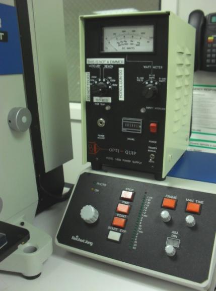
SOP: Polyvar Met Light Microscope
|
|
|
- August Wood
- 5 years ago
- Views:
Transcription
1 SOP Polyvar Met Light Microscope Page 1 of 8 SOP: Polyvar Met Light Microscope 1. Scope 1.1 This document describes the standard operating procedure (SOP) for the Polyvar Met Light Microscope. 2. Table of Contents 1. Scope Table of Contents Reference Documents Referenced within this Document Safety Setup Procedures Remove blue nylon cover from microscope Hardware Procedures Adjusting Stage Light Source Focusing Filtering Software Procedures Viewing an Image Measuring an Image Saving an Image Revision History Reference Documents 3.1 Referenced within this Document None 4. Safety 4.1 Follow all Nanofab safety procedures. 4.2 Be aware, the black fins on top of the microscope get very hot 5. Setup Procedures 5.1 Remove blue nylon cover from microscope 5.2 Remove black plastic eyepiece cap

 6.1.2.1 The smaller of these knobs contains tick marks, each of which correspond to one micron 6.")
2 SOP Polyvar Met Light Microscope Page 2 of 8 6. Hardware Procedures 6.1 Adjusting Stage Up and Down Forward to Backward Right to Left Figure 1: Stage Controls The right hand knobs beneath the stage control motion in the x y plane The top knob controls motion in the y direction (clockwise moves the stage towards you) The bottom knob controls motion in the x direction (clockwise moves the stage to the right) The left hand knobs beside the stage control motion in the z direction (clockwise moves the stage down) The smaller of these knobs contains tick marks, each of which correspond to one micron 6.2 Light Source Swap Knob Xenon Arc Lamp Control Quartz Lamp Control Indicator Light Figure 2: Light Controls
, and the Swap Knob (Fig. 2) 6.2.1.4 Touch NO other nearby knobs or buttons on the light source WARNING Wait for the Xenon Arc Lamp to warm up after turning on the Quartz Lamp before turning it on.")
3 6.2.1 Quartz Lamp SOP Polyvar Met Light Microscope Page 3 of Turn on the Quartz Lamp Control knob (Fig. 2) The Indicator Light (Fig. 2) should now be green Light should now be reaching your sample If not, check the filters are open (6.4), and the Swap Knob (Fig. 2) Touch NO other nearby knobs or buttons on the light source WARNING Wait for the Xenon Arc Lamp to warm up after turning on the Quartz Lamp before turning it on. If you hear multiple snapping sounds turn OFF the Arc Lamp immediately Xenon Arc Lamp First turn on the Quartz Lamp (see 6.2.1) Give the Arc Lamp a few minutes to warm up Press the Xenon Arc Lamp Control button (Fig. 2), nothing else You should hear ONE snap only after turning on this lamp To switch lamp sources, adjust the Swap Knob (Fig. 2) on the right hand side of the microscope closest to the back 6.3 Focusing Multiplier Wheel Objective Lenses Increase Objective Decrease Objective Always start at the lowest magnification Objective lenses Figure 3: Magnification Figure 4: Objective Lenses There are six Objective Lenses (Fig. 4): 2.5X, 5X, 20X, 50X, 100X, 150X To switch lenses use the red Objective Buttons on the left side of the microscope (Fig. 4)


4 The upper button increases the magnification The lower button decreases the magnification. SOP Polyvar Met Light Microscope Page 4 of 8 WARNING When changing the magnification, be sure the objective lens does not crash into your sample. This will destroy the lens and harm your sample The Multiplier Wheel (Fig.4) can multiply the magnification of the objective lenses by either :.8X, 1X, 1.25X, and 2X The stacked Focus Knobs are located on the right side of the microscope The larger knob is the Coarse Focus, which will focus quickly The smaller knob is the Fine Focus, which will focus in more detail 6.4 Filtering Each tick of the Fine Focus is one micron. L Low Medium Red None Green Iris Simple Filter Wave Filter High Figure 5: Wave Filter Wheel Figure 6: Filters The Wave Filter (Fig. 6) has six different filtering options (Fig. 5), selected by turning the filtering disk on the right side of the microscope The circle containing an X indicates no filter If you can t see a light on your sample after turning on the quartz lamp, first check and make sure the X filter is visible (Fig. 5) The red circle filters out red light The white circles are neutral density filters The large white circle filters out the least light from the lamp The medium white circle filters out a midlevel amount of light from the lamp The small white circle filters out the most light from the lamp The green circle filters out green light


 WARNING Do not pull the prism block past the red dot, you should not be able to see any of")
5 SOP Polyvar Met Light Microscope Page 5 of The first knob above the objective lenses is an Iris (Fig. 6), which will let more or less light in from your sample The second knob is a Simple Filter (Fig. 6), controlling the amount of light that reaches your sample The Nomarski filters can further filter the color fields, giving additional topographic information For ordinary imaging, the Prism Block should be pulled out so that the red dot is visible Lock Knob Twist Knob Figure 7: Prism Block (Engaged) Figure 8: Prism Block (Disengaged) Figure 9: Prism Block ERROR! To adjust the prism, slightly unscrew the Lock Knob (Fig. 7), then gently pull the Prism Block (Figs. 7-9) WARNING Do not pull the prism block past the red dot, you should not be able to see any of the hole cut into the metal Figure 10: Removing the Bright Field Filter Figure 11: Installing the Dark Field Filter The Bright Field Filter (Fig. 10), which should be in the microscope, may be used for samples of ordinary brightness To engage the Bright Field Filter manually pull the block to the right until it gently locks into place
 on the front of the prism block 6.4.4.3 The Dark Field Filter (Fig. 11) is for samples of unusually high brightness 6.4.4.3.1 The Dark Field Filter is kept in the top drawer of the desk behind you 6.")


6 SOP Polyvar Met Light Microscope Page 6 of Adjust the filter with the Twist Knob (Fig. 7) on the front of the prism block The Dark Field Filter (Fig. 11) is for samples of unusually high brightness The Dark Field Filter is kept in the top drawer of the desk behind you Remove the Bright Field Filter by gently pulling Replace with the Dark Field Filter so that it locks gently on the right Reinstall the Bright Field Filter when finished 7. Software Procedures Caliper Icon Ruler Icon Camera Icon Preview Camcorder Icon Snap Auto Expose Area Image Boxes Spatial Calibration Box Figure 12: Imaging software 7.1 Viewing an Image The computer should be on On the desktop, select the icon for the Q Capture Pro Software (Fig.13)
7 SOP Polyvar Met Light Microscope Page 7 of Once the software is open, select the Camcorder Icon, a new toolbar should appear (Fig. 12) Select the Preview button (Fig. 12) An Image Box should appear (Fig. 12) Click Auto Expose Area (Fig. 12) to automatically adjust the brightness If the image box is still dark review your light source and filters (6.2, 6.3) 7.2 Measuring an Image Before anything else, click the Snap (Fig. 12) button in the floating toolbar There should now be two Image Boxes (Fig. 12) Scale Bars Click the Caliper Icon (Fig.12) on the upper toolbar An Spatial Calibration Box should now appear (Fig. 12) In the same box, select the dropdown menu In the dropdown menu select your current objective lens and multiplier combination The box will then change to give you scale bar options Line Measurements Click the Ruler Icon (Fig. 12) on the upper toolbar Select Marker in the box that appears Free draw a line on your sample There is special procedure to save images with measurements (see 7.3.3) 7.3 Saving an Image Be sure you have selected the Snap image box (7.2.1) Save file to a folder under Labusers Saving images with measurements After drawing your measurements, select the Camera Icon on the upper toolbar (Fig. 12) Save from the Image Box that appears To retrieve your image, simply select the shortcut to Labusers Icon (Fig. 14) on the desktop The computer is not connected to the internet so remember to bring something to remove the files you want The computer will write to USBs, CDs, and ZIP disks Figure 13: Software Icon Figure 14: Labusers Icon
8 8. Revision History SOP Polyvar Met Light Microscope Page 8 of 8 Rev Date Originator Description of Changes 1 June 10, 2010 Kathryn Ecsedy First Draft
SOP: EDAX Eagle III Microspot XRF
 SOP: EDAX Eagle III Microspot XRF Page 1 of 6 SOP: EDAX Eagle III Microspot XRF 1. Scope 1.1 This document describes the standard operating procedure (SOP) for the EDAX Eagle III Microspot XRF. This X-ray
Brightfield Microscopy and Image Acquisition on Spotcam1. by Ryan Taylor/Nancy Kleene Last modified 10/02/05 by Birgit Ehmer
 Brightfield Microscopy and Image Acquisition on Spotcam1 by Ryan Taylor/Nancy Kleene Last modified 10/02/05 by Birgit Ehmer Log onto the computer. Enter your username and password to log onto the server.
Simplified Instructions: Zeiss Brightfield Microscope S1000
 Contents General Microscope Set-Up Adjust Illumination Focus Condenser Open Software Image Capture Settings Shading & Color Corrections Image Capture & Viewing Scaling and Measurements Synopsis of Other
Zeiss AxioImager.Z2 Brightfield Protocol
 Zeiss AxioImager.Z2 Brightfield Protocol 1) System Startup Please note put sign-up policy. You must inform the facility at least 24 hours beforehand if you can t come; otherwise, you will receive a charge
CAPTURING IMAGES ON THE HIGH-MAGNIFICATION MICROSCOPE
 University of Virginia ITC Academic Computing Health Sciences CAPTURING IMAGES ON THE HIGH-MAGNIFICATION MICROSCOPE Introduction The Olympus BH-2 microscope in ACHS s microscope lab has objectives from
Horiba LabRAM ARAMIS Raman Spectrometer Revision /28/2016 Page 1 of 11. Horiba Jobin-Yvon LabRAM Aramis - Raman Spectrometer
 Page 1 of 11 Horiba Jobin-Yvon LabRAM Aramis - Raman Spectrometer The Aramis Raman system is a software selectable multi-wavelength Raman system with mapping capabilities with a 400mm monochromator and
Standard Operating Procedure
 CENTER FOR NANOSCALE SCIENCE AND ENGINEERING Standard Operating Procedure Microscope Software Brian Wajdyk Page 1 of 6 Important Images are not to be saved to the computer. They will be deleted without
User Guide for TWAIN / DirectX interface for GRYPHAX USB 3.0 cameras
 User Guide for TWAIN / DirectX interface for GRYPHAX USB 3.0 cameras The TWAIN & DirectX driver for PROGRES GRYPHAX USB 3.0 cameras enables user to operate with TWAIN and DirectX supported 3 rd party software
Simplified Instructions: Olympus Widefield Microscope S1230
 Contents General Microscope Operation Simple Image Capture Multi-Wavelength Capture Z-Series Timelapse Combining Capture Modes Synopsis of Other Functions Pages 2-23 24-40 41-47 48-56 57-59 60-68 69-83
3D Capture. Using Fujifilm 3D Camera. Copyright Apis Footwear
 3D Capture Using Fujifilm 3D Camera Copyright 201 3 Apis Footwear Assembly and Settings 1. Assembly If your camera came without the projector attached, then you need to do it yourself. First remove the
Optika ISview. Image acquisition and processing software. Instruction Manual
 Optika ISview Image acquisition and processing software Instruction Manual Key to the Instruction Manual IS is shortened name used for OptikaISview Square brackets are used to indicate items such as menu
Zeiss Axioskop II. The AIF's "routine" light microscope. (Installed 8/24/04)AxioCam installed July 11th 2005
AxioCam installed July 11th 2005") Zeiss Axioskop II The AIF's "routine" light microscope. (Installed 8/24/04)AxioCam installed July 11th 2005 Featuring: Phase Contrast Darkfield DIC/Nomarski Brightfield Fluorescent filters for Dapi, FITC,Rhodamine
Zeiss Axioskop II The AIF's "routine" light microscope. (Installed 8/24/04)AxioCam installed July 11th 2005 Featuring: Phase Contrast Darkfield DIC/Nomarski Brightfield Fluorescent filters for Dapi, FITC,Rhodamine
3D Capture. Using Fujifilm 3D Camera. Copyright Apis Footwear
 3D Capture Using Fujifilm 3D Camera Copyright 201 4 Apis Footwear Camera Settings Before shooting 3D images, please make sure the camera is set as follows: a. Rotate the upper dial to position the red
Welcome 1. Precaution
 Table of Contents EN Precaution....2 Preparation.. 4 Standard accessories....4 Parts Names & Functions...5 Computer System requirements.... 6 Technical Specifications 7 Install the software.. 7 Start Microscope.8
Standard Operating Procedure (SOP) for Shared Equipment: Spinning Disk Confocal Microscope
 for Shared Equipment: Spinning Disk Confocal Microscope") Standard Operating Procedure (SOP) for Shared Equipment: Spinning Disk Confocal Microscope This document is to be used as a supplementary guide and not as a replacement for formal training. DO NOT operate
Standard Operating Procedure (SOP) for Shared Equipment: Spinning Disk Confocal Microscope This document is to be used as a supplementary guide and not as a replacement for formal training. DO NOT operate
GXCapture 8.1 Instruction Manual
 GT Vision image acquisition, managing and processing software GXCapture 8.1 Instruction Manual Contents of the Instruction Manual GXC is the shortened name used for GXCapture Square brackets are used to
Lab: Using a Compound Light Microscope
 Name Date Period Lab: Using a Compound Light Microscope Background: Microscopes are very important tools in biology. The term microscope can be translated as to view the tiny, because microscopes are used
KoPa Scanner. User's Manual A99. Ver 1.0. SHENZHEN OSTEC OPTO-ELECTRONIC TECHNOLOGY CO.,LTD.
 KoPa Scanner A99 User's Manual Ver 1.0 SHENZHEN OSTEC OPTO-ELECTRONIC TECHNOLOGY CO.,LTD. http://www.ostec.com.cn Content Chapter 1 Start... 1 1.1 Safety Warnings and Precautions... 1 1.2 Installation
Characterization Microscope Nikon LV150
 Characterization Microscope Nikon LV150 Figure 1: Microscope Nikon LV150 Introduction This upright optical microscope is designed for investigating up to 150 mm (6 inch) semiconductor wafers but can also
CHAPTER1: QUICK START...3 CAMERA INSTALLATION... 3 SOFTWARE AND DRIVER INSTALLATION... 3 START TCAPTURE...4 TCAPTURE PARAMETER SETTINGS... 5 CHAPTER2:
 Image acquisition, managing and processing software TCapture Instruction Manual Key to the Instruction Manual TC is shortened name used for TCapture. Help Refer to [Help] >> [About TCapture] menu for software
Zeiss LSM 880 Protocol
 Zeiss LSM 880 Protocol 1) System Startup Please note put sign-up policy. You must inform the facility at least 24 hours beforehand if you can t come; otherwise, you will receive a charge for unused time.
User Manual. Copyright 2010 Lumos. All rights reserved
 User Manual The contents of this document may not be copied nor duplicated in any form, in whole or in part, without prior written consent from Lumos. Lumos makes no warranties as to the accuracy of the
Dealer4 Beginner s Guide
 Dealer4 Beginner s Guide written by Cad Delworth, Carlton Bridge Club, Edinburgh This is revision number 6, saved at 09:42:00 on 23 October 2011. Dealer4 Beginner's Guide 2 Contents Introduction... 3 Do
Nikon E800 Operating Instructions.
 Nikon E800 Operating Instructions. You can request electronic copies of this manual by contacting lshats@jhsph.edu Copies are also available on the JHU MMI Department web site. Please send your comments
QUICKSTART GUIDE: WIDEFIELD WF3 Zeiss Cell Observer Live Cell Imaging System (SAF, ROOM 409) Imperial College London
 Imperial College London") Imperial College London Facility for Imaging by Light Microscopy QUICKSTART GUIDE: WIDEFIELD WF3 Zeiss Cell Observer Live Cell Imaging System (SAF, ROOM 409) Observing Life As It Happens Startup procedure...
Imperial College London Facility for Imaging by Light Microscopy QUICKSTART GUIDE: WIDEFIELD WF3 Zeiss Cell Observer Live Cell Imaging System (SAF, ROOM 409) Observing Life As It Happens Startup procedure...
ENSC 470/894 Lab 1 V1.4 (Oct )
") ENSC 470/894 Lab 1 V1.4 (Oct. 29 2010) Introduction: Lab 1 is designed to give students basic experience in optics. In the lab you will set up lenses on an optical table, with a LCD screen pattern as the
ENSC 470/894 Lab 1 V1.4 (Oct. 29 2010) Introduction: Lab 1 is designed to give students basic experience in optics. In the lab you will set up lenses on an optical table, with a LCD screen pattern as the
Guide to Confocal 5. Starting session
 Guide to Confocal 5 Remember that when booking and before starting session you can check for any problems at https://www.bris.ac.uk/biochemistry/uobonly/cif/index.html Starting session Switch on microscope
Match the microscope structures given in the left column with the statements in the right column that identify or describe them.
 49 Prelab for Name Match the microscope structures given in the left column with the statements in the right column that identify or describe them. Key: a. coarse adjustment knob f. turret or nosepiece
RENISHAW INVIA RAMAN SPECTROMETER
 STANDARD OPERATING PROCEDURE: RENISHAW INVIA RAMAN SPECTROMETER Purpose of this Instrument: The Renishaw invia Raman Spectrometer is an instrument used to analyze the Raman scattered light from samples
Using the AmScope Microscope Cameras
 Using the AmScope Microscope Cameras Part 1 Setup. In order to use the camera, you will need: a) the camera system; b) a computer running the camera software. The camera system is contained in a Pelican
Widefield 1. Switching on
 Widefield 1 Switching on 1. Ignite DG5 lamp - must be switched on first (if previous user has switched off, wait 30 min before igniting) 2. Wait 5s and then turn on the main DG5 controller switch. 3. DG5
Nikon Eclipse Ti A1-A Confocal Operating Manual. Start-up. Microscope
 Nikon Eclipse Ti A1-A Confocal Operating Manual Start-up 1. Turn on Excite Fluorescent light power supply- metal halide. a. Cool down as for mercury bulb b. Wheel closed liquid light guide 2. Turn on power
QUICKSTART GUIDE: WIDEFIELD HWF1 Zeiss Cell Observer Live Cell Imaging System (HAMMERSMITH, L BLOCK, ROOM 314) Imperial College London
 Imperial College London") Imperial College London Facility for Imaging by Light Microscopy QUICKSTART GUIDE: WIDEFIELD HWF1 Zeiss Cell Observer Live Cell Imaging System (HAMMERSMITH, L BLOCK, ROOM 314) Observing Life As It Happens
Imperial College London Facility for Imaging by Light Microscopy QUICKSTART GUIDE: WIDEFIELD HWF1 Zeiss Cell Observer Live Cell Imaging System (HAMMERSMITH, L BLOCK, ROOM 314) Observing Life As It Happens
Operating Instructions for Zeiss LSM 510
 Operating Instructions for Zeiss LSM 510 Location: GNL 6.312q (BSL3) Questions? Contact: Maxim Ivannikov, maivanni@utmb.edu 1 Attend A Complementary Training Before Using The Microscope All future users
Zeiss Axio Imager.A1 manual
 Zeiss Axio Imager.A1 manual Power-up protocol 1. Mercury lamp 2. Power strip on shelf 3. Computer The Mercury lamp should always be first-on and last-off. This prevents any electrical surges caused by
Instructions for Making On-Line Reservations for Microscopes in NB11-204
 Instructions for Making On-Line Reservations for Microscopes in NB11-204 1. Log into Mail using Mail.swmed.edu 2. Log in using your university id and password. 3. Click the Calendar Tab at the top right
ENSC 470/894 Lab 1 V2.0 (Sept )
") ENSC 470/894 Lab 1 V2.0 (Sept. 22 2013) Introduction: Lab 1 is designed to give students basic experience in optics. In the lab you will set up lenses on an optical table, with a LCD screen pattern as
ENSC 470/894 Lab 1 V2.0 (Sept. 22 2013) Introduction: Lab 1 is designed to give students basic experience in optics. In the lab you will set up lenses on an optical table, with a LCD screen pattern as
Photo Effects & Corrections with PhotoFiltre
 Photo Effects & Corrections with PhotoFiltre P 330 / 1 Fix Colour Problems and Apply Stylish Effects to Your Photos in Seconds with This Free Software If you re keen on digital photography, you probably
Digital Microscope. User Manual
 Digital Microscope User Manual Features The digital microscope provides 10~200X adjustable magnification range. The build-in high-performance white LED can illuminate the object without using any auxiliary
What is it? Study the mystery photos and try to identify each one! Have access to a computer?
 Station 1 Solve the Mystery What is it? Study the mystery photos and try to identify each one! They are all common objects that might be found in your home or a classroom. Write your guesses for the mystery
GlobiScope Analysis Software for the Globisens QX7 Digital Microscope. Quick Start Guide
 GlobiScope Analysis Software for the Globisens QX7 Digital Microscope Quick Start Guide Contents GlobiScope Overview... 1 Overview of home screen... 2 General Settings... 2 Measurements... 3 Movie capture...
Using a Compound Light Microscope Lab Pre-Lab Assignment
 Name: Block: Due Date: Using a Compound Light Microscope Lab Pre-Lab Assignment Pre-Lab Assignment This assignment must be completed by the next class period in order to be allowed to participate in the
Nasmyth Ultraview Vox User Protocol
 Nasmyth Ultraview Vox User Protocol Switch on all wall sockets labelled Nasmyth, switch camera on (power supply located on table behind monitor), switch on laser switch in laser rack, switch computer on
The ideal K-12 science microscope solution. User Guide. for use with the Nova5000
 The ideal K-12 science microscope solution User Guide for use with the Nova5000 NovaScope User Guide Information in this document is subject to change without notice. 2009 Fourier Systems Ltd. All rights
Basic Microscopy for Plant Biology
 Page 1 of 8 Basic Microscopy for Plant Biology OBJECTIVES After completing this exercise, you should be able to do the following: a. Name the parts of the compound microscope and the functions of each.
Contents STARTUP MICROSCOPE CONTROLS CAMERA CONTROLS SOFTWARE CONTROLS EXPOSURE AND CONTRAST MONOCHROME IMAGE HANDLING
 Operations Guide Contents STARTUP MICROSCOPE CONTROLS CAMERA CONTROLS SOFTWARE CONTROLS EXPOSURE AND CONTRAST MONOCHROME IMAGE HANDLING Nikon Eclipse 90i Operations Guide STARTUP Startup Powering Up Fluorescence
RENISHAW RAMAN MICROSCOPE STANDARD OPERATING PROCEDURE
 RENISHAW RAMAN MICROSCOPE STANDARD OPERATING PROCEDURE 09DJ00 CHRIS BUXEY (TECHNICIAN) X2715 C.BUXEY@SURREY.AC.UK V5.0 OCTOBER 2008 RENISHAW MICRORAMAN STANDARD OPERATING PROCEDURE 1.0 Foreword Chris Buxey:
Nikon E800 Operating Instructions.
 Nikon E800 Operating Instructions. You can request electronic copies of this manual by contacting imaging@fhcrc.org. Copies are also available on the Scientific Imaging web site. Please send your comments
Things to check before start-up.
 Byeong Cha Page 1 11/24/2009 Manual for Leica SP2 Confocal Microscope Enter you name, the date, the time, and the account number in the user log book. Things to check before start-up. Make sure that your
Jeol JEM Responsible personell: Endy ( ) Online booking is compulsory!
 Online booking is compulsory!") Jeol JEM 1230 Responsible personell: Endy (45279377) Online booking is compulsory! After training you will have access to working alone on the instrument. All insertion of samples is done by responsible
Jeol JEM 1230 Responsible personell: Endy (45279377) Online booking is compulsory! After training you will have access to working alone on the instrument. All insertion of samples is done by responsible
Nikon E800 Microscope. Operating Instructions
 Nikon E800 Microscope Operating Instructions B Watson 12/2005 Table of contents: 1. The Nikon E800 Microscope 2. Turning the system ON and OFF 3. Selecting the light path 4. Operating in transmitted light
AxioVision 4.5 Brightfield Image Capture Procedure
 AxioVision 4.5 Brightfield Image Capture Procedure 1. STARTING-UP PROCEDURE: Remove blue dust cover and place on shelf under microscope. Turn on the halogen lamp by pushing the switch at the back right
Olympus LEXT OLS 4000 Confocal Laser Microscope
 Olympus LEXT OLS 4000 Confocal Laser Microscope The Olympus LEXT OLS4000 is a confocal microscope capable of taking high-resolution 3D images. The magnification (Optical and Digital) of this microscope
Nikon Eclipse Ti2-E Widefield/Spinning Disk Confocal Microscope Standard Operation Protocol
 Nikon Eclipse Ti-E Widefield/Spinning Disk Confocal Microscope Standard Operation Protocol Please sign on the log sheet before switching on system. Turn on system Turn on A only if confocal mode or laser
Rachel
 http://gmv.cast.uark.edu A Method Store for Advanced Survey and Modeling Technologies Mon, 01 Apr 2013 03:29:18 +0000 en-us hourly 1 http://wordpress.org/?v=3.5.1 http://gmv.cast.uark.edu/scanning/guide-to-leveling-andaligning-the-breuckmann-tripod-and-smartscan-he-for-calibration/
OPERATION OF THE HITACHI S-450 SCANNING ELECTRON MICROSCOPE. by Doug Bray Department of Biological Sciences University of Lethbridge
 OPERATION OF THE HITACHI S-450 SCANNING ELECTRON MICROSCOPE by Doug Bray Department of Biological Sciences University of Lethbridge Revised September, 2000 Note: The terms in bold in this document represent
Zeiss Axiovert 135 Fluorescence Microscope Quick Guide / Operations Manual (v. 1.0 February 09)
") University of Chicago Integrated Light Microscopy Core Dr. Vytas Bindokas, Director http://digital.bsd.uchicago.edu By: Christine Labno, Assistant Director Room: AB-129 Phone: 4-9040 Zeiss Axiovert 135
University of Chicago Integrated Light Microscopy Core Dr. Vytas Bindokas, Director http://digital.bsd.uchicago.edu By: Christine Labno, Assistant Director Room: AB-129 Phone: 4-9040 Zeiss Axiovert 135
SWIFT SERIES M2252DGL MICROSCOPE
 SWIFT SERIES M2252DGL MICROSCOPE The M2252DGL Series is ideal for elementary to high school classrooms. Built to withstand student use, this series has locked-on eyepieces, objectives, illuminator housing
The image regions are all in focus, producing no contrast resulting from changes in signal level.
 StreamLineHR example Figure 6. Raman image of angled grid with no surface correction (5 µm step size, 50 objective). The bright image regions are in focus, darker regions are out of focus. (The centre
Photography Protocol. Software: ZEN Blue 2012
 Photography Protocol Hardware: Zeiss Axio Imager.A2 or Imager.M2 microscope 10x eye piece 20x / 0.45 dry lens 40x / 0.75 dry lens 63x / 0.95 dry lens 100x / 1.4 oil lens AxioCam MRc camera Software: ZEN
Confocal Raman Microscopy (WITec Alpha 300R)
") Confocal Raman Microscopy (WITec Alpha 300R) Please refer to Witec Alpha300R Confocal Raman Microscope User Manual for the details of the operating procedure. Sample preparation 1. Attach your sample on
Confocal Raman Microscopy (WITec Alpha 300R) Please refer to Witec Alpha300R Confocal Raman Microscope User Manual for the details of the operating procedure. Sample preparation 1. Attach your sample on
USING LEICA AS LASER MICRODISSECTION (LMD6000) MICROSCOPE Written By Jungim Hur
 MICROSCOPE Written By Jungim Hur") USING LEICA AS LASER MICRODISSECTION (LMD6000) MICROSCOPE Written By Jungim Hur Digital Video Camera Eyepieces Laser module Laser safety UV shield Specimen holder Smart move control LEICA CTR6500 electronics
USING LEICA AS LASER MICRODISSECTION (LMD6000) MICROSCOPE Written By Jungim Hur Digital Video Camera Eyepieces Laser module Laser safety UV shield Specimen holder Smart move control LEICA CTR6500 electronics
The invention of the microscope made it possible for scientists to view and study cells. Cells the basic units of all living organisms.
 The Discovery of Cells The invention of the microscope made it possible for scientists to view and study cells. Cells the basic units of all living organisms. The Cell Theory All living things are made
ML7520 ML7530 DIOPTER ADJUSTMENT RING BINOCULAR BODY, INCLINED 30. (a) Field Iris Control Lever. (c) Filter Slots EYEPIECES, KHW10X
 Field Iris Control Lever. (c) Filter Slots EYEPIECES, KHW10X") JAPAN DIOPTER ADJUSTMENT RING BINOCULAR BODY, INCLINED 30 (a) Field Iris Control Lever (c) Filter Slots EYEPIECES, KHW10X ANALYZER CONTROL LEVER (b) Aperture Iris Control Lever LIGHT SOURCE HOUSING VERTICAL
JAPAN DIOPTER ADJUSTMENT RING BINOCULAR BODY, INCLINED 30 (a) Field Iris Control Lever (c) Filter Slots EYEPIECES, KHW10X ANALYZER CONTROL LEVER (b) Aperture Iris Control Lever LIGHT SOURCE HOUSING VERTICAL
Zeiss LSM 780 Protocol
 Zeiss LSM 780 Protocol 1) System Startup F Please note the sign-up policy. You must inform the facility at least 24 hours beforehand if you can t come; otherwise, you will receive a charge for unused time.
Basic Microscopy. OBJECTIVES After completing this exercise, you should be able to do the following:
 Page 1 of 10 Basic Microscopy OBJECTIVES After completing this exercise, you should be able to do the following: a. Name the parts of the compound microscope and the functions of each. b. Describe how
REMEMBER: You have 5GB of disk space on this microscope. Check before you start if you have room for your experiment. If not delete your old data.
 1 Use of the Zeiss LSM 510 Inverted Firstly please be aware that this microscope should be treated with respect and care at all times. Rules of use: This Microscope can only be used by Masters by Research
JEOL 6500 User Manual
 LOG IN to your session on the computer to the left of the microscope. Starting Conditions 1. Press Ctrl-Alt-Del and log on to the microscope computer. Click on JEOL PC SEM 6500 icon. Click yes if message
Operation Guide for the Leica SP2 Confocal Microscope Bio-Imaging Facility Hunter College October 2009
 Operation Guide for the Leica SP2 Confocal Microscope Bio-Imaging Facility Hunter College October 2009 Introduction of Fluoresence Confocal Microscopy The first confocal microscope was invented by Princeton
DigiScope II v3 TM Aperture Scope User s Manual
 DigiScope II v3 TM Aperture Scope User s Manual Welcome Thank you for choosing DigiScope II v3 TM Aperture scope! The DigiScope II v3 TM Aperture Scope is an exciting new device to Capture and record the
Widefield-NikonEclipseTE200-ORCA Nikon Eclipse TE200 Inverted Microscope with Hamamatsu 1394 Orca-ER Cooled CCD Camera and Micromanager Software
 Widefield-NikonEclipseTE200-ORCA Nikon Eclipse TE200 Inverted Microscope with Hamamatsu 1394 Orca-ER Cooled CCD Camera and Micromanager Software September 2007 Check website for most current User Guide
Olympus Digital Microscope Camera (DP70) checklist
 checklist") Smith College - July 2005 Olympus Digital Microscope Camera (DP70) checklist CONTENT, page no. Camera Information, 1 Startup, 1 Retrieve an Image, 2 Microscope Setup, 2 Capture, 3 Preview. 3 Color Balans,
Smith College - July 2005 Olympus Digital Microscope Camera (DP70) checklist CONTENT, page no. Camera Information, 1 Startup, 1 Retrieve an Image, 2 Microscope Setup, 2 Capture, 3 Preview. 3 Color Balans,
SOP-P051. Scanning of Optical Filters With USB2000. Objective: To determine the spectral transmittance properties of an optical filter.
 Purdue University Cytometry Laboratories SOP-P051 Scanning of Optical Filters With USB2000 Objective: To determine the spectral transmittance properties of an optical filter. Procedure: 1. Ensure that
GlassSpection User Guide
 i GlassSpection User Guide GlassSpection User Guide v1.1a January2011 ii Support: Support for GlassSpection is available from Pyramid Imaging. Send any questions or test images you want us to evaluate
THE COMPOUND BRIGHTFIELD MICROSCOPE
 THE COMPOUND BRIGHTFIELD MICROSCOPE Microbiology is the study of microscopic organisms that are so small that they are below the limit of vision of the human eye. Bacteria are the smallest of microorganisms
CNC Using the FlexiCam CNC and HMI Software. Guldbergsgade 29N, P0 E: T:
 CNC Using the FlexiCam CNC and HMI Software Guldbergsgade 29N, P0 E: makerlab@kea.dk T: +46 46 03 90 This grey box is the NC controller. Let s start by turning the red switch to the ON position, then press
Quick Guide for Zeiss 710 Laser Scanning Confocal MGH Cancer Center
 Quick Guide for Zeiss 710 Laser Scanning Confocal MGH Cancer Center For any questions or concerns, please contact: Linda Nieman lnieman@mgh.harvard.edu Office: (617) 643-9684 Cell: (512) 565-8076 Chenyue
Nikon A1R. Multi-Photon & Laser Scanning Confocal Microscope. Kyle Marchuk Adam Fries Jordan Briscoe Kaitlin Corbin. April 2017.
 Nikon A1R Multi-Photon & Laser Scanning Confocal Microscope Kyle Marchuk Adam Fries Jordan Briscoe Kaitlin Corbin April 2017 Contents 1 Introduction 2 2 Start-Up 2 3 Imaging 4 3.1 Sample Alignment...........................................
BX-61: Brightfield Instruction /Continue to scroll for Fluorescent Instuctions
 BX-61: Brightfield Instruction /Continue to scroll for Fluorescent Instuctions Starting up: Schematic of Olympus BX-61. 1. Turn on Olympus microscope power box (left of microscope) with toggle switch on
Nikon C1si Spectral Laser Scanning Confocal Microscope. User Guide
 Nikon C1si Spectral Laser Scanning Confocal Microscope User Guide Contents: C1Si Turn-On/ShutDown Procedures... 2 Overview... 4 Setup for epi-illumination to view through the eyepieces:... 5 Setup for
JEOL 6700 User Manual 05/18/2009
 JEOL 6700 User Manual 05/18/2009 LOG IN to your session on the computer to the right of the microscope. Starting Conditions 1. Click the button and read the Penning Gauge to ensure that the microscope
User Manual. Digital Compound Binocular LED Microscope. MicroscopeNet.com
 User Manual Digital Compound Binocular LED Microscope Model MD82ES10 MicroscopeNet.com Table of Contents i. Caution... 1 ii. Care and Maintenance... 2 1. Components Illustration... 3 2. Installation...
SHORT GUIDE TO LASER MICRODISSECTION USING THE PALM COMBI SYSTEM
 SHORT GUIDE TO LASER MICRODISSECTION USING THE PALM COMBI SYSTEM Turning ON the PALM DuoFlex Combi system 1. Turn on the three power point switches on the wall. From right to left: mercury lamp, microscope
FE-SEM SU-8020 Operating manual (Preliminary version)
") FE-SEM SU-8020 Operating manual (Preliminary version) 2016/04/11 Seimitsu Bunseki sitsu lab. Starting up 1.Turn on the Display switch. Windows OS is starting up 2. Select the user SU-8000. 3. Click the
FE-SEM SU-8020 Operating manual (Preliminary version) 2016/04/11 Seimitsu Bunseki sitsu lab. Starting up 1.Turn on the Display switch. Windows OS is starting up 2. Select the user SU-8000. 3. Click the
The microscope is useful in making observations and collecting data in scientific experiments. Microscopy involves three basic concepts:
 AP BIOLOGY Chapter 6 NAME DATE Block MICROSCOPE LAB PART I: COMPOUND MICROSCOPE OBJECTIVES: After completing this exercise you should be able to: Demonstrate proper care and use of a compound microscope.
used for low power magnification of a sample image is 3 dimensional
 MICROSCOPES One of the most important inventions in the advancement of Biology 1. Simple Microscopes ie. magnifying glass, stereoscope (dissecting scope) have a single lens or a pair of lenses combined
Leica DB LB Research microscope and Studo Lite Imaging software
 Leica DB LB Research microscope and Studo Lite Imaging software Room B523 User Guide Molecular Imaging Unit University of Helsinki www.miu.helsinki.fi 9.4.2008 1 GENERAL USER INFORMATION... 1 2 SETTINGS
1.3. Before loading the holder into the TEM, make sure the X tilt is set to zero and the goniometer locked in place (this will make loading easier).
.") JEOL 200CX operating procedure Nicholas G. Rudawski ngr@ufl.edu (805) 252-4916 1. Specimen loading 1.1. Unlock the TUMI system. 1.2. Load specimen(s) into the holder. If using the double tilt holder, ensure
JEOL 200CX operating procedure Nicholas G. Rudawski ngr@ufl.edu (805) 252-4916 1. Specimen loading 1.1. Unlock the TUMI system. 1.2. Load specimen(s) into the holder. If using the double tilt holder, ensure
Part 2: Earpiece. Insert Protrusion (Internal Sketch) Hole Patterns Getting Started with Pro/ENGINEER Wildfire. Round extrusion.
 Hole Patterns Getting Started with Pro/ENGINEER Wildfire. Round extrusion.") Part 2: Earpiece 4 Round extrusion Radial pattern Chamfered edge To create this part, you'll use some of the same extrusion techniques you used in the lens part. The only difference in this part is that
Part 2: Earpiece 4 Round extrusion Radial pattern Chamfered edge To create this part, you'll use some of the same extrusion techniques you used in the lens part. The only difference in this part is that
SPINNING DISK CSU-X1 USER MANUAL
 SPINNING DISK CSU-X1 USER MANUAL Starting the temperature controller... 2 Starting the CO2 controller... 3 Start the spinning disk... 4 Sample observation with the oculars... 5 Spatial sampling, Pixel
Use of the HSW5 Spinning Disk Confocal Microscope Updated last May 25, 2010 OK
 Use of the HSW5 Spinning Disk Confocal Microscope Updated last May 25, 2010 OK Getting Started: 2 Starting Micromanager and Loading a Configuration 3 The Main Micromanager GUI 3 Configuration Settings
Manual for BMS E1 eplan series, compound microscope
 Manual for BMS E1 eplan series, compound microscope The compound microscope allows it to study, at cell level, structures of textures of botanical and zoological nature. (e.g. slides of roots, leaves and
Image-Pro Plus 7.0 Product Note
 Image-Pro Plus 7.0 Product Note Automated Microscope Configuration Introduction Cameras, software, shutters, stages, objectives, filters, turrets there are a lot of components in an automated microscopy
Orion m Series Welding System User Manual
 Orion m Series Welding System User Manual Table of Contents Chapter 1: Setup and Assembly... p.5 What is in the Box... p.5 Darkening Lens & Power Supply Setup... p.5 Microscope Arm Setup... p.6 Microscope
HP Designjet HD Scanner and T1200 HD Multifunction Printer
 HP Designjet HD Scanner and T1200 HD Multifunction Printer Introductory information USB 2.0 high-speed certified Introductory Information Other sources of information The User s Guide for your scanner
Lab: The Compound Microscope
 Lab: The Compound Microscope Purpose: To learn the parts of the compound microscope and to learn the basic skills needed to use the microscope properly. Materials: Microscope Colored paper Cover slips
Protist Microscope Lab
 Name: Block: Due Date: Protist Microscope Lab Pre-Lab Assignment 1. Fill out the table for question #4 on the second page of your lab packet. (You may use the Biology textbook pages R8 and R9 in the back
CALIBRATION OF MICROSCOPE EYEPIECE GRATICULE
 CALIBRATION OF MICROSCOPE EYEPIECE GRATICULE A typical eyepiece graticule looks like this: It is 10mm in length and each mm is divided into 10 parts So each small division = 0.1mm = 100µm The eyepiece
OPT3: Operating Procedure for Horiba Jobin Yvon LabRam Aramis Raman/PL System See LabSpec_6_2 General User Quick Start Guide on the computer desktop
 OPT3: Operating Procedure for Horiba Jobin Yvon LabRam Aramis Raman/PL System See LabSpec_6_2 General User Quick Start Guide on the computer desktop 1. Log in usage using the SMIF web site 2. Turn power
User instructions Compound laboratory microscope
 KERN & Sohn GmbH Ziegelei 1 D-72336 Balingen E-mail: info@kern-sohn.com User instructions Compound laboratory microscope Tel: +49-[0]7433-9933-0 Fax: +49-[0]7433-9933-149 Internet: www.kern-sohn.com KERN