used for low power magnification of a sample image is 3 dimensional
|
|
|
- Isabel Brown
- 5 years ago
- Views:
Transcription
1
 have a single lens or a pair of lenses combined uses reflected light = light rays shone on an object are refracted (spread) as they pass through the")
2 MICROSCOPES One of the most important inventions in the advancement of Biology 1. Simple Microscopes ie. magnifying glass, stereoscope (dissecting scope) have a single lens or a pair of lenses combined uses reflected light = light rays shone on an object are refracted (spread) as they pass through the convex lens(es) = enlarges object used for low power magnification of a sample image is 3 dimensional
3 2. Compound Microscope has 2 separate lenses: ocular (monocular / binocular) & the objective uses transmitted light = light passes through the object on the slide and is focused by the 2 lenses (mirrors help) Magnification = how many times the specimen has been enlarged Eyepiece x Objective = Total Magnification 10 x 40 = 400x Resolution = how clear the image is : as magnification increases, clarity decreases = stains and reducing light intensity are often used to make structures easier to see Field of View = is the diameter of the circle of light - varies depending on the microscope used & objective lens that is in place - measured in m ( 1 mm = 1000 m)
4 Care and Use of Microscopes 1. Always use both hands to carry the microscope; one on the arm, the other supporting the base. 2. Use only lens paper to clean the lenses. 3. Coarse focus knob is ONLY used when the Low (Scanning) power objective is in place. 4. Do not remove any parts. 5. When finished, make sure the Low (Scanning ) power objective is in place and the stage is completely lowered. 6. Wrap the cord around the arm and replace dust cover. Trouble Shooting Occasionally you may have trouble with working your microscope. Here are some common problems and solutions. 1. Image is too dark! Adjust the diaphragm, make sure your light is on. 2. There's a spot in my viewing field, even when I move the slide the spot stays in the same place! Your lens is dirty. Use lens paper, and only lens paper to carefully clean the objective and ocular lens. The ocular lens can be removed to clean the inside. The spot is probably a speck of dust. 3. I can't see anything under high power! Remember the steps, if you can't focus under scanning and then low power, you won't be able to focus anything under high power. Start at scanning and walk through the steps again. 4. Only half of my viewing field is lit. You probably don't have your objective fully clicked into place..
5
6 Guidelines for Microscope Drawings 1. Always use pencil and draw on 8 x 11 white paper portrait direction. 2. List Name, Date, Class, Group members in upper right hand corner. 3. Drawings must be large (no more than 2 per page), and single sided. 4. Drawing should be centered or slightly to the left. 5. Uses smooth, firm lines no sketching or shading. 6. Use parallel lines drawn with a ruler to label. 7. Label on the right side only; line up labels vertically. 8. Print labels in lower case letters. 9. Print a title that is precise and descriptive above the drawing. 10. Calculate the size of your specimen below your diagram. To calculate the size of the specimen Size of Specimen = Diameter of the field of view # of Times the Specimen fits Across BE SURE YOUR FINAL ANSWER IS IN m 3000 m = 100 m 30
7
8 MICROSCOPES One of the most important inventions in the advancement of Biology 1. Simple Microscopes ie. magnifying glass, stereoscope (dissecting scope) have a single lens or a pair of lenses combined uses light = light rays shone an object are as they pass through the convex lens(es) = enlarges object used for of a sample image is
9 2. Compound Microscope has 2 separate lenses: (monocular / binocular) & the uses = light passes the object on the slide and is focused by the 2 lenses (mirrors help) Magnification = how many times the specimen has been enlarged Eyepiece x Objective = Total Magnification Resolution = how clear the image is : as magnification increases, clarity = & intensity are often used to make structures easier to see Field of View = is the diameter of the circle of light - varies depending on the & that is in place - measured in m ( 1 mm = 1000 m)
10 Care and Use of Microscopes 1. Always use to carry the microscope; one on the arm, the other supporting the base. 2. Use only to clean the lenses. 3. Coarse focus knob is used when the Low (Scanning) power objective is in place. 4. Do remove any parts. 5. When finished, make sure the Low (Scanning ) power objective is in and the stage is lowered. 6. the cord around the arm and dust cover. Trouble Shooting Occasionally you may have trouble with working your microscope. Here are some common problems and solutions: 1. Image is too dark! Adjust the diaphragm, make sure your light is on. 2. There's a spot in my viewing field, even when I move the slide the spot stays in the same place! Your lens is dirty. Use lens paper, and only lens paper to carefully clean the objective and ocular lens. The ocular lens can be removed to clean the inside. The spot is probably a speck of dust. 3. I can't see anything under high power! Remember the steps, if you can't focus under scanning and then low power, you won't be able to focus anything under high power. Start at scanning and walk through the steps again. 4. Only half of my viewing field is lit. You probably don't have your objective fully clicked into place..
, and single sided. 4. Drawing should be or slightly to the left. 5. Uses smooth, firm lines 6. Use lines drawn with a to label. 7.")
11 Guidelines for Microscope Drawings 1. Always use and draw on 8 x 11 white paper portrait direction. 2. List Name, Date, Class, Group members in upper. 3. Drawings must be (no more than 2 per page), and single sided. 4. Drawing should be or slightly to the left. 5. Uses smooth, firm lines 6. Use lines drawn with a to label. 7. Label on the only; line up labels. 8. labels in lower case letters. 9. a title that is precise and descriptive the drawing. 10. Calculate the of your specimen below your diagram. To calculate the size of the specimen Size of Specimen = Diameter of the field of view # of Times the Specimen fits Across BE SURE YOUR FINAL ANSWER IS IN m 3000 m = 100 m 30
 = 2.")
12 MICROSCOPES One of the most important inventions in the advancement of Biology 1. Simple Microscopes ie. magnifying glass, stereoscope have a or combined uses light : light rays shone an object are refracted as they pass through the convex lens(es) = 2. Compound Microscope has 2 separate lenses - & the uses light passes through the object on the slide and is focused by the 2 lenses Magnification = how many times the specimen has been enlarged Eyepiece x Objective = Total Magnification Resolution = how clear the image is : as magnification, clarity = dyes and stains are used to make structures easier to see
13
The invention of the microscope made it possible for scientists to view and study cells. Cells the basic units of all living organisms.
 The Discovery of Cells The invention of the microscope made it possible for scientists to view and study cells. Cells the basic units of all living organisms. The Cell Theory All living things are made
Lab: The Compound Microscope
 Lab: The Compound Microscope Purpose: To learn the parts of the compound microscope and to learn the basic skills needed to use the microscope properly. Materials: Microscope Colored paper Cover slips
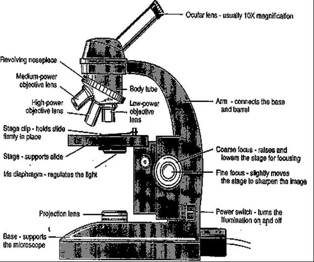
STRUCTURE OF THE MICROSCOPE
 STRUCTURE OF THE MICROSCOPE Use the word list to label the microscope below: Light Source Coarse adjustment knob Diaphragm Stage Clips Objectives Fine Adjustment Knob Base Stage Stage Clips Arm Revolving
The microscope is useful in making observations and collecting data in scientific experiments. Microscopy involves three basic concepts:
 AP BIOLOGY Chapter 6 NAME DATE Block MICROSCOPE LAB PART I: COMPOUND MICROSCOPE OBJECTIVES: After completing this exercise you should be able to: Demonstrate proper care and use of a compound microscope.
MICROSCOPE LAB. Resolving Power How well specimen detail is preserved during the magnifying process.
 AP BIOLOGY Cells ACTIVITY #2 MICROSCOPE LAB OBJECTIVES 1. Demonstrate proper care and use of a compound microscope. 2. Identify the parts of the microscope and describe the function of each part. 3. Compare
Basic Microscopy for Plant Biology
 Page 1 of 8 Basic Microscopy for Plant Biology OBJECTIVES After completing this exercise, you should be able to do the following: a. Name the parts of the compound microscope and the functions of each.
Microscope Review. 1. A compound light microscope is represented in the diagram below.
 Name Microscope Review Date 1. A compound light microscope is represented in the diagram below. 5. The diagram below represents a hydra as viewed with a compound light microscope. If the hydra moves toward
Name: Date Completed: Class: Lab Minutes: Teacher:
 Name: Date Completed: _ Class: Lab Minutes: _ Teacher: Introduction to the Microscope Lab Activity This lab was created by Mr. Buckley from Edward Knox High School. Credit is given for this original activity
Introduction. Instructional Objectives. Materials. Procedure. I. Microscope Parts and Function. Honors Biology
 Honors Biology Introduction to the Microscope Lab Activity This lab was created by Mr. Buckley from Edward Knox High School. Credit is given for this original activity to Mr. Buckley. Introduction "Micro"
Name: Period: Week of: January 21st-25th Root Words In-Class Homework. Picture: -Microscope Notes -Lesson on Focusing the Microscope
 Day 1/21: Monday Biology Week #21 Week of: January 21st-25th Root Words In-Class Homework Word: Definition: As in: - Picture: NO SCHOOL: MLK Day 1/22: Tuesday Word: Definition: As in: - Picture: -Microscope
Basic Microscopy. OBJECTIVES After completing this exercise, you should be able to do the following:
 Page 1 of 10 Basic Microscopy OBJECTIVES After completing this exercise, you should be able to do the following: a. Name the parts of the compound microscope and the functions of each. b. Describe how
Figure 3.4 Approximate size of various types of cells. ~10 um. Red Blood Cells = mm 1500 um. Width of penny Pearson Education, Inc.
 Figure 3.4 Approximate size of various types of cells. ~10 um Red Blood Cells 1.5mm 1500 um Width of penny = 1500 Figure 4.3 The limits of resolution (and some representative objects within those ranges)
A BRIEF INTRODUCTION TO MICROSCOPY The two key properties of a microscope that allow you to see microbes are resolution and magnification.
 A BRIEF INTRODUCTION TO MICROSCOPY The two key properties of a microscope that allow you to see microbes are resolution and magnification. Magnification refers to the enlargement of the specimen when seen
Microscope Tutorial. How to use a compound microscope
 Microscope Tutorial How to use a compound microscope Read this first Microscopes are extremely delicate and extremely expensive! You MUST be extremely careful when using the microscope. Always hold the
The Microscope. Packet #2. 10/17/2016 9:12:02 PM Ryan Barrow 2012
 1 The Microscope Packet #2 10/17/2016 9:12:02 PM Ryan Barrow 2012 2 Historical Timeline 1609 Galileo Galilei develops a compound microscope with a convex and a concave les. 1665 Robert Hooke publishes
Biology Lab #1: Using Microscopes to Observe and Measure Cells
 Biology Lab #1: Using Microscopes to Observe and Measure Cells Make sure you have signed and submitted the CDNIS Safety Contract before you start this experiment! PURPOSE: to review the use of the microscope
Anatomy: Introduction to the Light Microscope
 Anatomy: Introduction to the Light Microscope Background: Microscopes are very important tools in biology. The term microscope can be translated as to view the tiny, because microscopes are used to study
Match the microscope structures given in the left column with the statements in the right column that identify or describe them.
 49 Prelab for Name Match the microscope structures given in the left column with the statements in the right column that identify or describe them. Key: a. coarse adjustment knob f. turret or nosepiece
Microbiology Laboratory 2
 Microbiology Laboratory 2 Microscopy Background Microorganisms are too small to be seen with the naked eye. Thus a microscope is used to magnify objects so they can be observed. A lens consists of one
Marine Invertebrate Zoology Microscope Introduction
 Marine Invertebrate Zoology Microscope Introduction Introduction A laboratory tool that has become almost synonymous with biology is the microscope. As an extension of your eyes, the microscope is one
Bio 252: Microscopy Study THE COMPOUND MICROSCOPE
 Name: Date: Block: Microscope Number: Bio 252: Microscopy Study THE COMPOUND MICROSCOPE I. Introduction The compound microscope is one of the most important instruments used by biologists today. Through
What is it? Study the mystery photos and try to identify each one! Have access to a computer?
 Station 1 Solve the Mystery What is it? Study the mystery photos and try to identify each one! They are all common objects that might be found in your home or a classroom. Write your guesses for the mystery
History of microscopy
 History of microscopy Introduction Structure of microscope Care of microscope Use of microscope Magnification As we already know cells are microscopic. What does this mean? Scientists were able to see
Using a Compound Light Microscope Lab Pre-Lab Assignment
 Name: Block: Due Date: Using a Compound Light Microscope Lab Pre-Lab Assignment Pre-Lab Assignment This assignment must be completed by the next class period in order to be allowed to participate in the
Lab: Using a Compound Light Microscope
 Name Date Period Lab: Using a Compound Light Microscope Background: Microscopes are very important tools in biology. The term microscope can be translated as to view the tiny, because microscopes are used
Protist Microscope Lab
 Name: Block: Due Date: Protist Microscope Lab Pre-Lab Assignment 1. Fill out the table for question #4 on the second page of your lab packet. (You may use the Biology textbook pages R8 and R9 in the back
Using a Compound Light Microscope
 Name Class Date Laboratory Skills 5 Using a Compound Light Microscope Introduction Many objects are too small to be seen by the eye alone. They can be seen, however, with the use of an instrument that
MICROSCOPY MICROSCOPE TERMINOLOGY
 1 MICROSCOPY Most of the microorganisms that we talk about in this class are too small to be seen with the naked eye. The instruments we will use to visualize these microbes are microscopes. The laboratory
Unit Two Part II MICROSCOPY
 Unit Two Part II MICROSCOPY AVERETT 1 0 /9/2013 1 MICROSCOPES Microscopes are devices that produce magnified images of structures that are too small to see with the unaided eye Humans cannot see objects
Visual Anatomy ansd Physiology Lab Manual Pig Version 2nd Edition Sarikas TEST BANK
 Visual Anatomy ansd Physiology Lab Manual Pig Version 2nd Edition Sarikas TEST BANK https://testbankreal.com/download/visual-anatomy-ansd-physiology-labmanual-pig-version-2nd-edition-sarikas-test-bank/
1.When an object is sharply focused and the slide is moved towards you, in which direction does the
 image upright or inverted? Name: Date: _ BIOLOGY EXPERIMENT:Class: Using a Compound Light Microscope II: Depth Perception, resolution, field of view MATERIALS: Compound light microscopecolor magazine clipping
Scale. A Microscope s job in life. The Light Microscope. The Compound Microscope 9/24/12. Compound Microscope Anatomy
 The Study of Microbial Structure: Microscopy and Specimen Preparation Scale A Microscope s job in life 1.Magnify 2. Resolve ability to separate or distinguish between two points 3. Contrast How much or
AN INTRODUCTION TO THE MICROSCOPE
 AN INTRODUCTION TO THE MICROSCOPE INTRODUCTION In this exercise you will learn the components and operation of the compound microscope and the dissection microscope. This will be followed by a short exercise
CALIBRATION OF MICROSCOPE EYEPIECE GRATICULE
 CALIBRATION OF MICROSCOPE EYEPIECE GRATICULE A typical eyepiece graticule looks like this: It is 10mm in length and each mm is divided into 10 parts So each small division = 0.1mm = 100µm The eyepiece
Station 1 Solve the Mystery
 "Micro" (Greek!) refers to tiny, "scope" refers to view or look. Microscopes are tools used to enlarge images of small objects so they can be studied. The compound light microscope is an instrument containing
Microscope Notes. units of life.
 Microscope Notes Microscope an instrument that produces an enlarged image of an object. Biologists use microscopes to study cells, cell parts, and organisms that are too small to be seen with the naked
LAB ACTIVITY: USING A MICROSCOPE
 Name: Date: Period: Lab Partner(s): LAB ACTIVITY: USING A MICROSCOPE Objectives: Demonstrate the proper use and care of a compound light microscope and stereomicroscope. Focus the compound light microscope
Biology The Microscope. May 20 1:19 PM. Using a Microscope to Explore the Cell
 Biology 2201 1.2 The Microscope Using a Microscope to Explore the Cell Resolution or Resolving power The ability of the eye, or other instrument, to distinguish between two objects that are close together
The light microscope
 What is a microscope? The microscope is an essential tool in modern biology. It allows us to view structural details of organs, tissue, and cells not visible to the naked eye. The microscope should always
Ocular Lenses. Head. Arm. Objective Lenses. Slide Holder Stage. On / Off Switch. Condenser with Iris Diaphragm. Light Intensity Control
 BIOLOGY 211: HUMAN ANATOMY & PHYSIOLOGY ********************************************************************************************************* USE OF THE LIGHT MICROSCOPE **********************************************************************************************************
Perfecting Microscope Skills
 I. Introduction to the Microscope Perfecting Microscope Skills There are different types of microscopes used by biologists depending on the job they wish to accomplish, including dissecting (or "stereoscopic")
THE COMPOUND BRIGHTFIELD MICROSCOPE
 THE COMPOUND BRIGHTFIELD MICROSCOPE Microbiology is the study of microscopic organisms that are so small that they are below the limit of vision of the human eye. Bacteria are the smallest of microorganisms
The microscope is useful in making observations and collecting data in scientific experiments. Microscopy involves three basic concepts:
 Lab #4 Biology 10 BCC Topic: MICROSCOPE LAB PART I: COMPOUND LIGHT MICROSCOPE OBJECTIVES: After completing this exercise you should be able to: Demonstrate proper care and use of a compound microscope.
Microscope Skills. Scientific Skills the Microscope!
 Microscope Skills Scientific Skills the Microscope! T. Trimpe 2005 http://sciencespot.net/ Body Tube Ocular lens (Eyepiece) Nosepiece Objectives Stage Clips Diaphragm Light Always carry a microscope with
REVIEW FOR TEST ON MONDAY
 1. The diagram below shows an ameba moving out of the high-power field of view of a compound microscope in the direction indicated by the arrow. 4. The diagram below represents two cells next to a metric
The Care and Use of the Microscope. Lab Exercise #4
 Lab Safety No eating or drinking!!! Long hair must be tied back Clean up your workstation before you leave! Return all materials to the storage sites Clean glassware and wipe down countertops Follow directions
Microscope. & Measurements. Do Now
 Do Now Microscope & Measurements How many: 1. Centimeters (cm) in 4 meters (m)? m 2. Decimeters (dm) in 5 meters (m)? dm 3. Centimeters (cm) in 4,000 millimeters (mm) cm 4. Millimeters (mm) in 40 centimeters
MICROSCOPES. Magnification: Resolution: Field of View: Describes the visual picture seen when looking through the eyepiece of the microscope
 Microscopes MICROSCOPES Magnification: Resolution: Field of View: Describes the visual picture seen when looking through the eyepiece of the microscope 7X 45X 112.5X 225X 1 st crude microscope made by
PROPER USE OF LAB EQUIPMENT and DATA ANALYSIS SKILLS
 PROPER USE OF LAB EQUIPMENT and DATA ANALYSIS SKILLS Introduction: A good scientist must be able to use scientific tools to make accurate observations. While studying science in this class, you will be
1. A laboratory technique is illustrated in the diagram below. Explain why the coverslip is lowered at an angle.
 1. A laboratory technique is illustrated in the diagram below. Explain why the coverslip is lowered at an angle. 2. Base your answer to the following question on Which laboratory procedure is represented
Microbiology: Observing Bacteria Laboratory -1. Name Date
 Microbiology: Observing Bacteria Laboratory -1 Name Date Prelab: Part 1 Introduction to the microscope- please read through this handout and label the picture on the next page before starting the lab Care
Care and Use of the Compound Light Microscope
 EXERCISE 2 Care and Use of the Compound Light Microscope Time Estimates for Completing This Lab The activities in this laboratory exercise can be completed in 2 to 2.5 hours. Extra time will be required
Microscope & Measuring
 Name: ate: 1. microscope is supplied with 10 and 15 eyepieces, and with 10 and 44 objectives. What is the maximum magnification that can be obtained from this microscope?. 59. 150. 440. 660 3. student
LAB 1 Introduction to Microscopy
 I. Ubiquity of Microorganisms II. Microscopy LAB 1 Introduction to Microscopy I. UBIQUITY OF MICROORGANISMS Microorganisms are ubiquitous; that is, they are present nearly everywhere. In this lab you will
King Saud University Dept. of Bot. & Microbiology. General Microbiology 140 MIC
 King Saud University Dept. of Bot. & Microbiology General Microbiology 140 MIC Lab coat. Do not wearing the lab coat outside the lab. Gloves. Proper Clothing and closed shoes. Hair should be tied back.
Introduction to Microscopes
 INTRODUCTION TO THE MICROSCOPE Introduction to Microscopes The first microscopes worked by the same basic principle as the ones you will be using in lab. They are light microscopes. Visible light passes
VISUAL PHYSICS ONLINE DEPTH STUDY: ELECTRON MICROSCOPES
 VISUAL PHYSICS ONLINE DEPTH STUDY: ELECTRON MICROSCOPES Shortly after the experimental confirmation of the wave properties of the electron, it was suggested that the electron could be used to examine objects
Microscopy, Staining, and Classification
 PowerPoint Lecture Presentations prepared by Mindy Miller-Kittrell, North Carolina State University C H A P T E R 4 Microscopy, Staining, and Classification Figure 3.4 Approximate size of various types
Laboratory Introduction
 Laboratory Introduction There are two basic categories of microscopes: light microscopes and electron microscopes. Light, or optical, microscopes require light waves to provide the illumination while electron
Geometric Optics. Objective: To study the basics of geometric optics and to observe the function of some simple and compound optical devices.
 Geometric Optics Objective: To study the basics of geometric optics and to observe the function of some simple and compound optical devices. Apparatus: Pasco optical bench, mounted lenses (f= +100mm, +200mm,
Biology 29 Cell Structure and Function Spring, 2009 Springer LABORATORY 1: THE LIGHT MICROSCOPE
 Biology 29 Cell Structure and Function Spring, 2009 Springer LABORATORY 1: THE LIGHT MICROSCOPE Prior to lab: 1) Read these instructions (p 1-6) 2) Go through the online tutorial, the microscopy pre-lab
EXERCISE 3 The Microscope
 Instant download and all chapters Solutions Manual Human Anatomy Laboratory Manual with Cat Dissections 7th Edition Marieb Smith https://testbankdata.com/download/solutions-manual-human-anatomy-laboratorymanual-cat-dissections-7th-edition-marieb-smith/
How to Use a Microscope
 How to Use a Microscope Overview Welcome to our unit on microscopes! We re going to learn how to use our microscope to make things appear larger so we can study them more easily. If you ve ever wondered
Name Date Block LAB: Exploring Plant & Animal Cells
 Name Date Block LAB: Exploring Plant & Animal Cells Background Information: One of the first scientists to look at cells under a microscope was an English scientist by the name of Robert Hooke. He viewed
Microscope (and The Cell) Lab Exercise #1
 Lab Exercise #1") Lab Safety-General No eating or drinking Only registered students allowed in the class Long hair must be tied back Familiarize yourself with the emergency stations Do not mark on the models Inform me of
Lab Safety-General No eating or drinking Only registered students allowed in the class Long hair must be tied back Familiarize yourself with the emergency stations Do not mark on the models Inform me of
Observing Living Things
 Observing Living Things Textbook pages 8 21 Before You Read Section 1.1 Summary This section describes the signs that scientists look for to help them decide if something is living or non-living. On the
USING THE MICROSCOPE TO OBSERVE CELLS
 USING THE MICROSCOPE TO OBSERVE CELLS *****IMPORTANT!!!!! BEFORE VISITING YOUR LEARNING CENTER TO CARRY OUT THIS LAB ACTIVITY PLEASE READ BELOW Before you visit your Learning Center to use the microscope,
Key Points Refer to How to Use the Compound Light Microscope :
 MODULE 1 Objective 1.2 Lesson B Introduction to the Microscope Using the Light Microscope and Slide Preparation Course Advanced Biotechnology Unit Biotech Basics Essential Question How do scientists view
Test Review # 8. Physics R: Form TR8.17A. Primary colors of light
 Physics R: Form TR8.17A TEST 8 REVIEW Name Date Period Test Review # 8 Light and Color. Color comes from light, an electromagnetic wave that travels in straight lines in all directions from a light source
MICROSCOPY and CELL STRUCTURE
 MICROSCOPY and CELL STRUCTURE Readings: Review pp. 69-71, and Fig. 4.1 on p. 65 in your text (POHS, 5 th ed.). Introduction: Biologists rely on many different types of microscopic techniques to find out
What you should have learned from the microscope labs.
 What you should have learned from the microscope labs. Microscope Lab 1 Directionality Items appear backwards and inverted On Stage In Microscope NOT!!!! Microscope Lab 1 More Directionality Items move
Microscope. Dr. Leena Barhate Department of Microbiology M.J.College, Jalgaon
 Microscope Dr. Leena Barhate Department of Microbiology M.J.College, Jalgaon Acknowledgement http://www.cerebromente.org.br/n17/histor y/neurons1_i.htm Google Images http://science.howstuffworks.com/lightmicroscope1.htm
Complete the diagram to show what happens to the rays. ... (1) What word can be used to describe this type of lens? ... (1)
 What word can be used to describe this type of lens? ... (1)") Q1. (a) The diagram shows two parallel rays of light, a lens and its axis. Complete the diagram to show what happens to the rays. (2) Name the point where the rays come together. (iii) What word can be
Q1. (a) The diagram shows two parallel rays of light, a lens and its axis. Complete the diagram to show what happens to the rays. (2) Name the point where the rays come together. (iii) What word can be
The grade 6 English science unit, Lenses, meets the academic content standards set in the Korean curriculum, which state students should:
 This area covers the phenomena created by lenses. A lens is a tool of daily use that can concentrate light by creating refraction or make things appear larger, sparking interest and curiosity in students.
Microscopy. Danil Hammoudi.MD
 Microscopy Danil Hammoudi.MD Care and Handling of the Microscope: A microscope is a delicate piece of equipment and should be treated with care. Use two hands when carrying the microscope. Place one hand
MICROSCOPE (3 x 2 hour lesson)
") MICROSCOPE (3 x 2 hour lesson) 1ST WEEK (2 HOUR): PRINCIPLE OF MICROSCOPE AND BASIC QUIZ Principle of microscope Make a simple microscope using two convex lenses to learn the principle of microscope. Identification
MICROSCOPE (3 x 2 hour lesson) 1ST WEEK (2 HOUR): PRINCIPLE OF MICROSCOPE AND BASIC QUIZ Principle of microscope Make a simple microscope using two convex lenses to learn the principle of microscope. Identification
Microscopes & cells. 2. arm. 3. ocular lens. 4. objective lenses. 5. stage. 6. slide clamp. 7. stage controls
 Microscopes & cells Objectives: At the end of this lab you should be able to: o demonstrate the safe and proper handling of a microscope, including carrying a microscope, slide placement, and storage.
Title: Thinking with the Eyes Author(s): Elizabeth Haggerty Hutton Date Created: 8/5/2011 Subject: Biology Grade Level: 9 th Grade Honors Standards:
: Elizabeth Haggerty Hutton Date Created: 8/5/2011 Subject: Biology Grade Level: 9 th Grade Honors Standards:") Title: Thinking with the Eyes Author(s): Elizabeth Haggerty Hutton Date Created: 8/5/2011 Subject: Biology Grade Level: 9 th Grade Honors Standards: SC.912.N.1.1: The practice of science SC.912.L.14.4:
Title: Thinking with the Eyes Author(s): Elizabeth Haggerty Hutton Date Created: 8/5/2011 Subject: Biology Grade Level: 9 th Grade Honors Standards: SC.912.N.1.1: The practice of science SC.912.L.14.4:
The Compound Microscope and Calculations
 The Compound Microscope and Calculations The magnifying power of the eyepiece,(a.k.a.: ocular) is (10 x) The magnifying power of the low-power objective is: (40 x) The magnifying power of the medium-power
Exercise 2-A MICROSCOPIC TECHNIQUE & EXAMINATION OF MICROORGANISMS
 Exercise 2-A MICROSCOPIC TECHNIQUE & EXAMINATION OF MICROORGANISMS Introduction to Microscopic Technique Microbiology is the science or study of living organisms too small to be seen with the naked eye.
Physiology Honors Interactive Notebook
 0 Foothill Technology High School Physiology Honors Interactive Notebook DEPARTMENT STATEMENT: Students will actively experience science both the concepts and practices of the disciplines. Science requires
sclera pupil What happens to light that enters the eye?
 Human Vision Textbook pages 202 215 Before You Read Some people can see things clearly from a great distance. Other people can see things clearly only when they are nearby. Why might this be? Write your
Life Science Chapter 2 Study Guide
 Key concepts and definitions Waves and the Electromagnetic Spectrum Wave Energy Medium Mechanical waves Amplitude Wavelength Frequency Speed Properties of Waves (pages 40-41) Trough Crest Hertz Electromagnetic
I. The First Microscopes. Microscope Basics. II. The Bright Field Microscope. Confocal Laser Scanning Microscopy. A. The Compound Microscope
 Microscope Basics I. The First Microscopes NGSSS: SC.912.N.2.1 through N.4.2 A. About 1590, two Dutch spectacle makers, Zaccharias Janssen and his son Hans, while experimenting with several lenses in a
2/4/15. Brightfield Microscopy! It s all about Magnification..! or is it?!
 Brightfield Microscopy It s all about Magnification.. or is it? 1 What actually does go into chosing a microscope Choice depends on what you need the microscope to do. Do you want to magnify stained specimens?
Exercise 2-A MICROSCOPIC TECHNIQUE & EXAMINATION OF MICROORGANISMS
 Exercise 2-A MICROSCOPIC TECHNIQUE & EXAMINATION OF MICROORGANISMS Introduction to Microscopic Technique Microbiology is the science or study of living organisms too small to be seen with the naked eye.
The Microscopic Image
 The Microscopic Image Name: # Pretend you could travel back in time to the late 1500s. How would your life be different? Think of where you would live, how you would dress, and what you might do for fun.
SWIFT SERIES M2252DGL MICROSCOPE
 SWIFT SERIES M2252DGL MICROSCOPE The M2252DGL Series is ideal for elementary to high school classrooms. Built to withstand student use, this series has locked-on eyepieces, objectives, illuminator housing
GRADE 11-LESSON 2 PHENOMENA RELATED TO OPTICS
 REFLECTION OF LIGHT GRADE 11-LESSON 2 PHENOMENA RELATED TO OPTICS 1.i. What is reflection of light?.. ii. What are the laws of reflection? a...... b.... iii. Consider the diagram at the right. Which one
Using Microscopes. Life Science: Molecular
 Using Microscopes Life Science: Molecular Light Microscopy: Instrumentation and Principles A light microscope is so named because it uses visible light to produce a magnified image. Compound light microscopes
A&P 1 Histology Lab Week 1 In-lab Guide Epithelial Tissue ID: Stratified Squamous Tissue & Microscope Use
 A&P 1 Histology Lab Week 1 In-lab Guide Epithelial Tissue ID: Stratified Squamous Tissue & Microscope Use NOTE: if you have had microbiology, or some other college-level course that uses a microscope,
Option G 2: Lenses. The diagram below shows the image of a square grid as produced by a lens that does not cause spherical aberration.
 Name: Date: Option G 2: Lenses 1. This question is about spherical aberration. The diagram below shows the image of a square grid as produced by a lens that does not cause spherical aberration. In the
Using a Microscope. Year Group: BVSc1 + Document number: CSL_L07
 Year Group: BVSc1 + Document number: CSL_L07 Equipment list: Equipment for this station: Microscope Power supply and a level surface to work on Gloves The sample to examine Marker or pencil for labelling
User instructions Compound laboratory microscope
 KERN & Sohn GmbH Ziegelei 1 D-72336 Balingen E-mail: info@kern-sohn.com User instructions Compound laboratory microscope Tel: +49-[0]7433-9933-0 Fax: +49-[0]7433-9933-149 Internet: www.kern-sohn.com KERN
How Microscopes Work By Cindy Grigg
 By Cindy Grigg 1 Inventions often lead scientists to make new discoveries. One of the most important discoveries in life science was the microscope. A microscope is used for looking at things too small
Lab 12. Optical Instruments
 Lab 12. Optical Instruments Goals To construct a simple telescope with two positive lenses having known focal lengths, and to determine the angular magnification (analogous to the magnifying power of a
Observing Microorganisms through a Microscope
 2016/2/19 PowerPoint Lecture Presentations prepared by Bradley W. Christian, McLennan Community College CHAPTER 3 Observing Microorganisms through a Microscope 1 Figure 3.2 Microscopes and Magnification.
Introduction. Laboratory Equipment & Supplies. Model 1333PHi Shown (Phase Contrast) (2) Eyepieces (Eyecups installed) Diopter Adjustment Mechanism
 (2) Eyepieces (Eyecups installed) Diopter Adjustment Mechanism") Introduction With the invention of the microscope in the early 17th century, it was made possible to view objects which were too small for the human eye to see. As the microscope evolved, the structure
Introduction With the invention of the microscope in the early 17th century, it was made possible to view objects which were too small for the human eye to see. As the microscope evolved, the structure
Microscopy Primer. Fig A compound light microscope with important parts labeled.
 BIOL 221 Concepts of Botany Fall 2010 Microscopy Primer A. Introduction: The microscope is a vital scientific tool that will be used often to study plants. We shall begin our studies of plants with a brief
MICROSCOPE TERMS 7X 45X 112.5X 225X
 Microscopes MICROSCOPE TERMS Magnification- how much larger the image is Resolution- how clear the image is Field of View: Describes the visual picture seen when looking through the eyepiece of the microscope