I. The First Microscopes. Microscope Basics. II. The Bright Field Microscope. Confocal Laser Scanning Microscopy. A. The Compound Microscope
|
|
|
- Kerry Cameron
- 5 years ago
- Views:
Transcription

, plants, the teeming life in a drop of water, and the circulation of blood corpuscles in")


1 Microscope Basics I. The First Microscopes NGSSS: SC.912.N.2.1 through N.4.2 A. About 1590, two Dutch spectacle makers, Zaccharias Janssen and his son Hans, while experimenting with several lenses in a tube, discovered that nearby objects appeared greatly enlarged. C. Robert Hooke ( ) B. Anton van Leeuwenhoek k( ) 1723) 1. Also made his own microscopes. 2. He was the first to see and describe bacteria, yeast(fungi), plants, the teeming life in a drop of water, and the circulation of blood corpuscles in capillaries 1. Invented universal joint and iris diaphragm amazing scientist 2. His book Micrographia published in 1665 Also observed cork cells (same year) 3. Perfected the best microscope of his time Compound microscope II. The Bright Field Microscope A. The Compound Microscope 1. Most common microscope. 2. Light passes through a condenser lens, then through two more lenses. 3. Magnification up to 1000x 4. Limitations: a. Resolution, illumination and contrast Resolution can be improved using oil immersion lenses, and lighting and contrast can be dramatically improved using modifications such as dark field, phase contrast, and differential interference contrast. 5. Specialized types a. Fluorescence and confocal microscopes Confocal Laser Scanning Microscopy A technique for obtaining high resolution optical images with depth selectivity Key feature ability to acquire in focus images from selected depths process known as optical sectioning Images are acquired point by point and reconstructed with a computer 3 D reconstructions of topologically complex objects 1


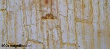
2 Compound Light Microscope Ocular lens (eyepiece) Objective lens Stage Glass slide Coverslip Diaphragm (regulates light) Base Fine adjustment knob Coarse adjustment knob Stage clips Arm Microscope Ocular lens: magnification 10x Objective lens: magnification up to 100x Gives up to 1000x total magnification 100 x 10 = 1000 Magnification 40 X Elodea 100 X 400 X Microscopes Magnify and Resolve two very different things Magnification \mag ne fe 'ka shen\ n Apparent enlargement of an object The ratio of image size to actual size Magnification of "100x" means the image is 100 times bigger than the actual object Resolution \rez e loo shen\ n Clarity, sharpness The ability of a microscope to show two very close points separately Resolution Examples Differential Interference Contrast 2


 Scanning electron")


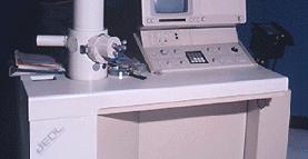
3 III. Electron Microscopes A. History 1. Electron microscopes were developed because of the limitations of compound microscopes. More on Microscopes Electron Microscopes overcomes resolution restrictions Transmission electron microscopes (TEM s) Scanning electron microscopes (SEM s) ESEM linkenvironmental SEM 2. First developed in the 1930 s. Photos: B. Transmission Electron Microscopes (TEM) 1. Operates on the same principle as a light microscope, but uses a stream of electrons instead of light. 2. Increases magnification to 200,000 x D images of 4. Can not observe living organisms. Electron Micrographs Electron Micrographs C. Scanning Electron Microscope(SEM) 1. Specimen coated with fine metal coating 2. Magnifies to 100,000x 3. 3 D images of surface/topography E.coli S. auerus 3


")


4 Image Comparison SEM micrographs Scanning Electron Microscopy Heart papillary muscle Heart papillary muscle (small muscles within the heart which anchor the heart valves) Heart collagen bundles website (Coppermine Photo Gallery, 2009) (Coppermine Photo Gallery, 2009) Compound vs. SEM Stigmatella Gram negative, rod shaped Saprophytic p bacteria of the phylum myxobacteria Typically found embedded in slime (Coppermine Photo Gallery, 2009) 4


5 Probe Microscopes Don t use lenses to produce images Ability to see atoms Moves a probe across the surface of specimen Records surface shape info on computer Centrifuge Spins down and separates culture Heavy parts settle on bottom Light parts rise to top Micromanipulation Technique used to dissect, remove, insert or manipulate parts of cells Invitro Types of Microscopes: ws/microscopes/types.html Cell Photos: Object Size and Magnifying Power of Microscopes (Leica Microsystems GmbH, 2009) Using the Microscope Place the Slide on the Microscope Use Stage Clips Click Nosepiece to the lowest (shortest) setting Look into the Eyepiece Use the Coarse Focus to find object High Power Focusing Find the specimen using low power, then Click the nosepiece to the longest objective DoNOT use the Coarse Focusing Knob Do NOT use the Coarse Focusing Knob Use the Fine Focus Knob to bring the slide into focus without breaking the slide or ruining the sample 5


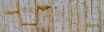

6 Ficus (dicot) leaf slide Prepared Slides Allium (onion) root tip slide Spirogrya Scalariform Conjugation 8/29/ Elodea Biological Drawings Refer to BioZone workbook for proper drawing and labeling techniques 6

. Optical microscopy primer.")
. Surgical microscopes.")
7 Drawing the Field of View link Let s Review Anatomy Virtual Sites Body Tube Revolving Nosepiece Objective Lens Stage Clips Diaphragm Light Eyepiece Arm Stage Coarse Focus Fine Focus Base Optical Microscopy Primer Histology: Human Anatomy Slides almicroscope/vmpage/very1st.htm Microscope Tutorial: Resources Coppermine Photo Gallery, (2009). Xtalent image gallery. Retrieved August 10, 2009, from Scanning Electron Microscopy Web site: Davidson, M. (2010). Optical microscopy primer. Retrieved August 23, 2010 from Molecular Expressions website Epel, D. (2010). Virtual urchin. Stanford University. Retrieved August 24, 2010 from website Leica Microsystems GmbH, (2009). Surgical microscopes. Retrieved August 11, 2009, from Leica Web site: microsystems.com/products/surgical microscopes/ Postlethwait, J. and Hopson, J.(2006). Modern biology. Austin, TX. Holt, Reinhart, Winston. 7
Microscope Notes. units of life.
 Microscope Notes Microscope an instrument that produces an enlarged image of an object. Biologists use microscopes to study cells, cell parts, and organisms that are too small to be seen with the naked
STRUCTURE OF THE MICROSCOPE
 STRUCTURE OF THE MICROSCOPE Use the word list to label the microscope below: Light Source Coarse adjustment knob Diaphragm Stage Clips Objectives Fine Adjustment Knob Base Stage Stage Clips Arm Revolving
History of microscopy
 History of microscopy Introduction Structure of microscope Care of microscope Use of microscope Magnification As we already know cells are microscopic. What does this mean? Scientists were able to see
The Microscope. Packet #2. 10/17/2016 9:12:02 PM Ryan Barrow 2012
 1 The Microscope Packet #2 10/17/2016 9:12:02 PM Ryan Barrow 2012 2 Historical Timeline 1609 Galileo Galilei develops a compound microscope with a convex and a concave les. 1665 Robert Hooke publishes
Microscope. Dr. Leena Barhate Department of Microbiology M.J.College, Jalgaon
 Microscope Dr. Leena Barhate Department of Microbiology M.J.College, Jalgaon Acknowledgement http://www.cerebromente.org.br/n17/histor y/neurons1_i.htm Google Images http://science.howstuffworks.com/lightmicroscope1.htm
Basic Microscopy for Plant Biology
 Page 1 of 8 Basic Microscopy for Plant Biology OBJECTIVES After completing this exercise, you should be able to do the following: a. Name the parts of the compound microscope and the functions of each.
Biology The Microscope. May 20 1:19 PM. Using a Microscope to Explore the Cell
 Biology 2201 1.2 The Microscope Using a Microscope to Explore the Cell Resolution or Resolving power The ability of the eye, or other instrument, to distinguish between two objects that are close together
The invention of the microscope made it possible for scientists to view and study cells. Cells the basic units of all living organisms.
 The Discovery of Cells The invention of the microscope made it possible for scientists to view and study cells. Cells the basic units of all living organisms. The Cell Theory All living things are made
Marine Invertebrate Zoology Microscope Introduction
 Marine Invertebrate Zoology Microscope Introduction Introduction A laboratory tool that has become almost synonymous with biology is the microscope. As an extension of your eyes, the microscope is one
Unit Two Part II MICROSCOPY
 Unit Two Part II MICROSCOPY AVERETT 1 0 /9/2013 1 MICROSCOPES Microscopes are devices that produce magnified images of structures that are too small to see with the unaided eye Humans cannot see objects
Microscopy Techniques that make it easy to see things this small.
 Microscopy Techniques that make it easy to see things this small. What is a Microscope? An instrument for viewing objects that are too small to be seen easily by the naked eye. Dutch spectacle-makers Hans
Scale. A Microscope s job in life. The Light Microscope. The Compound Microscope 9/24/12. Compound Microscope Anatomy
 The Study of Microbial Structure: Microscopy and Specimen Preparation Scale A Microscope s job in life 1.Magnify 2. Resolve ability to separate or distinguish between two points 3. Contrast How much or
Visual Anatomy ansd Physiology Lab Manual Pig Version 2nd Edition Sarikas TEST BANK
 Visual Anatomy ansd Physiology Lab Manual Pig Version 2nd Edition Sarikas TEST BANK https://testbankreal.com/download/visual-anatomy-ansd-physiology-labmanual-pig-version-2nd-edition-sarikas-test-bank/
Figure 3.4 Approximate size of various types of cells. ~10 um. Red Blood Cells = mm 1500 um. Width of penny Pearson Education, Inc.
 Figure 3.4 Approximate size of various types of cells. ~10 um Red Blood Cells 1.5mm 1500 um Width of penny = 1500 Figure 4.3 The limits of resolution (and some representative objects within those ranges)
Basic Microscopy. OBJECTIVES After completing this exercise, you should be able to do the following:
 Page 1 of 10 Basic Microscopy OBJECTIVES After completing this exercise, you should be able to do the following: a. Name the parts of the compound microscope and the functions of each. b. Describe how
Bio 252: Microscopy Study THE COMPOUND MICROSCOPE
 Name: Date: Block: Microscope Number: Bio 252: Microscopy Study THE COMPOUND MICROSCOPE I. Introduction The compound microscope is one of the most important instruments used by biologists today. Through
The microscope is useful in making observations and collecting data in scientific experiments. Microscopy involves three basic concepts:
 AP BIOLOGY Chapter 6 NAME DATE Block MICROSCOPE LAB PART I: COMPOUND MICROSCOPE OBJECTIVES: After completing this exercise you should be able to: Demonstrate proper care and use of a compound microscope.
CALIBRATION OF MICROSCOPE EYEPIECE GRATICULE
 CALIBRATION OF MICROSCOPE EYEPIECE GRATICULE A typical eyepiece graticule looks like this: It is 10mm in length and each mm is divided into 10 parts So each small division = 0.1mm = 100µm The eyepiece
Microscope (and The Cell) Lab Exercise #1
 Lab Exercise #1") Lab Safety-General No eating or drinking Only registered students allowed in the class Long hair must be tied back Familiarize yourself with the emergency stations Do not mark on the models Inform me of
Lab Safety-General No eating or drinking Only registered students allowed in the class Long hair must be tied back Familiarize yourself with the emergency stations Do not mark on the models Inform me of
Anatomy: Introduction to the Light Microscope
 Anatomy: Introduction to the Light Microscope Background: Microscopes are very important tools in biology. The term microscope can be translated as to view the tiny, because microscopes are used to study
Biology Lab #1: Using Microscopes to Observe and Measure Cells
 Biology Lab #1: Using Microscopes to Observe and Measure Cells Make sure you have signed and submitted the CDNIS Safety Contract before you start this experiment! PURPOSE: to review the use of the microscope
Microscope Tutorial. How to use a compound microscope
 Microscope Tutorial How to use a compound microscope Read this first Microscopes are extremely delicate and extremely expensive! You MUST be extremely careful when using the microscope. Always hold the
Observing Microorganisms through a Microscope
 2016/2/19 PowerPoint Lecture Presentations prepared by Bradley W. Christian, McLennan Community College CHAPTER 3 Observing Microorganisms through a Microscope 1 Figure 3.2 Microscopes and Magnification.
Introduction. Instructional Objectives. Materials. Procedure. I. Microscope Parts and Function. Honors Biology
 Honors Biology Introduction to the Microscope Lab Activity This lab was created by Mr. Buckley from Edward Knox High School. Credit is given for this original activity to Mr. Buckley. Introduction "Micro"
Laboratory Introduction
 Laboratory Introduction There are two basic categories of microscopes: light microscopes and electron microscopes. Light, or optical, microscopes require light waves to provide the illumination while electron
A BRIEF INTRODUCTION TO MICROSCOPY The two key properties of a microscope that allow you to see microbes are resolution and magnification.
 A BRIEF INTRODUCTION TO MICROSCOPY The two key properties of a microscope that allow you to see microbes are resolution and magnification. Magnification refers to the enlargement of the specimen when seen
The Care and Use of the Microscope. Lab Exercise #4
 Lab Safety No eating or drinking!!! Long hair must be tied back Clean up your workstation before you leave! Return all materials to the storage sites Clean glassware and wipe down countertops Follow directions
The microscope is useful in making observations and collecting data in scientific experiments. Microscopy involves three basic concepts:
 Lab #4 Biology 10 BCC Topic: MICROSCOPE LAB PART I: COMPOUND LIGHT MICROSCOPE OBJECTIVES: After completing this exercise you should be able to: Demonstrate proper care and use of a compound microscope.
The light microscope
 What is a microscope? The microscope is an essential tool in modern biology. It allows us to view structural details of organs, tissue, and cells not visible to the naked eye. The microscope should always
MICROSCOPE TERMS 7X 45X 112.5X 225X
 Microscopes MICROSCOPE TERMS Magnification- how much larger the image is Resolution- how clear the image is Field of View: Describes the visual picture seen when looking through the eyepiece of the microscope
The Compound Microscope. Brightfield: Köhler Illumination
 Outline History of Microscopy The Magnifying Glass The Compound Microscope Brightfield: Köhler Illumination Microscopy µικροσ (mikros): small σκοπειν (skopein): to observe History of Microscopy Well :
Microscopy. Danil Hammoudi.MD
 Microscopy Danil Hammoudi.MD Care and Handling of the Microscope: A microscope is a delicate piece of equipment and should be treated with care. Use two hands when carrying the microscope. Place one hand
LAB ACTIVITY: USING A MICROSCOPE
 Name: Date: Period: Lab Partner(s): LAB ACTIVITY: USING A MICROSCOPE Objectives: Demonstrate the proper use and care of a compound light microscope and stereomicroscope. Focus the compound light microscope
Ocular Lenses. Head. Arm. Objective Lenses. Slide Holder Stage. On / Off Switch. Condenser with Iris Diaphragm. Light Intensity Control
 BIOLOGY 211: HUMAN ANATOMY & PHYSIOLOGY ********************************************************************************************************* USE OF THE LIGHT MICROSCOPE **********************************************************************************************************
Microscope Review. 1. A compound light microscope is represented in the diagram below.
 Name Microscope Review Date 1. A compound light microscope is represented in the diagram below. 5. The diagram below represents a hydra as viewed with a compound light microscope. If the hydra moves toward
Care and Use of the Compound Light Microscope
 EXERCISE 2 Care and Use of the Compound Light Microscope Time Estimates for Completing This Lab The activities in this laboratory exercise can be completed in 2 to 2.5 hours. Extra time will be required
Name: Date Completed: Class: Lab Minutes: Teacher:
 Name: Date Completed: _ Class: Lab Minutes: _ Teacher: Introduction to the Microscope Lab Activity This lab was created by Mr. Buckley from Edward Knox High School. Credit is given for this original activity
Microscopes. A guide to use, general Maintenance, and repair tailored to the Olympus CX-21 microscope
 Microscopes A guide to use, general Maintenance, and repair tailored to the Olympus CX-21 microscope Topics Principles of Operation Diagrams Applications History Safety Operation Preventive Maintenance
used for low power magnification of a sample image is 3 dimensional
 MICROSCOPES One of the most important inventions in the advancement of Biology 1. Simple Microscopes ie. magnifying glass, stereoscope (dissecting scope) have a single lens or a pair of lenses combined
Name: Period: Week of: January 21st-25th Root Words In-Class Homework. Picture: -Microscope Notes -Lesson on Focusing the Microscope
 Day 1/21: Monday Biology Week #21 Week of: January 21st-25th Root Words In-Class Homework Word: Definition: As in: - Picture: NO SCHOOL: MLK Day 1/22: Tuesday Word: Definition: As in: - Picture: -Microscope
Seeing in Biology. Resolving Power of Optical Devices. Bio 101 Laboratory 2. Microscope Intro to Cell Cycle Mitosis
 Bio 101 Laboratory 2 Microscope Intro to Cell Cycle Mitosis 1 Seeing in Biology There are many different tools that biologists/anatomists can use to see biological samples at high resolution. Some include:
Using a Compound Light Microscope
 Name Class Date Laboratory Skills 5 Using a Compound Light Microscope Introduction Many objects are too small to be seen by the eye alone. They can be seen, however, with the use of an instrument that
MICROSCOPE LAB. Resolving Power How well specimen detail is preserved during the magnifying process.
 AP BIOLOGY Cells ACTIVITY #2 MICROSCOPE LAB OBJECTIVES 1. Demonstrate proper care and use of a compound microscope. 2. Identify the parts of the microscope and describe the function of each part. 3. Compare
THE COMPOUND BRIGHTFIELD MICROSCOPE
 THE COMPOUND BRIGHTFIELD MICROSCOPE Microbiology is the study of microscopic organisms that are so small that they are below the limit of vision of the human eye. Bacteria are the smallest of microorganisms
MICROSCOPES. Magnification: Resolution: Field of View: Describes the visual picture seen when looking through the eyepiece of the microscope
 Microscopes MICROSCOPES Magnification: Resolution: Field of View: Describes the visual picture seen when looking through the eyepiece of the microscope 7X 45X 112.5X 225X 1 st crude microscope made by
UNIT C: CYCLING OF MATTER IN LIVING SYSTEMS
 UNIT C: CYCLING OF MATTER IN LIVING SYSTEMS Aristotle is known as The Father of Biology. He was one of the first Greek philosophers who used the Scientific Method of observing, recording, reasoning, and
MICROSCOPY MICROSCOPE TERMINOLOGY
 1 MICROSCOPY Most of the microorganisms that we talk about in this class are too small to be seen with the naked eye. The instruments we will use to visualize these microbes are microscopes. The laboratory
Microbiology Laboratory 2
 Microbiology Laboratory 2 Microscopy Background Microorganisms are too small to be seen with the naked eye. Thus a microscope is used to magnify objects so they can be observed. A lens consists of one
AN INTRODUCTION TO THE MICROSCOPE
 AN INTRODUCTION TO THE MICROSCOPE INTRODUCTION In this exercise you will learn the components and operation of the compound microscope and the dissection microscope. This will be followed by a short exercise
 Microscopy http://www.microscopyu.com/articles/phasecontrast/phasemicroscopy.html http://micro.magnet.fsu.edu/primer/anatomy/anatomy.html 2005, Dr. Jack Ikeda & Dr. Gail Grabner 9 Nikon Labophot (Question
What is it? Study the mystery photos and try to identify each one! Have access to a computer?
 Station 1 Solve the Mystery What is it? Study the mystery photos and try to identify each one! They are all common objects that might be found in your home or a classroom. Write your guesses for the mystery
2018 MICROSCOPE REVIEW by Karen L. Lancour RELATIVE SIZE OF MICROBES
 2018 MICROSCOPE REVIEW by Karen L. Lancour RELATIVE SIZE OF MICROBES 1000 millimeters (mm) = 1 meter (m) 1000 micrometers (µm or mcm) = 1 millimeter (mm) 1000 nanometers (nm) = 1 micrometer (mcm) Size
Physiology Honors Interactive Notebook
 0 Foothill Technology High School Physiology Honors Interactive Notebook DEPARTMENT STATEMENT: Students will actively experience science both the concepts and practices of the disciplines. Science requires
USING THE MICROSCOPE TO OBSERVE CELLS
 USING THE MICROSCOPE TO OBSERVE CELLS *****IMPORTANT!!!!! BEFORE VISITING YOUR LEARNING CENTER TO CARRY OUT THIS LAB ACTIVITY PLEASE READ BELOW Before you visit your Learning Center to use the microscope,
REVIEW FOR TEST ON MONDAY
 1. The diagram below shows an ameba moving out of the high-power field of view of a compound microscope in the direction indicated by the arrow. 4. The diagram below represents two cells next to a metric
Match the microscope structures given in the left column with the statements in the right column that identify or describe them.
 49 Prelab for Name Match the microscope structures given in the left column with the statements in the right column that identify or describe them. Key: a. coarse adjustment knob f. turret or nosepiece
Burton's Microbiology for the Health Sciences
 Burton's Microbiology for the Health Sciences Chapter 2. Viewing the Microbial World Chapter 2 Outline Introduction Using the metric system to express the sizes of microbes Microscopes Simple microscopes
King Saud University Dept. of Bot. & Microbiology. General Microbiology 140 MIC
 King Saud University Dept. of Bot. & Microbiology General Microbiology 140 MIC Lab coat. Do not wearing the lab coat outside the lab. Gloves. Proper Clothing and closed shoes. Hair should be tied back.
If your worksheet is completed, get a sticker from a helper. You may check your answers and fix anything you missed.
 T. Tomm Updated 2015 http://sciencespot.net/ Images from http://www.tnmanning.com/id150.htm If your worksheet is completed, get a sticker from a helper. You may check your answers and fix anything you
Biology 29 Cell Structure and Function Spring, 2009 Springer LABORATORY 1: THE LIGHT MICROSCOPE
 Biology 29 Cell Structure and Function Spring, 2009 Springer LABORATORY 1: THE LIGHT MICROSCOPE Prior to lab: 1) Read these instructions (p 1-6) 2) Go through the online tutorial, the microscopy pre-lab
2017 MICROSCOPE REVIEW by Karen L. Lancour RELATIVE SIZE OF MICROBES
 2017 MICROSCOPE REVIEW by Karen L. Lancour RELATIVE SIZE OF MICROBES 1000 millimeters (mm) = 1 meter (m) 1000 micrometers (µm or mcm) = 1 millimeter (mm) 1000 nanometers (nm) = 1 micrometer (mcm) Size
MICROSCOPY and CELL STRUCTURE
 MICROSCOPY and CELL STRUCTURE Readings: Review pp. 69-71, and Fig. 4.1 on p. 65 in your text (POHS, 5 th ed.). Introduction: Biologists rely on many different types of microscopic techniques to find out
Lab: The Compound Microscope
 Lab: The Compound Microscope Purpose: To learn the parts of the compound microscope and to learn the basic skills needed to use the microscope properly. Materials: Microscope Colored paper Cover slips
Microbiology: Observing Bacteria Laboratory -1. Name Date
 Microbiology: Observing Bacteria Laboratory -1 Name Date Prelab: Part 1 Introduction to the microscope- please read through this handout and label the picture on the next page before starting the lab Care
Microscope Skills. Scientific Skills the Microscope!
 Microscope Skills Scientific Skills the Microscope! T. Trimpe 2005 http://sciencespot.net/ Body Tube Ocular lens (Eyepiece) Nosepiece Objectives Stage Clips Diaphragm Light Always carry a microscope with
Chapter 3. Observing Microorganisms Through a Microscope
 Chapter 3 Observing Microorganisms Through a Microscope Microbial Size Macroscopic organisms can be measured in the range from meters (m) to centimeters (cm) Microscopic organisms fall into the range
Observing Microorganisms through a Microscope LIGHT MICROSCOPY: This type of microscope uses visible light to observe specimens. Compound Light Micros
 PHARMACEUTICAL MICROBIOLOGY JIGAR SHAH INSTITUTE OF PHARMACY NIRMA UNIVERSITY Observing Microorganisms through a Microscope LIGHT MICROSCOPY: This type of microscope uses visible light to observe specimens.
Introduction to Microscopes
 INTRODUCTION TO THE MICROSCOPE Introduction to Microscopes The first microscopes worked by the same basic principle as the ones you will be using in lab. They are light microscopes. Visible light passes
Key Points Refer to How to Use the Compound Light Microscope :
 MODULE 1 Objective 1.2 Lesson B Introduction to the Microscope Using the Light Microscope and Slide Preparation Course Advanced Biotechnology Unit Biotech Basics Essential Question How do scientists view
Microscopy, Staining, and Classification
 PowerPoint Lecture Presentations prepared by Mindy Miller-Kittrell, North Carolina State University C H A P T E R 4 Microscopy, Staining, and Classification Figure 3.4 Approximate size of various types
Perfecting Microscope Skills
 I. Introduction to the Microscope Perfecting Microscope Skills There are different types of microscopes used by biologists depending on the job they wish to accomplish, including dissecting (or "stereoscopic")
How Microscopes Work By Cindy Grigg
 By Cindy Grigg 1 Inventions often lead scientists to make new discoveries. One of the most important discoveries in life science was the microscope. A microscope is used for looking at things too small
Exercise 2-A MICROSCOPIC TECHNIQUE & EXAMINATION OF MICROORGANISMS
 Exercise 2-A MICROSCOPIC TECHNIQUE & EXAMINATION OF MICROORGANISMS Introduction to Microscopic Technique Microbiology is the science or study of living organisms too small to be seen with the naked eye.
Microscopes & cells. 2. arm. 3. ocular lens. 4. objective lenses. 5. stage. 6. slide clamp. 7. stage controls
 Microscopes & cells Objectives: At the end of this lab you should be able to: o demonstrate the safe and proper handling of a microscope, including carrying a microscope, slide placement, and storage.
Exercise 2-A MICROSCOPIC TECHNIQUE & EXAMINATION OF MICROORGANISMS
 Exercise 2-A MICROSCOPIC TECHNIQUE & EXAMINATION OF MICROORGANISMS Introduction to Microscopic Technique Microbiology is the science or study of living organisms too small to be seen with the naked eye.
Microscopy Primer. Fig A compound light microscope with important parts labeled.
 BIOL 221 Concepts of Botany Fall 2010 Microscopy Primer A. Introduction: The microscope is a vital scientific tool that will be used often to study plants. We shall begin our studies of plants with a brief
Light Microscopy. Upon completion of this lecture, the student should be able to:
 Light Light microscopy is based on the interaction of light and tissue components and can be used to study tissue features. Upon completion of this lecture, the student should be able to: 1- Explain the
Name Date Block LAB: Exploring Plant & Animal Cells
 Name Date Block LAB: Exploring Plant & Animal Cells Background Information: One of the first scientists to look at cells under a microscope was an English scientist by the name of Robert Hooke. He viewed
DOWNLOAD OR READ : MICROSCOPE PDF EBOOK EPUB MOBI
 DOWNLOAD OR READ : MICROSCOPE PDF EBOOK EPUB MOBI Page 1 Page 2 microscope microscope pdf microscope We would like to show you a description here but the site wonâ t allow us. "Microscopy: Types of Microscopy"
VISUAL PHYSICS ONLINE DEPTH STUDY: ELECTRON MICROSCOPES
 VISUAL PHYSICS ONLINE DEPTH STUDY: ELECTRON MICROSCOPES Shortly after the experimental confirmation of the wave properties of the electron, it was suggested that the electron could be used to examine objects
Chapter 2 The Study of Microbial Structure: Microscopy and Specimen Preparation
 Chapter 2 The Study of Microbial Structure: Microscopy and Specimen Preparation 1 Lenses and the Bending of Light light is refracted (bent) when passing from one medium to another refractive index a measure
Observing Living Things
 Observing Living Things Textbook pages 8 21 Before You Read Section 1.1 Summary This section describes the signs that scientists look for to help them decide if something is living or non-living. On the
BIOLOGY 1101 LAB 2: MICROSCOPES AND CELLS
 BIOLOGY 1101 LAB 2: MICROSCOPES AND CELLS READING: Please read Chapter 4 in your text book to learn about the history of microscopy and basic cell structure. INTRODUCTION: The microscope is an important
Chapter 2 Alignment C. Robert Bagnell, Jr., Ph.D., 2012
 Chapter 2 Alignment C. Robert Bagnell, Jr., Ph.D., 2012 Figure 2.1 is an image of striated muscle taken with a misaligned microscope and figure 2.2 is with a properly aligned microscope. To the untrained
Manual for BMS E1 eplan series, compound microscope
 Manual for BMS E1 eplan series, compound microscope The compound microscope allows it to study, at cell level, structures of textures of botanical and zoological nature. (e.g. slides of roots, leaves and
EXERCISE 3 The Microscope
 Instant download and all chapters Solutions Manual Human Anatomy Laboratory Manual with Cat Dissections 7th Edition Marieb Smith https://testbankdata.com/download/solutions-manual-human-anatomy-laboratorymanual-cat-dissections-7th-edition-marieb-smith/
Using a Compound Light Microscope Lab Pre-Lab Assignment
 Name: Block: Due Date: Using a Compound Light Microscope Lab Pre-Lab Assignment Pre-Lab Assignment This assignment must be completed by the next class period in order to be allowed to participate in the
Protist Microscope Lab
 Name: Block: Due Date: Protist Microscope Lab Pre-Lab Assignment 1. Fill out the table for question #4 on the second page of your lab packet. (You may use the Biology textbook pages R8 and R9 in the back
tweezers Goggles Scalpel Tongs E G H K J F C L B D A I Aim #1 3 Safety, Instrumentation, Microscope Ruler Beaker Microscope Thermometer Graduated
 Ruler Beaker Microscope Thermometer Bunsen Burner (We use Hot plates) Eye Dropper/ Pipette Test tube Holder tweezers Goggles Scalpel Tongs Graduated cylinder C L B D A I E G H K J F Youtube: Powers of
LAB 1 Introduction to Microscopy
 I. Ubiquity of Microorganisms II. Microscopy LAB 1 Introduction to Microscopy I. UBIQUITY OF MICROORGANISMS Microorganisms are ubiquitous; that is, they are present nearly everywhere. In this lab you will
Title the next page in your notes: Cell Theory
 Title the next page in your notes: Cell Theory Write down AT LEAST the things in green Take Action! Look at the back of your hand What do you see? Describe your observation to a partner. Now look at the
Microscopy. Krishna Priya.K Lecturer Dept. of Microbiology
 Microscopy Krishna Priya.K Lecturer Dept. of Microbiology TERMS AND DEFINITIONS Principle Microscopy is to get a magnified image, in which structures may be resolved which could not be resolved with the
Today is Wednesday, October 7 th, 2015
 In This Lesson: Unit 2 Microscopes (Lesson 1 of 5) Today is Wednesday, October 7 th, 2015 Pre-Class: Write down three facts you know about microscopes. I will call on each of you for one of them. Please
Nikon Ti-E Microscope Manual. Rightmire Hall Ohio State University. Director: Tony Brown Rightmire
 Nikon Ti-E Microscope Manual Rightmire Hall Ohio State University Director: Tony Brown Rightmire 060 292-1205 brown.2302@osu.edu Facility Manager: Paula Monsma Rightmire 062 293-0939 292-1367 monsma.1@osu.edu
Lab: Using a Compound Light Microscope
 Name Date Period Lab: Using a Compound Light Microscope Background: Microscopes are very important tools in biology. The term microscope can be translated as to view the tiny, because microscopes are used
UNIT: THE MICROSCOPE AND CELLULAR DIVERSITY
 Course: Biology Agricultural Science & Technology UNIT: THE MICROSCOPE AND CELLULAR DIVERSITY State Standard: State Objectives: Unit Objectives: A. Learn how to use the compound microscope. B. Learn the
How to Use a Microscope
 How to Use a Microscope Overview Welcome to our unit on microscopes! We re going to learn how to use our microscope to make things appear larger so we can study them more easily. If you ve ever wondered
1. A laboratory technique is illustrated in the diagram below. Explain why the coverslip is lowered at an angle.
 1. A laboratory technique is illustrated in the diagram below. Explain why the coverslip is lowered at an angle. 2. Base your answer to the following question on Which laboratory procedure is represented
Systems Biology. Optical Train, Köhler Illumination
 McGill University Life Sciences Complex Imaging Facility Systems Biology Microscopy Workshop Tuesday December 7 th, 2010 Simple Lenses, Transmitted Light Optical Train, Köhler Illumination What Does a
DIC Imaging using Laser Scanning Microscopes (LSM) on Inverted Stands
 on Inverted Stands") DIC Imaging using Laser Scanning Microscopes (LSM) on Inverted Stands Differential Interference Contrast (DIC) imaging is a technique used to increase contrast in brightfield images. In confocal systems,
DIC Imaging using Laser Scanning Microscopes (LSM) on Inverted Stands Differential Interference Contrast (DIC) imaging is a technique used to increase contrast in brightfield images. In confocal systems,