History of microscopy
|
|
|
- Ronald Phillips
- 5 years ago
- Views:
Transcription


1 History of microscopy

2 Introduction Structure of microscope Care of microscope Use of microscope Magnification
3 As we already know cells are microscopic. What does this mean? Scientists were able to see and study the cells because of a discovery of microscopes. Initially botanist and zoologist were only able to study, describe, draw and label only the external structure of macroscopic organism. This is referred to as the macroscopic view. What are macroscopic organisms?
4 Microscopic: refers to organisms that are so small that they are not visible to the naked. Macroscopic: refers to organisms that are visible to the naked eye.
5 Robert Hooke, an English scientists looked at cork tissue under a very simple microscope as shown below. He observed these cells in As shown in the insert he saw many box like compartments. He called these compartments cells.
6 The cork tissue comes from the cork plant. However all he managed to see were the cell walls of the cells. The cork cells were dead, therefore there was nothing inside these cells.

7 He was a Dutch scientist. He observed the bacteria under a simple microscope in 1674 as shown below ( little animals in rain water).
8 Three scientist, Oken in 1805, Lamarck in 1809 and Dutrochet in 1824, independently stated that plants and animals were made up of cells. In 1838 Mathias Schleidan, a German scientist, was first to regard the cell as a structural unit of plants. Then in 1839, Theodor Schwann was the first to regard the cell as the structural unit of animals.
9 A German scientist, Purkinje, was the first to use the term protoplasm to describe the living contents of the cell. This was in Then 1859, another German scientist, Schultz, was the first to describe the protoplasm as the physical basis of life. Finally, Rudolf Virchow, put forward the idea that new cells formed when existing cells divided. These scientist were able to make these discoveries as a result of advancement in the development of the microscope.
10 These ideas mentioned in the previous two slides developed into what is known as the cell theory. According to this theory: 1. Every living organism is made up of cells (cells are the basic unit of life). 2. Every living cell comes from another. 3. All cells are basically similar. 4. Cells consist of a substance called protoplasm.
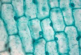
11 Compound light microscope (use 2 lenses) With your microscope at school you are able to see objects magnified 600 times, by using the 40X objective and 15X ocular lens. The nucleus, cell membrane, cytoplasm and chloroplast were observed and named using the light microscope. However the details of these structures were not visible because the microscope is not powerful enough.
12 However in the 1930s a more powerful microscope was invented by Zworykin. This was called the electron microscope There are 2 types of electron microscopes. They are the scanning electron microscope (SEM) and the transmission electron microscope (TEM) The SEM is used to scan and view the surface of objects. The TEM is used to see inside the objects by allowing light to pass through them.

13 With the electron microscope we are able to view objects to times. The image is clear and not blurred. They are able to provide such good images because they use electron beams instead of light
14
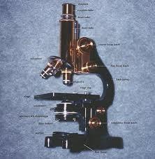
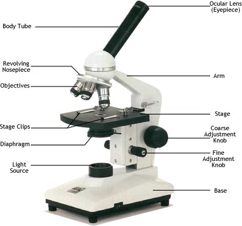
15 1. Base: supports the microscope. Always place your hand under the base when transporting the microscope. 2. Mirror: provides source of natural light. The mirror must be focused to reflect light. 3. Illuminator/lamp: it provides an electric source of light, it is much easier to use. 4. The condenser: it is found below the stage. Its function is to concentrate the light through the slide and specimen. 5. Iris diaphragm: this is an opening in the condenser, it controls the amount of light falling on the specimen.
16 6. Stage: this is the platform on which the slide is placed. 7. Stage/slide clips: these are metal clips that are used to hold the slide in position so that is does not move around when it is being focused. 8. Mechanical stage: this is found in only some microscopes, it allows easy movement of the slide.
17 9. Objectives: these are a combination of lenses used to magnify the specimen. There are 3 different types of objectives X objectives: this is the short objectives. It magnifies the objectives 4X X objectives: this is the medium objective. It magnifies the specimen 10X X objective: this is the long objective. It magnifies the specimen 40X 13. The revolving nose piece: the objectives are attached to this nose piece. Ensures the objective is in position when viewing the specimen.
18 14. Body tube: the eye piece and objectives are found on it. It also links the eye piece and objectives. In other words it links and supports the optical parts. 15. Coarse adjustment screw: used to make adjustments to focus the image. It moves the body tube up and down quickly, it provides quick focus. Used mainly at low magnification.
19 16. Fine adjustment screw: is used to make fine adjustments to focus the image. Used with higher magnification. Prevents damage to slide if any sudden movements are made during focusing. 17. Eye piece/ocular: combination of lenses that are used to magnify the specimen. There are 3 oculars 5X, 10X and 15X
20
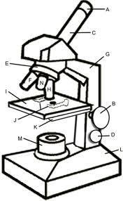
21 Use your notes and diagram of the microscope to label the diagram and provide the functions of the following parts: 1. A 2. C 3. E 4. I 5. M
22 A. Eyepiece B. Coarse adjustment screw C. Body tube D. Fine adjustment screw E. Nose piece F. Medium objective/10x G. Arm H. Long objective/40x I. Stage J. clip
23 K. Diaphragm L. Base M. Light source N. Short objective/4x 1. Eye piece-magnifies image 2. Body tube- supports and links optical parts 3. Nose piece- attachment of objectives 4. Stage- platform on which slide is place 5. Light source- provides electrical light
24 Follow these guidelines when handling a microscope: 1. Transport the microscope by placing one hand under the base and the other holding the arm, 2. Work one hand width away from the edge of the work bench. 3. Use only soft tissue to clean the lens. 4. Always ask for help if you are unsure of anything
25 Setting the light and condenser 1. Open the diaphragm fully. 2. Look at the mirror. Notice that it has two surfaces: a plane one and a concave one. If the microscope has a built in condenser then have the plane surface of the mirror facing up. If the there is no condenser then the concave surface must face up. 3. Adjust the mirror so that it faces a source of natural light. E.g. A window, open door.
26 Setting the light and condenser 4. Select the lowest power objective by turning the nose piece until you hear or feel the object click into position. 5. Place a sharp pencil on the mirror and focus until you obtain a sharp image of the pencil tip.
27 Focusing at low magnification 1. Place the slide on the stage. 2. Secure it using the clips. 3. Look through the eyepiece and slowly turn the coarse adjustment screw to focus the specimen. 4. Slowly turn the fine power objective to focus a clearer image of the specimen. 5. The slide may need to be moved to ensure the part of the specimen under examination is what you are seeing.
28 Focusing at higher magnification 1. Move the next objective into position by carefully moving the nose piece until the objective clicks into position. 2. If the specimen is not clearly visible then use the fine adjustment screw to obtain a clearer image. 3. You may repeat the process using the next high power object if more detail is required.
29 Changing the eyepiece 1. The magnification can be increased by using higher power eyepieces. 2. Simply remove the existing eyepiece and replace it with one that has higher magnification power.
30 Record what you see 1. Draw a diagram of what you see. 2. Draw and label exactly what you see, even if many aspects are missing. 3. In many cases you do not have to draw the entire image, only a portion of it. 4. Do not forget to indicate the scale of your drawing. For example if you used the medium power objective and the 5X eyepiece then your scale is 150 times(5x10) 5. Use a sharp pencil.
31 1. Remove the slide. 2. The 4x objective must be in position. 3. Replace the dust cover 4. Place the microscope in the correct box. 5. Store away.
32 Magnification of the microscope when viewing an objective: In order to determine how many times the specimen viewed is magnified by we need to calculate the magnification of the microscope. We can do this by using the following formula: Magnification power of = magnification of eyepiece X magnification of microscope lens
33 For example if you use the 10X eyepiece and the 40X objective then Magnification = 10 X 40 = 400X It is usually expressed as viewed under the microscope at 400X. To determine the actual size of an object viewed under the microscope using the field of view approach.
34 To determine the actual size of an object viewed under the microscope using the field of view approach. The field of view is the circle of light that you see when looking through the eyepiece. The diameter maybe measured by viewing a ruler under the microscope. The diameter for the field of view and the lens on your microscope is about 4.5 mm or 4500μm.
35 To calculate the length of the object you must determine the portion/fraction of the field it covers. This can only be done by determining the number of objects that can fit in the field of view.
36 For example look at the two field of views below. X X X X X In the one on the right 4 of the object can fit in the field of view. Therefore one object occupies ¼ of the field diameter.
37 Therefore the approximate length of the object can be calculated as follows: 4500 μm X ¼ = 1125 μm The approximate length maybe calculated using the formula: Approximate length of object = fraction X diameter of field
38 To determine the magnification of a drawing. 1. You need the actual size of the object drawn and the actual size of the drawing. 2. Magnification of the drawing can be calculated using the formula: Magnification of drawing = drawing size object size
39 3. For example if your object has an actual length of 1115 μm, and your drawing of that object has a length of 5 cm, then you,can calculate magnification of drawing as follows: First the 5cm must be converted into μm that is Then: 5 X = μm Magnification of drawing = drawing size object size = / 1115 = 45 μm
40 To determine the actual size of the object whose image or micrograph is viewed using a scale line. 1. Determine what the scale line measures. 2. Assume it represents 1 μm. 3. Measure the scale line given in the drawing or micrograph. ( lets say its 15mm) 4. Measure the length of the image in ;the drawing or micrograph. ( lets say its 50mm) 5. Now we can use the following formula
41 Actual size = measured length of object (mm) X length of scale line (μm) measured length of scale line (mm)) = 50mm X 1 μm 15mm = 3.3 μm
42 Macroscopic: refers to organisms that are visible to the naked eye. Microscopic: refers to organisms that are so small that they are not visible to the naked. Field of view is the circle of light that you see when looking through the eyepiece
43 1. The platform on which the slide sits is called the A. Base B. Stage C. Condenser D. diaphragm
44 2. Opening that controls the amount of light entering the microscope A. Base B. Stage C. Condenser D. diaphragm
45 3. It concentrates light through the slide and specimen A. Base B. Stage C. Condenser D. diaphragm
46 4. The combination of lens to magnify the image from objectives and specimen A. Eyepiece B. Body tube C. Illuminator D. Mirror
47 5. It supports and links the optical parts. A. Eyepiece B. Body tube C. Illuminator D. Mirror
48 6. Provides support for the microscope A. Base B. Stage C. Condenser D. diaphragm
49 7. Provides electrical light when switched on. A. Eyepiece B. Body tube C. Illuminator D. Mirror
50 8. Provides a source of natural light. A. Eyepiece B. Body tube C. Illuminator D. Mirror
51 9. Attachment of objectives. A. Nose piece B. Mechanical stage C. Stage clips D. Fine adjustment screw
52 10. Holds the slide in position on stage. A. Nose piece B. Mechanical stage C. Stage clips D. Fine adjustment screw
53 11. Allows for easy movement of slide A. Nose piece B. Mechanical stage C. Stage clips D. Fine adjustment screw
54 12. The picture below shows the microscope used by A. Robert Hooke B. Van Leeuwenhoek C. Oken D. Lamarck
55 13. The scientist who viewed cork cells under a simple microscope. A. Robert Hooke B. Van Leeuwenhoek C. Oken D. Lamarck
56 14. The scientist that observed and described single celled organisms. A. Robert Hooke B. Van Leeuwenhoek C. Oken D. Lamarck
57 15. The electron microscope was invented by A. Robert Hooke B. Van Leeuwenhoek C. Zworykin D. Lamarck
58 16. Calculate the magnification power of a microscope if you use the medium power objective and the 15X eyepiece. A. 150X B. 75X C. 25X D. 20X
59 17. Calculate the length of an object if it covers ½ of the field of view, assume that the diameter of the field of view is 4500μm A μm B μm C μm D. None of the above
60 18. Calculate the magnification of a drawing you have done if your drawing of the organism is 2500 μm and your drawing of it is 7 cm long. A μm B. 28X C. 0,0028 D. 28 μm
61 19. If you wanted to scan and view the surfaces of objects, you must use the A. Electron microscope B. SEM C. TEM D. Light microscope
62 20. If you wanted to see the inside of objects you would use the A. Electron microscope B. SEM C. TEM D. Light microscope
63 1. B 2. D 3. C 4. A 5. B 6. A 7. C 8. D 9. A 10. C
64 11. B 12. B 13. A 14. B 15. C 16. A 17. A 18. B 19. B 20. C
The invention of the microscope made it possible for scientists to view and study cells. Cells the basic units of all living organisms.
 The Discovery of Cells The invention of the microscope made it possible for scientists to view and study cells. Cells the basic units of all living organisms. The Cell Theory All living things are made
STRUCTURE OF THE MICROSCOPE
 STRUCTURE OF THE MICROSCOPE Use the word list to label the microscope below: Light Source Coarse adjustment knob Diaphragm Stage Clips Objectives Fine Adjustment Knob Base Stage Stage Clips Arm Revolving
Basic Microscopy. OBJECTIVES After completing this exercise, you should be able to do the following:
 Page 1 of 10 Basic Microscopy OBJECTIVES After completing this exercise, you should be able to do the following: a. Name the parts of the compound microscope and the functions of each. b. Describe how
Basic Microscopy for Plant Biology
 Page 1 of 8 Basic Microscopy for Plant Biology OBJECTIVES After completing this exercise, you should be able to do the following: a. Name the parts of the compound microscope and the functions of each.
Microscope Notes. units of life.
 Microscope Notes Microscope an instrument that produces an enlarged image of an object. Biologists use microscopes to study cells, cell parts, and organisms that are too small to be seen with the naked
Biology The Microscope. May 20 1:19 PM. Using a Microscope to Explore the Cell
 Biology 2201 1.2 The Microscope Using a Microscope to Explore the Cell Resolution or Resolving power The ability of the eye, or other instrument, to distinguish between two objects that are close together
used for low power magnification of a sample image is 3 dimensional
 MICROSCOPES One of the most important inventions in the advancement of Biology 1. Simple Microscopes ie. magnifying glass, stereoscope (dissecting scope) have a single lens or a pair of lenses combined
Microscope Tutorial. How to use a compound microscope
 Microscope Tutorial How to use a compound microscope Read this first Microscopes are extremely delicate and extremely expensive! You MUST be extremely careful when using the microscope. Always hold the
The Care and Use of the Microscope. Lab Exercise #4
 Lab Safety No eating or drinking!!! Long hair must be tied back Clean up your workstation before you leave! Return all materials to the storage sites Clean glassware and wipe down countertops Follow directions
MICROSCOPY MICROSCOPE TERMINOLOGY
 1 MICROSCOPY Most of the microorganisms that we talk about in this class are too small to be seen with the naked eye. The instruments we will use to visualize these microbes are microscopes. The laboratory
Biology Lab #1: Using Microscopes to Observe and Measure Cells
 Biology Lab #1: Using Microscopes to Observe and Measure Cells Make sure you have signed and submitted the CDNIS Safety Contract before you start this experiment! PURPOSE: to review the use of the microscope
The microscope is useful in making observations and collecting data in scientific experiments. Microscopy involves three basic concepts:
 AP BIOLOGY Chapter 6 NAME DATE Block MICROSCOPE LAB PART I: COMPOUND MICROSCOPE OBJECTIVES: After completing this exercise you should be able to: Demonstrate proper care and use of a compound microscope.
Microscope Review. 1. A compound light microscope is represented in the diagram below.
 Name Microscope Review Date 1. A compound light microscope is represented in the diagram below. 5. The diagram below represents a hydra as viewed with a compound light microscope. If the hydra moves toward
The microscope is useful in making observations and collecting data in scientific experiments. Microscopy involves three basic concepts:
 Lab #4 Biology 10 BCC Topic: MICROSCOPE LAB PART I: COMPOUND LIGHT MICROSCOPE OBJECTIVES: After completing this exercise you should be able to: Demonstrate proper care and use of a compound microscope.
CALIBRATION OF MICROSCOPE EYEPIECE GRATICULE
 CALIBRATION OF MICROSCOPE EYEPIECE GRATICULE A typical eyepiece graticule looks like this: It is 10mm in length and each mm is divided into 10 parts So each small division = 0.1mm = 100µm The eyepiece
Marine Invertebrate Zoology Microscope Introduction
 Marine Invertebrate Zoology Microscope Introduction Introduction A laboratory tool that has become almost synonymous with biology is the microscope. As an extension of your eyes, the microscope is one
MICROSCOPE LAB. Resolving Power How well specimen detail is preserved during the magnifying process.
 AP BIOLOGY Cells ACTIVITY #2 MICROSCOPE LAB OBJECTIVES 1. Demonstrate proper care and use of a compound microscope. 2. Identify the parts of the microscope and describe the function of each part. 3. Compare
The Microscope. Packet #2. 10/17/2016 9:12:02 PM Ryan Barrow 2012
 1 The Microscope Packet #2 10/17/2016 9:12:02 PM Ryan Barrow 2012 2 Historical Timeline 1609 Galileo Galilei develops a compound microscope with a convex and a concave les. 1665 Robert Hooke publishes
Microbiology Laboratory 2
 Microbiology Laboratory 2 Microscopy Background Microorganisms are too small to be seen with the naked eye. Thus a microscope is used to magnify objects so they can be observed. A lens consists of one
I. The First Microscopes. Microscope Basics. II. The Bright Field Microscope. Confocal Laser Scanning Microscopy. A. The Compound Microscope
 Microscope Basics I. The First Microscopes NGSSS: SC.912.N.2.1 through N.4.2 A. About 1590, two Dutch spectacle makers, Zaccharias Janssen and his son Hans, while experimenting with several lenses in a
Ocular Lenses. Head. Arm. Objective Lenses. Slide Holder Stage. On / Off Switch. Condenser with Iris Diaphragm. Light Intensity Control
 BIOLOGY 211: HUMAN ANATOMY & PHYSIOLOGY ********************************************************************************************************* USE OF THE LIGHT MICROSCOPE **********************************************************************************************************
Microscope & Measuring
 Name: ate: 1. microscope is supplied with 10 and 15 eyepieces, and with 10 and 44 objectives. What is the maximum magnification that can be obtained from this microscope?. 59. 150. 440. 660 3. student
Scale. A Microscope s job in life. The Light Microscope. The Compound Microscope 9/24/12. Compound Microscope Anatomy
 The Study of Microbial Structure: Microscopy and Specimen Preparation Scale A Microscope s job in life 1.Magnify 2. Resolve ability to separate or distinguish between two points 3. Contrast How much or
Introduction to Microscopes
 INTRODUCTION TO THE MICROSCOPE Introduction to Microscopes The first microscopes worked by the same basic principle as the ones you will be using in lab. They are light microscopes. Visible light passes
Using a Compound Light Microscope
 Name Class Date Laboratory Skills 5 Using a Compound Light Microscope Introduction Many objects are too small to be seen by the eye alone. They can be seen, however, with the use of an instrument that
Anatomy: Introduction to the Light Microscope
 Anatomy: Introduction to the Light Microscope Background: Microscopes are very important tools in biology. The term microscope can be translated as to view the tiny, because microscopes are used to study
Bio 252: Microscopy Study THE COMPOUND MICROSCOPE
 Name: Date: Block: Microscope Number: Bio 252: Microscopy Study THE COMPOUND MICROSCOPE I. Introduction The compound microscope is one of the most important instruments used by biologists today. Through
Name Date Block LAB: Exploring Plant & Animal Cells
 Name Date Block LAB: Exploring Plant & Animal Cells Background Information: One of the first scientists to look at cells under a microscope was an English scientist by the name of Robert Hooke. He viewed
Microscope (and The Cell) Lab Exercise #1
 Lab Exercise #1") Lab Safety-General No eating or drinking Only registered students allowed in the class Long hair must be tied back Familiarize yourself with the emergency stations Do not mark on the models Inform me of
Lab Safety-General No eating or drinking Only registered students allowed in the class Long hair must be tied back Familiarize yourself with the emergency stations Do not mark on the models Inform me of
Unit Two Part II MICROSCOPY
 Unit Two Part II MICROSCOPY AVERETT 1 0 /9/2013 1 MICROSCOPES Microscopes are devices that produce magnified images of structures that are too small to see with the unaided eye Humans cannot see objects
USING THE MICROSCOPE TO OBSERVE CELLS
 USING THE MICROSCOPE TO OBSERVE CELLS *****IMPORTANT!!!!! BEFORE VISITING YOUR LEARNING CENTER TO CARRY OUT THIS LAB ACTIVITY PLEASE READ BELOW Before you visit your Learning Center to use the microscope,
How Microscopes Work By Cindy Grigg
 By Cindy Grigg 1 Inventions often lead scientists to make new discoveries. One of the most important discoveries in life science was the microscope. A microscope is used for looking at things too small
Lab: The Compound Microscope
 Lab: The Compound Microscope Purpose: To learn the parts of the compound microscope and to learn the basic skills needed to use the microscope properly. Materials: Microscope Colored paper Cover slips
The light microscope
 What is a microscope? The microscope is an essential tool in modern biology. It allows us to view structural details of organs, tissue, and cells not visible to the naked eye. The microscope should always
Care and Use of the Compound Light Microscope
 EXERCISE 2 Care and Use of the Compound Light Microscope Time Estimates for Completing This Lab The activities in this laboratory exercise can be completed in 2 to 2.5 hours. Extra time will be required
A BRIEF INTRODUCTION TO MICROSCOPY The two key properties of a microscope that allow you to see microbes are resolution and magnification.
 A BRIEF INTRODUCTION TO MICROSCOPY The two key properties of a microscope that allow you to see microbes are resolution and magnification. Magnification refers to the enlargement of the specimen when seen
King Saud University Dept. of Bot. & Microbiology. General Microbiology 140 MIC
 King Saud University Dept. of Bot. & Microbiology General Microbiology 140 MIC Lab coat. Do not wearing the lab coat outside the lab. Gloves. Proper Clothing and closed shoes. Hair should be tied back.
LAB ACTIVITY: USING A MICROSCOPE
 Name: Date: Period: Lab Partner(s): LAB ACTIVITY: USING A MICROSCOPE Objectives: Demonstrate the proper use and care of a compound light microscope and stereomicroscope. Focus the compound light microscope
Perfecting Microscope Skills
 I. Introduction to the Microscope Perfecting Microscope Skills There are different types of microscopes used by biologists depending on the job they wish to accomplish, including dissecting (or "stereoscopic")
1. A laboratory technique is illustrated in the diagram below. Explain why the coverslip is lowered at an angle.
 1. A laboratory technique is illustrated in the diagram below. Explain why the coverslip is lowered at an angle. 2. Base your answer to the following question on Which laboratory procedure is represented
Microscope Skills. Scientific Skills the Microscope!
 Microscope Skills Scientific Skills the Microscope! T. Trimpe 2005 http://sciencespot.net/ Body Tube Ocular lens (Eyepiece) Nosepiece Objectives Stage Clips Diaphragm Light Always carry a microscope with
Microscope - Exercise 1
 Microscope - Exercise 1 Objectives -Familiarize parts and functions of the microscope. -Calculate total magnifications. -Determining the Diameter of the field of view for different magnifications. -Estimate
Name: Period: Week of: January 21st-25th Root Words In-Class Homework. Picture: -Microscope Notes -Lesson on Focusing the Microscope
 Day 1/21: Monday Biology Week #21 Week of: January 21st-25th Root Words In-Class Homework Word: Definition: As in: - Picture: NO SCHOOL: MLK Day 1/22: Tuesday Word: Definition: As in: - Picture: -Microscope
Key Points Refer to How to Use the Compound Light Microscope :
 MODULE 1 Objective 1.2 Lesson B Introduction to the Microscope Using the Light Microscope and Slide Preparation Course Advanced Biotechnology Unit Biotech Basics Essential Question How do scientists view
Visual Anatomy ansd Physiology Lab Manual Pig Version 2nd Edition Sarikas TEST BANK
 Visual Anatomy ansd Physiology Lab Manual Pig Version 2nd Edition Sarikas TEST BANK https://testbankreal.com/download/visual-anatomy-ansd-physiology-labmanual-pig-version-2nd-edition-sarikas-test-bank/
Microbiology: Observing Bacteria Laboratory -1. Name Date
 Microbiology: Observing Bacteria Laboratory -1 Name Date Prelab: Part 1 Introduction to the microscope- please read through this handout and label the picture on the next page before starting the lab Care
REVIEW FOR TEST ON MONDAY
 1. The diagram below shows an ameba moving out of the high-power field of view of a compound microscope in the direction indicated by the arrow. 4. The diagram below represents two cells next to a metric
UNIT C: CYCLING OF MATTER IN LIVING SYSTEMS
 UNIT C: CYCLING OF MATTER IN LIVING SYSTEMS Aristotle is known as The Father of Biology. He was one of the first Greek philosophers who used the Scientific Method of observing, recording, reasoning, and
Biology 29 Cell Structure and Function Spring, 2009 Springer LABORATORY 1: THE LIGHT MICROSCOPE
 Biology 29 Cell Structure and Function Spring, 2009 Springer LABORATORY 1: THE LIGHT MICROSCOPE Prior to lab: 1) Read these instructions (p 1-6) 2) Go through the online tutorial, the microscopy pre-lab
EXERCISE 3 The Microscope
 Instant download and all chapters Solutions Manual Human Anatomy Laboratory Manual with Cat Dissections 7th Edition Marieb Smith https://testbankdata.com/download/solutions-manual-human-anatomy-laboratorymanual-cat-dissections-7th-edition-marieb-smith/
What you should have learned from the microscope labs.
 What you should have learned from the microscope labs. Microscope Lab 1 Directionality Items appear backwards and inverted On Stage In Microscope NOT!!!! Microscope Lab 1 More Directionality Items move
MICROSCOPE TERMS 7X 45X 112.5X 225X
 Microscopes MICROSCOPE TERMS Magnification- how much larger the image is Resolution- how clear the image is Field of View: Describes the visual picture seen when looking through the eyepiece of the microscope
AN INTRODUCTION TO THE MICROSCOPE
 AN INTRODUCTION TO THE MICROSCOPE INTRODUCTION In this exercise you will learn the components and operation of the compound microscope and the dissection microscope. This will be followed by a short exercise
Introduction to the Compound Microscope Cell Structure & Function
 Introduction to the Compound Microscope Cell Structure & Function Revised Fall 2018 Laboratory Safety Lab coat, long pants, closed-toe shoes, safety goggles, and nitrile or latex gloves are required. **You
MICROSCOPES. Magnification: Resolution: Field of View: Describes the visual picture seen when looking through the eyepiece of the microscope
 Microscopes MICROSCOPES Magnification: Resolution: Field of View: Describes the visual picture seen when looking through the eyepiece of the microscope 7X 45X 112.5X 225X 1 st crude microscope made by
What is it? Study the mystery photos and try to identify each one! Have access to a computer?
 Station 1 Solve the Mystery What is it? Study the mystery photos and try to identify each one! They are all common objects that might be found in your home or a classroom. Write your guesses for the mystery
Match the microscope structures given in the left column with the statements in the right column that identify or describe them.
 49 Prelab for Name Match the microscope structures given in the left column with the statements in the right column that identify or describe them. Key: a. coarse adjustment knob f. turret or nosepiece
Burton's Microbiology for the Health Sciences
 Burton's Microbiology for the Health Sciences Chapter 2. Viewing the Microbial World Chapter 2 Outline Introduction Using the metric system to express the sizes of microbes Microscopes Simple microscopes
Laboratory Introduction
 Laboratory Introduction There are two basic categories of microscopes: light microscopes and electron microscopes. Light, or optical, microscopes require light waves to provide the illumination while electron
Figure 3.4 Approximate size of various types of cells. ~10 um. Red Blood Cells = mm 1500 um. Width of penny Pearson Education, Inc.
 Figure 3.4 Approximate size of various types of cells. ~10 um Red Blood Cells 1.5mm 1500 um Width of penny = 1500 Figure 4.3 The limits of resolution (and some representative objects within those ranges)
Exercise 2-A MICROSCOPIC TECHNIQUE & EXAMINATION OF MICROORGANISMS
 Exercise 2-A MICROSCOPIC TECHNIQUE & EXAMINATION OF MICROORGANISMS Introduction to Microscopic Technique Microbiology is the science or study of living organisms too small to be seen with the naked eye.
Title the next page in your notes: Cell Theory
 Title the next page in your notes: Cell Theory Write down AT LEAST the things in green Take Action! Look at the back of your hand What do you see? Describe your observation to a partner. Now look at the
Observing Living Things
 Observing Living Things Textbook pages 8 21 Before You Read Section 1.1 Summary This section describes the signs that scientists look for to help them decide if something is living or non-living. On the
Exercise 2-A MICROSCOPIC TECHNIQUE & EXAMINATION OF MICROORGANISMS
 Exercise 2-A MICROSCOPIC TECHNIQUE & EXAMINATION OF MICROORGANISMS Introduction to Microscopic Technique Microbiology is the science or study of living organisms too small to be seen with the naked eye.
Microscopy. Danil Hammoudi.MD
 Microscopy Danil Hammoudi.MD Care and Handling of the Microscope: A microscope is a delicate piece of equipment and should be treated with care. Use two hands when carrying the microscope. Place one hand
MICROSCOPY and CELL STRUCTURE
 MICROSCOPY and CELL STRUCTURE Readings: Review pp. 69-71, and Fig. 4.1 on p. 65 in your text (POHS, 5 th ed.). Introduction: Biologists rely on many different types of microscopic techniques to find out
Name: Date Completed: Class: Lab Minutes: Teacher:
 Name: Date Completed: _ Class: Lab Minutes: _ Teacher: Introduction to the Microscope Lab Activity This lab was created by Mr. Buckley from Edward Knox High School. Credit is given for this original activity
Using a Compound Light Microscope Lab Pre-Lab Assignment
 Name: Block: Due Date: Using a Compound Light Microscope Lab Pre-Lab Assignment Pre-Lab Assignment This assignment must be completed by the next class period in order to be allowed to participate in the
Lab: Using a Compound Light Microscope
 Name Date Period Lab: Using a Compound Light Microscope Background: Microscopes are very important tools in biology. The term microscope can be translated as to view the tiny, because microscopes are used
Manual for BMS E1 eplan series, compound microscope
 Manual for BMS E1 eplan series, compound microscope The compound microscope allows it to study, at cell level, structures of textures of botanical and zoological nature. (e.g. slides of roots, leaves and
Protist Microscope Lab
 Name: Block: Due Date: Protist Microscope Lab Pre-Lab Assignment 1. Fill out the table for question #4 on the second page of your lab packet. (You may use the Biology textbook pages R8 and R9 in the back
Microscopes. A guide to use, general Maintenance, and repair tailored to the Olympus CX-21 microscope
 Microscopes A guide to use, general Maintenance, and repair tailored to the Olympus CX-21 microscope Topics Principles of Operation Diagrams Applications History Safety Operation Preventive Maintenance
tweezers Goggles Scalpel Tongs E G H K J F C L B D A I Aim #1 3 Safety, Instrumentation, Microscope Ruler Beaker Microscope Thermometer Graduated
 Ruler Beaker Microscope Thermometer Bunsen Burner (We use Hot plates) Eye Dropper/ Pipette Test tube Holder tweezers Goggles Scalpel Tongs Graduated cylinder C L B D A I E G H K J F Youtube: Powers of
2018 MICROSCOPE REVIEW by Karen L. Lancour RELATIVE SIZE OF MICROBES
 2018 MICROSCOPE REVIEW by Karen L. Lancour RELATIVE SIZE OF MICROBES 1000 millimeters (mm) = 1 meter (m) 1000 micrometers (µm or mcm) = 1 millimeter (mm) 1000 nanometers (nm) = 1 micrometer (mcm) Size
Observing Microorganisms through a Microscope
 2016/2/19 PowerPoint Lecture Presentations prepared by Bradley W. Christian, McLennan Community College CHAPTER 3 Observing Microorganisms through a Microscope 1 Figure 3.2 Microscopes and Magnification.
Using a Microscope. Year Group: BVSc1 + Document number: CSL_L07
 Year Group: BVSc1 + Document number: CSL_L07 Equipment list: Equipment for this station: Microscope Power supply and a level surface to work on Gloves The sample to examine Marker or pencil for labelling
Microscope. Dr. Leena Barhate Department of Microbiology M.J.College, Jalgaon
 Microscope Dr. Leena Barhate Department of Microbiology M.J.College, Jalgaon Acknowledgement http://www.cerebromente.org.br/n17/histor y/neurons1_i.htm Google Images http://science.howstuffworks.com/lightmicroscope1.htm
Easy Kohler Illumination Method
 Easy Kohler Illumination Method ACADEMIC SKILLS CENTRE (ASC) A. Silverberg Completion of a Kohler illumination method is required before a microscope can be used efficiently. The Kohler method is designed
Components of the Microscope
 Swift M3 Microscope The Swift M3 is a versatile microscope designed for both microscopic (high magnification, small field of view) and macroscopic (low magnification, large field of view) applications.
Microscopy Techniques that make it easy to see things this small.
 Microscopy Techniques that make it easy to see things this small. What is a Microscope? An instrument for viewing objects that are too small to be seen easily by the naked eye. Dutch spectacle-makers Hans
Title: Thinking with the Eyes Author(s): Elizabeth Haggerty Hutton Date Created: 8/5/2011 Subject: Biology Grade Level: 9 th Grade Honors Standards:
: Elizabeth Haggerty Hutton Date Created: 8/5/2011 Subject: Biology Grade Level: 9 th Grade Honors Standards:") Title: Thinking with the Eyes Author(s): Elizabeth Haggerty Hutton Date Created: 8/5/2011 Subject: Biology Grade Level: 9 th Grade Honors Standards: SC.912.N.1.1: The practice of science SC.912.L.14.4:
Title: Thinking with the Eyes Author(s): Elizabeth Haggerty Hutton Date Created: 8/5/2011 Subject: Biology Grade Level: 9 th Grade Honors Standards: SC.912.N.1.1: The practice of science SC.912.L.14.4:
1.When an object is sharply focused and the slide is moved towards you, in which direction does the
 image upright or inverted? Name: Date: _ BIOLOGY EXPERIMENT:Class: Using a Compound Light Microscope II: Depth Perception, resolution, field of view MATERIALS: Compound light microscopecolor magazine clipping
The Compound Microscope and Calculations
 The Compound Microscope and Calculations The magnifying power of the eyepiece,(a.k.a.: ocular) is (10 x) The magnifying power of the low-power objective is: (40 x) The magnifying power of the medium-power
Microscopy Primer. Fig A compound light microscope with important parts labeled.
 BIOL 221 Concepts of Botany Fall 2010 Microscopy Primer A. Introduction: The microscope is a vital scientific tool that will be used often to study plants. We shall begin our studies of plants with a brief
Microscope Labs #1 and #2 e Lab and Hair Lab. Day 1 - e Lab
 Name: Date: Microscope Labs #1 and #2 e Lab and Hair Lab Day 1 - e Lab Purpose: To study the image formed by the microscope Procedure: You may begin once you go to the lab area! A. Preparation of the slide
Microscopes & cells. 2. arm. 3. ocular lens. 4. objective lenses. 5. stage. 6. slide clamp. 7. stage controls
 Microscopes & cells Objectives: At the end of this lab you should be able to: o demonstrate the safe and proper handling of a microscope, including carrying a microscope, slide placement, and storage.
Chapter 2 Alignment C. Robert Bagnell, Jr., Ph.D., 2012
 Chapter 2 Alignment C. Robert Bagnell, Jr., Ph.D., 2012 Figure 2.1 is an image of striated muscle taken with a misaligned microscope and figure 2.2 is with a properly aligned microscope. To the untrained
BIOLOGY 1101 LAB 2: MICROSCOPES AND CELLS
 BIOLOGY 1101 LAB 2: MICROSCOPES AND CELLS READING: Please read Chapter 4 in your text book to learn about the history of microscopy and basic cell structure. INTRODUCTION: The microscope is an important
Introduction. Instructional Objectives. Materials. Procedure. I. Microscope Parts and Function. Honors Biology
 Honors Biology Introduction to the Microscope Lab Activity This lab was created by Mr. Buckley from Edward Knox High School. Credit is given for this original activity to Mr. Buckley. Introduction "Micro"
The Compound Microscope. Brightfield: Köhler Illumination
 Outline History of Microscopy The Magnifying Glass The Compound Microscope Brightfield: Köhler Illumination Microscopy µικροσ (mikros): small σκοπειν (skopein): to observe History of Microscopy Well :
Laboratory 2: Microscopy and Observation of Cells authors: Dr. Ruth Dahlquist-Willard & Michael Kunz
 Laboratory 2: Microscopy and Observation of Cells authors: Dr. Ruth Dahlquist-Willard & Michael Kunz Corresponding Readings: Campbell Ch. 4 BIOL-100L Safety Information: We will be using laboratory glassware
2017 MICROSCOPE REVIEW by Karen L. Lancour RELATIVE SIZE OF MICROBES
 2017 MICROSCOPE REVIEW by Karen L. Lancour RELATIVE SIZE OF MICROBES 1000 millimeters (mm) = 1 meter (m) 1000 micrometers (µm or mcm) = 1 millimeter (mm) 1000 nanometers (nm) = 1 micrometer (mcm) Size
If your worksheet is completed, get a sticker from a helper. You may check your answers and fix anything you missed.
 T. Tomm Updated 2015 http://sciencespot.net/ Images from http://www.tnmanning.com/id150.htm If your worksheet is completed, get a sticker from a helper. You may check your answers and fix anything you
The Microscopic Image
 The Microscopic Image Name: # Pretend you could travel back in time to the late 1500s. How would your life be different? Think of where you would live, how you would dress, and what you might do for fun.
Cells Unit GOALS AND OBJECTIVES. The student will become familiar with the use of the compound microscope.
 Cells Unit GOALS AND OBJECTIVES GOALS The student will become familiar with the use of the compound microscope. The student will become familiar with the basic parts and functions of the cell. OBJECTIVES
Microscope. & Measurements. Do Now
 Do Now Microscope & Measurements How many: 1. Centimeters (cm) in 4 meters (m)? m 2. Decimeters (dm) in 5 meters (m)? dm 3. Centimeters (cm) in 4,000 millimeters (mm) cm 4. Millimeters (mm) in 40 centimeters
How to Use a Microscope
 How to Use a Microscope Overview Welcome to our unit on microscopes! We re going to learn how to use our microscope to make things appear larger so we can study them more easily. If you ve ever wondered
General Physics Experiment 5 Optical Instruments: Simple Magnifier, Microscope, and Newtonian Telescope
 General Physics Experiment 5 Optical Instruments: Simple Magnifier, Microscope, and Newtonian Telescope Objective: < To observe the magnifying properties of the simple magnifier, the microscope and the
MICROSCOPE (3 x 2 hour lesson)
") MICROSCOPE (3 x 2 hour lesson) 1ST WEEK (2 HOUR): PRINCIPLE OF MICROSCOPE AND BASIC QUIZ Principle of microscope Make a simple microscope using two convex lenses to learn the principle of microscope. Identification
MICROSCOPE (3 x 2 hour lesson) 1ST WEEK (2 HOUR): PRINCIPLE OF MICROSCOPE AND BASIC QUIZ Principle of microscope Make a simple microscope using two convex lenses to learn the principle of microscope. Identification
What Type Of Light Microscope Is Used To View Large Objects Such As Flowers
 What Type Of Light Microscope Is Used To View Large Objects Such As Flowers However, preparing objects for SEM and other common imaging methods can explains researcher Jacob Landis, "there are alternatives