Instructions for Making On-Line Reservations for Microscopes in NB11-204
|
|
|
- Jacob Wood
- 5 years ago
- Views:
Transcription
1 Instructions for Making On-Line Reservations for Microscopes in NB Log into Mail using Mail.swmed.edu 2. Log in using your university id and password. 3. Click the Calendar Tab at the top right of the screen. 4. Click MolePathScope Sign Up at the left of the screen. 5. Click New Event at the top left-hand corner of the screen. 6. In the Event line on the new screen enter the scope you wish to use (Compound, Inverted or Dissection). Also indicate if you will be using bright-field illumination or fluorescence. 7. On the Attendees Line write MolePathScopeSignUp. 8. Enter date and start time. 9. The maximum time allowed is 2 hours. When finished click Send at the upper left-hand corner of the screen.
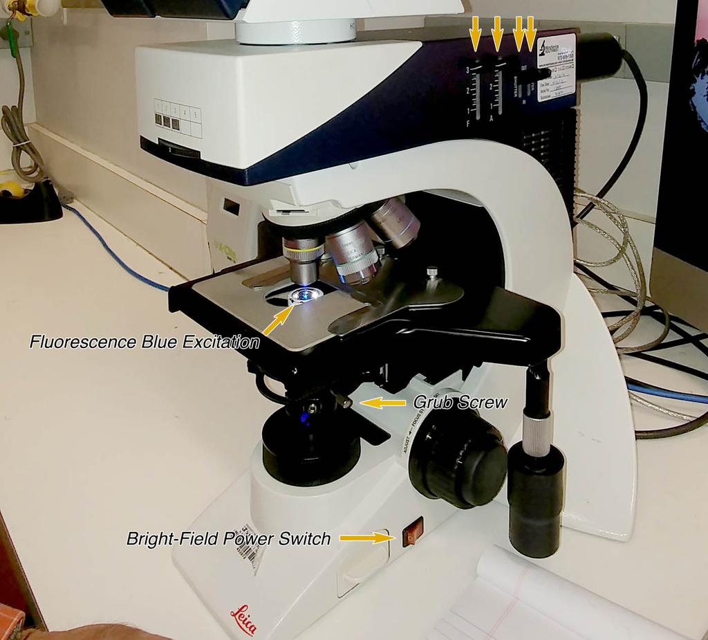
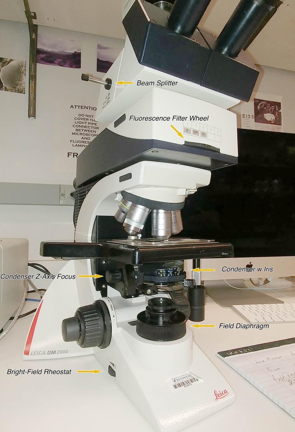
2 Operating Instructions Leica Microscope Room NB Bright field 1. Turn the power on using orange switch on the right-hand side of the base. 2. Pull the Beam Splitter on the upper left-hand side of the scope to 50/50 or Vis. 3. Adjust the light level to 8 by adjusting the Bright-Field Rheostat on the lower left-hand side of the base. 4. Use the correct condenser. One condenser will be mounted on the scope, the other is stored in the top drawer immediately to your left. a. For low magnification objectives (1.25X, 2.5X, 4X or 10X) use the Nikon Achro 0.13 condenser b. For high magnification (20X, 40X, 63X or 100X) use the Leica 0.9 PSI. The condenser is stored in the top drawer to your left. c. To replace the condenser, lower the condenser to the lowest Z positon using the Condenser Z-Axis Focus knob behind the condenser and loosen the silver grub screw on the right side of the condenser and remove the condenser towards you. 5. Replace the chosen condenser by sliding it into the support bracket. When using the high-magnification condenser, be careful to insert the pin on the back of the condenser into the notch at the back of the support. 6. Tighten the grub screw to secure the condenser in place. 7. Establish Kohler Illumination for the high-magnification condenser. a. Raise the condenser to near the top limit without hitting the slide. b. Place a slide on the stage to establish plane of focus using the 20X or 40X objective.
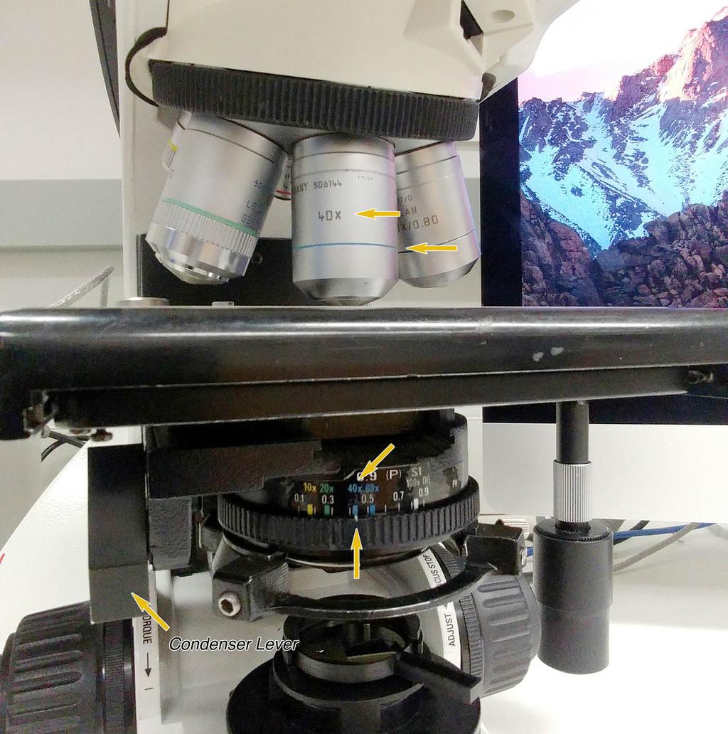
3 c. Close the Field Diaphragm so that you can see it through the objective. d. Adjust the Z axis of the condenser to bring the edge of the leaves of the field diaphragm into focus. e. Open the field diaphragm just beyond the limits of the field of view and then turn the Condenser Iris to match the color of the ring on objective you are using. 8. When using the low-magnification condenser, position it at the lowest Z position and open the Condenser Iris Diaphragm completely. To Acquire Data Log into computer by clicking on Camera User. Use Password: Password123. To save you files, open Documents, then Work, then open your lab folder.
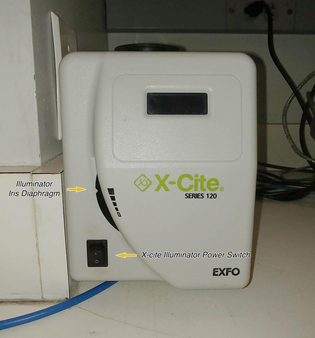
4 Operating Instructions Leica Microscope Room NB Fluorescence 1. Turn the bright-field light source off using orange Power Switch situated at the base on the right side. 2. Feel the X-Cite lamp housing. If it is hot wait ½ hour before using the lamp. 3. Turn the power on using the Illuminator Power Switch. 4. Check the following: a. Illuminator Iris Diaphragm on the lamp housing is open. b. All four light path levers on the right side of the scope housing are adjusted to the top. c. Fluorescence Filter Wheel at the front of the scope is set to position If all is well you now will see blue light coming through the lens. 6. Use 20X or higher objectives. 7. If the emission is dim then pull or push the Beam Splitter (upper left side of the scope housing) to Vis, or if acquiring an image set to Photo. 8. Adjust the Fluorescence Filter Wheel to excite the specific fluorophore you are using. a. #1 UV for Dapi, Hoechst, AMCA b. #2 GFP, FITZ, Alexa 488, Cy2 c. #3 Cy3, Texas Red, Rhodamine, Alexa To improve emission, keep the condenser lowered or flipped forward using the lever on the left of the condenser housing. 10. Leave the X-Cite illuminator on for a minimum of 30 minutes. To Acquire Data Log into computer by clicking on Camera User. Use Password: Password123. To save you files, open Documents, then Work, then open your lab folder.
5
6
7
8
BX-61: Brightfield Instruction /Continue to scroll for Fluorescent Instuctions
 BX-61: Brightfield Instruction /Continue to scroll for Fluorescent Instuctions Starting up: Schematic of Olympus BX-61. 1. Turn on Olympus microscope power box (left of microscope) with toggle switch on
Using the Nikon TE2000 Inverted Microscope
 Wellcome Trust Centre for Human Genetics Molecular Cytogenetics and Microscopy Core Using the Nikon TE2000 Inverted Microscope Fluorescence image acquisition using Scanalytic s IPLab software and the B&W
Operation Guide for the Leica SP2 Confocal Microscope Bio-Imaging Facility Hunter College October 2009
 Operation Guide for the Leica SP2 Confocal Microscope Bio-Imaging Facility Hunter College October 2009 Introduction of Fluoresence Confocal Microscopy The first confocal microscope was invented by Princeton
Leica SPEII confocal microscope. Short Manual
 Leica SPEII confocal microscope Short Manual Switching ON sequence: 1. Turn on the Workstation under the bench (top, far right). 2. Turn on the Supply Unit - Laser box (big green switch first and then
Zeiss Axio Imager.A1 manual
 Zeiss Axio Imager.A1 manual Power-up protocol 1. Mercury lamp 2. Power strip on shelf 3. Computer The Mercury lamp should always be first-on and last-off. This prevents any electrical surges caused by
Things to check before start-up.
 Byeong Cha Page 1 11/24/2009 Manual for Leica SP2 Confocal Microscope Enter you name, the date, the time, and the account number in the user log book. Things to check before start-up. Make sure that your
Nikon E800 Operating Instructions.
 Nikon E800 Operating Instructions. You can request electronic copies of this manual by contacting lshats@jhsph.edu Copies are also available on the JHU MMI Department web site. Please send your comments
Zeiss LSM 880 Protocol
 Zeiss LSM 880 Protocol 1) System Startup Please note put sign-up policy. You must inform the facility at least 24 hours beforehand if you can t come; otherwise, you will receive a charge for unused time.
Nikon E800 Microscope. Operating Instructions
 Nikon E800 Microscope Operating Instructions B Watson 12/2005 Table of contents: 1. The Nikon E800 Microscope 2. Turning the system ON and OFF 3. Selecting the light path 4. Operating in transmitted light
Nikon E800 Operating Instructions.
 Nikon E800 Operating Instructions. You can request electronic copies of this manual by contacting imaging@fhcrc.org. Copies are also available on the Scientific Imaging web site. Please send your comments
LEICA TCS SP5 AOBS TANDEM USER MANUAL
 LEICA TCS SP5 AOBS TANDEM USER MANUAL STARTING THE SYSTEM...2 THE LAS AF SOFTWARE...3 THE «ACQUIRE» MENU...5 CHOOSE AND CREATE A SETTING...6 THE CONTROL PANEL...8 THE DMI6000B MICROSCOPE...10 ACQUIRE ONE
Nikon Eclipse Ti2-E Widefield/Spinning Disk Confocal Microscope Standard Operation Protocol
 Nikon Eclipse Ti-E Widefield/Spinning Disk Confocal Microscope Standard Operation Protocol Please sign on the log sheet before switching on system. Turn on system Turn on A only if confocal mode or laser
Quick Start Guide. Leica SP5 X
 Quick Start Guide Leica SP5 X Please note: Some of the information in this guide was taken from Leica Microsystems Leica TCS SP5 LAS AF Guide for New Users. This work is licensed under the Creative Commons
CAPTURING IMAGES ON THE HIGH-MAGNIFICATION MICROSCOPE
 University of Virginia ITC Academic Computing Health Sciences CAPTURING IMAGES ON THE HIGH-MAGNIFICATION MICROSCOPE Introduction The Olympus BH-2 microscope in ACHS s microscope lab has objectives from
Motorized Axio Observer Start-up instructions
 Start-up instructions 1. If using fluorescence turn on Fluorescent light source. TL light Source (Hal 100) 2. Turn on microscope using switch on lower left side of the microscope. 3. If imaging, turn on
Characterization Microscope Nikon LV150
 Characterization Microscope Nikon LV150 Figure 1: Microscope Nikon LV150 Introduction This upright optical microscope is designed for investigating up to 150 mm (6 inch) semiconductor wafers but can also
Zeiss Axioskop II. The AIF's "routine" light microscope. (Installed 8/24/04)AxioCam installed July 11th 2005
AxioCam installed July 11th 2005") Zeiss Axioskop II The AIF's "routine" light microscope. (Installed 8/24/04)AxioCam installed July 11th 2005 Featuring: Phase Contrast Darkfield DIC/Nomarski Brightfield Fluorescent filters for Dapi, FITC,Rhodamine
Zeiss Axioskop II The AIF's "routine" light microscope. (Installed 8/24/04)AxioCam installed July 11th 2005 Featuring: Phase Contrast Darkfield DIC/Nomarski Brightfield Fluorescent filters for Dapi, FITC,Rhodamine
SHORT INSTRUCTIONS FOR OPERATING LSM1/2 (Zeiss LSM510) AT CIAN Version 1.4, September 2014
 AT CIAN Version 1.4, September 2014") CIAN LSM1 or LSM2 short instructions, version 1.4, September 2014 page 1 of 6 SHORT INSTRUCTIONS FOR OPERATING LSM1/2 (Zeiss LSM510) AT CIAN Version 1.4, September 2014 Before starting To work with LSM1
CIAN LSM1 or LSM2 short instructions, version 1.4, September 2014 page 1 of 6 SHORT INSTRUCTIONS FOR OPERATING LSM1/2 (Zeiss LSM510) AT CIAN Version 1.4, September 2014 Before starting To work with LSM1
Brightfield Microscopy and Image Acquisition on Spotcam1. by Ryan Taylor/Nancy Kleene Last modified 10/02/05 by Birgit Ehmer
 Brightfield Microscopy and Image Acquisition on Spotcam1 by Ryan Taylor/Nancy Kleene Last modified 10/02/05 by Birgit Ehmer Log onto the computer. Enter your username and password to log onto the server.
Quick Guide for Zeiss 710 Laser Scanning Confocal MGH Cancer Center
 Quick Guide for Zeiss 710 Laser Scanning Confocal MGH Cancer Center For any questions or concerns, please contact: Linda Nieman lnieman@mgh.harvard.edu Office: (617) 643-9684 Cell: (512) 565-8076 Chenyue
Ocular Lenses. Head. Arm. Objective Lenses. Slide Holder Stage. On / Off Switch. Condenser with Iris Diaphragm. Light Intensity Control
 BIOLOGY 211: HUMAN ANATOMY & PHYSIOLOGY ********************************************************************************************************* USE OF THE LIGHT MICROSCOPE **********************************************************************************************************
Nikon TE300 Eclipse Wide-Field Microscope
 Nikon TE300 Eclipse Wide-Field Microscope User Guide LSU Health Science Center-Shreveport Research Core Facility 1 User manual for Nikon Elements software Equipment: Nikon TE300 Eclipse microscope Photometrics
Widefield 1. Switching on
 Widefield 1 Switching on 1. Ignite DG5 lamp - must be switched on first (if previous user has switched off, wait 30 min before igniting) 2. Wait 5s and then turn on the main DG5 controller switch. 3. DG5
ZEISS LSM510META confocal manual
 ZEISS LSM510META confocal manual Switching on the system 1) Switch on the Remote Control button located on the table to the right of the microscope. This is the main switch for the whole system including
Zeiss Axiovert 135 Fluorescence Microscope Quick Guide / Operations Manual (v. 1.0 February 09)
") University of Chicago Integrated Light Microscopy Core Dr. Vytas Bindokas, Director http://digital.bsd.uchicago.edu By: Christine Labno, Assistant Director Room: AB-129 Phone: 4-9040 Zeiss Axiovert 135
University of Chicago Integrated Light Microscopy Core Dr. Vytas Bindokas, Director http://digital.bsd.uchicago.edu By: Christine Labno, Assistant Director Room: AB-129 Phone: 4-9040 Zeiss Axiovert 135
Operating Checklist for using the Laser Scanning Confocal Microscope. Leica TCS SP5.
 Smith College August 2010 Operating Checklist for using the Laser Scanning Confocal Microscope Leica TCS SP5. CONTENT, page no. Startup, 1 Initial set-up, 1 Software, 2 Microscope Specimen observation
Guide to Confocal 5. Starting session
 Guide to Confocal 5 Remember that when booking and before starting session you can check for any problems at https://www.bris.ac.uk/biochemistry/uobonly/cif/index.html Starting session Switch on microscope
User manual for Nikon Elements software
 User manual for Nikon Elements software Equipment: Nikon TE300 Eclipse microscope ANDOR Neo/Zyla B&W camera (default) DS Fi2 color camera Sign in on the sign in sheet; please use both your given name and
EPIFLUORESCENCE &/OR BRIGHTFIELD MICROSCOPY
 EPIFLUORESCENCE &/OR BRIGHTFIELD MICROSCOPY TURN ON THE FOLLOWING EQUIPMENT The fluorescent light (if needed) The power strip for the microscope and accessories The CoolSNAP HQ camera on the right (Turn
The Care and Use of the Microscope. Lab Exercise #4
 Lab Safety No eating or drinking!!! Long hair must be tied back Clean up your workstation before you leave! Return all materials to the storage sites Clean glassware and wipe down countertops Follow directions
Quick Guide for Zeiss 710 Laser Scanning Confocal MGH Cancer Center
 Quick Guide for Zeiss 710 Laser Scanning Confocal MGH Cancer Center For any questions or concerns, please contact: Linda Nieman lnieman@mgh.harvard.edu Office: (617) 643-9684 Cell: (512) 565-8076 Chenyue
Nikon C1si Spectral Laser Scanning Confocal Microscope. User Guide
 Nikon C1si Spectral Laser Scanning Confocal Microscope User Guide Contents: C1Si Turn-On/ShutDown Procedures... 2 Overview... 4 Setup for epi-illumination to view through the eyepieces:... 5 Setup for
Zeiss AxioImager.Z2 Brightfield Protocol
 Zeiss AxioImager.Z2 Brightfield Protocol 1) System Startup Please note put sign-up policy. You must inform the facility at least 24 hours beforehand if you can t come; otherwise, you will receive a charge
DIC Imaging using Laser Scanning Microscopes (LSM) on Inverted Stands
 on Inverted Stands") DIC Imaging using Laser Scanning Microscopes (LSM) on Inverted Stands Differential Interference Contrast (DIC) imaging is a technique used to increase contrast in brightfield images. In confocal systems,
DIC Imaging using Laser Scanning Microscopes (LSM) on Inverted Stands Differential Interference Contrast (DIC) imaging is a technique used to increase contrast in brightfield images. In confocal systems,
Leica SP8 TCS Users Manual
 Leica SP8 TCS Users Manual Follow the procedure for start up and log on as posted in the lab. Please log on with your account only and do not share your password with anyone. We track and confirm usage
Everest System / Slidebook Operating Procedures
 Everest System / Slidebook Operating Procedures NOTICE: This guide is meant to supplement training, not replace it. All users must be trained first hand by a core employee. Training of others in your lab
Leica SP8 TCS Users Manual
 Version : 07/08/0 Leica SP8 TCS Users Manual Start up:. Turn the PC Microscope, Scanner Power, Laser Power, and the Laser Emission key to on (bottom right of desk).. Turn on the fluorescent lamp (top left
Olympus Digital Microscope Camera (DP70) checklist
 checklist") Smith College - July 2005 Olympus Digital Microscope Camera (DP70) checklist CONTENT, page no. Camera Information, 1 Startup, 1 Retrieve an Image, 2 Microscope Setup, 2 Capture, 3 Preview. 3 Color Balans,
Smith College - July 2005 Olympus Digital Microscope Camera (DP70) checklist CONTENT, page no. Camera Information, 1 Startup, 1 Retrieve an Image, 2 Microscope Setup, 2 Capture, 3 Preview. 3 Color Balans,
DIC Imaging using Laser Scanning Microscopes (LSMs) on Axio Imager Stands
 on Axio Imager Stands") DIC Imaging using Laser Scanning Microscopes (LSMs) on Axio Imager Stands Differential Interference Contrast (DIC) imaging is a technique used to increase contrast in brightfield images. In confocal systems,
DIC Imaging using Laser Scanning Microscopes (LSMs) on Axio Imager Stands Differential Interference Contrast (DIC) imaging is a technique used to increase contrast in brightfield images. In confocal systems,
Zeiss LSM 780 Protocol
 Zeiss LSM 780 Protocol 1) System Startup F Please note the sign-up policy. You must inform the facility at least 24 hours beforehand if you can t come; otherwise, you will receive a charge for unused time.
Nikon Ti-E Microscope Manual. Rightmire Hall Ohio State University. Director: Tony Brown Rightmire
 Nikon Ti-E Microscope Manual Rightmire Hall Ohio State University Director: Tony Brown Rightmire 060 292-1205 brown.2302@osu.edu Facility Manager: Paula Monsma Rightmire 062 293-0939 292-1367 monsma.1@osu.edu
Easy Kohler Illumination Method
 Easy Kohler Illumination Method ACADEMIC SKILLS CENTRE (ASC) A. Silverberg Completion of a Kohler illumination method is required before a microscope can be used efficiently. The Kohler method is designed
Visual Anatomy ansd Physiology Lab Manual Pig Version 2nd Edition Sarikas TEST BANK
 Visual Anatomy ansd Physiology Lab Manual Pig Version 2nd Edition Sarikas TEST BANK https://testbankreal.com/download/visual-anatomy-ansd-physiology-labmanual-pig-version-2nd-edition-sarikas-test-bank/
Basic Microscopy. OBJECTIVES After completing this exercise, you should be able to do the following:
 Page 1 of 10 Basic Microscopy OBJECTIVES After completing this exercise, you should be able to do the following: a. Name the parts of the compound microscope and the functions of each. b. Describe how
LSM 710 Confocal Microscope Standard Operation Protocol
 LSM 710 Confocal Microscope Standard Operation Protocol Basic Operation Turning on the system 1. Switch on Main power switch 2. Switch on System / PC power button 3. Switch on Components power button 4.
2/4/15. Brightfield Microscopy! It s all about Magnification..! or is it?!
 Brightfield Microscopy It s all about Magnification.. or is it? 1 What actually does go into chosing a microscope Choice depends on what you need the microscope to do. Do you want to magnify stained specimens?
Operating the Hitachi 7100 Transmission Electron Microscope Electron Microscopy Core, University of Utah
 Operating the Hitachi 7100 Transmission Electron Microscope Electron Microscopy Core, University of Utah Follow the procedures below when you use the Hitachi 7100 TEM. Starting Session 1. Turn on the cold
Optics Day 3 Kohler Illumination (Philbert Tsai July 2004) Goal : To build an bright-field microscope with a Kohler illumination pathway
 Goal : To build an bright-field microscope with a Kohler illumination pathway") Optics Day 3 Kohler Illumination (Philbert Tsai July 2004) Goal : To build an bright-field microscope with a Kohler illumination pathway Prepare the Light source and Lenses Set up Light source Use 3 rail
Optics Day 3 Kohler Illumination (Philbert Tsai July 2004) Goal : To build an bright-field microscope with a Kohler illumination pathway Prepare the Light source and Lenses Set up Light source Use 3 rail
Olympus IX71 Microscope and DP71 Camera Instructions
 Olympus IX71 Microscope and DP71 Camera Instructions Microscopy in Medicine (MiM) Core Emory University Department of Medicine 1 Olympus IX71 Image Capture Procedure 2 3 1. STARTING-UP PROCEDURE: Remove
Operating Instructions for Zeiss LSM 510
 Operating Instructions for Zeiss LSM 510 Location: GNL 6.312q (BSL3) Questions? Contact: Maxim Ivannikov, maivanni@utmb.edu 1 Attend A Complementary Training Before Using The Microscope All future users
TABLE OF CONTENTS. Safety notes i. Care and Maintenance. ii. 1. Components Illustration Installation of Components.. 4
 TABLE OF CONTENTS Safety notes i Care and Maintenance. ii 1. Components Illustration... 1 2. Installation of Components.. 4 2.1 Installation Diagram... 4 2.2 Installation Procedures 5 3. Operation...11
Brief manual how to start and close the Leica sp2 Confocal. (TCS SP2 AOBS system mounted on a DM IRE2)
") Brief manual how to start and close the Leica sp2 Confocal (TCS SP2 AOBS system mounted on a DM IRE2) A. Switching on hardware B. Acquiring and saving images C. Switching off the microscope D. Good working
Brief manual how to start and close the Leica sp2 Confocal (TCS SP2 AOBS system mounted on a DM IRE2) A. Switching on hardware B. Acquiring and saving images C. Switching off the microscope D. Good working
Zeiss AxioObserver with ApoTome
 Zeiss AxioObserver with ApoTome Quick Start User Guide LSU Health Sciences Center-Shreveport Research Core Facility (RCF) Microscopy Table of Contents 1 Start up the system.. Page 3 2 Touch screen controller
AxioVision 4.5 Brightfield Image Capture Procedure
 AxioVision 4.5 Brightfield Image Capture Procedure 1. STARTING-UP PROCEDURE: Remove blue dust cover and place on shelf under microscope. Turn on the halogen lamp by pushing the switch at the back right
TouchBright Ver. 7.51
 TouchBright Ver. 7.51 High-Performance LED Excitation System Efficient Use Long Lifetime Brightest LEDs Compact Design High-Performance Live Cell Instrument Co., LTD www.touchbrightled.com TouchBright
Olympus Time-lapse Microscope Basic operation
 Olympus Time-lapse Microscope Basic operation To start up the microscope 1. Switch on the Olympus UCB. (label as ) 1 Power Switch 2 2. Switch on the MT10. (label as ) Power Switch Page 1 of 18 3 3. Switch
Microscope Confocal LSM510 META
 Microscope Confocal LSM510 META Welcome to the Zeiss LSM 510 Meta Confocal tutorial. Before using the LSM 510 META, Log off any other computer that is open with your personal login. You will need to put
Microscopy. Danil Hammoudi.MD
 Microscopy Danil Hammoudi.MD Care and Handling of the Microscope: A microscope is a delicate piece of equipment and should be treated with care. Use two hands when carrying the microscope. Place one hand
Marine Invertebrate Zoology Microscope Introduction
 Marine Invertebrate Zoology Microscope Introduction Introduction A laboratory tool that has become almost synonymous with biology is the microscope. As an extension of your eyes, the microscope is one
START-UP PROCEDURE 1 THE MICROSCOPE STAND 3 OBJECTIVES 5 STARTING WITH LAS (SOFTWARE) AND SETTING UP THE MICROSCOPE STAND 7
 AND SETTING UP THE MICROSCOPE STAND 7") Leica DMI AF6000LX Table of contents START-UP PROCEDURE 1 THE MICROSCOPE STAND 3 OBJECTIVES 5 STARTING WITH LAS (SOFTWARE) AND SETTING UP THE MICROSCOPE STAND 7 ACQUIRE MODULE 6 SETTING THE LIGHTPATH 6
Leica DMI AF6000LX Table of contents START-UP PROCEDURE 1 THE MICROSCOPE STAND 3 OBJECTIVES 5 STARTING WITH LAS (SOFTWARE) AND SETTING UP THE MICROSCOPE STAND 7 ACQUIRE MODULE 6 SETTING THE LIGHTPATH 6
Cell Biology and Bioimaging Core
 Cell Biology and Bioimaging Core Leica TCS SP5 Operating Instructions Starting up the instrument 1. First, log in the log book located on the confocal desk. Include your name, your lab s PI, an account
SHORT GUIDE TO LASER MICRODISSECTION USING THE PALM COMBI SYSTEM
 SHORT GUIDE TO LASER MICRODISSECTION USING THE PALM COMBI SYSTEM Turning ON the PALM DuoFlex Combi system 1. Turn on the three power point switches on the wall. From right to left: mercury lamp, microscope
Guide to Configuring Vectra 3 and inform for Compatibility with the DISCOVERY 5-Plex Procedure from Roche Tissue Diagnostics
 Vectra 3 Automated Quantitative Pathology Imaging System Guide to Configuring Vectra 3 and inform for Compatibility with the DISCOVERY 5-Plex Procedure from Roche Tissue Diagnostics Introduction Follow
User Operation of JEOL 1200 EX II
 **Log onto Computer** Open item program Start Up Procedure User Operation of JEOL 1200 EX II 1. If scope is not running, locate an electron microscopy technician (EMT) to find out why not. 2. Turn up brightness
Dante (Microscope) & Beatrice (Guide) Orth Lab
 & Beatrice (Guide) Orth Lab") Dante (Microscope) & Beatrice (Guide) Orth Lab Olympus IX81 Widefield Microscope User Guide v. 1.2 (11/2014) Objectives 4x/0.13NA UPLFLN Semi Apo 10x/0.4NA PH UPLAPO Plan Apo 20x/0.8NA PH UPLAPO Plan Apo
Dante (Microscope) & Beatrice (Guide) Orth Lab Olympus IX81 Widefield Microscope User Guide v. 1.2 (11/2014) Objectives 4x/0.13NA UPLFLN Semi Apo 10x/0.4NA PH UPLAPO Plan Apo 20x/0.8NA PH UPLAPO Plan Apo
Instruction Manual T Binocular Acromat Research Scope T Trinocular Acromat Research Scope
 Research Scope Instruction Manual T-29031 Binocular Acromat Research Scope T-29041 Trinocular Acromat Research Scope T-29032 Binocular Semi-Plan Research Scope T-29042 Trinocular Semi-Plan Research Scope
Manual for BMS E1 eplan series, compound microscope
 Manual for BMS E1 eplan series, compound microscope The compound microscope allows it to study, at cell level, structures of textures of botanical and zoological nature. (e.g. slides of roots, leaves and
Nature Protocols: doi: /nprot Supplementary Figure 1. Schematic diagram of Kőhler illumination.
 Supplementary Figure 1 Schematic diagram of Kőhler illumination. The green beam path represents the excitation path and the red represents the emission path. Supplementary Figure 2 Microscope base components
Olympus Fluoview 1000S Spectral Confocal Microscope Introduction to the NRI-MCDB Microscopy Facility Spectral Confocal Microscope
 Olympus Fluoview 1000S Spectral Confocal Microscope Introduction to the NRI-MCDB Microscopy Facility Spectral Confocal Microscope Improved Optics More Lasers 405 diode 440 diode 488 Argon 515 Argon 559
3. are adherent cells (ie. cells in suspension are too far away from the coverslip)
") Before you begin, make sure your sample... 1. is seeded on #1.5 coverglass (thickness = 0.17) 2. is an aqueous solution (ie. fixed samples mounted on a slide will not work - not enough difference in refractive
Before you begin, make sure your sample... 1. is seeded on #1.5 coverglass (thickness = 0.17) 2. is an aqueous solution (ie. fixed samples mounted on a slide will not work - not enough difference in refractive
OPERATING INSTRUCTIONS
 Zeiss LSM 510 M eta Confocal M icroscope OPERATING INSTRUCTIONS Starting the System: 1. Turn the black knob on the laser box one-quarter turn from Off to On. You will hear the laser cooling mechanisms
BRIGHTFIELD Olympus TH4-200 Olympus TH4-200 Please keep objectives off touching to avoid their loosening or damage.
 BRIGHTFIELD 1. Remove the microscope cover. 2. Dial the brightness adjustment knob (1) of Olympus TH4-200 halogen light source down to MIN. 3. Turn on the Olympus TH4-200 halogen light switch for transmitted
Basic Microscopy for Plant Biology
 Page 1 of 8 Basic Microscopy for Plant Biology OBJECTIVES After completing this exercise, you should be able to do the following: a. Name the parts of the compound microscope and the functions of each.
Simplified Instructions: Olympus Widefield Microscope S1230
 Contents General Microscope Operation Simple Image Capture Multi-Wavelength Capture Z-Series Timelapse Combining Capture Modes Synopsis of Other Functions Pages 2-23 24-40 41-47 48-56 57-59 60-68 69-83
MICROSCOPY FOR THE DEVELOPMENTAL BIOLOGY STUDENT...
 MICROSCOPY FOR THE DEVELOPMENTAL BIOLOGY STUDENT... You will be using two configurations of microscope during the course of the semester to observe specimens and record your results: compound microscopes
LSM 510 META in Chang Gung University
 Content LSM 510 META in Chang ung University LSM 510 META 路 理 The features and applications of LSM 510 META 01-09 Introduction of the hardware 10-12 Fluorescence observation in conventional microscope
Widefield-NikonEclipseTE200-ORCA Nikon Eclipse TE200 Inverted Microscope with Hamamatsu 1394 Orca-ER Cooled CCD Camera and Micromanager Software
 Widefield-NikonEclipseTE200-ORCA Nikon Eclipse TE200 Inverted Microscope with Hamamatsu 1394 Orca-ER Cooled CCD Camera and Micromanager Software September 2007 Check website for most current User Guide
Leica TCS SP2 User Manual 1.1. Markus Friedrich and James Lee. Department of Biological Sciences Wayne State University
 Leica TCS SP2 User Manual 1.1 Markus Friedrich and James Lee Department of Biological Sciences Wayne State University Confocal Manual 1 Helpful hints The confocal room is cold. Bring a sweater. Confocaling
personal DELTAVISION (pdv)
") GUIDELINES AND HINTS Version 1.3 (March 2015) personal DELTAVISION (pdv) Epifluorescence microscope from Applied Precision Inc.: The microscope can be found in room 1.320. For details see the architectural
GUIDELINES AND HINTS Version 1.3 (March 2015) personal DELTAVISION (pdv) Epifluorescence microscope from Applied Precision Inc.: The microscope can be found in room 1.320. For details see the architectural
User Manual. Digital Compound Binocular LED Microscope. MicroscopeNet.com
 User Manual Digital Compound Binocular LED Microscope Model MD82ES10 MicroscopeNet.com Table of Contents i. Caution... 1 ii. Care and Maintenance... 2 1. Components Illustration... 3 2. Installation...
JEOL JEM-1400 Transmission Electron Microscope Operating Instructions
 JEOL JEM-1400 Transmission Electron Microscope Operating Instructions Anti-contamination device Objective aperture Objective aperture translation knobs Specimen holder Pump/air switch Left hand control
Laboratory Introduction
 Laboratory Introduction There are two basic categories of microscopes: light microscopes and electron microscopes. Light, or optical, microscopes require light waves to provide the illumination while electron
QUICKSTART GUIDE: WIDEFIELD WF3 Zeiss Cell Observer Live Cell Imaging System (SAF, ROOM 409) Imperial College London
 Imperial College London") Imperial College London Facility for Imaging by Light Microscopy QUICKSTART GUIDE: WIDEFIELD WF3 Zeiss Cell Observer Live Cell Imaging System (SAF, ROOM 409) Observing Life As It Happens Startup procedure...
Imperial College London Facility for Imaging by Light Microscopy QUICKSTART GUIDE: WIDEFIELD WF3 Zeiss Cell Observer Live Cell Imaging System (SAF, ROOM 409) Observing Life As It Happens Startup procedure...
Zoom Stereo Microscope NYMCS-360 Instruction Manual
 Zoom Stereo Microscope NYMCS-60 Instruction Manual This manual is written for stereo microscope NYMCS-60. To ensure the safety, obtain optimum performance and to familiarize yourself fully with the use
SHORT GUIDE TO LASER MICRODISSECTION USING THE PALM COMBI SYSTEM
 SHORT GUIDE TO LASER MICRODISSECTION USING THE PALM COMBI SYSTEM Turning ON the PALM DuoFlex Combi system 1. Turn on the three power point switches on the wall. From right to left: mercury lamp, microscope
Simplified Instructions: Zeiss Brightfield Microscope S1000
 Contents General Microscope Set-Up Adjust Illumination Focus Condenser Open Software Image Capture Settings Shading & Color Corrections Image Capture & Viewing Scaling and Measurements Synopsis of Other
Omega D2V Operating Notes
 Omega D2V Operating Notes David Elden ccc03reg@magma.ca 1 Introduction... 2 2 Changing The Lens... 2 3 Adjusting the Condenser... 3 4 Contrast Filters... 3 5 Inserting the Negative... 4 6 Adjusting Magnification...
Nikon AZ100. Laser Scanning Macro Confocal Microscope. Jordan Briscoe Adam Fries Kyle Marchuk Kaitlin Corbin. May 2017.
 Nikon AZ100 Laser Scanning Macro Confocal Microscope Jordan Briscoe Adam Fries Kyle Marchuk Kaitlin Corbin May 2017 Contents 1 Introduction 2 2 Hardware - Startup 2 3 Software/Operation 4 3.1 Multidimensional
OM FL400. Reflected Light Fluorescence Microscope. Instruction Manual. Please read instructions carefully before using microscope.
 OM FL400 Reflected Light Fluorescence Microscope Instruction Manual Please read instructions carefully before using microscope. Contents Safety ---------------------------------------------- 2 Parts List
Biological Microscope Manual
 Version No.: V1.2 Series Biological Microscope Manual This manual expatiates the using method, troubleshooting and maintenance about MT-50 series biological microscope. Please study this manual thoroughly
OMM300. Inverted Metallurgical Microscope
 OMM300 Inverted Metallurgical Microscope Instruction Manual Please read the instructions carefully before operating CONTENTS Safety 2 Parts List 2 Features 3 Assembly 5 Operation 7 Maintenance 9 Specifications
Training Guide for Carl Zeiss LSM 5 LIVE Confocal Microscope
 Training Guide for Carl Zeiss LSM 5 LIVE Confocal Microscope AIM 4.2 Optical Imaging & Vital Microscopy Core Baylor College of Medicine (2017) Power ON Routine 1 2 Verify that main power switches on the
Zeiss Deconvolution Microscope: A Quick Guide
 Zeiss Deconvolution Microscope: A Quick Guide Start-up Uncover microscope. Do not put dust cover on the floor. Plug in both cameras. The default camera is the AxioCam HRm (monochrome camera) for fluorescence
JEOL 6500 User Manual
 LOG IN to your session on the computer to the left of the microscope. Starting Conditions 1. Press Ctrl-Alt-Del and log on to the microscope computer. Click on JEOL PC SEM 6500 icon. Click yes if message
O P E R A T I N G I N S T R U C T I O N S
 E N L A R G E R MODEL 3C - 2B O P E R A T I N G I N S T R U C T I O N S Manufactured in India by (Rajkot, Gujarat) Description: Modern Model 3C-2B is a precision enlarger designed to take negatives up
Chapter 2 Alignment C. Robert Bagnell, Jr., Ph.D., 2012
 Chapter 2 Alignment C. Robert Bagnell, Jr., Ph.D., 2012 Figure 2.1 is an image of striated muscle taken with a misaligned microscope and figure 2.2 is with a properly aligned microscope. To the untrained
User instructions Compound laboratory microscope
 KERN & Sohn GmbH Ziegelei 1 D-72336 Balingen E-mail: info@kern-sohn.com User instructions Compound laboratory microscope Tel: +49-[0]7433-9933-0 Fax: +49-[0]7433-9933-149 Internet: www.kern-sohn.com KERN
SOP: Polyvar Met Light Microscope
 SOP Polyvar Met Light Microscope Page 1 of 8 SOP: Polyvar Met Light Microscope 1. Scope 1.1 This document describes the standard operating procedure (SOP) for the Polyvar Met Light Microscope. 2. Table
Last updated: May 2014 Y.DeGraaf
 FLINDERS MICROSCOPY BIOMEDICAL SERVICES AVAILABLE MICROSCOPES AND SPECIFICATIONS & INFORMATION REGARDING TRAINING FOR NEW USERS Last updated: May 2014 Y.DeGraaf If you have new staff or students (Honours/Masters
MetaMorph Imaging Handbook Update 6/4/13
 MetaMorph Imaging Handbook Update 6/4/13 Startup FIRST turn on mercury lamp (Fluorescence) Computer and monitor Qimaging Camera (on top) Uniblitz Shutters-2 Halogen Lamp (Transmitted Light) Computer Login