Nikon E800 Operating Instructions.
|
|
|
- Dorcas McCarthy
- 6 years ago
- Views:
Transcription

1 Nikon E800 Operating Instructions. You can request electronic copies of this manual by contacting Copies are also available on the Scientific Imaging web site. Please send your comments and/or suggestions to Prepared by Scientific Imaging. 12/01/2003
2 Table of contents: 1. The Nikon E800 Microscope 3 2a. Turning ON the system 5 2b. Turning OFF the system 5 3. Selecting the light path 6 4. Operating in transmitted light mode 7 5. Adjusting the Microscope for Koehler illumination Setting the microscope for DIC Setting the microscope for fluorescence Starting imaging software (Metamorph) Turning OFF the system 17 2
3 Nikon E800 Operating Instructions. 1. The Nikon E800 Microscope. The Nikon E800 is a fixed-stage upright microscope configured to image in transmitted light, DIC, and epifluorescence modes. The Nikon is equipped with the following objectives: 10x/0.45 PlanApo DIC L /0.17 WD x/0.75 PlanApo DIC M /0.17 WD x/0.95 PlanApo DIC M /0.17 WD x/1.3 PlanFluor DIC H /0.17 WD 0.2 Oil 60x/1.4 PlanApo DIC H /0.17 WD 0.21 Oil 100x/1.3 PlanApo DIC H /0.17 WD 0.2 Oil The last three objectives (shaded) are Oil immersion objectives. The condenser is: C-CU with 0.9 N.A. dry lens (swing-out) And contains the following inserts: A(bright field) /DIC L/ DIC M/ DIC H/ø/ø/ø DIC lens prisms are inserted above each individual objective lens. The Nikon also has a 5-position filter cube slider with (generally) the following filter cubes (see attached spectra in the appendix): 1. empty (VIS) 2. UV2E/C DAPI /400/ FITC, HYQ /505/ TRITC, HTQ /575/ TripleBP(UV/D/F/TR) In addition, we have the two following cubes: Cy5, HYQ /660/ TexRed, HQ /595/ These cubes can be interchanged on slider position #5 (ask the staff) 3
4 4
 2b. Turning OFF the system.")
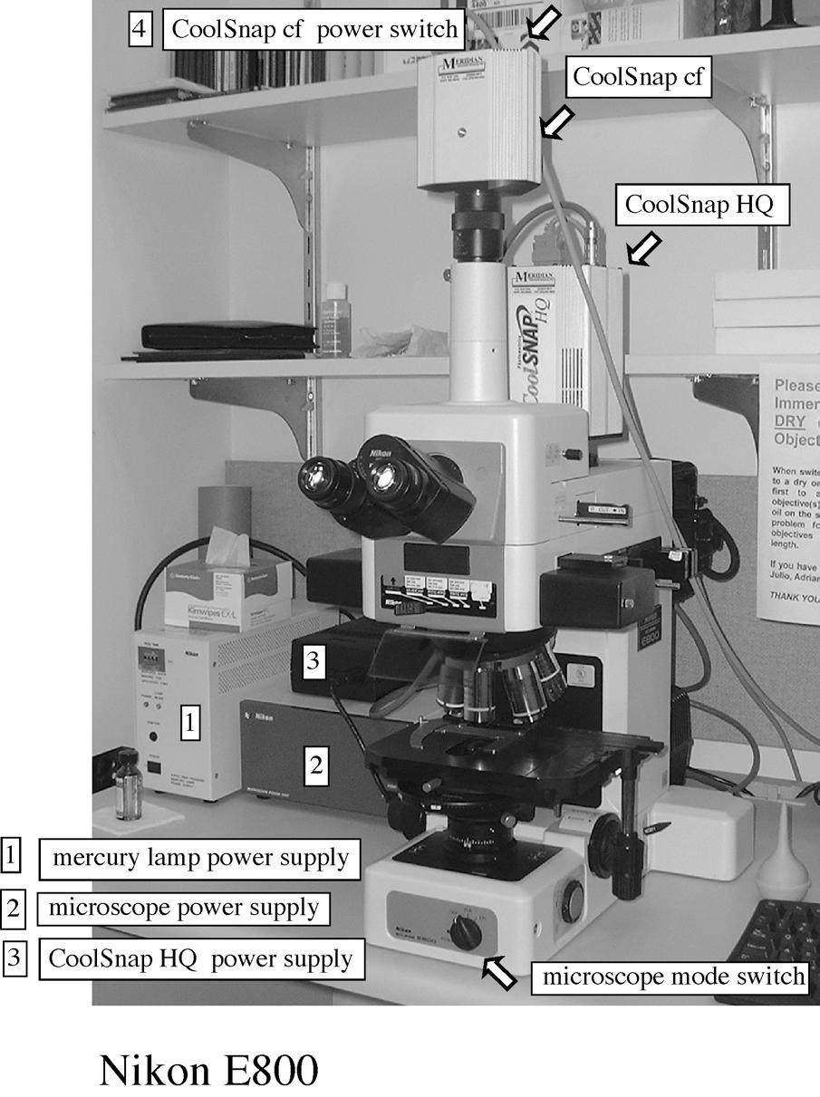
5 2a. Turning ON the system. 2.1 Power ON Mercury (Hg) arc lamp (if using fluorescence). 2.2 Power ON microscope. 2.3 Power ON CoolSnap HQ high-resolution monochrome camera (if necessary) 2.4. Power ON CoolSnap cf color camera (if necessary) 2b. Turning OFF the system. IMPORTANT: IF YOU ARE THE LAST USER, ESPECIALLY AFTER HOURS OR DURING WEEK-ENDS, ALWAYS REMEMBER TO POWER SYSTEM OFF WHEN DONE: 1. POWER OFF ALL SWITCHES (EXCEPT COMPUTER) 2. CLEAN UP IMMERSION OIL FROM OBJECTIVES USING LENS TISSUE (FOLD ONE OR TWO SHEETS OF TISSUE IN TWO ALONG THEIR LENGTH, HOLD BY THE EDGES, AND GENTLY WIPE OBJECTIVE; REPEAT WITH NEW TISSUE IF NECESSARY). 3. MAKE SURE ALL LAMPS AND CAMERAS ARE OFF BEFORE YOU COVER MICROSCOPE. 5
 IN: Coolsnap HQ High Resolution Monochrome CCD Camera Lower Slider (2) OUT: Coolsnap cf Color CCD Camera/Binocular viewing (eyepiece) Upper Slider (1) IN: Eyepiece Upper Slider (1)")
6 3. Selecting the light path. Two sliders on the right side of the eyepiece select the light path. Lower Slider (2) IN: Coolsnap HQ High Resolution Monochrome CCD Camera Lower Slider (2) OUT: Coolsnap cf Color CCD Camera/Binocular viewing (eyepiece) Upper Slider (1) IN: Eyepiece Upper Slider (1) OUT: Coolsnap cf color CCD Slider 3: Infra-Red protection for CCD camera (leave in the engaged (IN) position). 6
. 3.")
7 4. Operating in transmitted light mode. 3.1 Use switch on the front base of microscope to select transmitted light (DIA). 3.2 If Hg lamp is ON, engage Hg lamp block (pull slider OUT) to block Hg lamp illumination 3.3 Select blank filter cube (DIA-ILL) 7

.")
8 3.4. Choose objective. Apply a SMALL drop of immersion oil to sample if using immersion lens. Inspect objective markings and chart on the wall to make sure you do not apply immersion oil to a dry lens Focus on sample Adjust illumination intensity with power dial on left side of microscope 3.7. Engage filters for transmitted illumination if required (e.g. neutral density to attenuate light). Push lever down to engage filter. 8
 out; pull analyzer (2) out). 3.9.")
9 3.8 Remove polarizer and analyzer from the light path. (swing polarizer (1) out; pull analyzer (2) out) Set Condenser turret to position A (for normal transmitted light) 9

 5.3 Center field diaphragm using condenser centering screws (2) 5.4.")
10 5. Adjusting the microscope for Koehler illumination. Typically, the microscope will be properly adjusted. The only adjustments you need to do is to open the field diaphragm to just enclose the field of view for your objective, and adjust the condenser aperture. The full procedure is described below. 5.1 Stop down (close) field diaphragm by rotating dial on right side of microscope base 5.2 Focus field diaphragm by moving condenser UP or DOWN (1) 5.3 Center field diaphragm using condenser centering screws (2) 5.4. Open field diaphragm barely wider than the field of view (see Fig 4.1) 10

11 5.5 Open condenser aperture diaphragm to about 75% of the objective back aperture. You can examine the objective back aperture by removing one eyepiece and looking down the tube. As an approximation, the condenser aperture should be set to minimum for 10x objective and to max for 100x objective: 10x: setting ~2 20x ~5 <open-close> 40x ~7 O o 60x ~ x ~9 11

12 6. Adjusting the microscope for DIC. Typically, the microscope will be properly adjusted. Normally, you only need to select the condenser prism matching your objective, and engage the polarizer and analyzer. The whole procedure is described below. 6.1 Engage Polarizer (swing over light path; left) and Analyzer (slide IN; right) 6.2 Select position A on condenser turret (no DIC prism engaged) 12
. 6.4 Verify that field is dark.")
13 6.3 Disengage objective prism by pulling prism slider OUT (without removing completely; the DIC objective prisms are the little sliders located just above each objective). Note: make sure to disengage the prism directly above the objective in use (in the image below, the prism shown belongs to an objective that is NOT in use). 6.4 Verify that field is dark. Rotate polarizer for minimum transmission (normally when set to o-) 6.5 engage objective prism (push IN) 6.6 select matching condenser prism (H, L or M; read DIC label on objective or use Table in section 1) 6.7 for fine adjustments, rotate analyzer dial (make sure to unlock by rotating screw on top) 13
 Note: the polarizer is not in the epi-illumination light")
. 7.")
14 7. Setting the Microscope for Fluorescence. 7.1 Disengage Analyzer (1; pull OUT) Note: the polarizer is not in the epi-illumination light path, and therefore does not affect its quality. 7.2 Pull epi-illumination shutter OUT to block light (2). 7.3 select EPI setting on front base dial (will work on OFF too) 7.4 select fluorescence filter cube by moving slider 14
. [Note: Positions 1-4 are ge")
]: Position 1: blank (transmitted light) Position 2: DAPI Position 3: FITC Position 4: TRITC (Rhodamine) Position 5: Triple")
15 Filter positions (from left to right). [Note: Positions 1-4 are generally fixed. Position 5 may be occupied by a different filter (e.g. Texas Red or Cy5)]: Position 1: blank (transmitted light) Position 2: DAPI Position 3: FITC Position 4: TRITC (Rhodamine) Position 5: Triple DAPI/FITC/TexasRed 7.5. Open fluorescence illumination shutter by pushing IN 7.6. Select the smallest convenient illumination field aperture using the epifluorescence field diaphragm (1). Center if necessary using the two centering screws (2). 7.7 Always pull shutter OUT when not imaging or viewing to minimize photobleaching of your sample. 15
16 8. Starting Imaging Software (Metamorph). 8.1 Verify that the desired camera is ON (typically, HQ for fluorescence, cf for transmitted light); See section double-click on the camera icon on desktop. This will launch Metamorph acquisition software. 8.3 If one camera is not ON, will give error message: camera not found, do you want to try again? Answer NO 8.4 Adjust light path for the appropriate camera: Lower Slider (1) IN: Coolsnap HQ High Resolution Monochrome CCD Camera Lower Slider (1) OUT: Coolsnap cf Color CCD Camera/Binocular viewing (eyepiece) Upper Slider (2) IN: Eyepiece Upper Slider (2) OUT: Coolsnap cf color CCD 8.5 Click on Acquire Image. Will open acquire dialog window. On acquire window: use show live to show live image for centering and focusing your sample use acquire to acquire single exposure. Adjust exposure and collect Image. 16
17 Use Save as to save the image to your local folder. Move later to your fred account. Use control buttons on left of window to display image histogram, select display colors, and select magnification. Notes: To collect multiple color planes (e.g. in fluorescence) with Metamorph, you need to acquire a greyscale image for each channel, by manually selecting th eappropriate fluorescence filters. Once you have the desired images, you can combine them into an RGB color image with Metamorph, or other imaging software. Metamorph can collect time-lapse images into a multi-layered image file (stack). The stack can be saved as an AVI or QT movie. Metamorph does not record the type of objective used. For distance measurements on your image, you need to record that information (e.g. by adding it as a file info ). Metamorph can measure distances if you have the proper calibration file for the objective you used. 9. Turning OFF the system. IMPORTANT: IF YOU ARE THE LAST USER, ESPECIALLY AFTER HOURS OR DURING WEEK-ENDS, ALWAYS REMEMBER TO POWER SYSTEM OFF WHEN DONE: 9.1. POWER OFF ALL SWITCHES (EXCEPT COMPUTER) 9.2. CLEAN UP IMMERSION OIL FROM OBJECTIVES USING LENS TISSUE (FOLD ONE OR TWO SHEETS OF TISSUE IN TWO ALONG THEIR LENGTH, HOLD BY THE EDGES, AND GENTLY WIPE OBJECTIVE; REPEAT WITH NEW TISSUE IF NECESSARY) MAKE SURE ALL LAMPS AND CAMERAS ARE OFF BEFORE YOU COVER MICROSCOPE. 17
Nikon E800 Operating Instructions.
 Nikon E800 Operating Instructions. You can request electronic copies of this manual by contacting lshats@jhsph.edu Copies are also available on the JHU MMI Department web site. Please send your comments
Nikon E800 Microscope. Operating Instructions
 Nikon E800 Microscope Operating Instructions B Watson 12/2005 Table of contents: 1. The Nikon E800 Microscope 2. Turning the system ON and OFF 3. Selecting the light path 4. Operating in transmitted light
EPIFLUORESCENCE &/OR BRIGHTFIELD MICROSCOPY
 EPIFLUORESCENCE &/OR BRIGHTFIELD MICROSCOPY TURN ON THE FOLLOWING EQUIPMENT The fluorescent light (if needed) The power strip for the microscope and accessories The CoolSNAP HQ camera on the right (Turn
Zeiss Axio Imager.A1 manual
 Zeiss Axio Imager.A1 manual Power-up protocol 1. Mercury lamp 2. Power strip on shelf 3. Computer The Mercury lamp should always be first-on and last-off. This prevents any electrical surges caused by
Using the Nikon TE2000 Inverted Microscope
 Wellcome Trust Centre for Human Genetics Molecular Cytogenetics and Microscopy Core Using the Nikon TE2000 Inverted Microscope Fluorescence image acquisition using Scanalytic s IPLab software and the B&W
Characterization Microscope Nikon LV150
 Characterization Microscope Nikon LV150 Figure 1: Microscope Nikon LV150 Introduction This upright optical microscope is designed for investigating up to 150 mm (6 inch) semiconductor wafers but can also
Zeiss Axioskop II. The AIF's "routine" light microscope. (Installed 8/24/04)AxioCam installed July 11th 2005
AxioCam installed July 11th 2005") Zeiss Axioskop II The AIF's "routine" light microscope. (Installed 8/24/04)AxioCam installed July 11th 2005 Featuring: Phase Contrast Darkfield DIC/Nomarski Brightfield Fluorescent filters for Dapi, FITC,Rhodamine
Zeiss Axioskop II The AIF's "routine" light microscope. (Installed 8/24/04)AxioCam installed July 11th 2005 Featuring: Phase Contrast Darkfield DIC/Nomarski Brightfield Fluorescent filters for Dapi, FITC,Rhodamine
Nikon TE300 Eclipse Wide-Field Microscope
 Nikon TE300 Eclipse Wide-Field Microscope User Guide LSU Health Science Center-Shreveport Research Core Facility 1 User manual for Nikon Elements software Equipment: Nikon TE300 Eclipse microscope Photometrics
SHORT INSTRUCTIONS FOR OPERATING LSM1/2 (Zeiss LSM510) AT CIAN Version 1.4, September 2014
 AT CIAN Version 1.4, September 2014") CIAN LSM1 or LSM2 short instructions, version 1.4, September 2014 page 1 of 6 SHORT INSTRUCTIONS FOR OPERATING LSM1/2 (Zeiss LSM510) AT CIAN Version 1.4, September 2014 Before starting To work with LSM1
CIAN LSM1 or LSM2 short instructions, version 1.4, September 2014 page 1 of 6 SHORT INSTRUCTIONS FOR OPERATING LSM1/2 (Zeiss LSM510) AT CIAN Version 1.4, September 2014 Before starting To work with LSM1
Widefield-NikonEclipseTE200-ORCA Nikon Eclipse TE200 Inverted Microscope with Hamamatsu 1394 Orca-ER Cooled CCD Camera and Micromanager Software
 Widefield-NikonEclipseTE200-ORCA Nikon Eclipse TE200 Inverted Microscope with Hamamatsu 1394 Orca-ER Cooled CCD Camera and Micromanager Software September 2007 Check website for most current User Guide
Nikon Ti-E Microscope Manual. Rightmire Hall Ohio State University. Director: Tony Brown Rightmire
 Nikon Ti-E Microscope Manual Rightmire Hall Ohio State University Director: Tony Brown Rightmire 060 292-1205 brown.2302@osu.edu Facility Manager: Paula Monsma Rightmire 062 293-0939 292-1367 monsma.1@osu.edu
Guide to Confocal 5. Starting session
 Guide to Confocal 5 Remember that when booking and before starting session you can check for any problems at https://www.bris.ac.uk/biochemistry/uobonly/cif/index.html Starting session Switch on microscope
Olympus Fluoview 1000S Spectral Confocal Microscope Introduction to the NRI-MCDB Microscopy Facility Spectral Confocal Microscope
 Olympus Fluoview 1000S Spectral Confocal Microscope Introduction to the NRI-MCDB Microscopy Facility Spectral Confocal Microscope Improved Optics More Lasers 405 diode 440 diode 488 Argon 515 Argon 559
Nikon C1si Spectral Laser Scanning Confocal Microscope. User Guide
 Nikon C1si Spectral Laser Scanning Confocal Microscope User Guide Contents: C1Si Turn-On/ShutDown Procedures... 2 Overview... 4 Setup for epi-illumination to view through the eyepieces:... 5 Setup for
Motorized Axio Observer Start-up instructions
 Start-up instructions 1. If using fluorescence turn on Fluorescent light source. TL light Source (Hal 100) 2. Turn on microscope using switch on lower left side of the microscope. 3. If imaging, turn on
BX-61: Brightfield Instruction /Continue to scroll for Fluorescent Instuctions
 BX-61: Brightfield Instruction /Continue to scroll for Fluorescent Instuctions Starting up: Schematic of Olympus BX-61. 1. Turn on Olympus microscope power box (left of microscope) with toggle switch on
Nikon Eclipse Ti2-E Widefield/Spinning Disk Confocal Microscope Standard Operation Protocol
 Nikon Eclipse Ti-E Widefield/Spinning Disk Confocal Microscope Standard Operation Protocol Please sign on the log sheet before switching on system. Turn on system Turn on A only if confocal mode or laser
Zeiss Axiovert 135 Fluorescence Microscope Quick Guide / Operations Manual (v. 1.0 February 09)
") University of Chicago Integrated Light Microscopy Core Dr. Vytas Bindokas, Director http://digital.bsd.uchicago.edu By: Christine Labno, Assistant Director Room: AB-129 Phone: 4-9040 Zeiss Axiovert 135
University of Chicago Integrated Light Microscopy Core Dr. Vytas Bindokas, Director http://digital.bsd.uchicago.edu By: Christine Labno, Assistant Director Room: AB-129 Phone: 4-9040 Zeiss Axiovert 135
Olympus IX71 Microscope and DP71 Camera Instructions
 Olympus IX71 Microscope and DP71 Camera Instructions Microscopy in Medicine (MiM) Core Emory University Department of Medicine 1 Olympus IX71 Image Capture Procedure 2 3 1. STARTING-UP PROCEDURE: Remove
Zeiss AxioImager.Z2 Brightfield Protocol
 Zeiss AxioImager.Z2 Brightfield Protocol 1) System Startup Please note put sign-up policy. You must inform the facility at least 24 hours beforehand if you can t come; otherwise, you will receive a charge
Zeiss LSM 880 Protocol
 Zeiss LSM 880 Protocol 1) System Startup Please note put sign-up policy. You must inform the facility at least 24 hours beforehand if you can t come; otherwise, you will receive a charge for unused time.
Instructions for Making On-Line Reservations for Microscopes in NB11-204
 Instructions for Making On-Line Reservations for Microscopes in NB11-204 1. Log into Mail using Mail.swmed.edu 2. Log in using your university id and password. 3. Click the Calendar Tab at the top right
START-UP PROCEDURE 1 THE MICROSCOPE STAND 3 OBJECTIVES 5 STARTING WITH LAS (SOFTWARE) AND SETTING UP THE MICROSCOPE STAND 7
 AND SETTING UP THE MICROSCOPE STAND 7") Leica DMI AF6000LX Table of contents START-UP PROCEDURE 1 THE MICROSCOPE STAND 3 OBJECTIVES 5 STARTING WITH LAS (SOFTWARE) AND SETTING UP THE MICROSCOPE STAND 7 ACQUIRE MODULE 6 SETTING THE LIGHTPATH 6
Leica DMI AF6000LX Table of contents START-UP PROCEDURE 1 THE MICROSCOPE STAND 3 OBJECTIVES 5 STARTING WITH LAS (SOFTWARE) AND SETTING UP THE MICROSCOPE STAND 7 ACQUIRE MODULE 6 SETTING THE LIGHTPATH 6
b. Turn the power switch and key to on position for blue laser.
 OLYMPUS FLUOVIEW 300 CONFOCAL MICOSCOPE OPERATION PROCEDURE 1. Turn ON microscope in this order: 1) Turn on mercury lamp (Note: once the mercury lamp is turned off, DO NOT turn it back on for at least
DIC Imaging using Laser Scanning Microscopes (LSMs) on Axio Imager Stands
 on Axio Imager Stands") DIC Imaging using Laser Scanning Microscopes (LSMs) on Axio Imager Stands Differential Interference Contrast (DIC) imaging is a technique used to increase contrast in brightfield images. In confocal systems,
DIC Imaging using Laser Scanning Microscopes (LSMs) on Axio Imager Stands Differential Interference Contrast (DIC) imaging is a technique used to increase contrast in brightfield images. In confocal systems,
Operation Guide for the Leica SP2 Confocal Microscope Bio-Imaging Facility Hunter College October 2009
 Operation Guide for the Leica SP2 Confocal Microscope Bio-Imaging Facility Hunter College October 2009 Introduction of Fluoresence Confocal Microscopy The first confocal microscope was invented by Princeton
CAPTURING IMAGES ON THE HIGH-MAGNIFICATION MICROSCOPE
 University of Virginia ITC Academic Computing Health Sciences CAPTURING IMAGES ON THE HIGH-MAGNIFICATION MICROSCOPE Introduction The Olympus BH-2 microscope in ACHS s microscope lab has objectives from
Nikon Eclipse Ti A1-A Confocal Operating Manual. Start-up. Microscope
 Nikon Eclipse Ti A1-A Confocal Operating Manual Start-up 1. Turn on Excite Fluorescent light power supply- metal halide. a. Cool down as for mercury bulb b. Wheel closed liquid light guide 2. Turn on power
DIC Imaging using Laser Scanning Microscopes (LSM) on Inverted Stands
 on Inverted Stands") DIC Imaging using Laser Scanning Microscopes (LSM) on Inverted Stands Differential Interference Contrast (DIC) imaging is a technique used to increase contrast in brightfield images. In confocal systems,
DIC Imaging using Laser Scanning Microscopes (LSM) on Inverted Stands Differential Interference Contrast (DIC) imaging is a technique used to increase contrast in brightfield images. In confocal systems,
INSTRUCTIONS FOR COURSE WORK 4 (AxioVert) Instructor: Anne Vaahtokari (MIU) 1. Purpose of the work
 Instructor: Anne Vaahtokari (MIU) 1. Purpose of the work") INSTRUCTIONS FOR COURSE WORK 4 (AxioVert) Instructor: Anne Vaahtokari (MIU) 1. Purpose of the work In this work, you will get familiar with an inverted epifluorescence microscope. Also, you will learn
INSTRUCTIONS FOR COURSE WORK 4 (AxioVert) Instructor: Anne Vaahtokari (MIU) 1. Purpose of the work In this work, you will get familiar with an inverted epifluorescence microscope. Also, you will learn
Widefield 1. Switching on
 Widefield 1 Switching on 1. Ignite DG5 lamp - must be switched on first (if previous user has switched off, wait 30 min before igniting) 2. Wait 5s and then turn on the main DG5 controller switch. 3. DG5
personal DELTAVISION (pdv)
") GUIDELINES AND HINTS Version 1.3 (March 2015) personal DELTAVISION (pdv) Epifluorescence microscope from Applied Precision Inc.: The microscope can be found in room 1.320. For details see the architectural
GUIDELINES AND HINTS Version 1.3 (March 2015) personal DELTAVISION (pdv) Epifluorescence microscope from Applied Precision Inc.: The microscope can be found in room 1.320. For details see the architectural
Olympus Time-lapse Microscope Basic operation
 Olympus Time-lapse Microscope Basic operation To start up the microscope 1. Switch on the Olympus UCB. (label as ) 1 Power Switch 2 2. Switch on the MT10. (label as ) Power Switch Page 1 of 18 3 3. Switch
Contents STARTUP MICROSCOPE CONTROLS CAMERA CONTROLS SOFTWARE CONTROLS EXPOSURE AND CONTRAST MONOCHROME IMAGE HANDLING
 Operations Guide Contents STARTUP MICROSCOPE CONTROLS CAMERA CONTROLS SOFTWARE CONTROLS EXPOSURE AND CONTRAST MONOCHROME IMAGE HANDLING Nikon Eclipse 90i Operations Guide STARTUP Startup Powering Up Fluorescence
Olympus xcellence Software - basic user guide
 Olympus xcellence Software - basic user guide This is a basic overview of setting up time lapse experiments using Olympus's xcellence software on BIU's IX81 inverted phase contrast system - the software
User manual for Nikon Elements software
 User manual for Nikon Elements software Equipment: Nikon TE300 Eclipse microscope ANDOR Neo/Zyla B&W camera (default) DS Fi2 color camera Sign in on the sign in sheet; please use both your given name and
Things to check before start-up.
 Byeong Cha Page 1 11/24/2009 Manual for Leica SP2 Confocal Microscope Enter you name, the date, the time, and the account number in the user log book. Things to check before start-up. Make sure that your
Microscope ECLIPSE 80i Instructions <Reference>
 M318E 03.12.CF.1(2/2) Microscope ECLIPSE 80i Instructions Introduction Thank you for purchasing this Nikon product. This instruction manual, which describes basic microscope operations, is
Quick Start Guide. Leica SP5 X
 Quick Start Guide Leica SP5 X Please note: Some of the information in this guide was taken from Leica Microsystems Leica TCS SP5 LAS AF Guide for New Users. This work is licensed under the Creative Commons
AxioVision 4.5 Brightfield Image Capture Procedure
 AxioVision 4.5 Brightfield Image Capture Procedure 1. STARTING-UP PROCEDURE: Remove blue dust cover and place on shelf under microscope. Turn on the halogen lamp by pushing the switch at the back right
Brightfield Microscopy and Image Acquisition on Spotcam1. by Ryan Taylor/Nancy Kleene Last modified 10/02/05 by Birgit Ehmer
 Brightfield Microscopy and Image Acquisition on Spotcam1 by Ryan Taylor/Nancy Kleene Last modified 10/02/05 by Birgit Ehmer Log onto the computer. Enter your username and password to log onto the server.
Nikon AZ100. Laser Scanning Macro Confocal Microscope. Jordan Briscoe Adam Fries Kyle Marchuk Kaitlin Corbin. May 2017.
 Nikon AZ100 Laser Scanning Macro Confocal Microscope Jordan Briscoe Adam Fries Kyle Marchuk Kaitlin Corbin May 2017 Contents 1 Introduction 2 2 Hardware - Startup 2 3 Software/Operation 4 3.1 Multidimensional
TABLE OF CONTENTS. Safety notes i. Care and Maintenance. ii. 1. Components Illustration Installation of Components.. 4
 TABLE OF CONTENTS Safety notes i Care and Maintenance. ii 1. Components Illustration... 1 2. Installation of Components.. 4 2.1 Installation Diagram... 4 2.2 Installation Procedures 5 3. Operation...11
MetaMorph Imaging Handbook Update 6/4/13
 MetaMorph Imaging Handbook Update 6/4/13 Startup FIRST turn on mercury lamp (Fluorescence) Computer and monitor Qimaging Camera (on top) Uniblitz Shutters-2 Halogen Lamp (Transmitted Light) Computer Login
Operating Instructions for Zeiss LSM 510
 Operating Instructions for Zeiss LSM 510 Location: GNL 6.312q (BSL3) Questions? Contact: Maxim Ivannikov, maivanni@utmb.edu 1 Attend A Complementary Training Before Using The Microscope All future users
Quick Guide for Zeiss 710 Laser Scanning Confocal MGH Cancer Center
 Quick Guide for Zeiss 710 Laser Scanning Confocal MGH Cancer Center For any questions or concerns, please contact: Linda Nieman lnieman@mgh.harvard.edu Office: (617) 643-9684 Cell: (512) 565-8076 Chenyue
Zeiss AxioObserver with ApoTome
 Zeiss AxioObserver with ApoTome Quick Start User Guide LSU Health Sciences Center-Shreveport Research Core Facility (RCF) Microscopy Table of Contents 1 Start up the system.. Page 3 2 Touch screen controller
Reichert Univar Manual
 Reichert Univar Manual Translated from the 11/1975 German language edition, with slight modifications. William R. Porter San Marcos CA USA 2017 v 1.3 Notes This is a very slightly-modified, new (2017)
Leica DB LB Research microscope and Studo Lite Imaging software
 Leica DB LB Research microscope and Studo Lite Imaging software Room B523 User Guide Molecular Imaging Unit University of Helsinki www.miu.helsinki.fi 9.4.2008 1 GENERAL USER INFORMATION... 1 2 SETTINGS
Imaging Introduction. September 24, 2010
 Imaging Introduction September 24, 2010 What is a microscope? Merriam-Webster: an optical instrument consisting of a lens or combination of lenses for making enlarged images of minute objects; especially:
Leica SP8 TCS Users Manual
 Version : 07/08/0 Leica SP8 TCS Users Manual Start up:. Turn the PC Microscope, Scanner Power, Laser Power, and the Laser Emission key to on (bottom right of desk).. Turn on the fluorescent lamp (top left
Zeiss LSM 780 Protocol
 Zeiss LSM 780 Protocol 1) System Startup F Please note the sign-up policy. You must inform the facility at least 24 hours beforehand if you can t come; otherwise, you will receive a charge for unused time.
LSM 510 Meta Training Notes
 LSM 510 Meta Training Notes Turning on the system Turn on X-Cite power supply. This supplies light for epifluorescence for viewing your samples through the microscope. Turn on the remote control switch.
MICROSCOPY FOR THE DEVELOPMENTAL BIOLOGY STUDENT...
 MICROSCOPY FOR THE DEVELOPMENTAL BIOLOGY STUDENT... You will be using two configurations of microscope during the course of the semester to observe specimens and record your results: compound microscopes
Zeiss Deconvolution Microscope: A Quick Guide
 Zeiss Deconvolution Microscope: A Quick Guide Start-up Uncover microscope. Do not put dust cover on the floor. Plug in both cameras. The default camera is the AxioCam HRm (monochrome camera) for fluorescence
BRIGHTFIELD Olympus TH4-200 Olympus TH4-200 Please keep objectives off touching to avoid their loosening or damage.
 BRIGHTFIELD 1. Remove the microscope cover. 2. Dial the brightness adjustment knob (1) of Olympus TH4-200 halogen light source down to MIN. 3. Turn on the Olympus TH4-200 halogen light switch for transmitted
ZEISS LSM510META confocal manual
 ZEISS LSM510META confocal manual Switching on the system 1) Switch on the Remote Control button located on the table to the right of the microscope. This is the main switch for the whole system including
LSM 510 Training Notes
 LSM 510 Training Notes Turning on the system Turn on the arc lamp, found on the bench top left of the microscope. This supplies light for epifluorescence for viewing your samples through the microscope.
Nikon SIM-E & A1-R System
 Nikon SIM-E & A1-R System USER GUIDE LSU Health Sciences Center Shreveport Research Core Facility June 01 2017 Chaowei Shang 1 Table of Content 1. Start Up the System... Page 3 Hardware and microscope
Simplified Instructions: Olympus Widefield Microscope S1230
 Contents General Microscope Operation Simple Image Capture Multi-Wavelength Capture Z-Series Timelapse Combining Capture Modes Synopsis of Other Functions Pages 2-23 24-40 41-47 48-56 57-59 60-68 69-83
TRAINING MANUAL. Olympus FV1000
 TRAINING MANUAL Olympus FV1000 September 2014 TABLE OF CONTENTS A. Start-Up Procedure... 1 B. Visual Observation under the Microscope... 1 C. Image Acquisition... 4 A brief Overview of the Settings...
Indian Institute of technology Madras Presents NPTEL NATIONAL PROGRAMME ON TECHNOLOGY ENHANCED LEARNING
 Indian Institute of technology Madras Presents NPTEL NATIONAL PROGRAMME ON TECHNOLOGY ENHANCED LEARNING Lecture - 5 Materials Characterization Fundamentals of Optical microscopy Dr. S. Sankaran Associate
OMM300. Inverted Metallurgical Microscope
 OMM300 Inverted Metallurgical Microscope Instruction Manual Please read the instructions carefully before operating CONTENTS Safety 2 Parts List 2 Features 3 Assembly 5 Operation 7 Maintenance 9 Specifications
Simplified Instructions: Zeiss Brightfield Microscope S1000
 Contents General Microscope Set-Up Adjust Illumination Focus Condenser Open Software Image Capture Settings Shading & Color Corrections Image Capture & Viewing Scaling and Measurements Synopsis of Other
Brief manual how to start and close the Leica sp2 Confocal. (TCS SP2 AOBS system mounted on a DM IRE2)
") Brief manual how to start and close the Leica sp2 Confocal (TCS SP2 AOBS system mounted on a DM IRE2) A. Switching on hardware B. Acquiring and saving images C. Switching off the microscope D. Good working
Brief manual how to start and close the Leica sp2 Confocal (TCS SP2 AOBS system mounted on a DM IRE2) A. Switching on hardware B. Acquiring and saving images C. Switching off the microscope D. Good working
Quick Guide for Zeiss 710 Laser Scanning Confocal MGH Cancer Center
 Quick Guide for Zeiss 710 Laser Scanning Confocal MGH Cancer Center For any questions or concerns, please contact: Linda Nieman lnieman@mgh.harvard.edu Office: (617) 643-9684 Cell: (512) 565-8076 Chenyue
Fluorescence Imaging with Olympus IX81 Microscope Last updated December 1 st, 2017
 1 Fluorescence Imaging with Olympus IX81 Microscope Last updated December 1 st, 2017 Contents Mercury Lamp Precautions...1 Acknowledgment policies...2 Biosafety requirements and rules for work in the MIC...2
Microscope Confocal LSM510 META
 Microscope Confocal LSM510 META Welcome to the Zeiss LSM 510 Meta Confocal tutorial. Before using the LSM 510 META, Log off any other computer that is open with your personal login. You will need to put
User Manual. Digital Compound Binocular LED Microscope. MicroscopeNet.com
 User Manual Digital Compound Binocular LED Microscope Model MD82ES10 MicroscopeNet.com Table of Contents i. Caution... 1 ii. Care and Maintenance... 2 1. Components Illustration... 3 2. Installation...
Leica TCS SL Confocal Training. Neuroscience Imaging Core Staff. Core Director. Facility Manager
 Leica TCS SL Confocal Training Neuroscience Imaging Facility The Ohio State University Rightmire Hall 614-292-1367 Staff Core Director Anthony Brown, Ph. D. 060 Rightmire Hall 614-292-1205 brown.2302@osu.edu
Very short introduction to light microscopy and digital imaging
 Very short introduction to light microscopy and digital imaging Hernan G. Garcia August 1, 2005 1 Light Microscopy Basics In this section we will briefly describe the basic principles of operation and
Training Guide for Carl Zeiss AxioZoom V16 Stereo Microscope
 Training Guide for Carl Zeiss AxioZoom V16 Stereo Microscope ZEN 2012 Optical Imaging & Vital Microscopy Core Baylor College of Medicine (2017) Power ON Routine 1 2 If you require fluorescence imaging,
LEICA TCS SP5 AOBS TANDEM USER MANUAL
 LEICA TCS SP5 AOBS TANDEM USER MANUAL STARTING THE SYSTEM...2 THE LAS AF SOFTWARE...3 THE «ACQUIRE» MENU...5 CHOOSE AND CREATE A SETTING...6 THE CONTROL PANEL...8 THE DMI6000B MICROSCOPE...10 ACQUIRE ONE
STEINDORFF METALLURGICAL MICROSCOPE. NYMCS-620 Instruction Manual
 METALLURGICAL MICROSCOPE NYMCS-620 Instruction Manual It is recommended strongly that you study this manual thoroughly before using the microscope. Retain this manual in an easily accessible place near
Zeiss LSM 510 Multiphoton Confocal Microscope
 Zeiss LSM 510 Multiphoton Confocal Microscope Quick Start User Guide LSU Health Sciences Research Core Facility Table of Contents 1 Safety... Page 3 2 Turn On the System... Page 4 3 Start Up the ZEN Software.
Microscope Confocal Sp2 Upright.
 Microscope Confocal Sp2 Upright. Welcome to the Leica Sp2 Confocal Upright tutorial. Before using the Sp2 Invert, You will need to put down your name on the reservation system = http://svintranet.epfl.ch/index.php?optio
ZEISS LSM 710 CONFOCAL MICROSCOPE USER MANUAL
 ZEISS LSM 710 CONFOCAL MICROSCOPE USER MANUAL START THE SYSTEM... 2 START ZEN SOFTWARE... 3 SET THE TEMPERATURE AND THE CO2 CONTROLLERS... OBSERVATION AT OCULARS... 5 STATIF PRESENTATION... 6 ACQUIRE ONE
CMI STANDARD OPERATING PROCEDURE. Fluoview 300 laser scanning confocal microscope
 CMI STANDARD OPERATING PROCEDURE Fluoview 300 laser scanning confocal microscope CMI documentid:sop001 CONTACT INFORMATION: Peter Owens: 091 494036 (office) Peter.owens@nuigalway.ie Kerry Thompson: 091
Leica SPEII confocal microscope. Short Manual
 Leica SPEII confocal microscope Short Manual Switching ON sequence: 1. Turn on the Workstation under the bench (top, far right). 2. Turn on the Supply Unit - Laser box (big green switch first and then
Diskovery Spinning Disk Guide
 Diskovery Spinning Disk Guide qbi.microscopy@uq.edu.au Getting started The microscope and its peripherals (Fig. 1a) should always be turned on, but if they are not, turn them on in the following way: 1.
Nasmyth Ultraview Vox User Protocol
 Nasmyth Ultraview Vox User Protocol Switch on all wall sockets labelled Nasmyth, switch camera on (power supply located on table behind monitor), switch on laser switch in laser rack, switch computer on
Leica Confocal - 2. Instructions for Leica SP2 Confocal Equipped with Visible Laser Lines
 Leica Confocal - 2 Instructions for Leica SP2 Confocal Equipped with Visible Laser Lines Imaging Technology Group Revised as of 08/2011 by Dianwen Zhang (Email: zhangdw@illinois.edu) Revision History Authors
User Manual. Trinocular Metallurgical Microscope. MicroscopeNet.com
 User Manual Trinocular Metallurgical Microscope Model M83MPTR MicroscopeNet.com Table of Contents i. Caution.. 1 ii. Care and Maintenance... 2 1. Components Illustration..... 3 2. Installation...4 3. Operation
SWIFT SERIES M2252DGL MICROSCOPE
 SWIFT SERIES M2252DGL MICROSCOPE The M2252DGL Series is ideal for elementary to high school classrooms. Built to withstand student use, this series has locked-on eyepieces, objectives, illuminator housing
Manual for BMS E1 eplan series, compound microscope
 Manual for BMS E1 eplan series, compound microscope The compound microscope allows it to study, at cell level, structures of textures of botanical and zoological nature. (e.g. slides of roots, leaves and
INTRODUCTION TO OPTICAL MICROSCOPY
 Experimental Biophysics TEK265, FYST23, TNF060, FAF010F Lab Exercise Supervisor: Karl Adolfsson Written by Peter Jönsson and Jason Beech Updated by Henrik Persson, Karl Adolfsson and Zhen Li karl.adolfsson@ftf.lth.se
Therefore, all descriptions and illustrations in this instruction manual, including all specifications are subject to change without notice.
 We are constantly endeavouring to improve our instruments and to adapt them to the requirements of modern research techniques and testing methods. This involves modification to the mechanical structure
LSM 510 META in Chang Gung University
 Content LSM 510 META in Chang ung University LSM 510 META 路 理 The features and applications of LSM 510 META 01-09 Introduction of the hardware 10-12 Fluorescence observation in conventional microscope
Biological Microscope Manual
 Version No.: V1.2 Series Biological Microscope Manual This manual expatiates the using method, troubleshooting and maintenance about MT-50 series biological microscope. Please study this manual thoroughly
Leica SP8 TCS Users Manual
 Leica SP8 TCS Users Manual Follow the procedure for start up and log on as posted in the lab. Please log on with your account only and do not share your password with anyone. We track and confirm usage
Leica Sp5 II Confocal User Guide
 Leica Sp5 II Confocal User Guide Turning on the Confocal System (instructions are posted in the room) 1. Turn on Laser Power Button 2. Turn Key to On position 3. Turn on Scanner Power Button 4. Turn on
Dante (Microscope) & Beatrice (Guide) Orth Lab
 & Beatrice (Guide) Orth Lab") Dante (Microscope) & Beatrice (Guide) Orth Lab Olympus IX81 Widefield Microscope User Guide v. 1.2 (11/2014) Objectives 4x/0.13NA UPLFLN Semi Apo 10x/0.4NA PH UPLAPO Plan Apo 20x/0.8NA PH UPLAPO Plan Apo
Dante (Microscope) & Beatrice (Guide) Orth Lab Olympus IX81 Widefield Microscope User Guide v. 1.2 (11/2014) Objectives 4x/0.13NA UPLFLN Semi Apo 10x/0.4NA PH UPLAPO Plan Apo 20x/0.8NA PH UPLAPO Plan Apo
TE2000 T-FL Epi-Fl Attachment T-FL-E Motorized Epi-Fl Attachment Instructions
 M286 E 03.2.IF.4 TE2000 T-FL Epi-Fl Attachment T-FL-E Motorized Epi-Fl Attachment Instructions Thank you for purchasing the Nikon products. This instruction manual is written for the users of the T-FL
Practical Light Microscopy
 Biomedical & X-ray Physics Kjell Carlsson Important: Study the preparatory exercises carefully before the lab session starts! Practical Light Microscopy Laboratory instructions for course SK2500/01, Physics
Leica TCS SP2 User Manual 1.1. Markus Friedrich and James Lee. Department of Biological Sciences Wayne State University
 Leica TCS SP2 User Manual 1.1 Markus Friedrich and James Lee Department of Biological Sciences Wayne State University Confocal Manual 1 Helpful hints The confocal room is cold. Bring a sweater. Confocaling
CONFOCAL MICROSCOPE (Zeiss LSM 510 META v4.2)
") Wellcome Trust Centre for Human Genetics Molecular Cytogenetics and Microscopy Core CONFOCAL MICROSCOPE (Zeiss LSM 510 META v4.2) 1) STARTING THE SYSTEM Abridged INSTRUCTIONS Switch on the mercury bulb
Wellcome Trust Centre for Human Genetics Molecular Cytogenetics and Microscopy Core CONFOCAL MICROSCOPE (Zeiss LSM 510 META v4.2) 1) STARTING THE SYSTEM Abridged INSTRUCTIONS Switch on the mercury bulb
Zoom Stereo Microscope NYMCS-360 Instruction Manual
 Zoom Stereo Microscope NYMCS-60 Instruction Manual This manual is written for stereo microscope NYMCS-60. To ensure the safety, obtain optimum performance and to familiarize yourself fully with the use
SPINNING DISK CSU-X1 USER MANUAL
 SPINNING DISK CSU-X1 USER MANUAL Starting the temperature controller... 2 Starting the CO2 controller... 3 Start the spinning disk... 4 Sample observation with the oculars... 5 Spatial sampling, Pixel
CHAPTER1: QUICK START...3 CAMERA INSTALLATION... 3 SOFTWARE AND DRIVER INSTALLATION... 3 START TCAPTURE...4 TCAPTURE PARAMETER SETTINGS... 5 CHAPTER2:
 Image acquisition, managing and processing software TCapture Instruction Manual Key to the Instruction Manual TC is shortened name used for TCapture. Help Refer to [Help] >> [About TCapture] menu for software
WITec Alpha 300R Quick Operation Summary October 2018
 WITec Alpha 300R Quick Operation Summary October 2018 This document is frequently updated if you feel information should be added, please indicate that to the facility manager (currently Philip Carubia,