Microscopy. Krishna Priya.K Lecturer Dept. of Microbiology
|
|
|
- Posy Cunningham
- 5 years ago
- Views:
Transcription
1 Microscopy Krishna Priya.K Lecturer Dept. of Microbiology

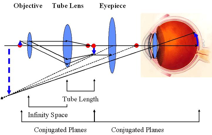
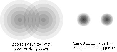
2 TERMS AND DEFINITIONS Principle Microscopy is to get a magnified image, in which structures may be resolved which could not be resolved with the help of an unaided eye. Magnification It is the ratio of the size of an object seen under microscope to the actual size observed with unaided eye. The total magnification of microscope is calculated by multiplying the magnifying power of the objective lens by that of eye piece. Resolving power It is the ability to differentiate two close points as separate. The resolving power of human eye is 0.25 mm The light microscope can separate dots that are 0.25µm apart.

3 Bright Field Microscope
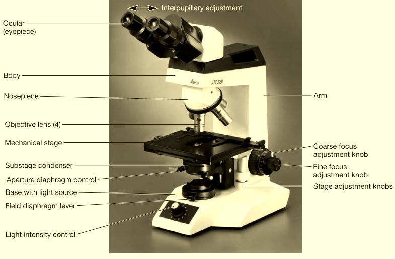
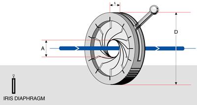
4 Light microscope Parts of microscope Illuminator - This is the light source located below the specimen. Condenser - Focuses the ray of light through the specimen. Stage - The fixed stage is a horizontal platform that holds the specimen. Objective - The lens that is directly above the stage. Nosepiece - The portion of the body that holds the objectives over the stage. Iris diaphragm - Regulates the amount of light into the condenser. Base Base supports the microscope which is horseshoe shaped. Coarse focusing knob - Used to make relatively wide focusing adjustments to the microscope. Fine focusing knob - Used to make relatively small adjustments to the microscope. Body - The microscope body. Ocular eyepiece - Lens on the top of the body tube. It has a magnification of 10 normal vision.


5 Light microscope In 1590 F.H Janssen & Z.Janssen constructed the first simple compound light microscope. In 1665 Robert Hooke developed a first laboratory compound microscope. Later, Kepler and galileo developed a modern class room microscope. In 1672 Leeuwenhoek developed a first simple microscope with a magnification of 200x 300x. He is called as Father of microscopy. The term microscope was coined by Faber in 1623.
6 Light microscope
7 Light microscope Objective PROPERTY LOW POWER HIGH POWER OIL IMMERSION Magnification of objective 10x 40-45x x Magnification of eyepiece 10x 10x 10x Total magnification 100x x x Numerical aperture Mirror used Concave Concave Plane Focal length (Approx) 16 mm 4 mm mm Working distance 4 8 mm mm 0.1 mm Iris diaphragm Partially closed Partially opened Fully opened Position of condenser Lowest Slightly raised Fully raised Maximum resolution(approx) 0.9 µm 0.35µm 0.18µm
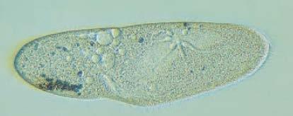
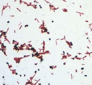
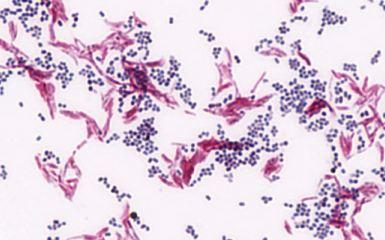
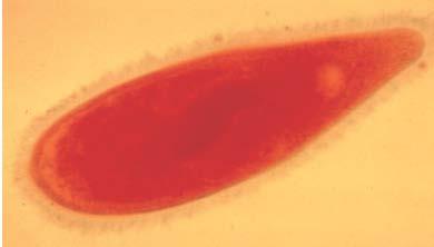
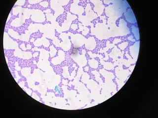
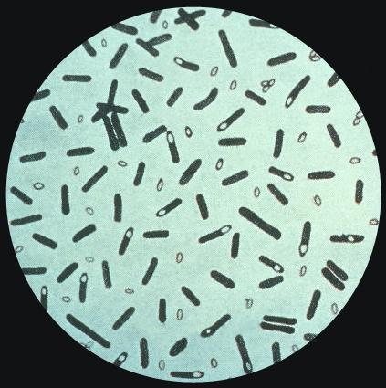
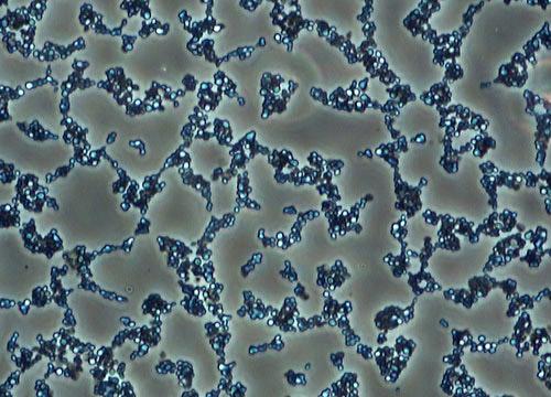
8 Light microscope Baccili and cocci under light microscope Paramecium specimen
9
10

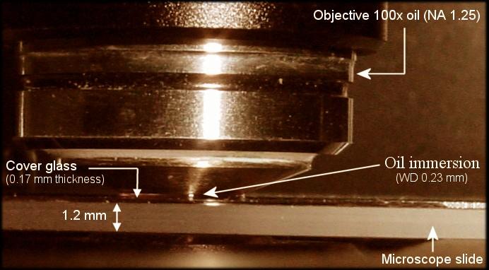
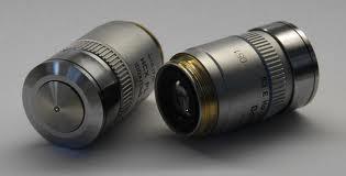
11 Oil immersion lens
12
13

14 Focal length

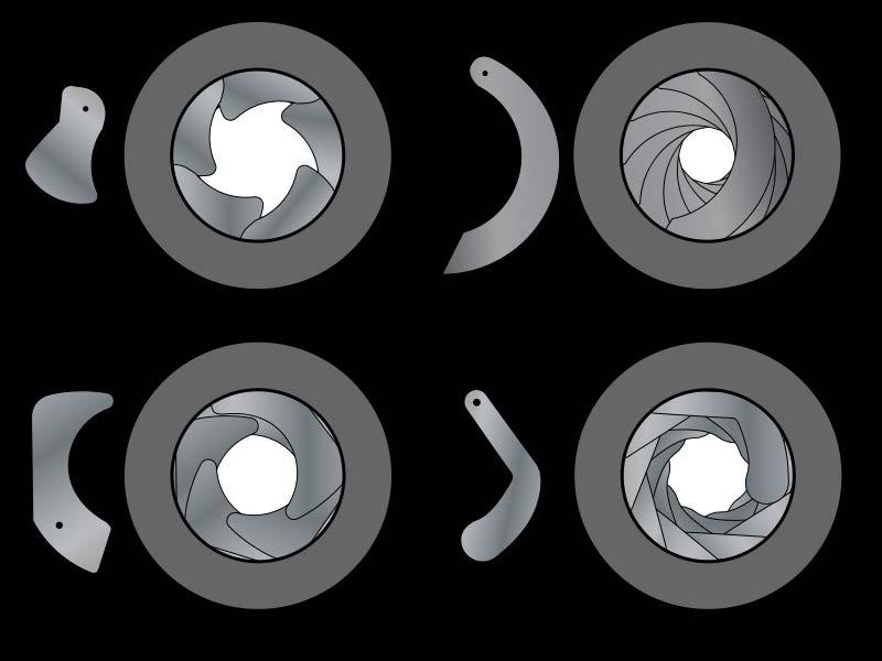
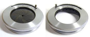
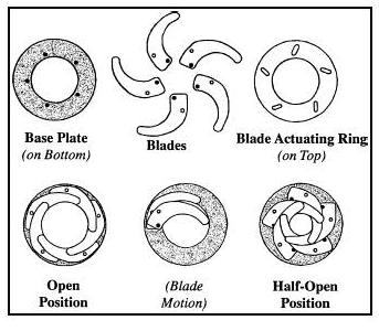
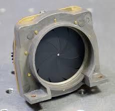
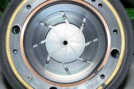
15 Iris diaphragm
16

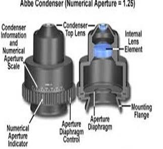
17 Condenser lens
18
19 Resolution power

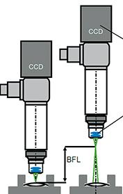
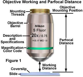
20 TERMS AND DEFINITIONS Limit of resolution It is the minimum distance between two points to identify them separately. It is calculated by Abbé equation. Limit of resolution is inversely proportional to power or resolution. If the wavelength is shorter then the resolution will be greater. Working distance It is the distance between the objective and the objective slide. The working distance decreases with increasing magnification.

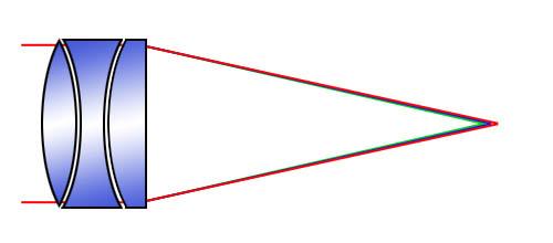
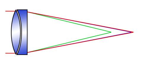
21 Objective lens Apochromatic lens Achromatic lens
22


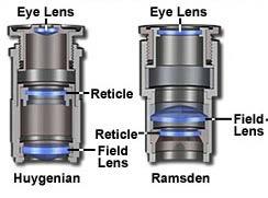
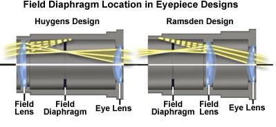
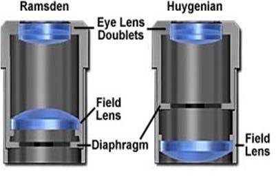
23 Occular lens

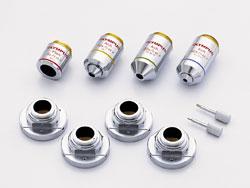
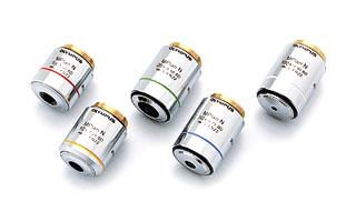
24 Objective lens system
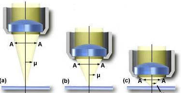
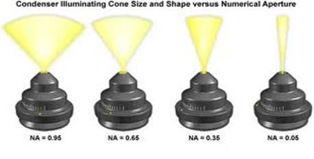
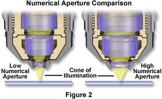
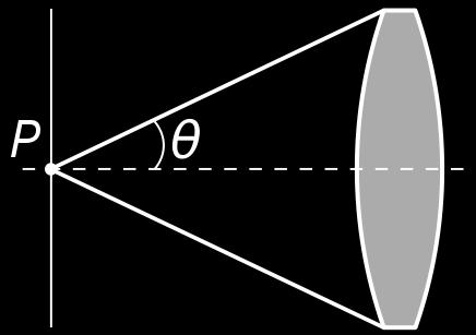
25 TERMS AND DEFINITIONS Numerical aperture(na) The numerical aperture of a lens is the ratio of the diameter of the lens to its focal length. NA of a lens is an index of the resolving power. NA can be decreased by decreasing the amount of light that passes through a lens. Diameter of the lens

26 Numerical aperture
27

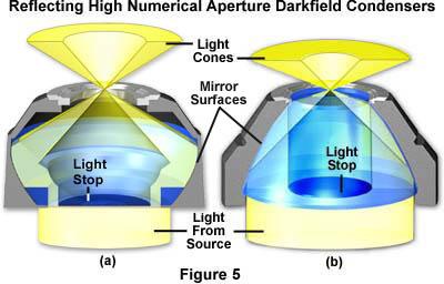
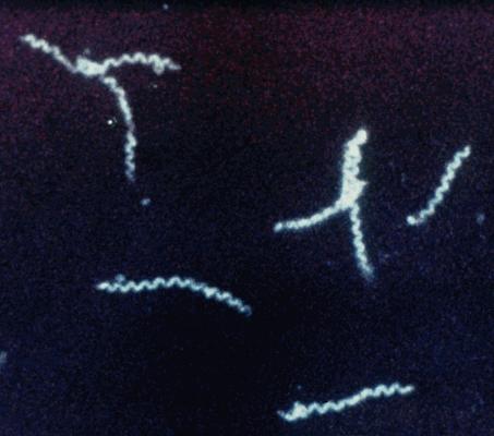
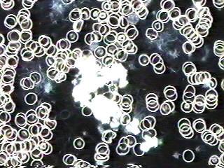
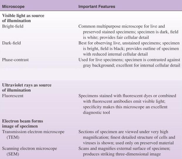
28 Dark field microscope A bright-field microscope can be adapted as a dark-field microscope by adding a special disc called a stop to the condenser. The stop blocks all light from entering the objective lens except peripheral light that is reflected off the sides of the specimen itself. The resulting image is a brightly illuminated specimens surrounded by a dark (black) field. Uses: This microscope is used to study spirochetes in the exudates form leptospiral or syphilitic Infections.

29 Dark filed Microscope


30 Dark field microscope Paramecium Treponema vincenti Volvox and Spirogyra
31
32
33

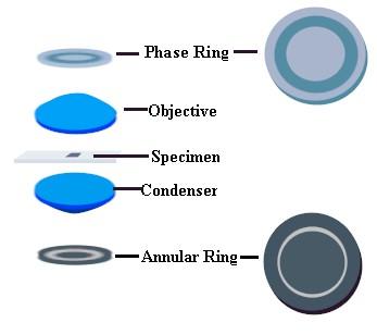
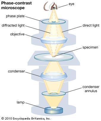
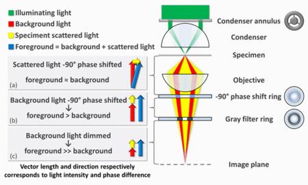
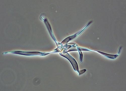
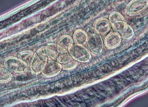
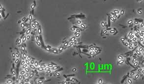
34 Phase contrast microscope In 1935 F.Zernike produced the phase contrast microscope. Phase-contrast microscope is also called as zernike microscope. Phase-contrast microscope uses a special condenser and objective lenses. This condenser lens on the light microscope splits a light beam and throws the light rays slightly out of phase. The separated beams of light then pass through and around the specimen, and small differences in the refractive index within the specimen show up as different degrees of brightness and contrast. Uses: Phase-contrast microscopy is especially useful for studying microbial motility, studying eukaryotic Cells, determining the shape of living cells, and detecting bacterial components such as endospores and Inclusion bodies that contain poly--hydroxyalkanoates (e.g., poly-hydroxybutyrate), polymetaphosphate, sulfur, or other substances.

35 Phase Contrast Microscopy


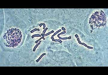
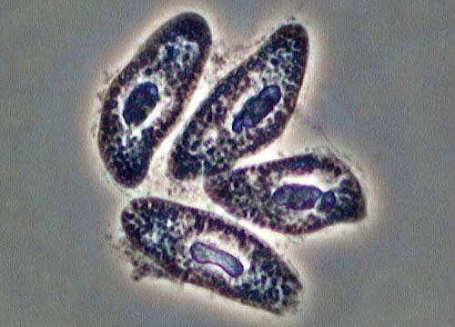
36 Phase contrast microscope Macronucleus Paramecium Micronucleus

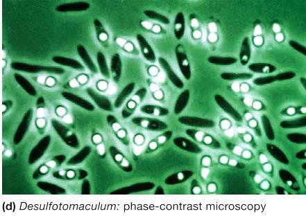
37 Phase contrast microscope Rhodospirillum rubrum
38
39
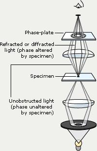
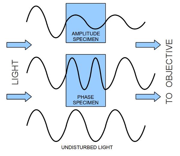
40 Principle
41

42 Principle Constructive interference corresponds to bright spots in the field of view Destructive interference corresponds to dark spots
43 Type of image produced The end result is a magnified and highly contrasted view of a living, unstained, normally transparent specimen
44
45
46

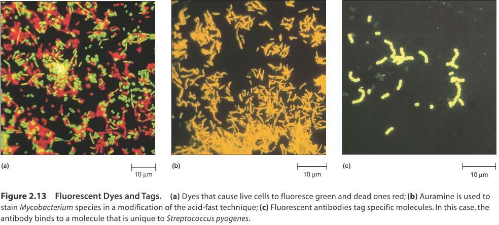
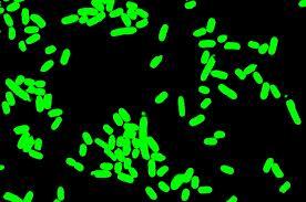
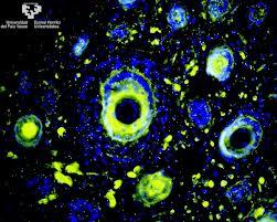
47 Fluorescence microscope It was developed by Haitinger and coons A fluorescence microscope differs from an ordinary brightfield microscope in several respects. It utilizes a powerful mercury vapor arc lamp for its light source. A darkfield condenser is usually used in place of the conventional Abbé brightfield condenser. It employs three sets of filters to alter the light that passes up through the instrument to the eye. Microbiological speciemen that is to be studied must be coated with special compounds that possess the quality of fluorescence. Such compounds are called fluorochromes. AuramineO, acridine orange, and fluorescein are well-known fluorochromes.
48 Fluorescence Microscope

49 Fluorescence microscope Uses: It is used to study the substance like chlorophylls, riboflavin, vitamin A, collagen which have the property of auto fluorescence. Some cellular components like cellulose, starch, glycogen, protein and Y chromosome can be made visible under this microscope by staining them with fluorochromes. It used to identify Y chromosome to determine sex, determination of microbial cells in the infected tissue and to study the structure of proteins.
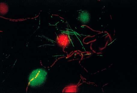
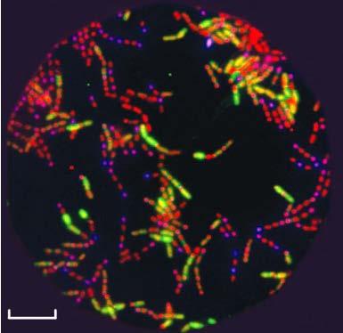
50 Fluorescence microscope Bacillus subtilis Oral cavity
51 Fluorescent Microscope Microorganisms or tissue cells are stained with dyes or compounds called fluorochromes. Examined under microscope with ultra violet radiation instead of visible light. They convert light of shorter (UV) wavelength into visible light and so become luminous Fluoresce. Wavelengths absorbed & emitted are specific for specific fluorochromes.
52 Fluorochromes Acridine orange : Orange Auramine-Rhodamine : Yellow Calcofluor white :White Fluorescein Isothiocyanate (FITC) : Green
53 Modification of Fluorescent Microscope Immunofluorescence : Antibodies labeled with fluorochrome used to specifically stain a particular bacterial species. Uses of IF : viruses, direct examination of C.trachomatis, B.pertussis

54
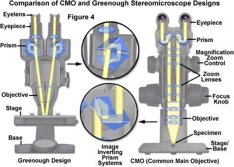
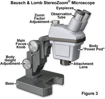
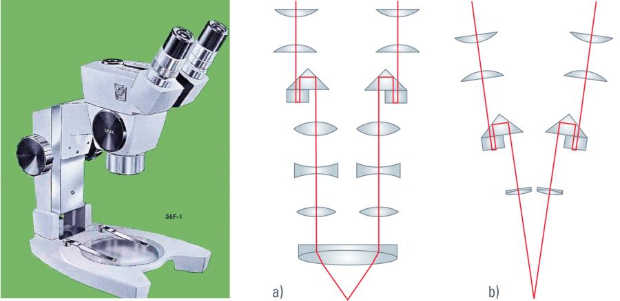
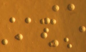
55 Stereomicroscope
56
57
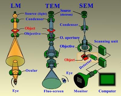
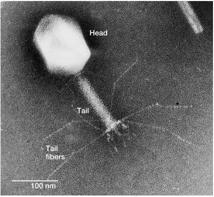
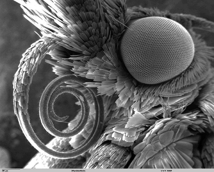
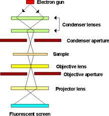
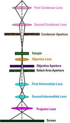
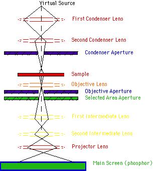
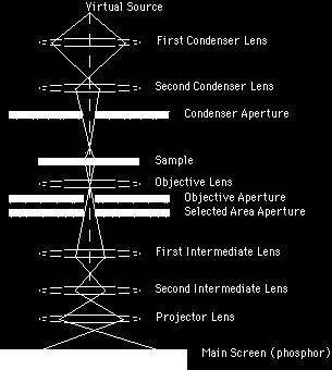
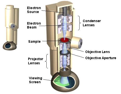
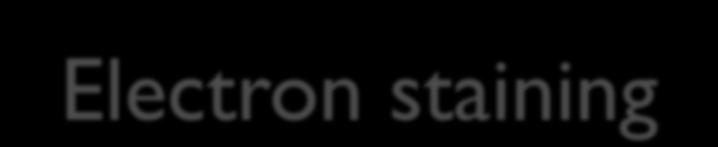
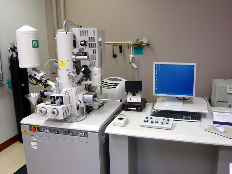
58 Electron microscope In 1932 Knoll and Ruska invented first electron microscope. The electron microscope uses a beam of electrons rather than visible light. The magnified image is visible on a fluorescent screen and can be recorded on a photographic film. The drawback of the electron microscope is specimen are killed in order to view the cells or organisms. Images produced by electrons lack color, electron micrographs are always shades of black, gray, and white. Two general forms of EM are the transmission electron microscope (TEM) and the scanning electron microscope (SEM). Transmission electron microscopes are the method of choice for viewing the detailed structure of cells and viruses. This microscope produces its image by transmitting electrons through the specimen. Because electrons cannot readily penetrate thick preparations, the specimen must be sectioned into extremely thin slices ( nm thick) and stained or coated with metals that will increase image contrast. The darkest areas of TEM micrographs represent the thicker (denser) parts, and the lighter areas indicate the more transparent and less dense parts.
59 Electron Microscopy
60 Electron microscope(tem)
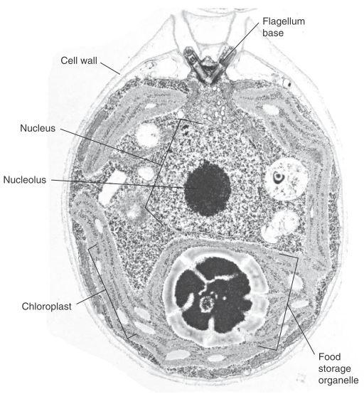
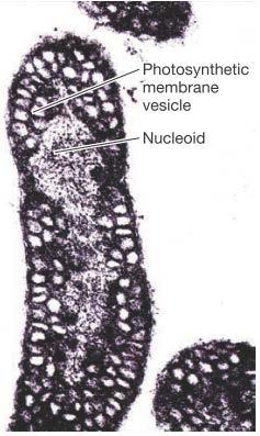
61 Electron microscope(tem) Chlamydomonas
62 Transmission Electron Microscope Illumination: electrons Magnification: ~100,000x How it works: Detect electrons scattered as they move through the sample. Image: Monotone (but may be color enhanced), 2-D structure of specimen

63 Source: u.au/hb313/main_pages/tim etable/lectures/image6.gif
64 Pros High magnification High resolution Shows small structures that cannot be seen under light microscopes Cons Needs specimen to be in vacuum Needs specimen to be covered in gold film Specimen <100nm thick (obviously cannot observe live specimen) No color Really. Big. And. Expensive. Equipment
65
66
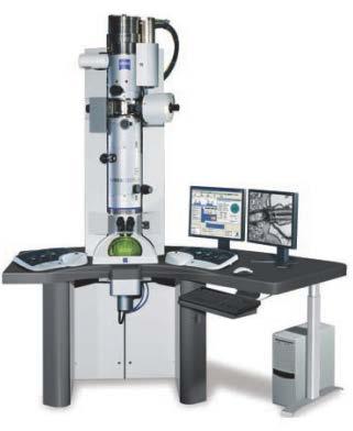
67 Transmission electron microscope
68
69
70
71
72
73
74
75
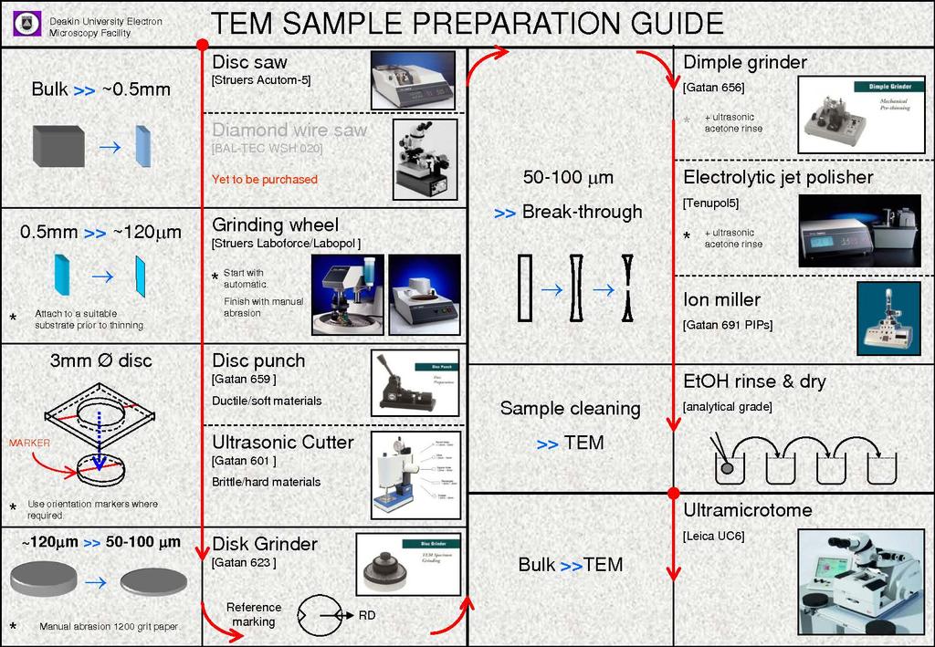
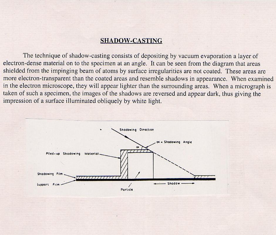
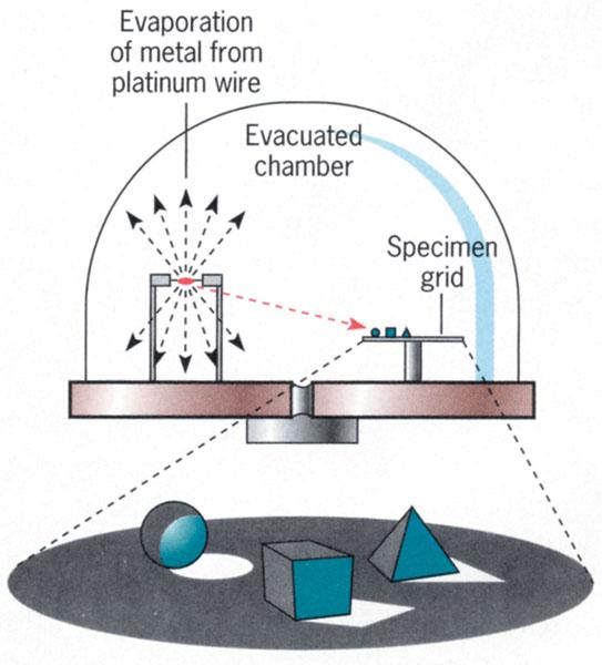
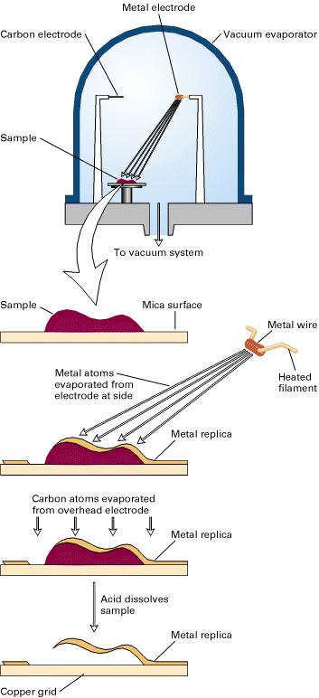
76 Surface replicas


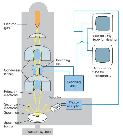
77 Negative staining- specimens are observed on an electron dense background

78 Electron staining

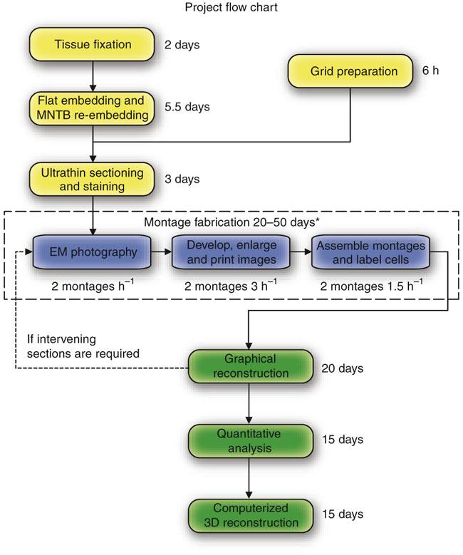
79 Ultra thin sectioning

80
81
82
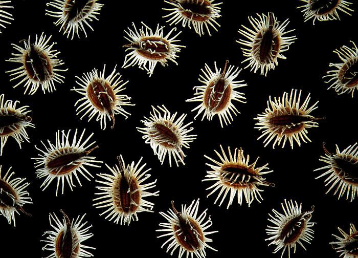
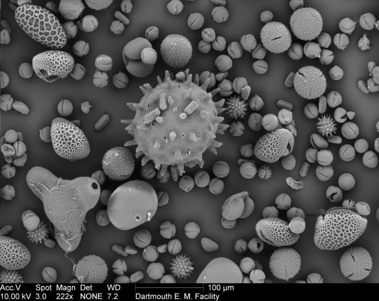
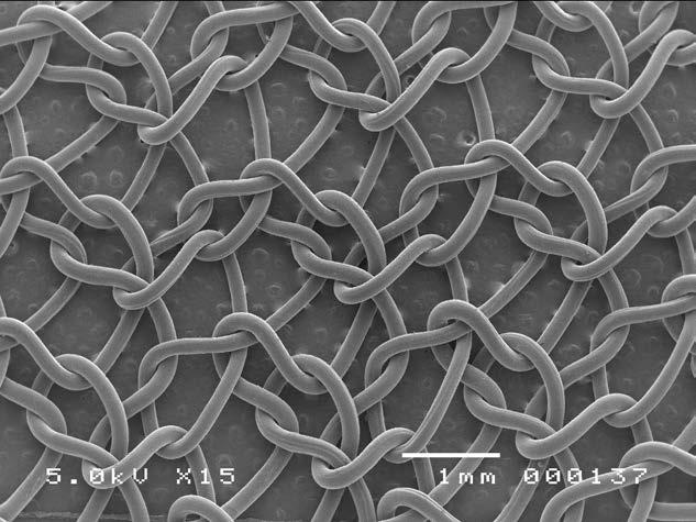
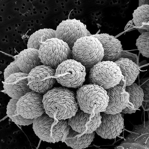
83 Scanning Electron Microscope Illumination: electrons Magnification: ~100,000x How it works: Detect electrons backscattered by the sample. Image: Monotone (but may be color enhanced), 3-D surface of specimen

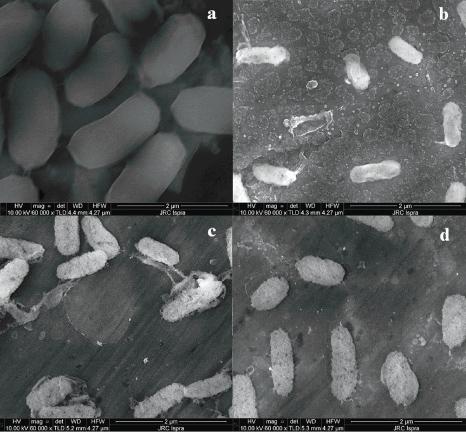
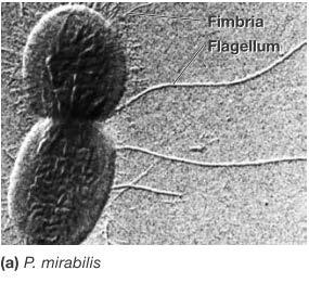
84 Electron microscope(sem) The specimen is placed in the vacuum chamber and covered with a thin coat of gold. The electron beam then scans across the specimen and knocks loose showers of electrons that are captured by a detector. An image builds line by line, as in a television receiver. Electrons that strike a sloping surface yield fewer electrons, thereby producing a darker contrasting spot and a sense of three dimensions. The resolving power of the conventional SEM is about 10 nm and magnifications with the SEM are limited to about 20,000x.
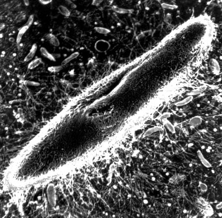
85 Electron microscope(sem) Paramecium SEM
86 Magnetic lens focuses electron beam Scanning coils for systematic scanning (left to right, then down) Backscattered Electron Detector detects electrons that bounced off the film Secondary Electron Detector detects electrons emitted by the film Source:
87 Pros High magnification High resolution Shows the surface of specimen Cons Needs specimen to be in vacuum Needs living cells and tissues and whole, softbodied organisms to be treated, usu. coated w/ gold film No color Cannot examine live specimen Really. Big. And Expensive. Equipment.

88 Scanning electron microscope
89

90 Light Vs Electron microscope

91 Uses
92 Thank you
Observing Microorganisms through a Microscope LIGHT MICROSCOPY: This type of microscope uses visible light to observe specimens. Compound Light Micros
 PHARMACEUTICAL MICROBIOLOGY JIGAR SHAH INSTITUTE OF PHARMACY NIRMA UNIVERSITY Observing Microorganisms through a Microscope LIGHT MICROSCOPY: This type of microscope uses visible light to observe specimens.
Observing Microorganisms through a Microscope
 2016/2/19 PowerPoint Lecture Presentations prepared by Bradley W. Christian, McLennan Community College CHAPTER 3 Observing Microorganisms through a Microscope 1 Figure 3.2 Microscopes and Magnification.
Burton's Microbiology for the Health Sciences
 Burton's Microbiology for the Health Sciences Chapter 2. Viewing the Microbial World Chapter 2 Outline Introduction Using the metric system to express the sizes of microbes Microscopes Simple microscopes
Microscopy Techniques that make it easy to see things this small.
 Microscopy Techniques that make it easy to see things this small. What is a Microscope? An instrument for viewing objects that are too small to be seen easily by the naked eye. Dutch spectacle-makers Hans
Figure 3.4 Approximate size of various types of cells. ~10 um. Red Blood Cells = mm 1500 um. Width of penny Pearson Education, Inc.
 Figure 3.4 Approximate size of various types of cells. ~10 um Red Blood Cells 1.5mm 1500 um Width of penny = 1500 Figure 4.3 The limits of resolution (and some representative objects within those ranges)
Compound Light Microscopy. Microscopy. Things to remember... 1/22/2017. This is what we use in the laboratory
 Compound Light Microscopy This is what we use in the laboratory Microscopy Chapter 3 BIO 440 A series of finely ground lenses is used to form a magnified image Specimen is illuminated with visible light
Microscopy, Staining, and Classification
 PowerPoint Lecture Presentations prepared by Mindy Miller-Kittrell, North Carolina State University C H A P T E R 4 Microscopy, Staining, and Classification Figure 3.4 Approximate size of various types
2018 MICROSCOPE REVIEW by Karen L. Lancour RELATIVE SIZE OF MICROBES
 2018 MICROSCOPE REVIEW by Karen L. Lancour RELATIVE SIZE OF MICROBES 1000 millimeters (mm) = 1 meter (m) 1000 micrometers (µm or mcm) = 1 millimeter (mm) 1000 nanometers (nm) = 1 micrometer (mcm) Size
2017 MICROSCOPE REVIEW by Karen L. Lancour RELATIVE SIZE OF MICROBES
 2017 MICROSCOPE REVIEW by Karen L. Lancour RELATIVE SIZE OF MICROBES 1000 millimeters (mm) = 1 meter (m) 1000 micrometers (µm or mcm) = 1 millimeter (mm) 1000 nanometers (nm) = 1 micrometer (mcm) Size
Chapter 2 The Study of Microbial Structure: Microscopy and Specimen Preparation
 Chapter 2 The Study of Microbial Structure: Microscopy and Specimen Preparation 1 Lenses and the Bending of Light light is refracted (bent) when passing from one medium to another refractive index a measure
Light Microscopy. Upon completion of this lecture, the student should be able to:
 Light Light microscopy is based on the interaction of light and tissue components and can be used to study tissue features. Upon completion of this lecture, the student should be able to: 1- Explain the
The microscope is useful in making observations and collecting data in scientific experiments. Microscopy involves three basic concepts:
 AP BIOLOGY Chapter 6 NAME DATE Block MICROSCOPE LAB PART I: COMPOUND MICROSCOPE OBJECTIVES: After completing this exercise you should be able to: Demonstrate proper care and use of a compound microscope.
The light microscope
 What is a microscope? The microscope is an essential tool in modern biology. It allows us to view structural details of organs, tissue, and cells not visible to the naked eye. The microscope should always
VISUAL PHYSICS ONLINE DEPTH STUDY: ELECTRON MICROSCOPES
 VISUAL PHYSICS ONLINE DEPTH STUDY: ELECTRON MICROSCOPES Shortly after the experimental confirmation of the wave properties of the electron, it was suggested that the electron could be used to examine objects
MICROSCOPY MICROSCOPE TERMINOLOGY
 1 MICROSCOPY Most of the microorganisms that we talk about in this class are too small to be seen with the naked eye. The instruments we will use to visualize these microbes are microscopes. The laboratory
STRUCTURE OF THE MICROSCOPE
 STRUCTURE OF THE MICROSCOPE Use the word list to label the microscope below: Light Source Coarse adjustment knob Diaphragm Stage Clips Objectives Fine Adjustment Knob Base Stage Stage Clips Arm Revolving
Chapter 3. Observing Microorganisms Through a Microscope
 Chapter 3 Observing Microorganisms Through a Microscope Microbial Size Macroscopic organisms can be measured in the range from meters (m) to centimeters (cm) Microscopic organisms fall into the range
The Microscope. Packet #2. 10/17/2016 9:12:02 PM Ryan Barrow 2012
 1 The Microscope Packet #2 10/17/2016 9:12:02 PM Ryan Barrow 2012 2 Historical Timeline 1609 Galileo Galilei develops a compound microscope with a convex and a concave les. 1665 Robert Hooke publishes
Unit Two Part II MICROSCOPY
 Unit Two Part II MICROSCOPY AVERETT 1 0 /9/2013 1 MICROSCOPES Microscopes are devices that produce magnified images of structures that are too small to see with the unaided eye Humans cannot see objects
THE COMPOUND BRIGHTFIELD MICROSCOPE
 THE COMPOUND BRIGHTFIELD MICROSCOPE Microbiology is the study of microscopic organisms that are so small that they are below the limit of vision of the human eye. Bacteria are the smallest of microorganisms
Scale. A Microscope s job in life. The Light Microscope. The Compound Microscope 9/24/12. Compound Microscope Anatomy
 The Study of Microbial Structure: Microscopy and Specimen Preparation Scale A Microscope s job in life 1.Magnify 2. Resolve ability to separate or distinguish between two points 3. Contrast How much or
The Care and Use of the Microscope. Lab Exercise #4
 Lab Safety No eating or drinking!!! Long hair must be tied back Clean up your workstation before you leave! Return all materials to the storage sites Clean glassware and wipe down countertops Follow directions
MICROSCOPE LAB. Resolving Power How well specimen detail is preserved during the magnifying process.
 AP BIOLOGY Cells ACTIVITY #2 MICROSCOPE LAB OBJECTIVES 1. Demonstrate proper care and use of a compound microscope. 2. Identify the parts of the microscope and describe the function of each part. 3. Compare
 Microscopy http://www.microscopyu.com/articles/phasecontrast/phasemicroscopy.html http://micro.magnet.fsu.edu/primer/anatomy/anatomy.html 2005, Dr. Jack Ikeda & Dr. Gail Grabner 9 Nikon Labophot (Question
Lab 1, 2 and 3: Stain, Observe and Identify the Microbes. BIOHAZARD Rules. VIOLATORS will lose points. A) Lab Safety Rules Lab Safety Form Signup
 Lab Safety Rules Lab Safety Form Signup") MICROLAB PREPARATIONS A) Lab Safety Rules Lab Safety Form Signup B) Lab Participation Instructor Review Peer Review Form C) Biohazard Rules How to dispose Trash REQUIRED Items: 1) LAB Manual/Journal 2)
MICROLAB PREPARATIONS A) Lab Safety Rules Lab Safety Form Signup B) Lab Participation Instructor Review Peer Review Form C) Biohazard Rules How to dispose Trash REQUIRED Items: 1) LAB Manual/Journal 2)
Laboratory Introduction
 Laboratory Introduction There are two basic categories of microscopes: light microscopes and electron microscopes. Light, or optical, microscopes require light waves to provide the illumination while electron
Microscope Notes. units of life.
 Microscope Notes Microscope an instrument that produces an enlarged image of an object. Biologists use microscopes to study cells, cell parts, and organisms that are too small to be seen with the naked
Microbiology Laboratory 2
 Microbiology Laboratory 2 Microscopy Background Microorganisms are too small to be seen with the naked eye. Thus a microscope is used to magnify objects so they can be observed. A lens consists of one
A BRIEF INTRODUCTION TO MICROSCOPY The two key properties of a microscope that allow you to see microbes are resolution and magnification.
 A BRIEF INTRODUCTION TO MICROSCOPY The two key properties of a microscope that allow you to see microbes are resolution and magnification. Magnification refers to the enlargement of the specimen when seen
Microscope. Dr. Leena Barhate Department of Microbiology M.J.College, Jalgaon
 Microscope Dr. Leena Barhate Department of Microbiology M.J.College, Jalgaon Acknowledgement http://www.cerebromente.org.br/n17/histor y/neurons1_i.htm Google Images http://science.howstuffworks.com/lightmicroscope1.htm
Microscopy. ( greek mikros = small; skopein = to observe)
") Microscopy ( greek mikros = small; skopein = to observe) Zacharias Jansen put several lenses in a tube (first compound microscope) and the object near the end of tube appeared to be greatly enlarged, much
Microscopy ( greek mikros = small; skopein = to observe) Zacharias Jansen put several lenses in a tube (first compound microscope) and the object near the end of tube appeared to be greatly enlarged, much
Biology 29 Cell Structure and Function Spring, 2009 Springer LABORATORY 1: THE LIGHT MICROSCOPE
 Biology 29 Cell Structure and Function Spring, 2009 Springer LABORATORY 1: THE LIGHT MICROSCOPE Prior to lab: 1) Read these instructions (p 1-6) 2) Go through the online tutorial, the microscopy pre-lab
Microscopy Training & Overview
 Microscopy Training & Overview Product Marketing October 2011 Stephan Briggs - PLE OVERVIEW AND PRESENTATION FLOW Glossary and Important Terms Introduction Timeline Innovation and Advancement Primary Components
Lecture 4 to 5 MICROSCOPY-PRINCIPLES AND TYPES
 Lecture 4 to 5 MICROSCOPY-PRINCIPLES AND TYPES Microorganisms are too small to be seen by our unaided eyes and the microscopes are of crucial importance as they help to view the microbes. A microscope
Microscopy. Danil Hammoudi.MD
 Microscopy Danil Hammoudi.MD Care and Handling of the Microscope: A microscope is a delicate piece of equipment and should be treated with care. Use two hands when carrying the microscope. Place one hand
S200 Course LECTURE 1 TEM
 S200 Course LECTURE 1 TEM Development of Electron Microscopy 1897 Discovery of the electron (J.J. Thompson) 1924 Particle and wave theory (L. de Broglie) 1926 Electromagnetic Lens (H. Busch) 1932 Construction
Microscope Review. 1. A compound light microscope is represented in the diagram below.
 Name Microscope Review Date 1. A compound light microscope is represented in the diagram below. 5. The diagram below represents a hydra as viewed with a compound light microscope. If the hydra moves toward
Exercise 2-A MICROSCOPIC TECHNIQUE & EXAMINATION OF MICROORGANISMS
 Exercise 2-A MICROSCOPIC TECHNIQUE & EXAMINATION OF MICROORGANISMS Introduction to Microscopic Technique Microbiology is the science or study of living organisms too small to be seen with the naked eye.
Biology The Microscope. May 20 1:19 PM. Using a Microscope to Explore the Cell
 Biology 2201 1.2 The Microscope Using a Microscope to Explore the Cell Resolution or Resolving power The ability of the eye, or other instrument, to distinguish between two objects that are close together
Visual Anatomy ansd Physiology Lab Manual Pig Version 2nd Edition Sarikas TEST BANK
 Visual Anatomy ansd Physiology Lab Manual Pig Version 2nd Edition Sarikas TEST BANK https://testbankreal.com/download/visual-anatomy-ansd-physiology-labmanual-pig-version-2nd-edition-sarikas-test-bank/
Ex 1: Introduction to the microscope
 Ex 1: Introduction to the microscope So what exactly is a microorganism? Microorganisms = any living thing that is too small to be seen with the unaided eye fungus protist bacteria virus Parasitic worm
Match the microscope structures given in the left column with the statements in the right column that identify or describe them.
 49 Prelab for Name Match the microscope structures given in the left column with the statements in the right column that identify or describe them. Key: a. coarse adjustment knob f. turret or nosepiece
Imaging Introduction. September 24, 2010
 Imaging Introduction September 24, 2010 What is a microscope? Merriam-Webster: an optical instrument consisting of a lens or combination of lenses for making enlarged images of minute objects; especially:
The microscope is useful in making observations and collecting data in scientific experiments. Microscopy involves three basic concepts:
 Lab #4 Biology 10 BCC Topic: MICROSCOPE LAB PART I: COMPOUND LIGHT MICROSCOPE OBJECTIVES: After completing this exercise you should be able to: Demonstrate proper care and use of a compound microscope.
History of microscopy
 History of microscopy Introduction Structure of microscope Care of microscope Use of microscope Magnification As we already know cells are microscopic. What does this mean? Scientists were able to see
Basics of Light Microscopy and Metallography
 ENGR45: Introduction to Materials Spring 2012 Laboratory 8 Basics of Light Microscopy and Metallography In this exercise you will: gain familiarity with the proper use of a research-grade light microscope
Exercise 2-A MICROSCOPIC TECHNIQUE & EXAMINATION OF MICROORGANISMS
 Exercise 2-A MICROSCOPIC TECHNIQUE & EXAMINATION OF MICROORGANISMS Introduction to Microscopic Technique Microbiology is the science or study of living organisms too small to be seen with the naked eye.
ANSWER KEY Lab 2 (IGB): Bright Field and Fluorescence Optical Microscopy and Sectioning
: Bright Field and Fluorescence Optical Microscopy and Sectioning") Phys598BP Spring 2016 University of Illinois at Urbana-Champaign ANSWER KEY Lab 2 (IGB): Bright Field and Fluorescence Optical Microscopy and Sectioning Location: IGB Core Microscopy Facility Microscope:
Phys598BP Spring 2016 University of Illinois at Urbana-Champaign ANSWER KEY Lab 2 (IGB): Bright Field and Fluorescence Optical Microscopy and Sectioning Location: IGB Core Microscopy Facility Microscope:
MICROSCOPY and CELL STRUCTURE
 MICROSCOPY and CELL STRUCTURE Readings: Review pp. 69-71, and Fig. 4.1 on p. 65 in your text (POHS, 5 th ed.). Introduction: Biologists rely on many different types of microscopic techniques to find out
Life Science Chapter 2 Study Guide
 Key concepts and definitions Waves and the Electromagnetic Spectrum Wave Energy Medium Mechanical waves Amplitude Wavelength Frequency Speed Properties of Waves (pages 40-41) Trough Crest Hertz Electromagnetic
Instruction Manual T Binocular Acromat Research Scope T Trinocular Acromat Research Scope
 Research Scope Instruction Manual T-29031 Binocular Acromat Research Scope T-29041 Trinocular Acromat Research Scope T-29032 Binocular Semi-Plan Research Scope T-29042 Trinocular Semi-Plan Research Scope
Applications of Optics
 Nicholas J. Giordano www.cengage.com/physics/giordano Chapter 26 Applications of Optics Marilyn Akins, PhD Broome Community College Applications of Optics Many devices are based on the principles of optics
Marine Invertebrate Zoology Microscope Introduction
 Marine Invertebrate Zoology Microscope Introduction Introduction A laboratory tool that has become almost synonymous with biology is the microscope. As an extension of your eyes, the microscope is one
ELECTRON MICROSCOPY AN OVERVIEW
 ELECTRON MICROSCOPY AN OVERVIEW Anjali Priya 1, Abhishek Singh 2, Nikhil Anand Srivastava 3 1,2,3 Department of Electrical & Instrumentation, Sant Longowal Institute of Engg. & Technology, Sangrur, India.
INTRODUCTION TO OPTICAL MICROSCOPY
 Experimental Biophysics TEK265, FYST23, TNF060, FAF010F Lab Exercise Supervisor: Karl Adolfsson Written by Peter Jönsson and Jason Beech Updated by Henrik Persson, Karl Adolfsson and Zhen Li karl.adolfsson@ftf.lth.se
Ocular Lenses. Head. Arm. Objective Lenses. Slide Holder Stage. On / Off Switch. Condenser with Iris Diaphragm. Light Intensity Control
 BIOLOGY 211: HUMAN ANATOMY & PHYSIOLOGY ********************************************************************************************************* USE OF THE LIGHT MICROSCOPE **********************************************************************************************************
Microscopy. Matti Hotokka Department of Physical Chemistry Åbo Akademi University
 Microscopy Matti Hotokka Department of Physical Chemistry Åbo Akademi University What s coming Anatomy of a microscope Modes of illumination Practicalities Special applications Basic microscope Ocular
Microscope (and The Cell) Lab Exercise #1
 Lab Exercise #1") Lab Safety-General No eating or drinking Only registered students allowed in the class Long hair must be tied back Familiarize yourself with the emergency stations Do not mark on the models Inform me of
Lab Safety-General No eating or drinking Only registered students allowed in the class Long hair must be tied back Familiarize yourself with the emergency stations Do not mark on the models Inform me of
I. The First Microscopes. Microscope Basics. II. The Bright Field Microscope. Confocal Laser Scanning Microscopy. A. The Compound Microscope
 Microscope Basics I. The First Microscopes NGSSS: SC.912.N.2.1 through N.4.2 A. About 1590, two Dutch spectacle makers, Zaccharias Janssen and his son Hans, while experimenting with several lenses in a
Microscopy and Staining
 Microscopy and Staining Figure 2.1 Different types of microscopy are used to visualize different structures. Brightfield microscopy (left) renders a darker image on a lighter background, producing a clear
King Saud University Dept. of Bot. & Microbiology. General Microbiology 140 MIC
 King Saud University Dept. of Bot. & Microbiology General Microbiology 140 MIC Lab coat. Do not wearing the lab coat outside the lab. Gloves. Proper Clothing and closed shoes. Hair should be tied back.
The invention of the microscope made it possible for scientists to view and study cells. Cells the basic units of all living organisms.
 The Discovery of Cells The invention of the microscope made it possible for scientists to view and study cells. Cells the basic units of all living organisms. The Cell Theory All living things are made
LlIGHT REVIEW PART 2 DOWNLOAD, PRINT and submit for 100 points
 WRITE ON SCANTRON WITH NUMBER 2 PENCIL DO NOT WRITE ON THIS TEST LlIGHT REVIEW PART 2 DOWNLOAD, PRINT and submit for 100 points Multiple Choice Identify the choice that best completes the statement or
! Because microbiology deals with organisms too small they cannot be seen distinctly with the unaided eye, the microscope is essential.
 Microscopy! Because microbiology deals with organisms too small they cannot be seen distinctly with the unaided eye, the microscope is essential.! The light microscope is the single most important research
Introduction. Laboratory Equipment & Supplies. Model 1333PHi Shown (Phase Contrast) (2) Eyepieces (Eyecups installed) Diopter Adjustment Mechanism
 (2) Eyepieces (Eyecups installed) Diopter Adjustment Mechanism") Introduction With the invention of the microscope in the early 17th century, it was made possible to view objects which were too small for the human eye to see. As the microscope evolved, the structure
Introduction With the invention of the microscope in the early 17th century, it was made possible to view objects which were too small for the human eye to see. As the microscope evolved, the structure
Education in Microscopy and Digital Imaging
 Contact Us Carl Zeiss Education in Microscopy and Digital Imaging ZEISS Home Products Solutions Support Online Shop ZEISS International ZEISS Campus Home Interactive Tutorials Basic Microscopy Spectral
used for low power magnification of a sample image is 3 dimensional
 MICROSCOPES One of the most important inventions in the advancement of Biology 1. Simple Microscopes ie. magnifying glass, stereoscope (dissecting scope) have a single lens or a pair of lenses combined
Chapter 1 Parts. Figure 1.1. Parts of a Compound Light Microscope
 Chapter 1 Parts Chapter 1 Parts Figure 1.1 illustrates the parts of an upright compound microscope and indicates the terminology that I use in these notes. Figure 1.1. Parts of a Compound Light Microscope
LSM 510 META in Chang Gung University
 Content LSM 510 META in Chang ung University LSM 510 META 路 理 The features and applications of LSM 510 META 01-09 Introduction of the hardware 10-12 Fluorescence observation in conventional microscope
Microscopy: Fundamental Principles and Practical Approaches
 Microscopy: Fundamental Principles and Practical Approaches Simon Atkinson Online Resource: http://micro.magnet.fsu.edu/primer/index.html Book: Murphy, D.B. Fundamentals of Light Microscopy and Electronic
The Nature of Light. Light and Energy
 The Nature of Light Light and Energy - dependent on energy from the sun, directly and indirectly - solar energy intimately associated with existence of life -light absorption: dissipate as heat emitted
Care and Use of the Compound Light Microscope
 EXERCISE 2 Care and Use of the Compound Light Microscope Time Estimates for Completing This Lab The activities in this laboratory exercise can be completed in 2 to 2.5 hours. Extra time will be required
Perfecting Microscope Skills
 I. Introduction to the Microscope Perfecting Microscope Skills There are different types of microscopes used by biologists depending on the job they wish to accomplish, including dissecting (or "stereoscopic")
CCAM Microscope Objectives
 CCAM Microscope Objectives Things to consider when selecting an objective Magnification Numerical Aperture (NA) resolving power and light intensity of the objective Working Distance distance between the
CALIBRATION OF MICROSCOPE EYEPIECE GRATICULE
 CALIBRATION OF MICROSCOPE EYEPIECE GRATICULE A typical eyepiece graticule looks like this: It is 10mm in length and each mm is divided into 10 parts So each small division = 0.1mm = 100µm The eyepiece
Very short introduction to light microscopy and digital imaging
 Very short introduction to light microscopy and digital imaging Hernan G. Garcia August 1, 2005 1 Light Microscopy Basics In this section we will briefly describe the basic principles of operation and
Reichert Univar Manual
 Reichert Univar Manual Translated from the 11/1975 German language edition, with slight modifications. William R. Porter San Marcos CA USA 2017 v 1.3 Notes This is a very slightly-modified, new (2017)
2/4/15. Brightfield Microscopy! It s all about Magnification..! or is it?!
 Brightfield Microscopy It s all about Magnification.. or is it? 1 What actually does go into chosing a microscope Choice depends on what you need the microscope to do. Do you want to magnify stained specimens?
Microscope anatomy, image formation and resolution
 Microscope anatomy, image formation and resolution Ian Dobbie Buy this book for your lab: D.B. Murphy, "Fundamentals of light microscopy and electronic imaging", ISBN 0-471-25391-X Visit these websites:
Instructional Resources/Materials: Light vocabulary cards printed (class set) Enough for each student (See card sort below)
 Enough for each student (See card sort below)") Grade Level/Course: Grade 7 Life Science Lesson/Unit Plan Name: Light Card Sort Rationale/Lesson Abstract: Light vocabulary building, students identify and share vocabulary meaning. Timeframe: 10 to 20
Grade Level/Course: Grade 7 Life Science Lesson/Unit Plan Name: Light Card Sort Rationale/Lesson Abstract: Light vocabulary building, students identify and share vocabulary meaning. Timeframe: 10 to 20
Chapter 23 Study Questions Name: Class:
 Chapter 23 Study Questions Name: Class: Multiple Choice Identify the letter of the choice that best completes the statement or answers the question. 1. When you look at yourself in a plane mirror, you
microscopy A great online resource Molecular Expressions, a Microscope Primer Partha Roy
 Fundamentals of optical microscopy A great online resource Molecular Expressions, a Microscope Primer http://micro.magnet.fsu.edu/primer/index.html Partha Roy 1 Why microscopy Topics Functions of a microscope
FLUORESCENCE MICROSCOPY. Matyas Molnar and Dirk Pacholsky
 FLUORESCENCE MICROSCOPY Matyas Molnar and Dirk Pacholsky 1 The human eye perceives app. 400-700 nm; best at around 500 nm (green) Has a general resolution down to150-300 μm (human hair: 40-250 μm) We need
INTRODUCTION THIN LENSES. Introduction. given by the paraxial refraction equation derived last lecture: Thin lenses (19.1) = 1. Double-lens systems
 = 1. Double-lens systems") Chapter 9 OPTICAL INSTRUMENTS Introduction Thin lenses Double-lens systems Aberrations Camera Human eye Compound microscope Summary INTRODUCTION Knowledge of geometrical optics, diffraction and interference,
Chapter 9 OPTICAL INSTRUMENTS Introduction Thin lenses Double-lens systems Aberrations Camera Human eye Compound microscope Summary INTRODUCTION Knowledge of geometrical optics, diffraction and interference,
Motorized Axio Observer Start-up instructions
 Start-up instructions 1. If using fluorescence turn on Fluorescent light source. TL light Source (Hal 100) 2. Turn on microscope using switch on lower left side of the microscope. 3. If imaging, turn on
Light microscopy BMB 173, Lecture 14, Feb. 21, 2018
 Light microscopy The Structural Biology Continuum Next two lectures: Light microscopy Many slides taken from Scott Fraser, Murphy s Fundamentals of light microscopy, Alberts Molecular Biology of the Cell,
Operation Guide for the Leica SP2 Confocal Microscope Bio-Imaging Facility Hunter College October 2009
 Operation Guide for the Leica SP2 Confocal Microscope Bio-Imaging Facility Hunter College October 2009 Introduction of Fluoresence Confocal Microscopy The first confocal microscope was invented by Princeton
Microbiology: Observing Bacteria Laboratory -1. Name Date
 Microbiology: Observing Bacteria Laboratory -1 Name Date Prelab: Part 1 Introduction to the microscope- please read through this handout and label the picture on the next page before starting the lab Care
Basic Microscopy. OBJECTIVES After completing this exercise, you should be able to do the following:
 Page 1 of 10 Basic Microscopy OBJECTIVES After completing this exercise, you should be able to do the following: a. Name the parts of the compound microscope and the functions of each. b. Describe how
MODULE I SCANNING ELECTRON MICROSCOPE (SEM)
") MODULE I SCANNING ELECTRON MICROSCOPE (SEM) Scanning Electron Microscope (SEM) Initially, the plan of SEM was offered by H. Stintzing in 1927 (a German patent application). His suggested procedure was
MODULE I SCANNING ELECTRON MICROSCOPE (SEM) Scanning Electron Microscope (SEM) Initially, the plan of SEM was offered by H. Stintzing in 1927 (a German patent application). His suggested procedure was
REVIEW FOR TEST ON MONDAY
 1. The diagram below shows an ameba moving out of the high-power field of view of a compound microscope in the direction indicated by the arrow. 4. The diagram below represents two cells next to a metric
Longitudinal No, Mechanical wave ~340 m/s (in air) 1,100 feet per second More elastic/denser medium = Greater speed of sound
 1,100 feet per second More elastic/denser medium = Greater speed of sound") Type of wave Travel in Vacuum? Speed Speed vs. Medium Light Sound vs. Sound Longitudinal No, Mechanical wave ~340 m/s (in air) 1,100 feet per second More elastic/denser medium = Greater speed of sound
Type of wave Travel in Vacuum? Speed Speed vs. Medium Light Sound vs. Sound Longitudinal No, Mechanical wave ~340 m/s (in air) 1,100 feet per second More elastic/denser medium = Greater speed of sound
Basic Microscopy for Plant Biology
 Page 1 of 8 Basic Microscopy for Plant Biology OBJECTIVES After completing this exercise, you should be able to do the following: a. Name the parts of the compound microscope and the functions of each.
User Manual. Cat.-No /1
 User Manual Cat.-No. 16100/1 No. DATE / Rev. REVISION DESCRIPTION 1 01/2004-07 First edition 2 02/2006-08 Addition of Chapter 4.2.1 / Köhler Illumination; Update Specifications i ii 1 INTRODUCTION This
Introduction to Microscopes
 INTRODUCTION TO THE MICROSCOPE Introduction to Microscopes The first microscopes worked by the same basic principle as the ones you will be using in lab. They are light microscopes. Visible light passes
The Compound Microscope. Brightfield: Köhler Illumination
 Outline History of Microscopy The Magnifying Glass The Compound Microscope Brightfield: Köhler Illumination Microscopy µικροσ (mikros): small σκοπειν (skopein): to observe History of Microscopy Well :
ABC Math Student Copy. N. May ABC Math Student Copy. Physics Week 13(Sem. 2) Name. Light Chapter Summary Cont d 2
 Name. Light Chapter Summary Cont d 2") Page 1 of 12 Physics Week 13(Sem. 2) Name Light Chapter Summary Cont d 2 Lens Abberation Lenses can have two types of abberation, spherical and chromic. Abberation occurs when the rays forming an image
Page 1 of 12 Physics Week 13(Sem. 2) Name Light Chapter Summary Cont d 2 Lens Abberation Lenses can have two types of abberation, spherical and chromic. Abberation occurs when the rays forming an image
Practical Light Microscopy
 Biomedical & X-ray Physics Kjell Carlsson Important: Study the preparatory exercises carefully before the lab session starts! Practical Light Microscopy Laboratory instructions for course SK2500/01, Physics
Resolution. Diffraction from apertures limits resolution. Rayleigh criterion θ Rayleigh = 1.22 λ/d 1 peak at 2 nd minimum. θ f D
 Microscopy Outline 1. Resolution and Simple Optical Microscope 2. Contrast enhancement: Dark field, Fluorescence (Chelsea & Peter), Phase Contrast, DIC 3. Newer Methods: Scanning Tunneling microscopy (STM),
Scanning Electron Microscopy SEM. Warren Straszheim, PhD MARL, 23 Town Engineering
 Scanning Electron Microscopy SEM Warren Straszheim, PhD MARL, 23 Town Engineering wesaia@iastate.edu 515-294-8187 How it works Create a focused electron beam Accelerate it Scan it across the sample Map