Observing Microorganisms through a Microscope LIGHT MICROSCOPY: This type of microscope uses visible light to observe specimens. Compound Light Micros
|
|
|
- Brandon Snow
- 5 years ago
- Views:
Transcription

1 PHARMACEUTICAL MICROBIOLOGY JIGAR SHAH INSTITUTE OF PHARMACY NIRMA UNIVERSITY
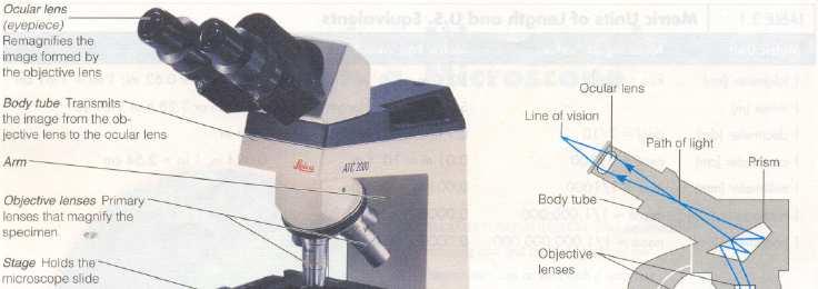
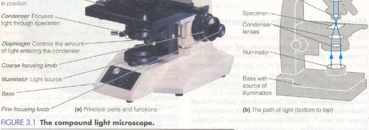
2 Observing Microorganisms through a Microscope LIGHT MICROSCOPY: This type of microscope uses visible light to observe specimens. Compound Light Microscopy: It has a series of lenses, uses visible light as source of illumination Parts of microscope: 1) Illuminator, light source, passes through a 2) Condenser, which has lenses that direct the light rays through the specimen. Light rays pass into the 3) Objective lenses, lenses closer to the specimen. The image of specimen is magnified again by the 4) Ocular lens or eyepiece. Total magnification of specimen: objective lens magnification power (10X, 45X, 100X) multiply by ocular lense magnification power (10). 100X (low power), 450X (high power), 1000X (oil immersion). Resolution: Ability of the lenses to distinguish fine detail and structure between two points a specified distance apart.
3
4 Microscope A general principle of microscopy is that the shorter the wavelength of light used in the instrument, greater the resolution. White light used in this microscope has relatively long wavelength and can not resolve structures smaller than about 0.2µm. To obtain a clear, finely detailed image under this microscope, specimens must be made to contrast sharply with their medium. To attain such contrast, we must change the refractive index of specimens from that of their medium. The refractive index is a measure of the light-bending ability of a medium. We change the refractive index of specimens by staining them. With different refractive indexes, the rays change directions (refract) from a straight path by bending an angle at the boundary between the materials and increase the image s contrast between specimen and medium.
5
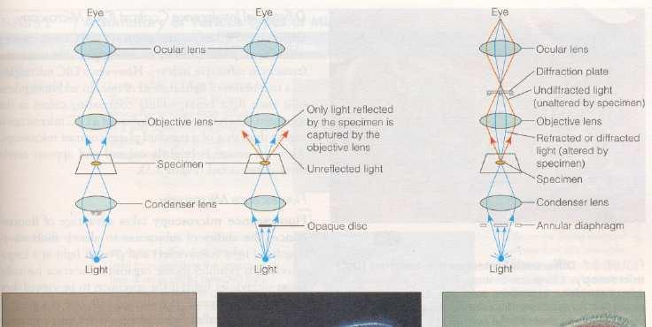
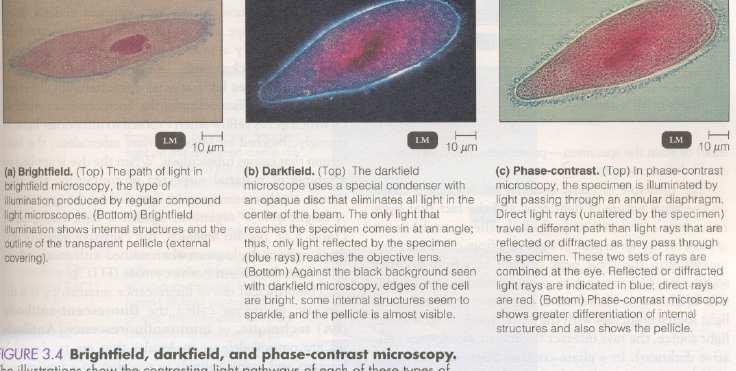
6 Microscope As the light rays travel away from the specimen, they spread out and enter the objective lens, and the image is magnified. To achieve high magnification (100X) with good resolution, the objective lens must be small. Use of oil immersion objective lens: To preserve the direction of light rays at the highest magnification, immersion oil is placed between the glass slide and the oil immersion objective lens. The immersion oil has the same refractive index as glass. In absence of immersion oil, light rays are refracted as they enter the air from slide, and the objective lens would have to be increased in diameter to capture them, & the image becomes fuzzy, with poor resolution. Under usual operating conditions, the field of vision in a compound light microscope is brightly illuminated, & produces brightfield illumination.
7
8 Darkfield Microscopy: Microscope A darkfield microscope is used for examining live microorganisms that either are invisible in the ordinary light microscope, cannot be stained by standard methods, or are so distorted by staining that their characteristics can not be identified. Instead of normal condenser, a darkfield microscope uses a darkfield condenser that contains an opaque disc. The disc blocks light that would enter the objective lens directly. Only light that is reflected off the specimen enters the objective lens. Because there is no direct background light, the specimen appears light against black background the dark field. This technique is used to examine unstained microorganisms suspended in liquid, examination of very thin spirochetes, such as Treponema pallidum (syphilis).
9 Phase-Contrast Microscopy: Microscope It permits detailed examination of internal structures in living microorganisms. Here, it is not necessary to fix or stain the specimen-procedures procedures that could distort or kill the microorganisms. The principle is based on the wave nature of light rays, and the fact that light rays can be in phase (their peaks and valleys match) or out of phase. If the wave peak of light rays from one source coincides with the wave peak of light rays from another source, the rays interact to produce reinforcement (relatively brightness). However, wave peak coincides with wave trough, rays produce interference (relative darkness). One set of light rays comes directly from the light source.
10 Microscope The other set comes from light that is reflected or diffracted from a particular structure in specimen. These two type of light rays form an image of the specimen on the ocular lens, containing area that are relatively light (in phase), through shades of gray, to black (out of phase). Differential Interference Contrast (DIC) Microscopy: Similar to phase-contrast microscopy, uses differences in refractive indexes. Uses two beams of light instead of one. Prisms split each light beam, adding contrasting colors to the specimen. So, resolution of a DIC microscope is higher than that of a standard phase-contrast microscope. The image is brightly colored and appears nearly three-dimensional.
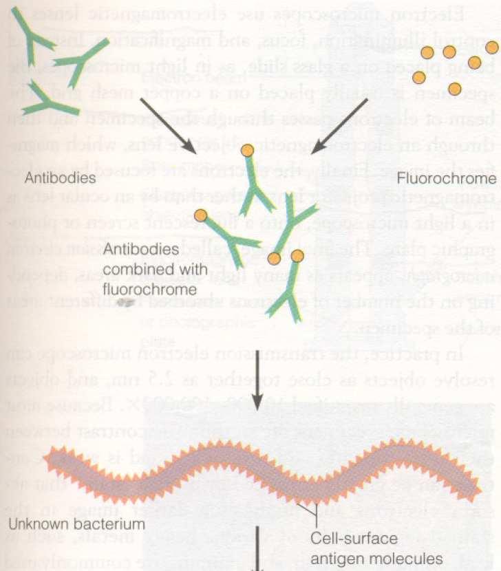
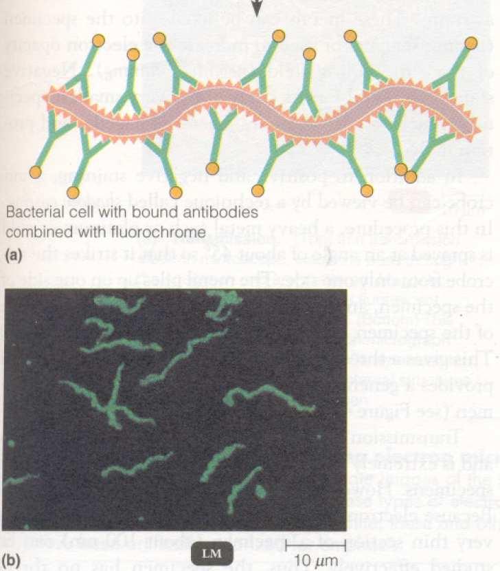
11 Fluorescence Microscopy: Microscope It takes the advantage of fluorescence, the ability of substance to absorb short wavelengths of light (ultraviolet) and give off light at a longer wavelength (visible). Some organisms fluoresce naturally under UV light. If specimen does not naturally fluoresce, it is stained with one of a group of fluorescent dyes called fluorochromes. E.g. Fluorochrome auramine O glows yellow when exposed to UV light, is strongly absorbed by M. Tuberculosis. Fluorescein isothiocyanate gives apple green with B. anthracis. Its principally used as a diagnostic technique called the fluorescent- antibody (FA) technique, or immunofluorescence. This technique can detect bacteria or other pathogenic microorganisms, even within cells, tissues or other clinical specimens. It is especially useful in diagnosing syphilis and rabies.
12
13 Confocal Microscopy: Microscope Here, one plane of a small region of specimen is illuminated with a laser, which passes the returned light through an aperture aligned with the illuminated region. Successive planes and regions are illuminated until the entire specimen has been scanned. Exceptionally clear three-dimensional images of entire cells and cellular components can be obtained, with improved resolution up to 40%, also to evaluate cellular physiology by monitoring the distributions and concentrations of substances such as ATP and calcium ions.
14 Electron Microscopy: Microscope Objects smaller than about 0.2µm can be examined. E.g. viruses. Use beam of free wave electrons as source of light. The resolving power is far greater than that of the other microscopes. Better resolution is due to the shorter wavelengths of electrons. Electromagnetic lenses are used to focus a beam of electrons onto a specimen and to control illumination, and magnification. Two types: 1) Transmission Electron Microscope (TEM), 2) Scanning Electron Microscope (SEM). Transmission Electron Microscope (TEM): Electron gun finely focused beam of electrons electromagnetic condenser lens specimen electromagnetic objective lens (magnifies the image) electromagnetic projector lens onto a fluorescent screen or photographic plate.
15 The specimen is usually placed on a copper Resolves objects close together as 2.5nm Magnified 10, ,000X. copper mesh grid. Microscope Shadow Casting technique: A heavy metal such as platinum or gold is sprayed at an angle of about 45º so that it strikes the microbe from one side, metal piles up on one side of specimen, and the uncoated area on the opposite side of specimen leaves a clear area behind it as a shadow. It provides a general idea of size and shape of specimen and gives three dimensional effect to the specimen. DISADVANTAGE: 1) electrons have limited penetrating power, 2) specimen viewed under a high vacuum to prevent electron scattering. These treatments kill the specimen, and also cause some shrinkage and distortion.
16
17 Microscope Scanning Electron Microscope (SEM): Provides striking 3 dimensional views of specimens. Electron gun finely focused beam of electrons (primary electron beam) electromagnetic condenser lens specimen primary electron beam knocks electrons out of surface and secondary electrons thus produced are transmitted electron collector amplified produce an image on a viewing screen or photographic plate. Useful in studying surface structures of intact cells and viruses. Resolves objects close together as 20 nm Magnified ,000X. Scanned-Probe Microscopy: Use various kinds of probes to examine the surface of specimen at very close range. Used to map atomic and molecular shapes, to characterize magnetic and chemical properties, to determine temperature variations inside cells. Two types: Scanning tunneling microscope, atomic force microscope.
18 Scanning Tunneling Microscopy: Microscope Uses a thin metal (tungsten) probe that scans a specimen and produces an image revealing bumps and depressions of the atoms on surface of specimen. The resolving power is much greater than other electron microscope. Used to provide detailed views of molecules such as DNA. Atomic Force Microscopy: A metal and diamond probe is forced down onto a specimen. As probe moves along the surface of the specimen, its movements are recorded and a three-dimensional image is produced. Doesn t require special specimen preparation. Used to image both biological substances (atomic detail) and molecular processes (a component of blood clot).
19
20
21
Observing Microorganisms through a Microscope
 2016/2/19 PowerPoint Lecture Presentations prepared by Bradley W. Christian, McLennan Community College CHAPTER 3 Observing Microorganisms through a Microscope 1 Figure 3.2 Microscopes and Magnification.
Compound Light Microscopy. Microscopy. Things to remember... 1/22/2017. This is what we use in the laboratory
 Compound Light Microscopy This is what we use in the laboratory Microscopy Chapter 3 BIO 440 A series of finely ground lenses is used to form a magnified image Specimen is illuminated with visible light
Microscopy Techniques that make it easy to see things this small.
 Microscopy Techniques that make it easy to see things this small. What is a Microscope? An instrument for viewing objects that are too small to be seen easily by the naked eye. Dutch spectacle-makers Hans
Burton's Microbiology for the Health Sciences
 Burton's Microbiology for the Health Sciences Chapter 2. Viewing the Microbial World Chapter 2 Outline Introduction Using the metric system to express the sizes of microbes Microscopes Simple microscopes
Chapter 2 The Study of Microbial Structure: Microscopy and Specimen Preparation
 Chapter 2 The Study of Microbial Structure: Microscopy and Specimen Preparation 1 Lenses and the Bending of Light light is refracted (bent) when passing from one medium to another refractive index a measure
Chapter 3. Observing Microorganisms Through a Microscope
 Chapter 3 Observing Microorganisms Through a Microscope Microbial Size Macroscopic organisms can be measured in the range from meters (m) to centimeters (cm) Microscopic organisms fall into the range
Figure 3.4 Approximate size of various types of cells. ~10 um. Red Blood Cells = mm 1500 um. Width of penny Pearson Education, Inc.
 Figure 3.4 Approximate size of various types of cells. ~10 um Red Blood Cells 1.5mm 1500 um Width of penny = 1500 Figure 4.3 The limits of resolution (and some representative objects within those ranges)
Microscopy. Krishna Priya.K Lecturer Dept. of Microbiology
 Microscopy Krishna Priya.K Lecturer Dept. of Microbiology TERMS AND DEFINITIONS Principle Microscopy is to get a magnified image, in which structures may be resolved which could not be resolved with the
2017 MICROSCOPE REVIEW by Karen L. Lancour RELATIVE SIZE OF MICROBES
 2017 MICROSCOPE REVIEW by Karen L. Lancour RELATIVE SIZE OF MICROBES 1000 millimeters (mm) = 1 meter (m) 1000 micrometers (µm or mcm) = 1 millimeter (mm) 1000 nanometers (nm) = 1 micrometer (mcm) Size
2018 MICROSCOPE REVIEW by Karen L. Lancour RELATIVE SIZE OF MICROBES
 2018 MICROSCOPE REVIEW by Karen L. Lancour RELATIVE SIZE OF MICROBES 1000 millimeters (mm) = 1 meter (m) 1000 micrometers (µm or mcm) = 1 millimeter (mm) 1000 nanometers (nm) = 1 micrometer (mcm) Size
S200 Course LECTURE 1 TEM
 S200 Course LECTURE 1 TEM Development of Electron Microscopy 1897 Discovery of the electron (J.J. Thompson) 1924 Particle and wave theory (L. de Broglie) 1926 Electromagnetic Lens (H. Busch) 1932 Construction
Light Microscopy. Upon completion of this lecture, the student should be able to:
 Light Light microscopy is based on the interaction of light and tissue components and can be used to study tissue features. Upon completion of this lecture, the student should be able to: 1- Explain the
 Microscopy http://www.microscopyu.com/articles/phasecontrast/phasemicroscopy.html http://micro.magnet.fsu.edu/primer/anatomy/anatomy.html 2005, Dr. Jack Ikeda & Dr. Gail Grabner 9 Nikon Labophot (Question
VISUAL PHYSICS ONLINE DEPTH STUDY: ELECTRON MICROSCOPES
 VISUAL PHYSICS ONLINE DEPTH STUDY: ELECTRON MICROSCOPES Shortly after the experimental confirmation of the wave properties of the electron, it was suggested that the electron could be used to examine objects
Lecture 4 to 5 MICROSCOPY-PRINCIPLES AND TYPES
 Lecture 4 to 5 MICROSCOPY-PRINCIPLES AND TYPES Microorganisms are too small to be seen by our unaided eyes and the microscopes are of crucial importance as they help to view the microbes. A microscope
Scale. A Microscope s job in life. The Light Microscope. The Compound Microscope 9/24/12. Compound Microscope Anatomy
 The Study of Microbial Structure: Microscopy and Specimen Preparation Scale A Microscope s job in life 1.Magnify 2. Resolve ability to separate or distinguish between two points 3. Contrast How much or
Microscopy. ( greek mikros = small; skopein = to observe)
") Microscopy ( greek mikros = small; skopein = to observe) Zacharias Jansen put several lenses in a tube (first compound microscope) and the object near the end of tube appeared to be greatly enlarged, much
Microscopy ( greek mikros = small; skopein = to observe) Zacharias Jansen put several lenses in a tube (first compound microscope) and the object near the end of tube appeared to be greatly enlarged, much
Unit Two Part II MICROSCOPY
 Unit Two Part II MICROSCOPY AVERETT 1 0 /9/2013 1 MICROSCOPES Microscopes are devices that produce magnified images of structures that are too small to see with the unaided eye Humans cannot see objects
Biology The Microscope. May 20 1:19 PM. Using a Microscope to Explore the Cell
 Biology 2201 1.2 The Microscope Using a Microscope to Explore the Cell Resolution or Resolving power The ability of the eye, or other instrument, to distinguish between two objects that are close together
The light microscope
 What is a microscope? The microscope is an essential tool in modern biology. It allows us to view structural details of organs, tissue, and cells not visible to the naked eye. The microscope should always
The Microscope. Packet #2. 10/17/2016 9:12:02 PM Ryan Barrow 2012
 1 The Microscope Packet #2 10/17/2016 9:12:02 PM Ryan Barrow 2012 2 Historical Timeline 1609 Galileo Galilei develops a compound microscope with a convex and a concave les. 1665 Robert Hooke publishes
Microscopy, Staining, and Classification
 PowerPoint Lecture Presentations prepared by Mindy Miller-Kittrell, North Carolina State University C H A P T E R 4 Microscopy, Staining, and Classification Figure 3.4 Approximate size of various types
MICROSCOPE LAB. Resolving Power How well specimen detail is preserved during the magnifying process.
 AP BIOLOGY Cells ACTIVITY #2 MICROSCOPE LAB OBJECTIVES 1. Demonstrate proper care and use of a compound microscope. 2. Identify the parts of the microscope and describe the function of each part. 3. Compare
MICROSCOPY MICROSCOPE TERMINOLOGY
 1 MICROSCOPY Most of the microorganisms that we talk about in this class are too small to be seen with the naked eye. The instruments we will use to visualize these microbes are microscopes. The laboratory
! Because microbiology deals with organisms too small they cannot be seen distinctly with the unaided eye, the microscope is essential.
 Microscopy! Because microbiology deals with organisms too small they cannot be seen distinctly with the unaided eye, the microscope is essential.! The light microscope is the single most important research
The microscope is useful in making observations and collecting data in scientific experiments. Microscopy involves three basic concepts:
 AP BIOLOGY Chapter 6 NAME DATE Block MICROSCOPE LAB PART I: COMPOUND MICROSCOPE OBJECTIVES: After completing this exercise you should be able to: Demonstrate proper care and use of a compound microscope.
Microbiology Laboratory 2
 Microbiology Laboratory 2 Microscopy Background Microorganisms are too small to be seen with the naked eye. Thus a microscope is used to magnify objects so they can be observed. A lens consists of one
Biology 29 Cell Structure and Function Spring, 2009 Springer LABORATORY 1: THE LIGHT MICROSCOPE
 Biology 29 Cell Structure and Function Spring, 2009 Springer LABORATORY 1: THE LIGHT MICROSCOPE Prior to lab: 1) Read these instructions (p 1-6) 2) Go through the online tutorial, the microscopy pre-lab
STRUCTURE OF THE MICROSCOPE
 STRUCTURE OF THE MICROSCOPE Use the word list to label the microscope below: Light Source Coarse adjustment knob Diaphragm Stage Clips Objectives Fine Adjustment Knob Base Stage Stage Clips Arm Revolving
Test Review # 8. Physics R: Form TR8.17A. Primary colors of light
 Physics R: Form TR8.17A TEST 8 REVIEW Name Date Period Test Review # 8 Light and Color. Color comes from light, an electromagnetic wave that travels in straight lines in all directions from a light source
The Care and Use of the Microscope. Lab Exercise #4
 Lab Safety No eating or drinking!!! Long hair must be tied back Clean up your workstation before you leave! Return all materials to the storage sites Clean glassware and wipe down countertops Follow directions
Ex 1: Introduction to the microscope
 Ex 1: Introduction to the microscope So what exactly is a microorganism? Microorganisms = any living thing that is too small to be seen with the unaided eye fungus protist bacteria virus Parasitic worm
Light microscopy BMB 173, Lecture 14, Feb. 21, 2018
 Light microscopy The Structural Biology Continuum Next two lectures: Light microscopy Many slides taken from Scott Fraser, Murphy s Fundamentals of light microscopy, Alberts Molecular Biology of the Cell,
Basics of Light Microscopy and Metallography
 ENGR45: Introduction to Materials Spring 2012 Laboratory 8 Basics of Light Microscopy and Metallography In this exercise you will: gain familiarity with the proper use of a research-grade light microscope
Applications of Optics
 Nicholas J. Giordano www.cengage.com/physics/giordano Chapter 26 Applications of Optics Marilyn Akins, PhD Broome Community College Applications of Optics Many devices are based on the principles of optics
LlIGHT REVIEW PART 2 DOWNLOAD, PRINT and submit for 100 points
 WRITE ON SCANTRON WITH NUMBER 2 PENCIL DO NOT WRITE ON THIS TEST LlIGHT REVIEW PART 2 DOWNLOAD, PRINT and submit for 100 points Multiple Choice Identify the choice that best completes the statement or
Light and Applications of Optics
 UNIT 4 Light and Applications of Optics Topic 4.1: What is light and how is it produced? Topic 4.6: What are lenses and what are some of their applications? Topic 4.2 : How does light interact with objects
Microscopy Training & Overview
 Microscopy Training & Overview Product Marketing October 2011 Stephan Briggs - PLE OVERVIEW AND PRESENTATION FLOW Glossary and Important Terms Introduction Timeline Innovation and Advancement Primary Components
Life Science Chapter 2 Study Guide
 Key concepts and definitions Waves and the Electromagnetic Spectrum Wave Energy Medium Mechanical waves Amplitude Wavelength Frequency Speed Properties of Waves (pages 40-41) Trough Crest Hertz Electromagnetic
Microscope (and The Cell) Lab Exercise #1
 Lab Exercise #1") Lab Safety-General No eating or drinking Only registered students allowed in the class Long hair must be tied back Familiarize yourself with the emergency stations Do not mark on the models Inform me of
Lab Safety-General No eating or drinking Only registered students allowed in the class Long hair must be tied back Familiarize yourself with the emergency stations Do not mark on the models Inform me of
Test Review # 9. Physics R: Form TR9.15A. Primary colors of light
 Physics R: Form TR9.15A TEST 9 REVIEW Name Date Period Test Review # 9 Light and Color. Color comes from light, an electromagnetic wave that travels in straight lines in all directions from a light source
Resolution. Diffraction from apertures limits resolution. Rayleigh criterion θ Rayleigh = 1.22 λ/d 1 peak at 2 nd minimum. θ f D
 Microscopy Outline 1. Resolution and Simple Optical Microscope 2. Contrast enhancement: Dark field, Fluorescence (Chelsea & Peter), Phase Contrast, DIC 3. Newer Methods: Scanning Tunneling microscopy (STM),
Figure 1. Relative intensity of solar energy of different wavelength at the earth's surface.
 Spectrum of light from the sun: Fig.1 Figure 1. Relative intensity of solar energy of different wavelength at the earth's surface. Properties of light 1-The speed of light changes when it goes from one
THE COMPOUND BRIGHTFIELD MICROSCOPE
 THE COMPOUND BRIGHTFIELD MICROSCOPE Microbiology is the study of microscopic organisms that are so small that they are below the limit of vision of the human eye. Bacteria are the smallest of microorganisms
Microscopy. Matti Hotokka Department of Physical Chemistry Åbo Akademi University
 Microscopy Matti Hotokka Department of Physical Chemistry Åbo Akademi University What s coming Anatomy of a microscope Modes of illumination Practicalities Special applications Basic microscope Ocular
Exercise 2-A MICROSCOPIC TECHNIQUE & EXAMINATION OF MICROORGANISMS
 Exercise 2-A MICROSCOPIC TECHNIQUE & EXAMINATION OF MICROORGANISMS Introduction to Microscopic Technique Microbiology is the science or study of living organisms too small to be seen with the naked eye.
Microscopy and Staining
 Microscopy and Staining Figure 2.1 Different types of microscopy are used to visualize different structures. Brightfield microscopy (left) renders a darker image on a lighter background, producing a clear
A BRIEF INTRODUCTION TO MICROSCOPY The two key properties of a microscope that allow you to see microbes are resolution and magnification.
 A BRIEF INTRODUCTION TO MICROSCOPY The two key properties of a microscope that allow you to see microbes are resolution and magnification. Magnification refers to the enlargement of the specimen when seen
Microscopy: Fundamental Principles and Practical Approaches
 Microscopy: Fundamental Principles and Practical Approaches Simon Atkinson Online Resource: http://micro.magnet.fsu.edu/primer/index.html Book: Murphy, D.B. Fundamentals of Light Microscopy and Electronic
Chapter 23 Study Questions Name: Class:
 Chapter 23 Study Questions Name: Class: Multiple Choice Identify the letter of the choice that best completes the statement or answers the question. 1. When you look at yourself in a plane mirror, you
LAB 1 Introduction to Microscopy
 I. Ubiquity of Microorganisms II. Microscopy LAB 1 Introduction to Microscopy I. UBIQUITY OF MICROORGANISMS Microorganisms are ubiquitous; that is, they are present nearly everywhere. In this lab you will
Exercise 2-A MICROSCOPIC TECHNIQUE & EXAMINATION OF MICROORGANISMS
 Exercise 2-A MICROSCOPIC TECHNIQUE & EXAMINATION OF MICROORGANISMS Introduction to Microscopic Technique Microbiology is the science or study of living organisms too small to be seen with the naked eye.
The Nature of Light. Light and Energy
 The Nature of Light Light and Energy - dependent on energy from the sun, directly and indirectly - solar energy intimately associated with existence of life -light absorption: dissipate as heat emitted
Imaging Introduction. September 24, 2010
 Imaging Introduction September 24, 2010 What is a microscope? Merriam-Webster: an optical instrument consisting of a lens or combination of lenses for making enlarged images of minute objects; especially:
Tissue Preparation ORGANISM IMAGE TISSUE PREPARATION. 1) Fixation: halts cell metabolism, preserves cell/tissue structure
 Fixation: halts cell metabolism, preserves cell/tissue structure") Lab starts this week! ANNOUNCEMENTS - Tuesday or Wednesday 1:25 ISB 264 - Read Lab 1: Microscopy and Imaging (see Web Page) - Getting started on Lab Group project - Organ for investigation - Lab project
Lab starts this week! ANNOUNCEMENTS - Tuesday or Wednesday 1:25 ISB 264 - Read Lab 1: Microscopy and Imaging (see Web Page) - Getting started on Lab Group project - Organ for investigation - Lab project
Very short introduction to light microscopy and digital imaging
 Very short introduction to light microscopy and digital imaging Hernan G. Garcia August 1, 2005 1 Light Microscopy Basics In this section we will briefly describe the basic principles of operation and
Lab 1, 2 and 3: Stain, Observe and Identify the Microbes. BIOHAZARD Rules. VIOLATORS will lose points. A) Lab Safety Rules Lab Safety Form Signup
 Lab Safety Rules Lab Safety Form Signup") MICROLAB PREPARATIONS A) Lab Safety Rules Lab Safety Form Signup B) Lab Participation Instructor Review Peer Review Form C) Biohazard Rules How to dispose Trash REQUIRED Items: 1) LAB Manual/Journal 2)
MICROLAB PREPARATIONS A) Lab Safety Rules Lab Safety Form Signup B) Lab Participation Instructor Review Peer Review Form C) Biohazard Rules How to dispose Trash REQUIRED Items: 1) LAB Manual/Journal 2)
Match the microscope structures given in the left column with the statements in the right column that identify or describe them.
 49 Prelab for Name Match the microscope structures given in the left column with the statements in the right column that identify or describe them. Key: a. coarse adjustment knob f. turret or nosepiece
SUBJECT: PHYSICS. Use and Succeed.
 SUBJECT: PHYSICS I hope this collection of questions will help to test your preparation level and useful to recall the concepts in different areas of all the chapters. Use and Succeed. Navaneethakrishnan.V
Laboratory Introduction
 Laboratory Introduction There are two basic categories of microscopes: light microscopes and electron microscopes. Light, or optical, microscopes require light waves to provide the illumination while electron
Conceptual Physics Fundamentals
 Conceptual Physics Fundamentals Chapter 13: LIGHT WAVES This lecture will help you understand: Electromagnetic Spectrum Transparent and Opaque Materials Color Why the Sky is Blue, Sunsets are Red, and
Introduction: Why electrons?
 Introduction: Why electrons? 1 Radiations Visible light X-rays Electrons Neutrons Advantages Not very damaging Easily focused Eye wonderful detector Small wavelength (Angstroms) Good penetration Small
OPTICAL SYSTEMS OBJECTIVES
 101 L7 OPTICAL SYSTEMS OBJECTIVES Aims Your aim here should be to acquire a working knowledge of the basic components of optical systems and understand their purpose, function and limitations in terms
Chapter 29: Light Waves
 Lecture Outline Chapter 29: Light Waves This lecture will help you understand: Huygens' Principle Diffraction Superposition and Interference Polarization Holography Huygens' Principle Throw a rock in a
Chapter 16 Light Waves and Color
 Chapter 16 Light Waves and Color Lecture PowerPoint Copyright The McGraw-Hill Companies, Inc. Permission required for reproduction or display. What causes color? What causes reflection? What causes color?
Refraction, Lenses, and Prisms
 CHAPTER 16 14 SECTION Sound and Light Refraction, Lenses, and Prisms KEY IDEAS As you read this section, keep these questions in mind: What happens to light when it passes from one medium to another? How
ELECTRON MICROSCOPY AN OVERVIEW
 ELECTRON MICROSCOPY AN OVERVIEW Anjali Priya 1, Abhishek Singh 2, Nikhil Anand Srivastava 3 1,2,3 Department of Electrical & Instrumentation, Sant Longowal Institute of Engg. & Technology, Sangrur, India.
Bio 407. Applied microscopy. Introduction into light microscopy. José María Mateos. Center for Microscopy and Image Analysis
 Center for Microscopy and Image Analysis Bio 407 Applied Introduction into light José María Mateos Fundamentals of light Compound microscope Microscope composed of an objective and an additional lens (eyepiece,
CHAPTER TWO METALLOGRAPHY & MICROSCOPY
 CHAPTER TWO METALLOGRAPHY & MICROSCOPY 1. INTRODUCTION: Materials characterisation has two main aspects: Accurately measuring the physical, mechanical and chemical properties of materials Accurately measuring
King Saud University Dept. of Bot. & Microbiology. General Microbiology 140 MIC
 King Saud University Dept. of Bot. & Microbiology General Microbiology 140 MIC Lab coat. Do not wearing the lab coat outside the lab. Gloves. Proper Clothing and closed shoes. Hair should be tied back.
ANSWER KEY Lab 2 (IGB): Bright Field and Fluorescence Optical Microscopy and Sectioning
: Bright Field and Fluorescence Optical Microscopy and Sectioning") Phys598BP Spring 2016 University of Illinois at Urbana-Champaign ANSWER KEY Lab 2 (IGB): Bright Field and Fluorescence Optical Microscopy and Sectioning Location: IGB Core Microscopy Facility Microscope:
Phys598BP Spring 2016 University of Illinois at Urbana-Champaign ANSWER KEY Lab 2 (IGB): Bright Field and Fluorescence Optical Microscopy and Sectioning Location: IGB Core Microscopy Facility Microscope:
Using Microscopes. Life Science: Molecular
 Using Microscopes Life Science: Molecular Light Microscopy: Instrumentation and Principles A light microscope is so named because it uses visible light to produce a magnified image. Compound light microscopes
microscopy A great online resource Molecular Expressions, a Microscope Primer Partha Roy
 Fundamentals of optical microscopy A great online resource Molecular Expressions, a Microscope Primer http://micro.magnet.fsu.edu/primer/index.html Partha Roy 1 Why microscopy Topics Functions of a microscope
Reflection! Reflection and Virtual Image!
 1/30/14 Reflection - wave hits non-absorptive surface surface of a smooth water pool - incident vs. reflected wave law of reflection - concept for all electromagnetic waves - wave theory: reflected back
NANO 703-Notes. Chapter 9-The Instrument
 1 Chapter 9-The Instrument Illumination (condenser) system Before (above) the sample, the purpose of electron lenses is to form the beam/probe that will illuminate the sample. Our electron source is macroscopic
FLUORESCENCE MICROSCOPY. Matyas Molnar and Dirk Pacholsky
 FLUORESCENCE MICROSCOPY Matyas Molnar and Dirk Pacholsky 1 The human eye perceives app. 400-700 nm; best at around 500 nm (green) Has a general resolution down to150-300 μm (human hair: 40-250 μm) We need
Chapter 17: Wave Optics. What is Light? The Models of Light 1/11/13
 Chapter 17: Wave Optics Key Terms Wave model Ray model Diffraction Refraction Fringe spacing Diffraction grating Thin-film interference What is Light? Light is the chameleon of the physical world. Under
Visual Anatomy ansd Physiology Lab Manual Pig Version 2nd Edition Sarikas TEST BANK
 Visual Anatomy ansd Physiology Lab Manual Pig Version 2nd Edition Sarikas TEST BANK https://testbankreal.com/download/visual-anatomy-ansd-physiology-labmanual-pig-version-2nd-edition-sarikas-test-bank/
Microscope Notes. units of life.
 Microscope Notes Microscope an instrument that produces an enlarged image of an object. Biologists use microscopes to study cells, cell parts, and organisms that are too small to be seen with the naked
Chapter 36: diffraction
 Chapter 36: diffraction Fresnel and Fraunhofer diffraction Diffraction from a single slit Intensity in the single slit pattern Multiple slits The Diffraction grating X-ray diffraction Circular apertures
Vocabulary. Unit 9 Forms of Energy. ENERGY: The capacity for doing work.
 Unit 9 Forms of Energy Main Idea: There are many forms of energy, including radiant energy and chemical energy. Energy can change form. ENERGY: The capacity for doing work. Heat, Light and Radiant Energy
Uses of Electromagnetic Waves
 Uses of Electromagnetic Waves 1 of 42 Boardworks Ltd 2016 Uses of Electromagnetic Waves 2 of 42 Boardworks Ltd 2016 What are radio waves? 3 of 42 Boardworks Ltd 2016 The broadcast of every radio and television
INTRODUCTION TO MICROSCOPY. Urs Ziegler THE PROBLEM
 INTRODUCTION TO MICROSCOPY Urs Ziegler ziegler@zmb.uzh.ch THE PROBLEM 1 ORGANISMS ARE LARGE LIGHT AND ELECTRONS: ELECTROMAGNETIC WAVES v = Wavelength ( ) Speed (v) Frequency ( ) Amplitude (A) Propagation
Low Voltage Electron Microscope
 LVEM5 Low Voltage Electron Microscope Nanoscale from your benchtop LVEM5 Delong America DELONG INSTRUMENTS COMPACT BUT POWERFUL The LVEM5 is designed to excel across a broad range of applications in material
Introduction. Laboratory Equipment & Supplies. Model 1333PHi Shown (Phase Contrast) (2) Eyepieces (Eyecups installed) Diopter Adjustment Mechanism
 (2) Eyepieces (Eyecups installed) Diopter Adjustment Mechanism") Introduction With the invention of the microscope in the early 17th century, it was made possible to view objects which were too small for the human eye to see. As the microscope evolved, the structure
Introduction With the invention of the microscope in the early 17th century, it was made possible to view objects which were too small for the human eye to see. As the microscope evolved, the structure
Invitation for a walk through microscopy. Sebastian Schuchmann Jörg Rösner
 Invitation for a walk through microscopy Sebastian Schuchmann Jörg Rösner joerg.roesner@charite.de Techniques in microscopy Conventional (light) microscopy bright & dark field, phase & interference contrast
Microscope. Dr. Leena Barhate Department of Microbiology M.J.College, Jalgaon
 Microscope Dr. Leena Barhate Department of Microbiology M.J.College, Jalgaon Acknowledgement http://www.cerebromente.org.br/n17/histor y/neurons1_i.htm Google Images http://science.howstuffworks.com/lightmicroscope1.htm
2/4/15. Brightfield Microscopy! It s all about Magnification..! or is it?!
 Brightfield Microscopy It s all about Magnification.. or is it? 1 What actually does go into chosing a microscope Choice depends on what you need the microscope to do. Do you want to magnify stained specimens?
INTRODUCTION THIN LENSES. Introduction. given by the paraxial refraction equation derived last lecture: Thin lenses (19.1) = 1. Double-lens systems
 = 1. Double-lens systems") Chapter 9 OPTICAL INSTRUMENTS Introduction Thin lenses Double-lens systems Aberrations Camera Human eye Compound microscope Summary INTRODUCTION Knowledge of geometrical optics, diffraction and interference,
Chapter 9 OPTICAL INSTRUMENTS Introduction Thin lenses Double-lens systems Aberrations Camera Human eye Compound microscope Summary INTRODUCTION Knowledge of geometrical optics, diffraction and interference,
Introduction to Light Microscopy. (Image: T. Wittman, Scripps)
") Introduction to Light Microscopy (Image: T. Wittman, Scripps) The Light Microscope Four centuries of history Vibrant current development One of the most widely used research tools A. Khodjakov et al. Major
Introduction to Light Microscopy (Image: T. Wittman, Scripps) The Light Microscope Four centuries of history Vibrant current development One of the most widely used research tools A. Khodjakov et al. Major
Name. Light Chapter Summary Cont d. Refraction
 Page 1 of 17 Physics Week 12(Sem. 2) Name Light Chapter Summary Cont d with a smaller index of refraction to a material with a larger index of refraction, the light refracts towards the normal line. Also,
MODULE I SCANNING ELECTRON MICROSCOPE (SEM)
") MODULE I SCANNING ELECTRON MICROSCOPE (SEM) Scanning Electron Microscope (SEM) Initially, the plan of SEM was offered by H. Stintzing in 1927 (a German patent application). His suggested procedure was
MODULE I SCANNING ELECTRON MICROSCOPE (SEM) Scanning Electron Microscope (SEM) Initially, the plan of SEM was offered by H. Stintzing in 1927 (a German patent application). His suggested procedure was
Optical and digital microscopic imaging techniques and applications in pathology
 Analytical Cellular Pathology 34 (2011) 5 18 DOI 10.3233/ACP-2011-0006 IOS Press Optical and digital microscopic imaging techniques and applications in pathology 5 Xiaodong Chen a,c, Bin Zheng b and Hong
Longitudinal No, Mechanical wave ~340 m/s (in air) 1,100 feet per second More elastic/denser medium = Greater speed of sound
 1,100 feet per second More elastic/denser medium = Greater speed of sound") Type of wave Travel in Vacuum? Speed Speed vs. Medium Light Sound vs. Sound Longitudinal No, Mechanical wave ~340 m/s (in air) 1,100 feet per second More elastic/denser medium = Greater speed of sound
Type of wave Travel in Vacuum? Speed Speed vs. Medium Light Sound vs. Sound Longitudinal No, Mechanical wave ~340 m/s (in air) 1,100 feet per second More elastic/denser medium = Greater speed of sound
PHYSICS. Chapter 35 Lecture FOR SCIENTISTS AND ENGINEERS A STRATEGIC APPROACH 4/E RANDALL D. KNIGHT
 PHYSICS FOR SCIENTISTS AND ENGINEERS A STRATEGIC APPROACH 4/E Chapter 35 Lecture RANDALL D. KNIGHT Chapter 35 Optical Instruments IN THIS CHAPTER, you will learn about some common optical instruments and
Topic 1 - What is Light? 1. Radiation is the type of energy transfer which does not require... A matter B heat C waves D light
 Grade 8 Unit 1 Test Student Class Topic 1 - What is Light? 1. Radiation is the type of energy transfer which does not require... A matter B heat C waves D light 2. Light-producing technologies, such as
Waves. A wave is a disturbance which travels through a vacuum or medium (air, water, etc) that contains matter A wave transports ENERGY not matter
 that contains matter A wave transports ENERGY not matter") Waves and Optics Waves A wave is a disturbance which travels through a vacuum or medium (air, water, etc) that contains matter A wave transports ENERGY not matter Waves Some waves do not need a medium
Waves and Optics Waves A wave is a disturbance which travels through a vacuum or medium (air, water, etc) that contains matter A wave transports ENERGY not matter Waves Some waves do not need a medium
Term Info Picture. A wave that has both electric and magnetic fields. They travel through empty space (a vacuum).
.") Waves S8P4. Obtain, evaluate, and communicate information to support the claim that electromagnetic (light) waves behave differently than mechanical (sound) waves. A. Ask questions to develop explanations
Waves S8P4. Obtain, evaluate, and communicate information to support the claim that electromagnetic (light) waves behave differently than mechanical (sound) waves. A. Ask questions to develop explanations
Physics Test Review Reflection/Refraction/Diffraction & Lenses Session: Name:
 Multiple Choice 1. The law of reflection says that a. the angle of reflection from a mirror equals the angle of incidence. b. waves incident on a mirror are partially reflected. c. all waves incident on
Microscopic Structures
 Microscopic Structures Image Analysis Metal, 3D Image (Red-Green) The microscopic methods range from dark field / bright field microscopy through polarisation- and inverse microscopy to techniques like