Microscopy Training & Overview
|
|
|
- Barbra Waters
- 5 years ago
- Views:
Transcription

1 Microscopy Training & Overview Product Marketing October 2011 Stephan Briggs - PLE
2 OVERVIEW AND PRESENTATION FLOW Glossary and Important Terms Introduction Timeline Innovation and Advancement Primary Components Edmund Optics Product Offering Illumination Techniques Microscopy Techniques Brightfield Darkfield Phase Contrast Microscope Objectives Finite Conjugate Infinity Corrected 2

3 GLOSSARY AND IMPORTANT TERMS 3 Numerical Aperture (NA) Function of the focal length and entrance pupil diameter. NA determines resolving power, depth of field, and contrast of the image. The higher the NA, the greater the resolving power and smaller the depth of field. NA can be calculated using Equation 1. NA = n*sinθ Equation 1 Where n is the index of refraction of the medium in which the lens is working (n=1.0 for air) and θ is the half-angle of the maximum cone of light that can enter or exit the lens. Resolving Power (R) Minimum distance between points or lines that are just distinguishable as separate entities. The resolving power of a system is determined by N.A. and wavelength of light (λ), as shown in Equation 2. Working Distance (WD) R = 0.61*λ /N.A. Equation 2 Distance between the surface of the specimen and the front face of the objective when in focus


4 GLOSSARY AND IMPORTANT TERMS 4 Parfocal Length Distance between the surface of the specimen and the objective mounting position when in focus. Infinity Corrected Optical System An optical system in which the image is formed by an objective and a tube lens with an Infinity Space between them, into which optical accessories can be inserted. Finite Conjugate Optical System An optical system in which the image is formed only by an objective.
5 GLOSSARY AND IMPORTANT TERMS 5 Focal length (F) Distance between a principal point and a focal point. F1 is the focal length of an objective. F2 is the focal length of a tube lens. For infinity-corrected systems, magnification is determined by the ratio of the focal length of the tube lens to that of the objective. Magnification of Objective = Focal length of tube lens / Focal length of objective Field Number and Field of View (FOV) The field number of an eyepiece (expressed in mm) is determined by the field stop diameter of the eyepiece. FOV is the area of the specimen that is observable, and is determined by the field number of the eyepiece and magnification of the objective. Depth of Field (DOF) FOV = Field number of eyepiece / magnification of objective Vertical distance in the specimen, measured from above and below the exact plane of focus, which still yields an acceptable image. The larger the NA, the smaller the depth of field. ± DOF = λ / (2*(NA) 2 ) - standard wavelength of 550nm Aperture Diaphragm Adjusts the amount of light passing through, and is related to the brightness and resolving power of an optical system. This diaphragm is especially useful in width dimension measurement of cylindrical objects with contour illumination, and provides the highest degree of correct measurement/observation by suppressing diffraction in an optimal aperture.
6 GLOSSARY AND IMPORTANT TERMS 6 Field Stop Used for blocking out unwanted light and preventing it from degrading the image Vignetting This unwanted effect is the reduction of an image s brightness or saturation at the periphery compared to the image center. May be caused by external (lens hood) or internal features (dimensions of a multielement objective). Double Image An image degrading phenomenon in which an image appears as if it is a double image due to redundant light projection and optical interference within the optical system. Flare Lens flare is typically seen as several starbursts, rings, or circles in a row across the image or view, and is caused by unwanted image formation mechanisms, such as internal reflection and scattering of light.
7 7 INNOVATION AND ADVANCEMENT IN OPTICAL MICROSCOPY Approximate microscopy conception date by lens grinders in Middleburg, Netherlands Term microscope is coined from Galileo s design First living tissue is examined: insect eyes Extensive research begins in Italy, Holland, and England Robert Hooke s Micrographia is microscopy s first big release Antoni van Leeuwenhoek discovers micro-organisms and analyzes red-blood cells and bacteria Joseph Jackson Lister combines weak lenses, forming doublets and triplets to eliminate spherical aberration Ernst Abbe formulates the relationship between wavelength and resolution August Kohler develops innovative illumination technique that is thought to be central to modern day optical microscopy Fritz Zernike and George Nomarski advance illumination with phase contrast and Differential Interference Contrast (DIC)
8 8 PRIMARY COMPONENTS OF A MICROSCOPE Eyepiece(s) Camera Port Objective Lenses Turret Condenser Diaphragm Light source Stage Focus XY Adjustment


9 EDMUND OPTICS PRODUCT OFFERING 9 Infinity Corrected Objectives Finite Conjugate Objectives Reflective Objectives Stereo Microscopes Miscellaneous Accessories, eyepieces, relay lenses, couplers, reticles, micrometers, pocket and direct microscopes, simple magnifiers

10 MICROSCOPE ILLUMINATION 10 Afocal/Nonfocused Illumination Systems that do not form an image of the light source at a specific point in the optical pathway. Early microscopes utilized natural sunlight and cloud cover for crude diffusion. Critical/Nelsonian Illumination Relies on substage condenser to produce a focused image from a homogeneous light source along the specimen plane to achieve even illumination over the entire field of view Current Techniques Incandescent Lamps Arc Lamps LEDs Lasers
11 KOEHLER ILLUMINATION 11 Koehler illumination overcomes the disadvantages of other illumination setups by causing parallel rays to light the specimen so that, because they will not be in focus, the image of the specimen will not include an image of the light source.
12 TELECENTRIC ILLUMINATION 12 Telecentric illumination is designed so that principal light passes through the focal point. This system has the advantage of retaining the size of the image center even if it is out of focus (although the circumference of the image is defocused). This illumination system provides an even illumination intensity over the entire field of view, and is typically used for lower magnifications: 2x-5x.
13 MICROSCOPY TECHNIQUES 13 Brightfield Darkfield Phase Contrast Polarization Contrast Differential Interference Contrast (DIC) Fluorescence Interference Reflection (TIRF)

14 BRIGHTFIELD - DARKFIELD 14 Brightfield Illumination Sample contrast comes from absorbance of light in the sample One of the simplest techniques in light microscopy Typical appearance is a dark sample on a bright background Darkfield Illumination Sample contrast comes from light scattered by the sample Technique used to enhance contrast in unstained samples Typical appearance is a dark/black background with bright objects up front
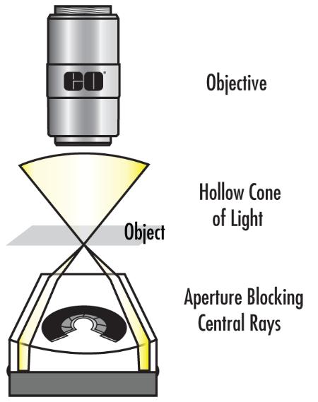
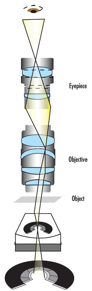
15 BRIGHTFIELD AND DARKFIELD SETUP 15 Brightfield Darkfield

16 PHASE CONTRAST 16 Displays proportional differences in optical density Shows differences in refractive index as contrast difference Nucleus appears darker than surrounding ECM Typically a grey background with light and dark features - these features represent change in optical density

17 POLARIZATION CONTRAST 17 Any optical microscope involving polarized light Complimentary to DIC and TIRF Identifies varying refractive indices much like brightfield Can distinguish between isotropic and anisotropic substances Must be equipped with a polarizer in the light path and an analyzer in the image path Image contrast arises from interaction with a birefringent specimen
18 DIFFERENTIAL INTERFERENCE CONTRAST (DIC) 18 Enhances the contrast in unstained, transparent samples. This technique works on the principle of interferometry to access the optical path length of the sample and enhances what were once invisible features. Similar to phase contrast without the illuminated diffraction halo. Fully utilizes NA of the system No confusing halos as seen in Phase images Images appear vibrant in color due to optical staining 3D shadowed like appearance Low cost plan achromats or achromats can be utilized Very high resolution system Can focus on a thin section of thicker specimen while avoiding noise around the image plane Micrasterias furcata - Transmitted DIC Microscopy

19 DIC OPTICAL PATHWAY 19


20 FLUORESCENCE 20 Photoluminescence when specimens absorb and subsequently re-radiate light Phosphorescence when the emission of light persists for up to a few seconds after excitation has ceased Fluorescence describes light emission that continues only during the absorption of excitation light Head lice Round Worm Specimen emits a longer wavelength than the light used to excite for fluorescence. Utilizes three filters for best image: 1. Excitation 2. Emission 3. Dichroic

21 TOTAL INTERFERENCE REFLECTION FLUORESCENCE (TIRF) 21 TIRF is an extremely elegant optical microscopy technique primarily used to observe single molecule fluorescence at adhesion points on a surface. This interaction between molecules and a surface is an area of importance for multiple disciplines such as cellular and molecular biology. Employs the properties of an induced evanescent wave to illuminate/excite fluorophores Molecules/fluorophores are immediately adjacent to the glasswater interface in most cases Simple concept requiring only an excitation light beam at high incident angle to sample Refractive index difference between glass and water regulate light refraction/reflection The evanescent wave/field intensity exponentially decays as distance from the surface increases

22 TIRF OPTICAL PATHWAY 22
Imaging Introduction. September 24, 2010
 Imaging Introduction September 24, 2010 What is a microscope? Merriam-Webster: an optical instrument consisting of a lens or combination of lenses for making enlarged images of minute objects; especially:
Microscopy: Fundamental Principles and Practical Approaches
 Microscopy: Fundamental Principles and Practical Approaches Simon Atkinson Online Resource: http://micro.magnet.fsu.edu/primer/index.html Book: Murphy, D.B. Fundamentals of Light Microscopy and Electronic
The Compound Microscope. Brightfield: Köhler Illumination
 Outline History of Microscopy The Magnifying Glass The Compound Microscope Brightfield: Köhler Illumination Microscopy µικροσ (mikros): small σκοπειν (skopein): to observe History of Microscopy Well :
microscopy A great online resource Molecular Expressions, a Microscope Primer Partha Roy
 Fundamentals of optical microscopy A great online resource Molecular Expressions, a Microscope Primer http://micro.magnet.fsu.edu/primer/index.html Partha Roy 1 Why microscopy Topics Functions of a microscope
Microscopy Techniques that make it easy to see things this small.
 Microscopy Techniques that make it easy to see things this small. What is a Microscope? An instrument for viewing objects that are too small to be seen easily by the naked eye. Dutch spectacle-makers Hans
Lecture 23 MNS 102: Techniques for Materials and Nano Sciences
 Lecture 23 MNS 102: Techniques for Materials and Nano Sciences Reference: #1 C. R. Brundle, C. A. Evans, S. Wilson, "Encyclopedia of Materials Characterization", Butterworth-Heinemann, Toronto (1992),
 Microscopy http://www.microscopyu.com/articles/phasecontrast/phasemicroscopy.html http://micro.magnet.fsu.edu/primer/anatomy/anatomy.html 2005, Dr. Jack Ikeda & Dr. Gail Grabner 9 Nikon Labophot (Question
Systems Biology. Optical Train, Köhler Illumination
 McGill University Life Sciences Complex Imaging Facility Systems Biology Microscopy Workshop Tuesday December 7 th, 2010 Simple Lenses, Transmitted Light Optical Train, Köhler Illumination What Does a
BASICS IN BIOIMAGING AND OPTICS PLATFORM EPFL SV PTBIOP LIGHT MICROSCOPY
 BASICS IN LIGHT MICROSCOPY OVERVIEW 1. Motivation 2. Basic in optics 3. How microscope works 4. Illumination and resolution 5. Microscope optics 6. Contrasting methods -2- MOTIVATION Why do we need microscopy?
VISUAL PHYSICS ONLINE DEPTH STUDY: ELECTRON MICROSCOPES
 VISUAL PHYSICS ONLINE DEPTH STUDY: ELECTRON MICROSCOPES Shortly after the experimental confirmation of the wave properties of the electron, it was suggested that the electron could be used to examine objects
FLUORESCENCE MICROSCOPY. Matyas Molnar and Dirk Pacholsky
 FLUORESCENCE MICROSCOPY Matyas Molnar and Dirk Pacholsky 1 The human eye perceives app. 400-700 nm; best at around 500 nm (green) Has a general resolution down to150-300 μm (human hair: 40-250 μm) We need
Observing Microorganisms through a Microscope LIGHT MICROSCOPY: This type of microscope uses visible light to observe specimens. Compound Light Micros
 PHARMACEUTICAL MICROBIOLOGY JIGAR SHAH INSTITUTE OF PHARMACY NIRMA UNIVERSITY Observing Microorganisms through a Microscope LIGHT MICROSCOPY: This type of microscope uses visible light to observe specimens.
Introduction. Laboratory Equipment & Supplies. Model 1333PHi Shown (Phase Contrast) (2) Eyepieces (Eyecups installed) Diopter Adjustment Mechanism
 (2) Eyepieces (Eyecups installed) Diopter Adjustment Mechanism") Introduction With the invention of the microscope in the early 17th century, it was made possible to view objects which were too small for the human eye to see. As the microscope evolved, the structure
Introduction With the invention of the microscope in the early 17th century, it was made possible to view objects which were too small for the human eye to see. As the microscope evolved, the structure
Optical Design of. Microscopes. George H. Seward. Tutorial Texts in Optical Engineering Volume TT88. SPIE PRESS Bellingham, Washington USA
 Optical Design of Microscopes George H. Seward Tutorial Texts in Optical Engineering Volume TT88 SPIE PRESS Bellingham, Washington USA Preface xiii Chapter 1 Optical Design Concepts /1 1.1 A Value Proposition
Biology 29 Cell Structure and Function Spring, 2009 Springer LABORATORY 1: THE LIGHT MICROSCOPE
 Biology 29 Cell Structure and Function Spring, 2009 Springer LABORATORY 1: THE LIGHT MICROSCOPE Prior to lab: 1) Read these instructions (p 1-6) 2) Go through the online tutorial, the microscopy pre-lab
Education in Microscopy and Digital Imaging
 Contact Us Carl Zeiss Education in Microscopy and Digital Imaging ZEISS Home Products Solutions Support Online Shop ZEISS International ZEISS Campus Home Interactive Tutorials Basic Microscopy Spectral
Introduction to Light Microscopy. (Image: T. Wittman, Scripps)
") Introduction to Light Microscopy (Image: T. Wittman, Scripps) The Light Microscope Four centuries of history Vibrant current development One of the most widely used research tools A. Khodjakov et al. Major
Introduction to Light Microscopy (Image: T. Wittman, Scripps) The Light Microscope Four centuries of history Vibrant current development One of the most widely used research tools A. Khodjakov et al. Major
Observing Microorganisms through a Microscope
 2016/2/19 PowerPoint Lecture Presentations prepared by Bradley W. Christian, McLennan Community College CHAPTER 3 Observing Microorganisms through a Microscope 1 Figure 3.2 Microscopes and Magnification.
Very short introduction to light microscopy and digital imaging
 Very short introduction to light microscopy and digital imaging Hernan G. Garcia August 1, 2005 1 Light Microscopy Basics In this section we will briefly describe the basic principles of operation and
Light Microscopy. Upon completion of this lecture, the student should be able to:
 Light Light microscopy is based on the interaction of light and tissue components and can be used to study tissue features. Upon completion of this lecture, the student should be able to: 1- Explain the
2/4/15. Brightfield Microscopy! It s all about Magnification..! or is it?!
 Brightfield Microscopy It s all about Magnification.. or is it? 1 What actually does go into chosing a microscope Choice depends on what you need the microscope to do. Do you want to magnify stained specimens?
Properties of optical instruments. Visual optical systems part 2: focal visual instruments (microscope type)
") Properties of optical instruments Visual optical systems part 2: focal visual instruments (microscope type) Examples of focal visual instruments magnifying glass Eyepieces Measuring microscopes from the
Properties of optical instruments Visual optical systems part 2: focal visual instruments (microscope type) Examples of focal visual instruments magnifying glass Eyepieces Measuring microscopes from the
a) How big will that physical image of the cells be your camera sensor?
 How big will that physical image of the cells be your camera sensor?") 1. Consider a regular wide-field microscope set up with a 60x, NA = 1.4 objective and a monochromatic digital camera with 8 um pixels, properly positioned in the primary image plane. This microscope is
1. Consider a regular wide-field microscope set up with a 60x, NA = 1.4 objective and a monochromatic digital camera with 8 um pixels, properly positioned in the primary image plane. This microscope is
Reflection! Reflection and Virtual Image!
 1/30/14 Reflection - wave hits non-absorptive surface surface of a smooth water pool - incident vs. reflected wave law of reflection - concept for all electromagnetic waves - wave theory: reflected back
Microscope anatomy, image formation and resolution
 Microscope anatomy, image formation and resolution Ian Dobbie Buy this book for your lab: D.B. Murphy, "Fundamentals of light microscopy and electronic imaging", ISBN 0-471-25391-X Visit these websites:
Chapter 2 The Study of Microbial Structure: Microscopy and Specimen Preparation
 Chapter 2 The Study of Microbial Structure: Microscopy and Specimen Preparation 1 Lenses and the Bending of Light light is refracted (bent) when passing from one medium to another refractive index a measure
Final Exam, 150 points PMB 185: Techniques in Light Microscopy
 Final Exam, 150 points Name PMB 185: Techniques in Light Microscopy Point value is in parentheses at the end of each question. Note: GFP = green fluorescent protein ; CFP = cyan fluorescent protein ; YFP
Motorized Axio Observer Start-up instructions
 Start-up instructions 1. If using fluorescence turn on Fluorescent light source. TL light Source (Hal 100) 2. Turn on microscope using switch on lower left side of the microscope. 3. If imaging, turn on
Rates of excitation, emission, ISC
 Bi177 Lecture 4 Fluorescence Microscopy Phenomenon of Fluorescence Energy Diagram Rates of excitation, emission, ISC Practical Issues Lighting, Filters More on diffraction Point Spread Functions Thus Far,
The Microscope. Packet #2. 10/17/2016 9:12:02 PM Ryan Barrow 2012
 1 The Microscope Packet #2 10/17/2016 9:12:02 PM Ryan Barrow 2012 2 Historical Timeline 1609 Galileo Galilei develops a compound microscope with a convex and a concave les. 1665 Robert Hooke publishes
Microscopy. Matti Hotokka Department of Physical Chemistry Åbo Akademi University
 Microscopy Matti Hotokka Department of Physical Chemistry Åbo Akademi University What s coming Anatomy of a microscope Modes of illumination Practicalities Special applications Basic microscope Ocular
Bio 407. Applied microscopy. Introduction into light microscopy. José María Mateos. Center for Microscopy and Image Analysis
 Center for Microscopy and Image Analysis Bio 407 Applied Introduction into light José María Mateos Fundamentals of light Compound microscope Microscope composed of an objective and an additional lens (eyepiece,
The Nature of Light. Light and Energy
 The Nature of Light Light and Energy - dependent on energy from the sun, directly and indirectly - solar energy intimately associated with existence of life -light absorption: dissipate as heat emitted
Burton's Microbiology for the Health Sciences
 Burton's Microbiology for the Health Sciences Chapter 2. Viewing the Microbial World Chapter 2 Outline Introduction Using the metric system to express the sizes of microbes Microscopes Simple microscopes
Microscopy BIU Equipment Center course for M.Sc. Students
 Microscopy BIU Equipment Center course for M.Sc. Students Avi Jacob, Ph.D. Head of Light Microscopy Part1: History Most of the slides in this lecture were taken from Paul Robinson J. Paul Robinson, Ph.D.
INTRODUCTION TO OPTICAL MICROSCOPY
 Experimental Biophysics TEK265, FYST23, TNF060, FAF010F Lab Exercise Supervisor: Karl Adolfsson Written by Peter Jönsson and Jason Beech Updated by Henrik Persson, Karl Adolfsson and Zhen Li karl.adolfsson@ftf.lth.se
Compound Light Microscopy. Microscopy. Things to remember... 1/22/2017. This is what we use in the laboratory
 Compound Light Microscopy This is what we use in the laboratory Microscopy Chapter 3 BIO 440 A series of finely ground lenses is used to form a magnified image Specimen is illuminated with visible light
G1 THE NATURE OF EM WAVES AND LIGHT SOURCES
 G1 THE NATURE OF EM WAVES AND LIGHT SOURCES G2 OPTICAL INSTRUMENTS HW/Study Packet Required: READ Tsokos, pp 598-620 SL/HL Supplemental: Hamper, pp 411-450 DO Questions p 605 #1,3 pp 621-623 #6,8,15,18,19,24,26
BIOIMAGING AND OPTICS PLATFORM EPFL SV PTBIOP BASICS IN LIGHT MICROSCOPY
 BASICS IN LIGHT MICROSCOPY INTERNAL COURSE 2014 13 TH JANUARY OVERVIEW 1. Motivation 2. Basic in optics 3. How microscope works 4. Illumination and resolution 5. Microscope optics 6. Contrasting methods
Overview of the Microscope Objective
 Overview of the Microscope Objective Item Type text; Electronic Thesis Authors Niu, Ruijuan Publisher The University of Arizona. Rights Copyright is held by the author. Digital access to this material
Light microscopy BMB 173, Lecture 14, Feb. 21, 2018
 Light microscopy The Structural Biology Continuum Next two lectures: Light microscopy Many slides taken from Scott Fraser, Murphy s Fundamentals of light microscopy, Alberts Molecular Biology of the Cell,
Microscopy. Lecture 2: Optical System of the Microscopy II Herbert Gross. Winter term
 Microscopy Lecture 2: Optical System of the Microscopy II 212-1-22 Herbert Gross Winter term 212 www.iap.uni-jena.de Preliminary time schedule 2 No Date Main subject Detailed topics Lecturer 1 15.1. Optical
Instruction Manual T Binocular Acromat Research Scope T Trinocular Acromat Research Scope
 Research Scope Instruction Manual T-29031 Binocular Acromat Research Scope T-29041 Trinocular Acromat Research Scope T-29032 Binocular Semi-Plan Research Scope T-29042 Trinocular Semi-Plan Research Scope
Opti 415/515. Introduction to Optical Systems. Copyright 2009, William P. Kuhn
 Opti 415/515 Introduction to Optical Systems 1 Optical Systems Manipulate light to form an image on a detector. Point source microscope Hubble telescope (NASA) 2 Fundamental System Requirements Application
ANSWER KEY Lab 2 (IGB): Bright Field and Fluorescence Optical Microscopy and Sectioning
: Bright Field and Fluorescence Optical Microscopy and Sectioning") Phys598BP Spring 2016 University of Illinois at Urbana-Champaign ANSWER KEY Lab 2 (IGB): Bright Field and Fluorescence Optical Microscopy and Sectioning Location: IGB Core Microscopy Facility Microscope:
Phys598BP Spring 2016 University of Illinois at Urbana-Champaign ANSWER KEY Lab 2 (IGB): Bright Field and Fluorescence Optical Microscopy and Sectioning Location: IGB Core Microscopy Facility Microscope:
Figure 3.4 Approximate size of various types of cells. ~10 um. Red Blood Cells = mm 1500 um. Width of penny Pearson Education, Inc.
 Figure 3.4 Approximate size of various types of cells. ~10 um Red Blood Cells 1.5mm 1500 um Width of penny = 1500 Figure 4.3 The limits of resolution (and some representative objects within those ranges)
OPTICS GLOSSARY A BRIEF EXPLANATION OF CERTAIN OPTICAL TERMS PRE1305 OPTICAL MEASURING
 OPTICS GLOSSARY A BRIEF EXPLANATION OF CERTAIN OPTICAL TERMS OPTICAL MEASURING PRE1305 INTRODUCTION INTRODUCTION With seven decades of experience and more than 5,000 specialized products, Mitutoyo is
Chapter 3 Op,cal Instrumenta,on
 Imaging by an Op,cal System Change in curvature of wavefronts by a thin lens Chapter 3 Op,cal Instrumenta,on 3-1 Stops, Pupils, and Windows 3-4 The Camera 3-5 Simple Magnifiers and Eyepieces 1. Magnifiers
Chapter 3 Op+cal Instrumenta+on
 Chapter 3 Op+cal Instrumenta+on 3-1 Stops, Pupils, and Windows 3-4 The Camera 3-5 Simple Magnifiers and Eyepieces 3-6 Microscopes 3-7 Telescopes Today (2011-09-22) 1. Magnifiers 2. Camera 3. Resolution
OPTICAL PRINCIPLES OF MICROSCOPY. Interuniversity Course 28 December 2003 Aryeh M. Weiss Bar Ilan University
 OPTICAL PRINCIPLES OF MICROSCOPY Interuniversity Course 28 December 2003 Aryeh M. Weiss Bar Ilan University FOREWORD This slide set was originally presented at the ISM Workshop on Theoretical and Experimental
Basics of Light Microscopy and Metallography
 ENGR45: Introduction to Materials Spring 2012 Laboratory 8 Basics of Light Microscopy and Metallography In this exercise you will: gain familiarity with the proper use of a research-grade light microscope
Introduction to light microscopy
 Center for Microscopy and Image Anaylsis Introduction to light microscopy Basic concepts of imaging with light Urs Ziegler ziegler@zmb.uzh.ch Light interacting with matter Absorbtion Refraction Diffraction
CCAM Microscope Objectives
 CCAM Microscope Objectives Things to consider when selecting an objective Magnification Numerical Aperture (NA) resolving power and light intensity of the objective Working Distance distance between the
BASICS IN LIGHT MICROSCOPY
 BASICS IN LIGHT MICROSCOPY INTERNAL COURSE 2015 26 TH JANUARY OVERVIEW Light microscopy Why do we need it? How does it work? What are its limitations? What do we need to consider? - 2 - HUMAN EYE Normal
MICROSCOPE LAB. Resolving Power How well specimen detail is preserved during the magnifying process.
 AP BIOLOGY Cells ACTIVITY #2 MICROSCOPE LAB OBJECTIVES 1. Demonstrate proper care and use of a compound microscope. 2. Identify the parts of the microscope and describe the function of each part. 3. Compare
Katarina Logg, Kristofer Bodvard, Mikael Käll. Dept. of Applied Physics. 12 September Optical Microscopy. Supervisor s signature:...
 Katarina Logg, Kristofer Bodvard, Mikael Käll Dept. of Applied Physics 12 September 2007 O1 Optical Microscopy Name:.. Date:... Supervisor s signature:... Introduction Over the past decades, the number
Script Bio 407 Applied Microscopy Light microscopy
 Center for Microscopy and Image Analysis Dr. José María Mateos Center for Microscopy and Image Analysis Winterthurerstrasse 190 CH-8057 Zurich Phone 044 635 98 20 mateos@zmb.uzh.ch www.zmb.uzh.ch Script
PHY385H1F Introductory Optics Term Test 2 November 6, 2012 Duration: 50 minutes. NAME: Student Number:.
 PHY385H1F Introductory Optics Term Test 2 November 6, 2012 Duration: 50 minutes NAME: Student Number:. Aids allowed: A pocket calculator with no communication ability. One 8.5x11 aid sheet, written on
Examination, TEN1, in courses SK2500/SK2501, Physics of Biomedical Microscopy,
 KTH Applied Physics Examination, TEN1, in courses SK2500/SK2501, Physics of Biomedical Microscopy, 2009-06-05, 8-13, FB51 Allowed aids: Compendium Imaging Physics (handed out) Compendium Light Microscopy
Digital Camera Technologies for Scientific Bio-Imaging. Part 2: Sampling and Signal
 Digital Camera Technologies for Scientific Bio-Imaging. Part 2: Sampling and Signal Yashvinder Sabharwal, 1 James Joubert 2 and Deepak Sharma 2 1. Solexis Advisors LLC, Austin, TX, USA 2. Photometrics
EE119 Introduction to Optical Engineering Spring 2002 Final Exam. Name:
 EE119 Introduction to Optical Engineering Spring 2002 Final Exam Name: SID: CLOSED BOOK. FOUR 8 1/2 X 11 SHEETS OF NOTES, AND SCIENTIFIC POCKET CALCULATOR PERMITTED. TIME ALLOTTED: 180 MINUTES Fundamental
Practical Light Microscopy
 Biomedical & X-ray Physics Kjell Carlsson Important: Study the preparatory exercises carefully before the lab session starts! Practical Light Microscopy Laboratory instructions for course SK2500/01, Physics
Microscopy. Krishna Priya.K Lecturer Dept. of Microbiology
 Microscopy Krishna Priya.K Lecturer Dept. of Microbiology TERMS AND DEFINITIONS Principle Microscopy is to get a magnified image, in which structures may be resolved which could not be resolved with the
ECEN 4606, UNDERGRADUATE OPTICS LAB
 ECEN 4606, UNDERGRADUATE OPTICS LAB Lab 2: Imaging 1 the Telescope Original Version: Prof. McLeod SUMMARY: In this lab you will become familiar with the use of one or more lenses to create images of distant
Resolution. Diffraction from apertures limits resolution. Rayleigh criterion θ Rayleigh = 1.22 λ/d 1 peak at 2 nd minimum. θ f D
 Microscopy Outline 1. Resolution and Simple Optical Microscope 2. Contrast enhancement: Dark field, Fluorescence (Chelsea & Peter), Phase Contrast, DIC 3. Newer Methods: Scanning Tunneling microscopy (STM),
Microscope. Dr. Leena Barhate Department of Microbiology M.J.College, Jalgaon
 Microscope Dr. Leena Barhate Department of Microbiology M.J.College, Jalgaon Acknowledgement http://www.cerebromente.org.br/n17/histor y/neurons1_i.htm Google Images http://science.howstuffworks.com/lightmicroscope1.htm
INTRODUCTION THIN LENSES. Introduction. given by the paraxial refraction equation derived last lecture: Thin lenses (19.1) = 1. Double-lens systems
 = 1. Double-lens systems") Chapter 9 OPTICAL INSTRUMENTS Introduction Thin lenses Double-lens systems Aberrations Camera Human eye Compound microscope Summary INTRODUCTION Knowledge of geometrical optics, diffraction and interference,
Chapter 9 OPTICAL INSTRUMENTS Introduction Thin lenses Double-lens systems Aberrations Camera Human eye Compound microscope Summary INTRODUCTION Knowledge of geometrical optics, diffraction and interference,
Chapter 25. Optical Instruments
 Chapter 25 Optical Instruments Optical Instruments Analysis generally involves the laws of reflection and refraction Analysis uses the procedures of geometric optics To explain certain phenomena, the wave
Optics B. Science Olympiad North Regional Tournament at the University of Florida DO NOT WRITE ON THIS BOOKLET. THIS IS AN TEST SET.
 Optics B Science Olympiad North Regional Tournament at the University of Florida 1 DO NOT WRITE ON THIS BOOKLET. THIS IS AN TEST SET. Part I: General Body Knowledge Questions 2 1) (3 PTS) For much of the
Applications of Optics
 Nicholas J. Giordano www.cengage.com/physics/giordano Chapter 26 Applications of Optics Marilyn Akins, PhD Broome Community College Applications of Optics Many devices are based on the principles of optics
The microscope is useful in making observations and collecting data in scientific experiments. Microscopy involves three basic concepts:
 AP BIOLOGY Chapter 6 NAME DATE Block MICROSCOPE LAB PART I: COMPOUND MICROSCOPE OBJECTIVES: After completing this exercise you should be able to: Demonstrate proper care and use of a compound microscope.
PHYSICS. Chapter 35 Lecture FOR SCIENTISTS AND ENGINEERS A STRATEGIC APPROACH 4/E RANDALL D. KNIGHT
 PHYSICS FOR SCIENTISTS AND ENGINEERS A STRATEGIC APPROACH 4/E Chapter 35 Lecture RANDALL D. KNIGHT Chapter 35 Optical Instruments IN THIS CHAPTER, you will learn about some common optical instruments and
Introduction to light microscopy
 Center for Microscopy and Image Anaylsis Introduction to light Basic concepts of imaging with light Urs Ziegler ziegler@zmb.uzh.ch Microscopy with light 1 Light interacting with matter Absorbtion Refraction
Basics of confocal imaging (part I)
") Basics of confocal imaging (part I) Swiss Institute of Technology (EPFL) Faculty of Life Sciences Head of BIOIMAGING AND OPTICS BIOP arne.seitz@epfl.ch Lateral resolution BioImaging &Optics Platform Light
Basics of confocal imaging (part I) Swiss Institute of Technology (EPFL) Faculty of Life Sciences Head of BIOIMAGING AND OPTICS BIOP arne.seitz@epfl.ch Lateral resolution BioImaging &Optics Platform Light
Differential Interference Contrast (DIC) Verses Dark Field and Phase Contrast Microscopy. E. D. Salmon University of North Carolina at Chapel Hill
 Verses Dark Field and Phase Contrast Microscopy. E. D. Salmon University of North Carolina at Chapel Hill") Differential Interference Contrast (DIC) Verses Dark Field and Phase Contrast Microscopy E. D. Salmon University of North Carolina at Chapel Hill How Does Contrast in DIC Differ from Phase and Pol? n e
Differential Interference Contrast (DIC) Verses Dark Field and Phase Contrast Microscopy E. D. Salmon University of North Carolina at Chapel Hill How Does Contrast in DIC Differ from Phase and Pol? n e
Lecture 2: Geometrical Optics. Geometrical Approximation. Lenses. Mirrors. Optical Systems. Images and Pupils. Aberrations.
 Lecture 2: Geometrical Optics Outline 1 Geometrical Approximation 2 Lenses 3 Mirrors 4 Optical Systems 5 Images and Pupils 6 Aberrations Christoph U. Keller, Leiden Observatory, keller@strw.leidenuniv.nl
EE119 Introduction to Optical Engineering Fall 2009 Final Exam. Name:
 EE119 Introduction to Optical Engineering Fall 2009 Final Exam Name: SID: CLOSED BOOK. THREE 8 1/2 X 11 SHEETS OF NOTES, AND SCIENTIFIC POCKET CALCULATOR PERMITTED. TIME ALLOTTED: 180 MINUTES Fundamental
Applied Optics. , Physics Department (Room #36-401) , ,
 , ,") Applied Optics Professor, Physics Department (Room #36-401) 2290-0923, 019-539-0923, shsong@hanyang.ac.kr Office Hours Mondays 15:00-16:30, Wednesdays 15:00-16:30 TA (Ph.D. student, Room #36-415) 2290-0921,
Applied Optics Professor, Physics Department (Room #36-401) 2290-0923, 019-539-0923, shsong@hanyang.ac.kr Office Hours Mondays 15:00-16:30, Wednesdays 15:00-16:30 TA (Ph.D. student, Room #36-415) 2290-0921,
Advanced Optical Microscopy
 Nanosystems I - Seminar TU München 8th December 2008 1 Introduction to Classical Optical Microscopy Denitions in Optical Microscopy Contrast and Contrast Enhancement 1 Introduction to Classical Optical
Chapter Ray and Wave Optics
 109 Chapter Ray and Wave Optics 1. An astronomical telescope has a large aperture to [2002] reduce spherical aberration have high resolution increase span of observation have low dispersion. 2. If two
Practical Flatness Tech Note
 Practical Flatness Tech Note Understanding Laser Dichroic Performance BrightLine laser dichroic beamsplitters set a new standard for super-resolution microscopy with λ/10 flatness per inch, P-V. We ll
Lecture 2: Geometrical Optics. Geometrical Approximation. Lenses. Mirrors. Optical Systems. Images and Pupils. Aberrations.
 Lecture 2: Geometrical Optics Outline 1 Geometrical Approximation 2 Lenses 3 Mirrors 4 Optical Systems 5 Images and Pupils 6 Aberrations Christoph U. Keller, Leiden Observatory, keller@strw.leidenuniv.nl
GEOMETRICAL OPTICS Practical 1. Part I. BASIC ELEMENTS AND METHODS FOR CHARACTERIZATION OF OPTICAL SYSTEMS
 GEOMETRICAL OPTICS Practical 1. Part I. BASIC ELEMENTS AND METHODS FOR CHARACTERIZATION OF OPTICAL SYSTEMS Equipment and accessories: an optical bench with a scale, an incandescent lamp, matte, a set of
MICROSCOPY FOR THE DEVELOPMENTAL BIOLOGY STUDENT...
 MICROSCOPY FOR THE DEVELOPMENTAL BIOLOGY STUDENT... You will be using two configurations of microscope during the course of the semester to observe specimens and record your results: compound microscopes
Point Spread Function. Confocal Laser Scanning Microscopy. Confocal Aperture. Optical aberrations. Alternative Scanning Microscopy
 Bi177 Lecture 5 Adding the Third Dimension Wide-field Imaging Point Spread Function Deconvolution Confocal Laser Scanning Microscopy Confocal Aperture Optical aberrations Alternative Scanning Microscopy
SETTING UP OF A TOTAL INTERNAL REFLECTION FLUORESCENT MICROSCOPE (TIRFM) SYSTEM: A DETAILED OVERVIEW
 SYSTEM: A DETAILED OVERVIEW") PK ISSN 0022-2941; CODEN JNSMAC Vol. 51, (2011) PP 31-45 SETTING UP OF A TOTAL INTERNAL REFLECTION FLUORESCENT MICROSCOPE (TIRFM) SYSTEM: A DETAILED OVERVIEW A. R. KHAN 1 *, S. AKHLAQ 1, M. N. B. ABID
PK ISSN 0022-2941; CODEN JNSMAC Vol. 51, (2011) PP 31-45 SETTING UP OF A TOTAL INTERNAL REFLECTION FLUORESCENT MICROSCOPE (TIRFM) SYSTEM: A DETAILED OVERVIEW A. R. KHAN 1 *, S. AKHLAQ 1, M. N. B. ABID
Test Review # 8. Physics R: Form TR8.17A. Primary colors of light
 Physics R: Form TR8.17A TEST 8 REVIEW Name Date Period Test Review # 8 Light and Color. Color comes from light, an electromagnetic wave that travels in straight lines in all directions from a light source
Supplementary Information. Stochastic Optical Reconstruction Microscopy Imaging of Microtubule Arrays in Intact Arabidopsis thaliana Seedling Roots
 Supplementary Information Stochastic Optical Reconstruction Microscopy Imaging of Microtubule Arrays in Intact Arabidopsis thaliana Seedling Roots Bin Dong 1,, Xiaochen Yang 2,, Shaobin Zhu 1, Diane C.
Topic 1 - What is Light? 1. Radiation is the type of energy transfer which does not require... A matter B heat C waves D light
 Grade 8 Unit 1 Test Student Class Topic 1 - What is Light? 1. Radiation is the type of energy transfer which does not require... A matter B heat C waves D light 2. Light-producing technologies, such as
Optical Components for Laser Applications. Günter Toesko - Laserseminar BLZ im Dezember
 Günter Toesko - Laserseminar BLZ im Dezember 2009 1 Aberrations An optical aberration is a distortion in the image formed by an optical system compared to the original. It can arise for a number of reasons
OPTICAL SYSTEMS OBJECTIVES
 101 L7 OPTICAL SYSTEMS OBJECTIVES Aims Your aim here should be to acquire a working knowledge of the basic components of optical systems and understand their purpose, function and limitations in terms
User Manual. Cat.-No /1
 User Manual Cat.-No. 16100/1 No. DATE / Rev. REVISION DESCRIPTION 1 01/2004-07 First edition 2 02/2006-08 Addition of Chapter 4.2.1 / Köhler Illumination; Update Specifications i ii 1 INTRODUCTION This
LSM 510 META in Chang Gung University
 Content LSM 510 META in Chang ung University LSM 510 META 路 理 The features and applications of LSM 510 META 01-09 Introduction of the hardware 10-12 Fluorescence observation in conventional microscope
Lens Design I. Lecture 3: Properties of optical systems II Herbert Gross. Summer term
 Lens Design I Lecture 3: Properties of optical systems II 207-04-20 Herbert Gross Summer term 207 www.iap.uni-jena.de 2 Preliminary Schedule - Lens Design I 207 06.04. Basics 2 3.04. Properties of optical
Microscopy. Danil Hammoudi.MD
 Microscopy Danil Hammoudi.MD Care and Handling of the Microscope: A microscope is a delicate piece of equipment and should be treated with care. Use two hands when carrying the microscope. Place one hand
Materials And Structure Property Correlations Characterization Technique: Optical Microscopy
 1 Materials And Structure Property Correlations Characterization Technique: Optical Microscopy Submitted By: Abhishek Verma (09367) Subrat Majhi (09230) R.Karthikeyan (09322) 2 OPTICAL MICROSCOPE Introduction
Lens Design I. Lecture 3: Properties of optical systems II Herbert Gross. Summer term
 Lens Design I Lecture 3: Properties of optical systems II 205-04-8 Herbert Gross Summer term 206 www.iap.uni-jena.de 2 Preliminary Schedule 04.04. Basics 2.04. Properties of optical systrems I 3 8.04.
Using Microscopes. Life Science: Molecular
 Using Microscopes Life Science: Molecular Light Microscopy: Instrumentation and Principles A light microscope is so named because it uses visible light to produce a magnified image. Compound light microscopes
EXC500p-- PATHOLOGY MICROSCOPE. EXC500hd -- HD DIGITAL PATHOLOGY MICROSCOPE. EXC500r -- RESEARCH MICROSCOPE EXC500-LABORATORY SCOPE
 EXC500p-- PATHOLOGY MICROSCOPE EXC500hd -- HD DIGITAL PATHOLOGY MICROSCOPE EXC500r -- RESEARCH MICROSCOPE EXC500-LABORATORY SCOPE The EXC500 Pathology and Laboratory Microscope is the most optically advanced
Optoliner NV. Calibration Standard for Sighting & Imaging Devices West San Bernardino Road West Covina, California 91790
 Calibration Standard for Sighting & Imaging Devices 2223 West San Bernardino Road West Covina, California 91790 Phone: (626) 962-5181 Fax: (626) 962-5188 www.davidsonoptronics.com sales@davidsonoptronics.com