Chapter 3. Observing Microorganisms Through a Microscope
|
|
|
- Bertha Richard
- 6 years ago
- Views:
Transcription

1 Chapter 3 Observing Microorganisms Through a Microscope

2
3 Microbial Size Macroscopic organisms can be measured in the range from meters (m) to centimeters (cm) Microscopic organisms fall into the range from millimeters (mm) to micrometers (μm) to nanometers (nm) Viruses measure between nm Smallest bacteria measure around 200 nm Average bacteria measure 1 um Human cells average 10 to 15 um Protozoa and algae measure 3 4 mm

4 -6 1 micron = 1 x 10 meters = meters
 Range of light microscope 10 µm 1 µm Red blood cell Most")
5 The Size of Things Copyright The McGraw-Hill Companies, Inc. Permission required for reproduction or display. Macroscopic View 1 mm Louse Range of human eye Microscopic View Reproductive structure of bread mold 100 µm Colonial alga (Pediastrum) Range of light microscope 10 µm 1 µm Red blood cell Most bacteria fall between 1 and 10 µm insize Escherichia coli bacteria 200 nm 100 nm Range 10 nm of electron microscope 1 nm Require special microscopes 0.1 nm (1 Angstrom) Mycoplasma bacteria AIDS virus Polio virus Large protein Diameter of DNA Amino acid (small molecule) Hydrogen atom Flagellum

6 Microscopes and Magnification. Unaided eye 200 μm Light microscope 200 nm 10 mm Scanning electron microscope 10 nm 1 mm Tick Actual size Red blood cells Transmission electron microscope 10 pm 100 μ m E. coli bacteria T-even bacteriophages (viruses) Atomic force microscope 0.1 nm 10nm DNA double helix

7
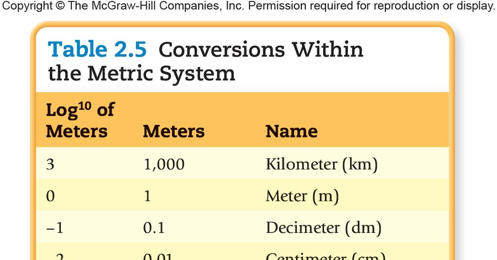
8 Units of Measurement 1 µm = 10 6 m = 10 3 mm 1 nm = 10 9 m = 10 6 mm 1000 nm = 1 µm µm = 1 nm Note: the average human cell is 10 to 15 um and the average prokaryote is 1 um

9 A simple microscope has only one lens Anton van Leeuwenhoek s microscopic He made observations of pond water & fabric fibers Lens Location of specimen on pin Specimen-positioning screw Focusing control Stage-positioning screw Microscope replica
10 Light Microscopy The use of any kind of microscope that uses visible light to observe specimens // simple staining required to increase contrast Types of light microscopy Compound light microscopy Darkfield microscopy Phase-contrast microscopy Differential interference contrast microscopy Fluorescence microscopy Confocal microscopy
 Remagnifies the image formed by the objective lens Body tube Transmits the image from the objective lens to the ocular lens Arm Fine focusing knob Coarse focusing knob")
11 The compound light microscope. Ocular lens (eyepiece) Remagnifies the image formed by the objective lens Body tube Transmits the image from the objective lens to the ocular lens Arm Fine focusing knob Coarse focusing knob Objective lenses Primary lenses that magnify the specimen Stage Holds the microscope slide in position Condenser Focuses light through specimen Diaphragm Controls the amount of light entering the condenser Illuminator Light source Base Principal parts and functions
12 Compound Light Microscopy In a compound microscope, the image from the objective lens is magnified again by the ocular lens Total magnification = objective lens ocular lens
13 Principles of Light Microscopy Power of Objective Usual power of ocular Total magnification 4x scanning objective 10x 40x 10x low power objective 10x 100x 40x high dry objective 10x 400x 100x oil immersion objective 10x 1000x
14 The compound light microscope. Ocular lens Line of vision Path of light Prism The path of light (bottom to top) Body tube Objective lenses Specimen Condenser lenses Illuminator Base with source of illumination
15 Compound Light Microscopy Resolution is the ability of the lenses to distinguish two points A microscope with a resolving power of 0.4 nm can distinguish between two points 0.4 nm Shorter wavelengths of light provide greater resolution the human eye can resolve two objects that are no closer than 0.2 mm apart
 Low resolution (b) High resolution")
16 The Effect of Wavelength on Resolution (a) Low resolution (b) High resolution
17 Compound Light Microscopy The refractive index is a measure of the light-bending ability of a medium The light may bend in air so much that it misses the small high-magnification lens Immersion oil is used to keep light from bending

18 Refraction in the compound microscope using an oil immersion objective lens. Unrefracted light Immersion oil Oil immersion objective lens Without immersion oil most light is refracted and lost Air Glass slide Condenser lenses Condenser Light source Iris diaphragm
19 Brightfield, darkfield, and phase-contrast microscopy. Eye Eye Eye Ocular lens Objective lens Specimen Condenser lens Only light reflected by the specimen is captured by the objective lens Unreflected light Opaque disk Ocular lens Diffraction plates Undiffracted light (unaltered by specimen) Objective lens Refracted or diffracted light (altered by specimen) Specimen Condenser lens Annular diaphragm Light Light Light Dark objects are visible against a bright background Brightfield Light reflected off the specimen does not enter the objective lens
20 Brightfield, darkfield, and phase-contrast microscopy. Eye Eye Eye Ocular lens Objective lens Specimen Condenser lens Only light reflected by the specimen is captured by the objective lens Unreflected light Opaque disk Ocular lens Diffraction plates Undiffracted light (unaltered by specimen) Objective lens Refracted or diffracted light (altered by specimen) Specimen Condenser lens Annular diaphragm Light Light Light Light objects are visible against a dark background Light reflected off the specimen enters the objective lens Darkfield

21 Brightfield, darkfield, and phase-contrast microscopy. Eye Eye Eye Ocular lens Objective lens Specimen Condenser lens Only light reflected by the specimen is captured by the objective lens Unreflected light Opaque disk Ocular lens Diffraction plates Undiffracted light (unaltered by specimen) Objective lens Refracted or diffracted light (altered by specimen) Specimen Condenser lens Annular diaphragm Light Light Light Accentuates diffraction of the light that passes through a specimen Phase-contrast


22 Differential Interference Contrast Microscopy Accentuates diffraction of the light that passes through a specimen; uses two beams of light

 The principle")
23 Fluorescence Microscopy Uses UV light Fluorescent substances absorb UV light and emit visible light Cells may be stained with fluorescent dyes (fluorochromes) The principle of immunofluorescence.
 light is used to excite")

24 Confocal Microscopy Cells are stained with fluorochrome dyes Short-wavelength (blue) light is used to excite the dyes The light illuminates each plane in a specimen to produce a three-dimensional image // Up to 100 µm deep


25 Two-Photon Microscopy Cells are stained with fluorochrome dyes Two photons of long-wavelength light are used to excite the dyes Used to study cells attached to a surface // Up to 1 mm deep

 of a bacterial")
26 Scanning Acoustic Microscopy (SAM) Measures sound waves that are reflected back from an object Used to study cells attached to a surface Resolution 1 µm Scanning acoustic microscopy (SAM) of a bacterial biofilm on glass.
27 Electron Microscopy Uses electrons instead of light The shorter wavelength of electrons gives greater resolution Transmission Electron Microscopy (TEM) Used to observe viruses and internal structures of cells Ultrathin sections of specimens Light passes through specimen, then an electromagnetic lens, to a screen or film Specimens may be stained with heavy-metal salts 10, ,000 ; resolution 2.5 nm
28 Transmission and scanning electron microscopy. Electron beam Electromagnetic condenser lens Specimen Electromagnetic objective lens Electromagnetic projector lens Fluorescent screen or photographic plate Electron gun Viewing eyepiece Secondary electrons Specimen Primary electron beam Electromagnetic lenses Viewing screen Electron collector Amplifier Transmission
29 Scanning Electron Microscopy (SEM) An electron gun produces a beam of electrons that scans the surface of a whole specimen Secondary electrons emitted from the specimen produce the image 1,000 10,000 /// resolution 20 nm

30 Transmission and scanning electron microscopy. Electron beam Electromagnetic condenser lens Specimen Electromagnetic objective lens Electromagnetic projector lens Fluorescent screen or photographic plate Electron gun Viewing eyepiece Secondary electrons Specimen Primary electron beam Electromagnetic lenses Viewing screen Electron collector Amplifier Scanning

31 Scanned-Probe Microscopy Scanning tunneling microscopy (STM) uses a metal probe to scan a specimen Resolution 1/100 of an atom

32 Scanned-Probe Microscopy Atomic force microscopy (AFM) uses a metal-anddiamond probe inserted into the specimen Produces threedimensional images
33 Preparing Specimens for the Microscope Specimens are usually prepared by mounting a sample on a suitable glass slide that sits on the stage between the condenser and the objective lens The manner in which it is prepared depends on the condition of the specimen, either living or preserved the aims of the examiner: to observe overall structure, identify microorganisms, or see movement the type of microscopy available: bright field, dark field, phase contrast, or fluorescence
34 Preparing Smears for Staining Smear: a thin film of a solution of microbes on a slide A smear is usually fixed to attach the microbes to the slide /// Note: Gently heating slide will kill and fix (attach) bacteria to the slide Staining: coloring the microbe with a dye that emphasizes certain structures
35 Preparing Smears for Staining Live or unstained cells have little contrast with the surrounding medium. Researchers do make discoveries about cell behavior by observing live specimens. Stains (are salts) consisting of a positive and negative ion basic dyes have a positive charge // In a basic dye, the chromophore is a cation (cells have mostly negative charges associated with their structures) acidic dyes have a negative charge // In an acidic dye, the chromophore is an anion Staining the background instead of the cell is called negative staining
36 Three Staining Procedures: Simple Stains, Differential, and Special Simple stain: use of a single basic dye A mordant may be used to hold the stain or coat the specimen to enlarge it
 Methylene blue stain of")
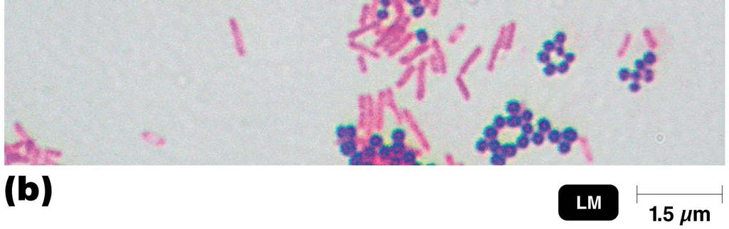
37 Simple Stains Simple Stains (a) Crystal violet stain of Escherichia coli (b) Methylene blue stain of Corynebacterium
38 Differential Stains Used to distinguish different types of bacteria Two different stains used in these procedures Used in the identification of bacteria E.g. Gram stain (distinguishes the type of cell wall // gram positive or negative) Note: Gram stain most useful in medicine to determine which antibiotic to prescribe! E.g. Acid-fast stain (detects unique lipid common in pathogenic bacteria // tuberculosis and lepersy)
 Acid-fast stain. Red cells are acid-fast. Blue cells are non-acidfast.")
39 Differential Stains Differential Stains (a) Gram stain. Purple cells are gram-positive. Pink cells are gram-negative. (b) Acid-fast stain. Red cells are acid-fast. Blue cells are non-acidfast. (c) Spore stain, showing endospores (red) and vegetative cells (blue)
40 Gram Stain Classifies bacteria into gram-positive or gram-negative Gram-positive bacteria tend to be killed by penicillin and detergents Gram-negative bacteria are more resistant to antibiotics Tells us something about structure of the bacteria s cell wall
41 Gram Stain Primary Stain: Crystal Violet Mordant: Iodine Decolorizing Agent: Alcohol-Acetone Counterstain: Safranin Color of Gram-Positive Cells Purple Purple Purple Purple Color of Gram-Negative Cells Purple Purple Colorless Red

42 The Gram Stain: Developed by Hans Christian Gram (1884) Copyright The McGraw-Hill Companies, Inc. Permission required for reproduction or display. Step 1. Crystal violet First, crystal violet is added to the cells in a smear. It stains them all the same purple color. Microscopic Appearance of Cell Gram (+) Gram ( ) Chemical Reaction in Cell Wall (very magnified view) Gram (+) Gram ( ) Both cell walls affix the dye 2. Gram s iodine Then, the mordant, Gram s iodine, is added. This is a stabilizer that causes the dye to form large complexes in the peptidoglycan meshwork of the cell wall. The thicker gram-positive cell walls are able to more firmly trap the large complexes than those of the gram-negative cells. Dye complex trapped in wall No effect of iodine 3. Alcohol Application of alcohol dissolves lipids in the outer membrane and removes the dye from the peptidoglycan layer only in the gram-negative cells. 4. Safranin (red dye) Because gram-negative bacteria are colorless after decolorization, their presence is demonstrated by applying the counterstain safranin in the final step. McGraw-Hill Companies, Inc. Crystals remain in cell wall Red dye masked by violet Outer membrane weakened; wall loses dye Red dye stains the colorless cell

43 Gram Staining Rod (gram-negative) Cocci (gram-positive)
44 Acid-Fast Stain Originated as a method to detect Mycobacterium tuberculosis // Also used to identify Leprosy Differentiates acid fast bacteria (pink) from non acid fast bacteria (blue) These bacteria have cell walls that are particularly impervious that holds fast (tightly or tenaciously) to the dye (carbol fuschin pink color) when washed with an acid alcohol decolorizer Stained waxy cell wall is not decolorized by acid-alcohol Also used in identification of other medically important bacteria, fungi, and protozoa
45 Acid-Fast Stain Color of Acid-Fast Color of Non Acid-Fast Primary Stain: Carbolfuchsin Red Red Decolorizing Agent: Acid-alcohol Red Colorless Counterstain: Methylene Blue Red Blue


46 Acid-fast bacteria. M. bovis Pink cells are acid fast bacteria // Cells counter stained with blue dye are not acid fast bacteria.
47 Special Stains Used to distinguish parts of cells Capsule stain Endospore stain Flagella stain

48 Special Stain / Negative Staining Illustrates Capsules Capsules Capsules
49 Special Staining // Endospore Staining Primary stain: malachite green, usually with heat Decolorize cells: water Counterstain: safranin

50 Special Staining // Endospore Staining Endospore Endospore staining
51 Special Staining // Flagella Staining Mordant used to attach stain to flagella so it appears thicker Carbolfuchsin attached to mordant stains flagella


52 Special Staining // Flagella Staining Flagellum Flagella staining
Observing Microorganisms through a Microscope
 2016/2/19 PowerPoint Lecture Presentations prepared by Bradley W. Christian, McLennan Community College CHAPTER 3 Observing Microorganisms through a Microscope 1 Figure 3.2 Microscopes and Magnification.
Chapter 2 The Study of Microbial Structure: Microscopy and Specimen Preparation
 Chapter 2 The Study of Microbial Structure: Microscopy and Specimen Preparation 1 Lenses and the Bending of Light light is refracted (bent) when passing from one medium to another refractive index a measure
Compound Light Microscopy. Microscopy. Things to remember... 1/22/2017. This is what we use in the laboratory
 Compound Light Microscopy This is what we use in the laboratory Microscopy Chapter 3 BIO 440 A series of finely ground lenses is used to form a magnified image Specimen is illuminated with visible light
Microscopy Techniques that make it easy to see things this small.
 Microscopy Techniques that make it easy to see things this small. What is a Microscope? An instrument for viewing objects that are too small to be seen easily by the naked eye. Dutch spectacle-makers Hans
Observing Microorganisms through a Microscope LIGHT MICROSCOPY: This type of microscope uses visible light to observe specimens. Compound Light Micros
 PHARMACEUTICAL MICROBIOLOGY JIGAR SHAH INSTITUTE OF PHARMACY NIRMA UNIVERSITY Observing Microorganisms through a Microscope LIGHT MICROSCOPY: This type of microscope uses visible light to observe specimens.
Scale. A Microscope s job in life. The Light Microscope. The Compound Microscope 9/24/12. Compound Microscope Anatomy
 The Study of Microbial Structure: Microscopy and Specimen Preparation Scale A Microscope s job in life 1.Magnify 2. Resolve ability to separate or distinguish between two points 3. Contrast How much or
Burton's Microbiology for the Health Sciences
 Burton's Microbiology for the Health Sciences Chapter 2. Viewing the Microbial World Chapter 2 Outline Introduction Using the metric system to express the sizes of microbes Microscopes Simple microscopes
Figure 3.4 Approximate size of various types of cells. ~10 um. Red Blood Cells = mm 1500 um. Width of penny Pearson Education, Inc.
 Figure 3.4 Approximate size of various types of cells. ~10 um Red Blood Cells 1.5mm 1500 um Width of penny = 1500 Figure 4.3 The limits of resolution (and some representative objects within those ranges)
! Because microbiology deals with organisms too small they cannot be seen distinctly with the unaided eye, the microscope is essential.
 Microscopy! Because microbiology deals with organisms too small they cannot be seen distinctly with the unaided eye, the microscope is essential.! The light microscope is the single most important research
Microscopy, Staining, and Classification
 PowerPoint Lecture Presentations prepared by Mindy Miller-Kittrell, North Carolina State University C H A P T E R 4 Microscopy, Staining, and Classification Figure 3.4 Approximate size of various types
Ex 1: Introduction to the microscope
 Ex 1: Introduction to the microscope So what exactly is a microorganism? Microorganisms = any living thing that is too small to be seen with the unaided eye fungus protist bacteria virus Parasitic worm
2018 MICROSCOPE REVIEW by Karen L. Lancour RELATIVE SIZE OF MICROBES
 2018 MICROSCOPE REVIEW by Karen L. Lancour RELATIVE SIZE OF MICROBES 1000 millimeters (mm) = 1 meter (m) 1000 micrometers (µm or mcm) = 1 millimeter (mm) 1000 nanometers (nm) = 1 micrometer (mcm) Size
2017 MICROSCOPE REVIEW by Karen L. Lancour RELATIVE SIZE OF MICROBES
 2017 MICROSCOPE REVIEW by Karen L. Lancour RELATIVE SIZE OF MICROBES 1000 millimeters (mm) = 1 meter (m) 1000 micrometers (µm or mcm) = 1 millimeter (mm) 1000 nanometers (nm) = 1 micrometer (mcm) Size
Lab 1, 2 and 3: Stain, Observe and Identify the Microbes. BIOHAZARD Rules. VIOLATORS will lose points. A) Lab Safety Rules Lab Safety Form Signup
 Lab Safety Rules Lab Safety Form Signup") MICROLAB PREPARATIONS A) Lab Safety Rules Lab Safety Form Signup B) Lab Participation Instructor Review Peer Review Form C) Biohazard Rules How to dispose Trash REQUIRED Items: 1) LAB Manual/Journal 2)
MICROLAB PREPARATIONS A) Lab Safety Rules Lab Safety Form Signup B) Lab Participation Instructor Review Peer Review Form C) Biohazard Rules How to dispose Trash REQUIRED Items: 1) LAB Manual/Journal 2)
The microscope is useful in making observations and collecting data in scientific experiments. Microscopy involves three basic concepts:
 AP BIOLOGY Chapter 6 NAME DATE Block MICROSCOPE LAB PART I: COMPOUND MICROSCOPE OBJECTIVES: After completing this exercise you should be able to: Demonstrate proper care and use of a compound microscope.
Microbiology: Observing Bacteria Laboratory -1. Name Date
 Microbiology: Observing Bacteria Laboratory -1 Name Date Prelab: Part 1 Introduction to the microscope- please read through this handout and label the picture on the next page before starting the lab Care
The microscope is useful in making observations and collecting data in scientific experiments. Microscopy involves three basic concepts:
 Lab #4 Biology 10 BCC Topic: MICROSCOPE LAB PART I: COMPOUND LIGHT MICROSCOPE OBJECTIVES: After completing this exercise you should be able to: Demonstrate proper care and use of a compound microscope.
 Microscopy http://www.microscopyu.com/articles/phasecontrast/phasemicroscopy.html http://micro.magnet.fsu.edu/primer/anatomy/anatomy.html 2005, Dr. Jack Ikeda & Dr. Gail Grabner 9 Nikon Labophot (Question
The Microscope. Packet #2. 10/17/2016 9:12:02 PM Ryan Barrow 2012
 1 The Microscope Packet #2 10/17/2016 9:12:02 PM Ryan Barrow 2012 2 Historical Timeline 1609 Galileo Galilei develops a compound microscope with a convex and a concave les. 1665 Robert Hooke publishes
MICROSCOPE LAB. Resolving Power How well specimen detail is preserved during the magnifying process.
 AP BIOLOGY Cells ACTIVITY #2 MICROSCOPE LAB OBJECTIVES 1. Demonstrate proper care and use of a compound microscope. 2. Identify the parts of the microscope and describe the function of each part. 3. Compare
STRUCTURE OF THE MICROSCOPE
 STRUCTURE OF THE MICROSCOPE Use the word list to label the microscope below: Light Source Coarse adjustment knob Diaphragm Stage Clips Objectives Fine Adjustment Knob Base Stage Stage Clips Arm Revolving
MICROSCOPY MICROSCOPE TERMINOLOGY
 1 MICROSCOPY Most of the microorganisms that we talk about in this class are too small to be seen with the naked eye. The instruments we will use to visualize these microbes are microscopes. The laboratory
Microscopy. Krishna Priya.K Lecturer Dept. of Microbiology
 Microscopy Krishna Priya.K Lecturer Dept. of Microbiology TERMS AND DEFINITIONS Principle Microscopy is to get a magnified image, in which structures may be resolved which could not be resolved with the
Lecture 4 to 5 MICROSCOPY-PRINCIPLES AND TYPES
 Lecture 4 to 5 MICROSCOPY-PRINCIPLES AND TYPES Microorganisms are too small to be seen by our unaided eyes and the microscopes are of crucial importance as they help to view the microbes. A microscope
Microbiology Laboratory 2
 Microbiology Laboratory 2 Microscopy Background Microorganisms are too small to be seen with the naked eye. Thus a microscope is used to magnify objects so they can be observed. A lens consists of one
THE COMPOUND BRIGHTFIELD MICROSCOPE
 THE COMPOUND BRIGHTFIELD MICROSCOPE Microbiology is the study of microscopic organisms that are so small that they are below the limit of vision of the human eye. Bacteria are the smallest of microorganisms
Microscope Notes. units of life.
 Microscope Notes Microscope an instrument that produces an enlarged image of an object. Biologists use microscopes to study cells, cell parts, and organisms that are too small to be seen with the naked
Microscope (and The Cell) Lab Exercise #1
 Lab Exercise #1") Lab Safety-General No eating or drinking Only registered students allowed in the class Long hair must be tied back Familiarize yourself with the emergency stations Do not mark on the models Inform me of
Lab Safety-General No eating or drinking Only registered students allowed in the class Long hair must be tied back Familiarize yourself with the emergency stations Do not mark on the models Inform me of
Exercise 2-A MICROSCOPIC TECHNIQUE & EXAMINATION OF MICROORGANISMS
 Exercise 2-A MICROSCOPIC TECHNIQUE & EXAMINATION OF MICROORGANISMS Introduction to Microscopic Technique Microbiology is the science or study of living organisms too small to be seen with the naked eye.
Exercise 2-A MICROSCOPIC TECHNIQUE & EXAMINATION OF MICROORGANISMS
 Exercise 2-A MICROSCOPIC TECHNIQUE & EXAMINATION OF MICROORGANISMS Introduction to Microscopic Technique Microbiology is the science or study of living organisms too small to be seen with the naked eye.
Unit Two Part II MICROSCOPY
 Unit Two Part II MICROSCOPY AVERETT 1 0 /9/2013 1 MICROSCOPES Microscopes are devices that produce magnified images of structures that are too small to see with the unaided eye Humans cannot see objects
Biology The Microscope. May 20 1:19 PM. Using a Microscope to Explore the Cell
 Biology 2201 1.2 The Microscope Using a Microscope to Explore the Cell Resolution or Resolving power The ability of the eye, or other instrument, to distinguish between two objects that are close together
Microscopy. ( greek mikros = small; skopein = to observe)
") Microscopy ( greek mikros = small; skopein = to observe) Zacharias Jansen put several lenses in a tube (first compound microscope) and the object near the end of tube appeared to be greatly enlarged, much
Microscopy ( greek mikros = small; skopein = to observe) Zacharias Jansen put several lenses in a tube (first compound microscope) and the object near the end of tube appeared to be greatly enlarged, much
The Care and Use of the Microscope. Lab Exercise #4
 Lab Safety No eating or drinking!!! Long hair must be tied back Clean up your workstation before you leave! Return all materials to the storage sites Clean glassware and wipe down countertops Follow directions
S200 Course LECTURE 1 TEM
 S200 Course LECTURE 1 TEM Development of Electron Microscopy 1897 Discovery of the electron (J.J. Thompson) 1924 Particle and wave theory (L. de Broglie) 1926 Electromagnetic Lens (H. Busch) 1932 Construction
Tissue Preparation ORGANISM IMAGE TISSUE PREPARATION. 1) Fixation: halts cell metabolism, preserves cell/tissue structure
 Fixation: halts cell metabolism, preserves cell/tissue structure") Lab starts this week! ANNOUNCEMENTS - Tuesday or Wednesday 1:25 ISB 264 - Read Lab 1: Microscopy and Imaging (see Web Page) - Getting started on Lab Group project - Organ for investigation - Lab project
Lab starts this week! ANNOUNCEMENTS - Tuesday or Wednesday 1:25 ISB 264 - Read Lab 1: Microscopy and Imaging (see Web Page) - Getting started on Lab Group project - Organ for investigation - Lab project
VISUAL PHYSICS ONLINE DEPTH STUDY: ELECTRON MICROSCOPES
 VISUAL PHYSICS ONLINE DEPTH STUDY: ELECTRON MICROSCOPES Shortly after the experimental confirmation of the wave properties of the electron, it was suggested that the electron could be used to examine objects
The light microscope
 What is a microscope? The microscope is an essential tool in modern biology. It allows us to view structural details of organs, tissue, and cells not visible to the naked eye. The microscope should always
Light Microscopy. Upon completion of this lecture, the student should be able to:
 Light Light microscopy is based on the interaction of light and tissue components and can be used to study tissue features. Upon completion of this lecture, the student should be able to: 1- Explain the
Laboratory Introduction
 Laboratory Introduction There are two basic categories of microscopes: light microscopes and electron microscopes. Light, or optical, microscopes require light waves to provide the illumination while electron
LAB 1 Introduction to Microscopy
 I. Ubiquity of Microorganisms II. Microscopy LAB 1 Introduction to Microscopy I. UBIQUITY OF MICROORGANISMS Microorganisms are ubiquitous; that is, they are present nearly everywhere. In this lab you will
MICROSCOPY and CELL STRUCTURE
 MICROSCOPY and CELL STRUCTURE Readings: Review pp. 69-71, and Fig. 4.1 on p. 65 in your text (POHS, 5 th ed.). Introduction: Biologists rely on many different types of microscopic techniques to find out
MICROSCOPES. Magnification: Resolution: Field of View: Describes the visual picture seen when looking through the eyepiece of the microscope
 Microscopes MICROSCOPES Magnification: Resolution: Field of View: Describes the visual picture seen when looking through the eyepiece of the microscope 7X 45X 112.5X 225X 1 st crude microscope made by
Match the microscope structures given in the left column with the statements in the right column that identify or describe them.
 49 Prelab for Name Match the microscope structures given in the left column with the statements in the right column that identify or describe them. Key: a. coarse adjustment knob f. turret or nosepiece
MICROSCOPE TERMS 7X 45X 112.5X 225X
 Microscopes MICROSCOPE TERMS Magnification- how much larger the image is Resolution- how clear the image is Field of View: Describes the visual picture seen when looking through the eyepiece of the microscope
Marine Invertebrate Zoology Microscope Introduction
 Marine Invertebrate Zoology Microscope Introduction Introduction A laboratory tool that has become almost synonymous with biology is the microscope. As an extension of your eyes, the microscope is one
King Saud University Dept. of Bot. & Microbiology. General Microbiology 140 MIC
 King Saud University Dept. of Bot. & Microbiology General Microbiology 140 MIC Lab coat. Do not wearing the lab coat outside the lab. Gloves. Proper Clothing and closed shoes. Hair should be tied back.
Lab 2 T. Microbes in Everyday Life; Pure Culture Project; Hand Washing; Light Microscopy
 Microbes in Everyday Life; Pure Culture Project; Hand Washing; Light Microscopy Lab 2 T oday s lab looks at the results of microbial diversity and continues the project of working toward producing a pure
Resolution. Diffraction from apertures limits resolution. Rayleigh criterion θ Rayleigh = 1.22 λ/d 1 peak at 2 nd minimum. θ f D
 Microscopy Outline 1. Resolution and Simple Optical Microscope 2. Contrast enhancement: Dark field, Fluorescence (Chelsea & Peter), Phase Contrast, DIC 3. Newer Methods: Scanning Tunneling microscopy (STM),
Introduction to Microscopes
 INTRODUCTION TO THE MICROSCOPE Introduction to Microscopes The first microscopes worked by the same basic principle as the ones you will be using in lab. They are light microscopes. Visible light passes
A BRIEF INTRODUCTION TO MICROSCOPY The two key properties of a microscope that allow you to see microbes are resolution and magnification.
 A BRIEF INTRODUCTION TO MICROSCOPY The two key properties of a microscope that allow you to see microbes are resolution and magnification. Magnification refers to the enlargement of the specimen when seen
Biology 29 Cell Structure and Function Spring, 2009 Springer LABORATORY 1: THE LIGHT MICROSCOPE
 Biology 29 Cell Structure and Function Spring, 2009 Springer LABORATORY 1: THE LIGHT MICROSCOPE Prior to lab: 1) Read these instructions (p 1-6) 2) Go through the online tutorial, the microscopy pre-lab
I. The First Microscopes. Microscope Basics. II. The Bright Field Microscope. Confocal Laser Scanning Microscopy. A. The Compound Microscope
 Microscope Basics I. The First Microscopes NGSSS: SC.912.N.2.1 through N.4.2 A. About 1590, two Dutch spectacle makers, Zaccharias Janssen and his son Hans, while experimenting with several lenses in a
History of microscopy
 History of microscopy Introduction Structure of microscope Care of microscope Use of microscope Magnification As we already know cells are microscopic. What does this mean? Scientists were able to see
Microscopy. Matti Hotokka Department of Physical Chemistry Åbo Akademi University
 Microscopy Matti Hotokka Department of Physical Chemistry Åbo Akademi University What s coming Anatomy of a microscope Modes of illumination Practicalities Special applications Basic microscope Ocular
Microscope Review. 1. A compound light microscope is represented in the diagram below.
 Name Microscope Review Date 1. A compound light microscope is represented in the diagram below. 5. The diagram below represents a hydra as viewed with a compound light microscope. If the hydra moves toward
UNIT C: CYCLING OF MATTER IN LIVING SYSTEMS
 UNIT C: CYCLING OF MATTER IN LIVING SYSTEMS Aristotle is known as The Father of Biology. He was one of the first Greek philosophers who used the Scientific Method of observing, recording, reasoning, and
What you should have learned from the microscope labs.
 What you should have learned from the microscope labs. Microscope Lab 1 Directionality Items appear backwards and inverted On Stage In Microscope NOT!!!! Microscope Lab 1 More Directionality Items move
tweezers Goggles Scalpel Tongs E G H K J F C L B D A I Aim #1 3 Safety, Instrumentation, Microscope Ruler Beaker Microscope Thermometer Graduated
 Ruler Beaker Microscope Thermometer Bunsen Burner (We use Hot plates) Eye Dropper/ Pipette Test tube Holder tweezers Goggles Scalpel Tongs Graduated cylinder C L B D A I E G H K J F Youtube: Powers of
Test Review # 8. Physics R: Form TR8.17A. Primary colors of light
 Physics R: Form TR8.17A TEST 8 REVIEW Name Date Period Test Review # 8 Light and Color. Color comes from light, an electromagnetic wave that travels in straight lines in all directions from a light source
1.When an object is sharply focused and the slide is moved towards you, in which direction does the
 image upright or inverted? Name: Date: _ BIOLOGY EXPERIMENT:Class: Using a Compound Light Microscope II: Depth Perception, resolution, field of view MATERIALS: Compound light microscopecolor magazine clipping
Introduction. Laboratory Equipment & Supplies. Model 1333PHi Shown (Phase Contrast) (2) Eyepieces (Eyecups installed) Diopter Adjustment Mechanism
 (2) Eyepieces (Eyecups installed) Diopter Adjustment Mechanism") Introduction With the invention of the microscope in the early 17th century, it was made possible to view objects which were too small for the human eye to see. As the microscope evolved, the structure
Introduction With the invention of the microscope in the early 17th century, it was made possible to view objects which were too small for the human eye to see. As the microscope evolved, the structure
Microscope. & Measurements. Do Now
 Do Now Microscope & Measurements How many: 1. Centimeters (cm) in 4 meters (m)? m 2. Decimeters (dm) in 5 meters (m)? dm 3. Centimeters (cm) in 4,000 millimeters (mm) cm 4. Millimeters (mm) in 40 centimeters
Introduction to the Compound Microscope Cell Structure & Function
 Introduction to the Compound Microscope Cell Structure & Function Revised Fall 2018 Laboratory Safety Lab coat, long pants, closed-toe shoes, safety goggles, and nitrile or latex gloves are required. **You
Bio 407. Applied microscopy. Introduction into light microscopy. José María Mateos. Center for Microscopy and Image Analysis
 Center for Microscopy and Image Analysis Bio 407 Applied Introduction into light José María Mateos Fundamentals of light Compound microscope Microscope composed of an objective and an additional lens (eyepiece,
microscopy A great online resource Molecular Expressions, a Microscope Primer Partha Roy
 Fundamentals of optical microscopy A great online resource Molecular Expressions, a Microscope Primer http://micro.magnet.fsu.edu/primer/index.html Partha Roy 1 Why microscopy Topics Functions of a microscope
CALIBRATION OF MICROSCOPE EYEPIECE GRATICULE
 CALIBRATION OF MICROSCOPE EYEPIECE GRATICULE A typical eyepiece graticule looks like this: It is 10mm in length and each mm is divided into 10 parts So each small division = 0.1mm = 100µm The eyepiece
Care and Use of the Compound Light Microscope
 EXERCISE 2 Care and Use of the Compound Light Microscope Time Estimates for Completing This Lab The activities in this laboratory exercise can be completed in 2 to 2.5 hours. Extra time will be required
Applications of Optics
 Nicholas J. Giordano www.cengage.com/physics/giordano Chapter 26 Applications of Optics Marilyn Akins, PhD Broome Community College Applications of Optics Many devices are based on the principles of optics
Microscope. Dr. Leena Barhate Department of Microbiology M.J.College, Jalgaon
 Microscope Dr. Leena Barhate Department of Microbiology M.J.College, Jalgaon Acknowledgement http://www.cerebromente.org.br/n17/histor y/neurons1_i.htm Google Images http://science.howstuffworks.com/lightmicroscope1.htm
Visual Anatomy ansd Physiology Lab Manual Pig Version 2nd Edition Sarikas TEST BANK
 Visual Anatomy ansd Physiology Lab Manual Pig Version 2nd Edition Sarikas TEST BANK https://testbankreal.com/download/visual-anatomy-ansd-physiology-labmanual-pig-version-2nd-edition-sarikas-test-bank/
Test Review # 9. Physics R: Form TR9.15A. Primary colors of light
 Physics R: Form TR9.15A TEST 9 REVIEW Name Date Period Test Review # 9 Light and Color. Color comes from light, an electromagnetic wave that travels in straight lines in all directions from a light source
INTRODUCTION TO MICROSCOPY. Urs Ziegler THE PROBLEM
 INTRODUCTION TO MICROSCOPY Urs Ziegler ziegler@zmb.uzh.ch THE PROBLEM 1 ORGANISMS ARE LARGE LIGHT AND ELECTRONS: ELECTROMAGNETIC WAVES v = Wavelength ( ) Speed (v) Frequency ( ) Amplitude (A) Propagation
USING THE MICROSCOPE TO OBSERVE CELLS
 USING THE MICROSCOPE TO OBSERVE CELLS *****IMPORTANT!!!!! BEFORE VISITING YOUR LEARNING CENTER TO CARRY OUT THIS LAB ACTIVITY PLEASE READ BELOW Before you visit your Learning Center to use the microscope,
Imaging Introduction. September 24, 2010
 Imaging Introduction September 24, 2010 What is a microscope? Merriam-Webster: an optical instrument consisting of a lens or combination of lenses for making enlarged images of minute objects; especially:
Optical and digital microscopic imaging techniques and applications in pathology
 Analytical Cellular Pathology 34 (2011) 5 18 DOI 10.3233/ACP-2011-0006 IOS Press Optical and digital microscopic imaging techniques and applications in pathology 5 Xiaodong Chen a,c, Bin Zheng b and Hong
Perfecting Microscope Skills
 I. Introduction to the Microscope Perfecting Microscope Skills There are different types of microscopes used by biologists depending on the job they wish to accomplish, including dissecting (or "stereoscopic")
LAB 3 Use of the Microscope
 LAB 3 Use of the Microscope Introduction In this laboratory you will be learning how to use one of the most important tools in biology the compound light microscope to view a variety of specimens. You
INTRODUCTION THIN LENSES. Introduction. given by the paraxial refraction equation derived last lecture: Thin lenses (19.1) = 1. Double-lens systems
 = 1. Double-lens systems") Chapter 9 OPTICAL INSTRUMENTS Introduction Thin lenses Double-lens systems Aberrations Camera Human eye Compound microscope Summary INTRODUCTION Knowledge of geometrical optics, diffraction and interference,
Chapter 9 OPTICAL INSTRUMENTS Introduction Thin lenses Double-lens systems Aberrations Camera Human eye Compound microscope Summary INTRODUCTION Knowledge of geometrical optics, diffraction and interference,
Microscopy Training & Overview
 Microscopy Training & Overview Product Marketing October 2011 Stephan Briggs - PLE OVERVIEW AND PRESENTATION FLOW Glossary and Important Terms Introduction Timeline Innovation and Advancement Primary Components
UNIT: THE MICROSCOPE AND CELLULAR DIVERSITY
 Course: Biology Agricultural Science & Technology UNIT: THE MICROSCOPE AND CELLULAR DIVERSITY State Standard: State Objectives: Unit Objectives: A. Learn how to use the compound microscope. B. Learn the
Microscopes & cells. 2. arm. 3. ocular lens. 4. objective lenses. 5. stage. 6. slide clamp. 7. stage controls
 Microscopes & cells Objectives: At the end of this lab you should be able to: o demonstrate the safe and proper handling of a microscope, including carrying a microscope, slide placement, and storage.
Light microscopy BMB 173, Lecture 14, Feb. 21, 2018
 Light microscopy The Structural Biology Continuum Next two lectures: Light microscopy Many slides taken from Scott Fraser, Murphy s Fundamentals of light microscopy, Alberts Molecular Biology of the Cell,
Bio 252: Microscopy Study THE COMPOUND MICROSCOPE
 Name: Date: Block: Microscope Number: Bio 252: Microscopy Study THE COMPOUND MICROSCOPE I. Introduction The compound microscope is one of the most important instruments used by biologists today. Through
Microscope anatomy, image formation and resolution
 Microscope anatomy, image formation and resolution Ian Dobbie Buy this book for your lab: D.B. Murphy, "Fundamentals of light microscopy and electronic imaging", ISBN 0-471-25391-X Visit these websites:
used for low power magnification of a sample image is 3 dimensional
 MICROSCOPES One of the most important inventions in the advancement of Biology 1. Simple Microscopes ie. magnifying glass, stereoscope (dissecting scope) have a single lens or a pair of lenses combined
LAB ACTIVITY: USING A MICROSCOPE
 Name: Date: Period: Lab Partner(s): LAB ACTIVITY: USING A MICROSCOPE Objectives: Demonstrate the proper use and care of a compound light microscope and stereomicroscope. Focus the compound light microscope
The Compound Microscope. Brightfield: Köhler Illumination
 Outline History of Microscopy The Magnifying Glass The Compound Microscope Brightfield: Köhler Illumination Microscopy µικροσ (mikros): small σκοπειν (skopein): to observe History of Microscopy Well :
AN INTRODUCTION TO THE MICROSCOPE
 AN INTRODUCTION TO THE MICROSCOPE INTRODUCTION In this exercise you will learn the components and operation of the compound microscope and the dissection microscope. This will be followed by a short exercise
Ocular Lenses. Head. Arm. Objective Lenses. Slide Holder Stage. On / Off Switch. Condenser with Iris Diaphragm. Light Intensity Control
 BIOLOGY 211: HUMAN ANATOMY & PHYSIOLOGY ********************************************************************************************************* USE OF THE LIGHT MICROSCOPE **********************************************************************************************************
Basic Microscopy. OBJECTIVES After completing this exercise, you should be able to do the following:
 Page 1 of 10 Basic Microscopy OBJECTIVES After completing this exercise, you should be able to do the following: a. Name the parts of the compound microscope and the functions of each. b. Describe how
Microscopy: Fundamental Principles and Practical Approaches
 Microscopy: Fundamental Principles and Practical Approaches Simon Atkinson Online Resource: http://micro.magnet.fsu.edu/primer/index.html Book: Murphy, D.B. Fundamentals of Light Microscopy and Electronic
Life Science Chapter 2 Study Guide
 Key concepts and definitions Waves and the Electromagnetic Spectrum Wave Energy Medium Mechanical waves Amplitude Wavelength Frequency Speed Properties of Waves (pages 40-41) Trough Crest Hertz Electromagnetic
Microscopy and Staining
 Microscopy and Staining Figure 2.1 Different types of microscopy are used to visualize different structures. Brightfield microscopy (left) renders a darker image on a lighter background, producing a clear
Key Points Refer to How to Use the Compound Light Microscope :
 MODULE 1 Objective 1.2 Lesson B Introduction to the Microscope Using the Light Microscope and Slide Preparation Course Advanced Biotechnology Unit Biotech Basics Essential Question How do scientists view
Education in Microscopy and Digital Imaging
 Contact Us Carl Zeiss Education in Microscopy and Digital Imaging ZEISS Home Products Solutions Support Online Shop ZEISS International ZEISS Campus Home Interactive Tutorials Basic Microscopy Spectral
III III 0 IIOI DID IIO 1101 I II 0II II 100 III IID II DI II
 (19) United States III III 0 IIOI DID IIO 1101 I0 1101 0II 0II II 100 III IID II DI II US 200902 19549A1 (12) Patent Application Publication (10) Pub. No.: US 2009/0219549 Al Nishizaka et al. (43) Pub.
Components of the Microscope
 Swift M3 Microscope The Swift M3 is a versatile microscope designed for both microscopic (high magnification, small field of view) and macroscopic (low magnification, large field of view) applications.
Using Microscopes. Life Science: Molecular
 Using Microscopes Life Science: Molecular Light Microscopy: Instrumentation and Principles A light microscope is so named because it uses visible light to produce a magnified image. Compound light microscopes
REVIEW FOR TEST ON MONDAY
 1. The diagram below shows an ameba moving out of the high-power field of view of a compound microscope in the direction indicated by the arrow. 4. The diagram below represents two cells next to a metric