MR Advance Techniques. Flow Phenomena. Class II
|
|
|
- Bryce Conley
- 6 years ago
- Views:
Transcription
1 MR Advance Techniques Flow Phenomena Class II
2 Flow Phenomena In this class we will explore different phenomenona produced from nuclei that move during the acquisition of data. Flowing nuclei exhibit different contrast characteristics from their neighboring stationary nuclei, and originate primarily from hydrogens in blood and CSF.
3 Types of flow There are four principal types of flow: Laminar flow Turbulent flow Vortex flow Spiral flow Stagnant flow
4 Laminar Flow Laminar flow also known as parabolic flow is flow that is at different but consisted velocities across a vessel. The flow in the center of the vessel move faster than at the vessel walls, where resistance slows down the flow. However, the velocity difference across the vessel is constant.
5 Spiral Flow Spiral flow is where the direction of the flow is spiral.
6 Vortex Flow Vortex flow is flow that is initially laminar but then passes through a stricture or stenosis in the vessel. Flow in the center of the lumen has high velocity, but near the walls, the flow spiral.
7 Turbulent Flow Turbulent flow is flow at different velocities that fluctuates randomly. The velocity difference across the vessel changes erratically.
8 Stagnant flow Stagnant flow is describe as no flow circulation due to blockage of a blood vessel
9 Blood Flow Velocity Blood flow is faster in arteries and slower in veins. The velocity will vary depending on the proximity to the heart and the caliber of the blood vessel. The definition of blood flow velocity as given in the medical dictionary is, "a value equal to the total volume flow divided by the cross-sectional area of vascular bed". Velocity of blood is measured in cm/s. Flow Velocity = Flow Volume / Vessel Area
10 Flow Velocity Blood Vessel Aorta Pulmonary Artery Inferior Vena Cava Superior Vena Cava Portal Vein Carotid Artery Middle Cerebral Artery Flow Velocity 92 cm/sec 63 cm/sec cm/sec cm/sec 20 cm/sec cm/sec 58.3 cm/sec
 Blood Vessel Aorta IVC Flow Velocity 92 cm/sec 30-45")
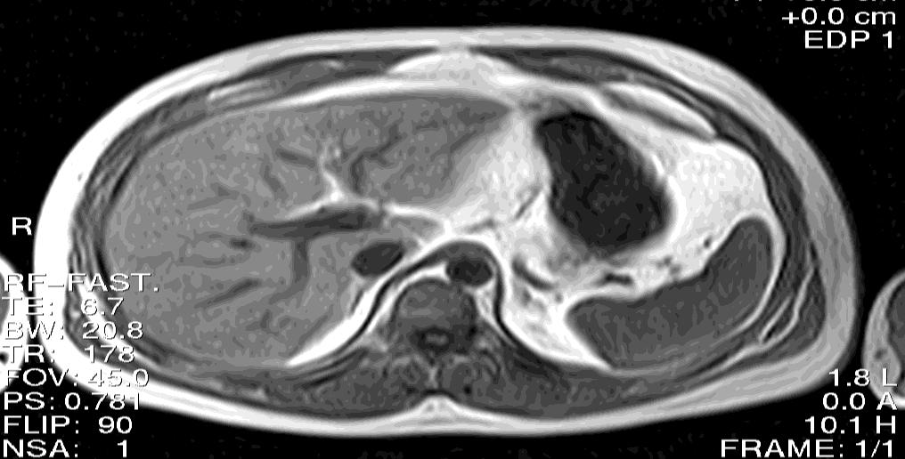
11 Abdominals Blood Vessels Abdominal Aorta Inferior Vena Cava (IVC) Blood Vessel Aorta IVC Flow Velocity 92 cm/sec cm/sec
12 Flowing Protons There two types of signal intensities from flowing protons: Dark / Hypo-intense / Black / signal void / signal loss Bright / Hyper-intense / enhance

13 Flow Phenomena The motion of flowing nuclei causes several artifacts effects on the image: Entry Slice Phase misshaping Signal Void
14 Flow Phenomena The causes of flow artifact are collectively know as Flow Phenomena. The principal Flow phenomena are: Time of flight Entry slice Intra-voxel dephasing
15 Stationary Protons Remember that RF excitation pulses are slice selected 90º
16 Stationary Protons To produce a signal the protons must receive a RF excitation and a rephasing pulse. 90º ½ TE (20 ms) 180º
17 Stationary Protons If protons receive both excitation pulses, they will have high signal intensity on the image. 90º 180º
18 TOF Effects in SE If the nucleus receive only one of the two pulses, it does not produce signal. This is call Time of Flight Phenomenon (TOF). 90º 180º
19 Time of Flight Phenomenon in SE Stationary nucleus will received both the excitation pulse plus the rephasing pulse. As result stationary protons will produce signal, but flowing protons will not produce any signal. 90º ½ TE (20 ms) 180º
20 TOF Effects in SE The TOF phenomenon depends on the following parameters: Velocity of Flow (intrinsic) TE (extrinsic) Slice Thickness (extrinsic)
21 Time of flight effects in SE
22 TOF Effects in SE Velocity of flow: as the velocity of the flow increases the time of flight effect increases due to protons will move faster and stay less time within the slice. This is called high velocity signal loss. 90º 180º ½ TE Rapid flow
23 TOF Effects As the velocity of the flow decreases, a higher proportion of flowing nuclei are present in the slice for both 90 and 180 RF pulses. As the velocity decreases the TOF decreases. This is called flow related enhancement. 90º 180º ½ TE Slow flow
24 90º 180º 90º 180º
25 TOF Effects TE: as the TE increases, a higher proportion of flowing protons have exited the slice between the excitation pulses, and the TOF effect increases. 90º 180º ½ TE ½ TE
26 TOF Effects TE: as the TE decreases, a smaller proportion of flowing protons have exited the slice between the excitation pulses, and the TOF effect decreases. 90º 180º ½ TE ½ TE
27 90º 180º 90º 180º

28 TOF Effects As the thickness of the slice decreases, the nuclei are more likely to receive only one pulse and the signal void increases. As the slice thickness is decreased TOF increases. 90º 180º ½ TE
29 TOF Effects Slice thickness: for a given constant flow velocity, protons take longer to travel through a thick slice compared with a thin slice. Therefore in a thick slice nuclei are more likely to receive both 90 and 180 RF pulses. In a thick slice the time of flight effect decreases. 90º 180º ½ TE
30 90º 180º 90º 180º
31 Flow related enhancement increases as: Velocity of flow TE Decreases Decreases Slice thickness Increases

32 High velocity signal void increases as: Velocity of flow TE Increases Increases Slice thickness Decreases
33 Time of Flight Phenomenon in GRE In gradient echo pulse sequences each slice is selectively exited by the RF pulse, the rephasing gradients applied to the entire body. Therefore, a flowing proton that receives an excitation pulse is rephased regardless of its slice position and produces a signal. 90º ½ TE (20 ms)
34 90º 180º 90º

35 Time of Flight Phenomenon in GRE In addition, the very short TR usually associated with gradient echo sequences tends to saturate stationary protons which receive repeated RF pulses so that flowing protons appear to have a higher signal intensity. GRE pulse sequences are often said to be flowsensitive.

36 Entry Slice Phenomenon The entry slice phenomenon is related to the excitation history of the protons. 12
37 Entry Slice Phenomenon The rate of which the flowing nuclei receive the excitation pulses determines the magnitude of the entry slice phenomenon. Any factor that affects the rate at which a nucleus receives repeated RF pulses affects this phenomenon.
38 Entry Slice Phenomenon The magnitude of the entry slice phenomenon depends on four factors: TR Slice thickness Velocity of flow Direction of flow 1 2 3
39 Entry Slice Phenomenon & TR 90º RF TR 90º RF TR 90º RF As the TR Increases the Entry Slice Phenomenon also Increases
40 Entry Slice Phenomenon & TR 90º RF TR 90º RF TR 90º RF TR 90º RF TR 90º RF TR 90º RF As the TR Decreases the Entry Slice Phenomenon Decreases
41 Entry Slice Phenomenon & TR 90º RF TR 90º RF TR 90º RF º RF TR 90º RF TR 90º RF TR 90º RF TR 90º RF TR 90º RF 1 2 3
42 Short TR Long TR RF RF RF RF RF RF RF RF RF RF RF RF RF RF RF
43 Entry Slice & Slice Thickness As the slice thickness increases the entry slice phenomenon decreases. Since slices are thicker entry slice will be sees in less slices when thicker slices are used.
44 Thicker Slice Thin Slice RF RF RF RF RF RF RF RF RF RF RF RF RF RF
45 Entry Slice & Slice Thickness As the velocity of the flow increases the entry slice phenomenon increases. When flowing protons move faster they receive less RF pulses and take longer to saturate.

46 Fast Flowing Protons Slow Flowing Protons RF RF RF RF RF RF RF RF RF RF RF RF RF RF
47 Entry Slice Phenomenon Direction of flow: Flow that is in the same direction as the slice acquisition Co-current flow Flow that is in the opposite direction of to the slice acquisition is called counter-current flow
48 Entry Slice Phenomenon Co-current flow: The flowing nuclei are more likely to receive repeated RF pulses as they move from slice to the next. Protons become saturated relatively quickly, and so the entry slice phenomenon decreases rapidly
49 Entry Slice Phenomenon Flowing protons stay fresh, as when they enter a slice they are less likely to have received previous excitation pulses. The entry slice phenomenon does not decrease rapidly, and may still be present in all the slices
50 Intra-Voxel Dephasing Protons that are not under a gradient magnetic field have the same precessional frequency and the same phase within a Voxel.
51 Intra-voxel dephasing Protons under a gradient magnetic field will result in a difference in their precessional frequency. The phase encoding gradient is utilize to produce this change and dephase the protons.
52 Flowing protons are adjacent to stationary protons in the voxel. During the application of the phase encoding gradient, flowing protons move under the gradient altering their frequency. When the gradient is turn off, flowing protons acquire a different phase compared to the stationary ones.
53 Intra-voxel dephasing Therefore nuclei within the same voxel are out of phase with each other, which results in a reduction of total signal amplitude from the voxel. This is called intra-voxel dephasing. Flow Flow
54 Intra-voxel dephasing When the net magnetization of flowing protons is phase shifted, the signal generated by the net magnetization of protons is mismapped and place elsewhere along the phase encoding direction of the image. When this occurs, the vessel on the resulting image appears dark (no signal) or an artifact called flow artifact. Flow Artifact.
55 Phase Encoding Gradient 90º
56 Intra-Voxel Dephasing Factors Affecting Intra-Voxel Dephasing Velocity of the Flow TE Voxel size The amplitude of the Phase Encoding Gradient
57 Intra-Voxel Dephasing Velocity of the Flow: as the velocity of the flow increases the intra-voxel dephasing also increases.
58 Intra-Voxel Dephasing TE: as the TE increases the intra-voxel dephasing increases). 90º 180º 90º 180º
59 Intra-Voxel Dephasing Voxel size: as the Voxel size increases the intra-voxel dephasing increases. Increase Slice Thickness Increase FOV Decrease Matrix

60 Intra-Voxel Dephasing PES: as the phase gradient increases the intra-voxel dephasing also increases.
61 Flow Phenomena Compensation There are three methods used to compensate for the flow phenomena. Even echo rephasing Gradient moment nulling Spatial pre-saturation
62 Even Echo Rephasing Even-echo rephasing is a flow phenomenon that is observed in SE images in which multiple evenly spaced echoes (e.g., 25/50/75/100) have been acquired. For blood flowing at constant velocity in such an environment, phase dispersion is lower on the evennumbered echoes (i.e., 50/100) than on the odd echoes (i.e., 25/75). TE 25 TE 50 TE 75 TE 100
63 Gradient Moment Nulling Gradient moment nulling also known as flow compensation (Flow Comp) technique compensates for the altered phase values of the protons by applying an additional gradient to correct the altered phases back to their original values.
64 Gradient moment nulling technique is achieved by the frequency or the slice select gradient. These gradients alter the polarity from negative to positive (Balance Gradient Echo). PES 25% 25% %
65 Gradient Moment Nulling Gradient moment nulling assures that the flowing protons are in phase with the stationary protons at the time of the echo; this will result in a bright signal from flowing protons without artifact.
66 Phase Encoding Gradient 90º
67 Phase Encoding Gradient 25% 25% + 50% + 90º
68 Gradient Moment Nulling Since gradient moment nulling technique uses extra gradients, it will force an increases of the minimum TE. 10 TE No gradient moment nulling application 15 TE Gradient moment nulling application
69 As a result of increasing the TE, fewer slices may be available for a given TR or the TR and therefore the scan time may be automatically increased. No gradient moment nulling application 500 TR Slice 1 Slice 2 Slice 3 Slice 4 Gradient moment nulling application 600 TR Slice 1 Slice 2 Slice 2 Slice 2
70 Spatial Pre-Saturation Bands Spatial Pre-saturation bands also known as, Pre-Sats or Sat Bands, are used to saturate specific areas within the image. Pres-Sats can be used to: Nullify the signal from flowing nuclei: Minimize Entry Slice Minimize TOF Minimize intra voxel
71 Pre-Saturation Bands Pre Sats deliver a 90º RF pulse to a volume of tissue that is desired to eliminate. Pre sat Pulse
72 GRE
73 GRE
74 Slice Pre Saturation Band

75 Pre Saturation Band Sat Band Slice Sat Band

76 Pre Saturation Band Sat Band Slice Sat Band

77 Spatial Pre-Saturation To be effective, presaturation bands should be placed between the flow and the slice been acquired. Signal from flowing protons entering the slice will be nullified. Pre-saturation band


78 Pre-Saturation bands are use to reduce flow artifact Pre-saturation band Pre-saturation band


79 Pre-sat used to reduce entry slice phenomenon Pre-saturation band

80 Spatial Pre-Saturation The use of pre-saturation pulses will increase the RF deposition on the patient (SAR), which may increase heating effects. The use of pre-saturation pulses may also decreases the number of slices available Pre sat No pre sat


81 Pre-Sat The use of pre-saturation bands cam be used to limit our view to either the arteries or the veins.
82 Pre-saturation band
83 Pre-Saturation Bands 83

84 Blood Flow Enhancement GRE Counter-current flow Flow Comp Short TE Signal Void Spin Echo GRE Sat Bands
85 Sat Bands Spatial pre-saturation pulses can be brought into the FOV itself. This permits artifacts producing areas such as pulsation, breathing, cardiac, peristaltic to be pre-saturated so that the phase misshaping can be reduced.


86 Spatial Pre-Saturation Bands Nullify signal from moving structures or undesired anatomy within the FOV.
87
88
89
90
Pulse Sequence Design and Image Procedures
 Pulse Sequence Design and Image Procedures 1 Gregory L. Wheeler, BSRT(R)(MR) MRI Consultant 2 A pulse sequence is a timing diagram designed with a series of RF pulses, gradients switching, and signal readout
Cardiac MR. Dr John Ridgway. Leeds Teaching Hospitals NHS Trust, UK
 Cardiac MR Dr John Ridgway Leeds Teaching Hospitals NHS Trust, UK Cardiac MR Physics for clinicians: Part I Journal of Cardiovascular Magnetic Resonance 2010, 12:71 http://jcmr-online.com/content/12/1/71
H 2 O and fat imaging
 H 2 O and fat imaging Xu Feng Outline Introduction benefit from the separation of water and fat imaging Chemical Shift definition of chemical shift origin of chemical shift equations of chemical shift
MR Basics: Module 8 Image Quality
 Module 8 Transcript For educational and institutional use. This transcript is licensed for noncommercial, educational inhouse or online educational course use only in educational and corporate institutions.
1 Introduction. 2 The basic principles of NMR
 1 Introduction Since 1977 when the first clinical MRI scanner was patented nuclear magnetic resonance imaging is increasingly being used for medical diagnosis and in scientific research and application
Pulse Sequence Design Made Easier
 Pulse Sequence Design Made Easier Gregory L. Wheeler, BSRT(R)(MR) MRI Consultant gurumri@gmail.com 1 2 Pulse Sequences generally have the following characteristics: An RF line characterizing RF Pulse applications
MRI at a Glance. Catherine Westbrook. Blackwell Science
 MRI at a Glance Catherine Westbrook Blackwell Science MRI at a Glance MRI at a Glance CATHERINE WESTBROOK MSC DCRR CTC Director of Training and Education Lodestone Patient Care Ltd Blackwell Science 2002
M R I Physics Course. Jerry Allison Ph.D., Chris Wright B.S., Tom Lavin B.S., Nathan Yanasak Ph.D. Department of Radiology Medical College of Georgia
 M R I Physics Course Jerry Allison Ph.D., Chris Wright B.S., Tom Lavin B.S., Nathan Yanasak Ph.D. Department of Radiology Medical College of Georgia M R I Physics Course Magnetic Resonance Imaging Spatial
MR Basics: Module 6 Pulse Sequences
 Module 6 Transcript For educational and institutional use. This transcript is licensed for noncommercial, educational inhouse or online educational course use only in educational and corporate institutions.
HETERONUCLEAR IMAGING. Topics to be Discussed:
 HETERONUCLEAR IMAGING BioE-594 Advanced MRI By:- Rajitha Mullapudi 04/06/2006 Topics to be Discussed: What is heteronuclear imaging. Comparing the hardware of MRI and heteronuclear imaging. Clinical applications
Advanced MSK MRI Protocols at 3.0T. Garry E. Gold, M.D. Associate Professor Department of Radiology Stanford University
 Advanced MSK MRI Protocols at 3.0T Garry E. Gold, M.D. Associate Professor Department of Radiology Stanford University Outline Why High Field for MSK? SNR and Relaxation Times Technical Issues Example
MRI Summer Course Lab 2: Gradient Echo T1 & T2* Curves
 MRI Summer Course Lab 2: Gradient Echo T1 & T2* Curves Experiment 1 Goal: Examine the effect caused by changing flip angle on image contrast in a simple gradient echo sequence and derive T1-curves. Image
Magnetic Resonance Imaging Principles, Methods, and Techniques
 Magnetic Resonance Imaging Principles, Methods, and Techniques Perry Sprawls Jr., Emory University Publisher: Medical Physics Publishing Corporation Publication Place: Madison, Wisconsin Publication Date:
Magnetic Resonance Imaging
 Magnetic Resonance Imaging Principles, Methods, and Techniques Perry Sprawls, Ph.D., FACR, FAAPM, FIOMP Distinguished Emeritus Professor Department of Radiology Emory University Atlanta, Georgia Medical
Gradient Spoiling. Average balanced SSFP magnetization Reduce sensitivity to off-resonance. FFE, FISP, GRASS, GRE, FAST, Field Echo
 Gradient Spoiling Average balanced SSFP magnetization Reduce sensitivity to off-resonance FFE, FISP, GRASS, GRE, FAST, Field Echo 1 Gradient-Spoiled Sequence (GRE, FFE, FISP, GRASS) RF TR G z G y G x Signal
Chapter 2. The Physics of Magnetic Resonance Imaging
 Chapter 2. The Physics of Magnetic Resonance Imaging 2.1. Introduction The origins of the Nuclear Magnetic Resonance (NMR) signal and how it is manipulated to form images are the subjects of this chapter.
MRI Metal Artifact Reduction
 MRI Metal Artifact Reduction PD Dr. med. Reto Sutter University Hospital Balgrist Zurich University of Zurich OUTLINE Is this Patient suitable for MR Imaging? Metal artifact reduction Is this Patient suitable
Index COPYRIGHTED MATERIAL. Note: Page number followed by italics are for figures and bold are for tables, respectively.
 Note: Page number followed by italics are for figures and bold are for tables, respectively. abdominal imaging aliasing along the phase axis of abdomen, 256 entry-slice phenomenon (ESP) in, 283, 283 5
The Enlightened Choice for High-field MRI
 The Enlightened Choice for High-field MRI ECHELON heralds the dawn of a new standard for 1.5T superconductive MRI. The ECHELON features a small footprint with economics that do not compromise diagnostic
Chapter 11 Coherence Editing: Pulse-field Gradients and Phase Cycling
 Chapter 11 Coherence Editing: Pulse-field Gradients and Phase Cycling Coherence editing is used to remove unwanted signals from NMR spectra. For example, in the double quantum filtered COSY experiment,
Doppler in Obstetrics: book by K Nicolaides, G Rizzo, K Hecher. Chapter on Doppler ultrasound: principles and practice by Colin Deane
 Doppler in Obstetrics: book by K Nicolaides, G Rizzo, K Hecher Chapter on Doppler ultrasound: principles and practice by Colin Deane INTRODUCTION Competent use of Doppler ultrasound techniques requires
(N)MR Imaging. Lab Course Script. FMP PhD Autumn School. Location: C81, MRI Lab B0.03 (basement) Instructor: Leif Schröder. Date: November 3rd, 2010
MR Imaging. Lab Course Script. FMP PhD Autumn School. Location: C81, MRI Lab B0.03 (basement) Instructor: Leif Schröder. Date: November 3rd, 2010") (N)MR Imaging Lab Course Script FMP PhD Autumn School Location: C81, MRI Lab B0.03 (basement) Instructor: Leif Schröder Date: November 3rd, 2010 1 Purpose: Understanding the basic principles of MR imaging
(N)MR Imaging Lab Course Script FMP PhD Autumn School Location: C81, MRI Lab B0.03 (basement) Instructor: Leif Schröder Date: November 3rd, 2010 1 Purpose: Understanding the basic principles of MR imaging
BOLD fmri: signal source, data acquisition, and interpretation
 BOLD fmri: signal source, data acquisition, and interpretation Cheryl Olman 4 th year student, Department of Neuroscience and Center for Magnetic Resonance Research Discussion series Week 1: Biological
MRI: From Signal to Image
 MRI: From Signal to Image Johannes Koch physics654 2013-05-06 1 / 27 Tomography Magnetic Resonance Tomography Tomography: tomos: section graphein: to write Signal measured as function of space 2 / 27 Tomography
Simultaneous Multi-Slice (Slice Accelerated) Diffusion EPI
 Diffusion EPI") Simultaneous Multi-Slice (Slice Accelerated) Diffusion EPI Val M. Runge, MD Institute for Diagnostic and Interventional Radiology Clinics for Neuroradiology and Nuclear Medicine University Hospital Zurich
Simultaneous Multi-Slice (Slice Accelerated) Diffusion EPI Val M. Runge, MD Institute for Diagnostic and Interventional Radiology Clinics for Neuroradiology and Nuclear Medicine University Hospital Zurich
Background (~EE369B)
") Background (~EE369B) Magnetic Resonance Imaging D. Nishimura Overview of NMR Hardware Image formation and k-space Excitation k-space Signals and contrast Signal-to-Noise Ratio (SNR) Pulse Sequences 13
Background (~EE369B) Magnetic Resonance Imaging D. Nishimura Overview of NMR Hardware Image formation and k-space Excitation k-space Signals and contrast Signal-to-Noise Ratio (SNR) Pulse Sequences 13
3T Unlimited. ipat on MAGNETOM Allegra The Importance of ipat at 3T. medical
 3T Unlimited ipat on MAGNETOM Allegra The Importance of ipat at 3T s medical ipat on MAGNETOM Allegra The Importance of ipat at 3T The rise of 3T MR imaging Ultra High Field MR (3T) has flourished during
MRI Anatomy and Positioning Series Module 12: Fat Suppression Techniques
 MRI Anatomy and Positioning Series Module 12: Fat Suppression Techniques 1 Introduction... 3 RF FatSat... 4 HOAST... 4 FatSat... 5 Segment FS... 8 PhaseCycle... 9 Water Excitation... 10 STIR... 12 FatSep...
functional MRI: A primer
 Activation Leads to: functional MRI: A primer CBF Increased +ΔR CBV Increased +ΔR (C+) O Utilization Increased slightly? Venous [O ] Increased -ΔR* Glucose Utilization Increased? Lactate BOLD R=/T R=/T
Module 2. Artefacts and Imaging Optimisation for single shot methods. Content: Introduction. Phase error. Phase bandwidth. Chemical shift review
 MRES 7005 - Fast Imaging Techniques Module 2 Artefacts and Imaging Optimisation for single shot methods Content: Introduction Phase error Phase bandwidth Chemical shift review Chemical shift in pixels
TimTX TrueShape. The parallel transmit architecture of the future. Answers for life.
 www.siemens.com/trueshape TimTX TrueShape The parallel transmit architecture of the future. The product/feature (mentioned herein) is not commercially available. Due to regulatory reasons its future availability
Passive Tracking Exploiting Local Signal Conservation: The White Marker Phenomenon
 Passive Tracking Exploiting Local Signal Conservation: The White Marker Phenomenon Jan-Henry Seppenwoolde,* Max A. Viergever, and Chris J.G. Bakker Magnetic Resonance in Medicine 50:784 790 (2003) This
SIEMENS MAGNETOM Skyra syngo MR D13
 Page 1 of 12 SIEMENS MAGNETOM Skyra syngo MR D13 \\USER\CIND\StudyProtocols\PTSA\*ep2d_M0Map_p2_TE15 TA:7.9 s PAT:2 Voxel size:2.5 2.5 3.0 mm Rel. SNR:1.00 :epfid Properties Routine Contrast Prio Recon
Principles of MRI EE225E / BIO265. Lecture 21. Instructor: Miki Lustig UC Berkeley, EECS. M. Lustig, EECS UC Berkeley
 Principles of MRI Lecture 21 EE225E / BIO265 Instructor: Miki Lustig UC Berkeley, EECS Question What is the difference between the images? Answer Both T1-weighted spin-echo gradient-echo Lower SNR Meniscus
High Field MRI: Technology, Applications, Safety, and Limitations
 High Field MRI: Technology, Applications, Safety, and Limitations R. Jason Stafford, Ph.D. The University of Texas M. D. Anderson Cancer Center, Houston, TX Introduction The amount of available signal
Architecture of Quality Imaging Mary K. Henne, MS, CNMT, RDMS, RVT Ultrasound Education Specialist GE Healthcare
 Architecture of Quality Imaging Mary K. Henne, MS, CNMT, RDMS, RVT Ultrasound Education Specialist GE Healthcare 2 DOC1292532 Architecture of Quality Imaging Agile Acoustic Architecture E-Series and XDclear
Works-in-Progress package Version 1.0. For the SIEMENS Magnetom. Installation and User s Guide NUMARIS/4VA21B. January 22, 2003
 Works-in-Progress package Version 1.0 For the Installation and User s Guide NUMARIS/4VA21B January 22, 2003 Section of Medical Physics, University Hospital Freiburg, Germany Contact: Klaus Scheffler PhD,
10. Phase Cycling and Pulsed Field Gradients Introduction to Phase Cycling - Quadrature images
 10. Phase Cycling and Pulsed Field Gradients 10.1 Introduction to Phase Cycling - Quadrature images The selection of coherence transfer pathways (CTP) by phase cycling or PFGs is the tool that allows the
Lesson 06: Pulse-echo Imaging and Display Modes. These lessons contain 26 slides plus 15 multiple-choice questions.
 Lesson 06: Pulse-echo Imaging and Display Modes These lessons contain 26 slides plus 15 multiple-choice questions. These lesson were derived from pages 26 through 32 in the textbook: ULTRASOUND IMAGING
25 CP Generalize Concepts in Abstract Multi-dimensional Image Model Component Semantics Page 1
 25 CP-1390 - Generalize Concepts in Abstract Multi-dimensional Image Model Component Semantics Page 1 1 STATUS Letter Ballot 2 Date of Last Update 2014/09/08 3 Person Assigned David Clunie 4 mailto:dclunie@dclunie.com
k y 2k y,max k x 2k x,max
 EE225E/BIOE265 Spring 2012 Principles of MRI Miki Lustig Assignment 5 Due Feb 26, 2012 1. Finish reading Nishimura Ch. 5. 2. For the 16 turn spiral trajectory, plotted below, what is the a) Spatial resolution,
Lab 8 6.S02 Spring 2013 MRI Projection Imaging
 1. Spin Echos 1.1 Find f0, TX amplitudes, and shim settings In order to acquire spin echos, we first need to find the appropriate scanner settings using the FID GUI. This was all done last week, but these
2014 M.S. Cohen all rights reserved
 2014 M.S. Cohen all rights reserved mscohen@g.ucla.edu IMAGE QUALITY / ARTIFACTS SYRINGOMYELIA Source http://gait.aidi.udel.edu/res695/homepage/pd_ortho/educate/clincase/syrsco.htm Surgery is usually recommended
Cover Page. The handle holds various files of this Leiden University dissertation
 Cover Page The handle http://hdl.handle.net/1887/49562 holds various files of this Leiden University dissertation Author: Schmid, Sophie Title: Arterial spin labeling in space and time : new MRI sequences
Half-Pulse Excitation Pulse Design and the Artifact Evaluation
 Half-Pulse Excitation Pulse Design and the Artifact Evaluation Phillip Cho. INRODUCION A conventional excitation scheme consists of a slice-selective RF excitation followed by a gradient-refocusing interval
Pulse Sequences: Rapid Gradient Echo
 Pulse Sequences: Rapid Gradient Echo M229 Advanced Topics in MRI Holden H. Wu, Ph.D. 2018.04.17 Department of Radiological Sciences David Geffen School of Medicine at UCLA Class Business Office hours -
Nuove tecnologie per ecografia ad ultrasuoni: da 2D a 4D
 DINFO Dipartimento di Ingegneria dell Informazione Department of Information Engineering Nuove tecnologie per ecografia ad ultrasuoni: da 2D a 4D Piero Tortoli Microelectronics Systems Design Lab 1 Introduction
MARP. MR Accreditation Program Quality Control Beyond Just the Scans and Measurements July 2005
 ACR MRI accreditation program MR Accreditation Program Quality Control Beyond Just the Scans and Measurements July 2005 Carl R. Keener, Ph.D., DABMP, DABR keener@marpinc.com MARP Medical & Radiation Physics,
Image Quality/Artifacts Frequency (MHz)
") The Larmor Relation 84 Image Quality/Artifacts (MHz) 42 ω = γ X B = 2πf 84 0.0 1.0 2.0 Magnetic Field (Tesla) 1 A 1D Image Magnetic Field Gradients Magnet Field Strength Field Strength / Gradient Coil
The Larmor Relation 84 Image Quality/Artifacts (MHz) 42 ω = γ X B = 2πf 84 0.0 1.0 2.0 Magnetic Field (Tesla) 1 A 1D Image Magnetic Field Gradients Magnet Field Strength Field Strength / Gradient Coil
IR/SR TrueFISP. Works-in-Progress package Version 1.2. For the SIEMENS Magnetom. Installation and User s Guide NUMARIS/4VA21B.
 Works-in-Progress package Version 1.2 For the Installation and User s Guide NUMARIS/4VA21B January 22, 2003 Section of Medical Physics, University Hospital Freiburg, Germany Contact: Klaus Scheffler PhD
MRI imaging in neuroscience Dr. Thom Oostendorp Lab class: 2 hrs
 MRI imaging in neuroscience Dr. Thom Oostendorp Lab class: 2 hrs 1 Introduction In tomographic imaging techniques, such as MRI, a certain tissue property within a slice is imaged. For each voxel (volume
monitoring device ought to find wide clinical application. available, while section three describes the Doppler interface with the B-scan machine.
 THE YALE JOURNAL OF BIOLOGY AND MEDICINE 50 (1977), 367-373 Pulse-Doppler Ultrasound and Its Clinical Application PETER ATKINSON AND PETER N.T. WELLS Yale University School of Medicine, New Haven, Connecticut
Photomultiplier Tube
 Nuclear Medicine Uses a device known as a Gamma Camera. Also known as a Scintillation or Anger Camera. Detects the release of gamma rays from Radionuclide. The radionuclide can be injected, inhaled or
Gradients. Effects of B0 gradients on transverse magnetisation Similar to figure 10 of Sattler review Progr. NMR 34 (1999), 93
, 93") Gradients 1. What are gradients? Modern high-resolution NMR probes contain -besides the RF coils - additional coils that can be fed a DC current. The coils are built so that a pulse (~1 ms long) of DC
Gradients 1. What are gradients? Modern high-resolution NMR probes contain -besides the RF coils - additional coils that can be fed a DC current. The coils are built so that a pulse (~1 ms long) of DC
2 Hardware for Magnetic Resonance Imaging
 Hardware for Magnetic Resonance Imaging 13 2 Hardware for Magnetic Resonance Imaging Kenneth W. Fishbein, Joseph C. McGowan, and Richard G. Spencer CONTENTS 2.1 Introduction 13 2.2 Magnets 13 2.2.1 Permanent
12/21/2016. Siemens Medical Systems Research Agreement Philips Healthcare Research Agreement AAN and ASN Committees
 Joseph V. Fritz, PhD Nandor Pintor, MD Dent Neurologic Institute ASN 2017 Friday, January 20, 2017 Siemens Medical Systems Research Agreement Philips Healthcare Research Agreement AAN and ASN Committees
Experience in implementing continuous arterial spin labeling on a commercial MR scanner
 JOURNAL OF APPLIED CLINICAL MEDICAL PHYSICS, VOLUME 6, NUMBER 1, WINTER 2005 Experience in implementing continuous arterial spin labeling on a commercial MR scanner Theodore R. Steger and Edward F. Jackson
Improving high-field MRI using parallel excitation
 review Improving high-field MRI using parallel excitation MRI at high magnetic field strengths promises to deliver clearer images of the body s structure and function. However, high-field MRI currently
Automatic Selection of Mask and Arterial Phase Images for Temporally-Resolved MR Digital Subtraction Angiography
 Automatic Selection of Mask and Arterial Phase Images for Temporally-Resolved MR Digital Subtraction Angiography 21 May 2002, ISMRM 2002 Junhwan Kim, Martin R. Prince, Ramin Zabih,, Jeff Bezanson, Richard
ISSN X CODEN (USA): PCHHAX. The role of dual spin echo in increasing resolution in diffusion weighted imaging of brain
: PCHHAX. The role of dual spin echo in increasing resolution in diffusion weighted imaging of brain") Available online at www.derpharmachemica.com ISSN 0975-413X CODEN (USA): PCHHAX Der Pharma Chemica, 2016, 8(17):15-20 (http://derpharmachemica.com/archive.html) The role of in increasing resolution in
Available online at www.derpharmachemica.com ISSN 0975-413X CODEN (USA): PCHHAX Der Pharma Chemica, 2016, 8(17):15-20 (http://derpharmachemica.com/archive.html) The role of in increasing resolution in
Page 1 of 9. Protocol: adult_other_adni3_study_human_ge_3t_25w_ _ _1. 3 Plane Localizer. 3 Plane Localizer PATIENT POSITION
 3 Localizer FOV 26.0 Slice Thickness 5.0 Slice Spacing 0.0 Freq 256 Phase 128 3-PLANE 3 Localizer Unswap Phase Correction Gradient Echo Imaging Options Seq, Fast Recon All Images Contrast Yes/ 3 Localizer
MRI MRI REGISTRY REVIEW PHYSICAL PRINCIPLES OF IMAGE FORMATION ARTIFACTS SUPERCONDUCTIVE MAGNET ANAIBI MOLINA(R) (RT) (MR) (CT) T2 DEPHASING
 (RT) (MR) (CT) T2 DEPHASING") MRI ANAIBI MOLINA(R) (RT) (MR) (CT) T2 DEPHASING SUPERCONDUCTIVE MAGNET FREE INDUCTION DECAY ARTIFACTS MRI REGISTRY REVIEW PHYSICAL PRINCIPLES OF IMAGE FORMATION Mri Registry Review Physical Principles
MRI ANAIBI MOLINA(R) (RT) (MR) (CT) T2 DEPHASING SUPERCONDUCTIVE MAGNET FREE INDUCTION DECAY ARTIFACTS MRI REGISTRY REVIEW PHYSICAL PRINCIPLES OF IMAGE FORMATION Mri Registry Review Physical Principles
Fundamentals Behind the 10 Most Common Magnetic Resonance Imaging Artifacts with Correction Strategies and 10 High-Yield Points
 Fundamentals Behind the 10 Most Common Magnetic Resonance Imaging Artifacts with Correction Strategies and 10 High-Yield Points Award: Magna Cum Laude Poster No.: C-1248 Congress: ECR 2011 Type: Educational
The Script of ZST + Presentation. MIS Upstream Marketing Team [ 日期 ]
![The Script of ZST + Presentation. MIS Upstream Marketing Team [ 日期 ]](/thumbs/94/119182132.jpg "The Script of ZST + Presentation. MIS Upstream Marketing Team [ 日期 ]") 1 The Script of ZST + Presentation MIS Upstream Marketing Team [ 日期 ] 1 The Script of ZST + Presentation Since Mindray was founded to develop ultrasound business, core technology has always been the engine
1 The Script of ZST + Presentation MIS Upstream Marketing Team [ 日期 ] 1 The Script of ZST + Presentation Since Mindray was founded to develop ultrasound business, core technology has always been the engine
Radionuclide Imaging MII Single Photon Emission Computed Tomography (SPECT)
") Radionuclide Imaging MII 3073 Single Photon Emission Computed Tomography (SPECT) Single Photon Emission Computed Tomography (SPECT) The successful application of computer algorithms to x-ray imaging in
Radionuclide Imaging MII 3073 Single Photon Emission Computed Tomography (SPECT) Single Photon Emission Computed Tomography (SPECT) The successful application of computer algorithms to x-ray imaging in
4 Working With Scan Modes
 4 Working With Scan Modes Scan Modes Overview All of the information in this chapter pertains to live imaging. Many of the controls and functions change when you freeze the scan. For information on using
Hardware. MRI System. MRI system Multicoil Microstrip. Part1
 Hardware MRI system Multicoil Microstrip MRI System Part1 1 The MRI system is made up of a variety of subsystems. the Operator Workspace Gradient Driver subsystem The Physiological Acquisition Controller
The SENSE Ghost: Field-of-View Restrictions for SENSE Imaging
 JOURNAL OF MAGNETIC RESONANCE IMAGING 20:1046 1051 (2004) Technical Note The SENSE Ghost: Field-of-View Restrictions for SENSE Imaging James W. Goldfarb, PhD* Purpose: To describe a known (but undocumented)
Potential Risks of MRI in Device Patients
 Outline Potential Risks of MRI in Device Patients Redha Boubertakh r.boubertakh@qmul.ac.uk MRI and cardiac implantable electronic devices (CIED) Components of an MRI scanner MRI implant and device safety
Clear delineation of optic radiation and very small vessels using phase difference enhanced imaging (PADRE)
") Clear delineation of optic radiation and very small vessels using phase difference enhanced imaging (PADRE) Poster No.: C-2459 Congress: ECR 2010 Type: Scientific Exhibit Topic: Neuro Authors: T. Yoneda,
Clear delineation of optic radiation and very small vessels using phase difference enhanced imaging (PADRE) Poster No.: C-2459 Congress: ECR 2010 Type: Scientific Exhibit Topic: Neuro Authors: T. Yoneda,
2015 Spin echoes and projection imaging
 1. Spin Echoes 1.1 Find f0, transmit amplitudes, and shim settings In order to acquire spin echoes, we first need to find the appropriate scanner settings using the FID GUI. This was all done last week,
COMPUTED TOMOGRAPHY 1
 COMPUTED TOMOGRAPHY 1 Why CT? Conventional X ray picture of a chest 2 Introduction Why CT? In a normal X-ray picture, most soft tissue doesn't show up clearly. To focus in on organs, or to examine the
INSTRUCTIONS FOR THE CAROTID MRI COMPLETION FORM, CMR, Version B, 12/08/2005
 INSTRUCTIONS FOR THE CAROTID MRI COMPLETION FORM, CMR, Version B, 12/08/2005 I. General Instructions: This form is used to document the conduct and completeness of the MRI examination. The MRI examination
磁振影像學 MRI 磁振假影與磁振安全 磁振假影. 本週課程內容 Hardware-related Artifacts 盧家鋒助理教授 磁振假影 磁振安全
 本週課程內容 http://www.ym.edu.tw/~cflu 磁振假影 磁振安全 磁振影像學 MRI 磁振假影與磁振安全 盧家鋒助理教授 國立陽明大學生物醫學影像暨放射科學系 alvin4016@ym.edu.tw MRI The Basics (3rd edition) Chapter 18: Artifacts in MRI MRI in Practice, (4th edition) Chapter
Answer: TGC is needed to amplify echoes from deeper structures so that they appear as bright as similar structures located at more shallow depths.
 Q47. When performing a sonogram why the sonographer needs to use the TGC? TGC is needed to amplify echoes from deeper structures so that they appear as bright as similar structures located at more shallow
Software and Hardware in CCTA. Elly Castellano PhD
 Software and Hardware in CCTA Elly Castellano PhD Outline technical requirements for coronary CTA the modern cardiac CT scanner ECG-gating technology image reconstruction algorithms 2 Technical requirements
160-slice CT SCANNER / New Standard for the Future
 TECHNOLOGY HISTORY For over 130 years, Toshiba has been a world leader in developing technology to improve the quality of life. Our 50,000 global patents demonstrate a long, rich history of leading innovation.
EE469B: Assignment 1 Solutions
 EE469B Fall 26-7 RF Pulse Design for MRI EE469B: Assignment Solutions Due Thursday Oct 6 Introduction This assignment concerns typical Fourier transform designs of excitation pulses. This includes designing
MRI SYSTEM COMPONENTS Module One
 MRI SYSTEM COMPONENTS Module One 1 MAIN COMPONENTS Magnet Gradient Coils RF Coils Host Computer / Electronic Support System Operator Console and Display Systems 2 3 4 5 Magnet Components 6 The magnet The
NIH Public Access Author Manuscript Magn Reson Med. Author manuscript; available in PMC 2010 July 21.
 NIH Public Access Author Manuscript Published in final edited form as: Magn Reson Med. 2010 April ; 63(4): 1092 1097. doi:10.1002/mrm.22223. Spatially Varying Fat-Water Excitation Using Short 2DRF Pulses
High-Field Surface-Coil MR Imaging of Localized Anatomy
 181 High-Field Surface-Coil MR Imaging of Localized Anatomy John F. Schenck,' Thomas H. Foster,' John l. Henkes,' William J. Adams,' Cecil Hayes,2 Howard R. Hart, Jr.,' William A. Edelstein,' Paul A. Bottomley,'
COMPUTER PHANTOMS FOR SIMULATING ULTRASOUND B-MODE AND CFM IMAGES
 Paper presented at the 23rd Acoustical Imaging Symposium, Boston, Massachusetts, USA, April 13-16, 1997: COMPUTER PHANTOMS FOR SIMULATING ULTRASOUND B-MODE AND CFM IMAGES Jørgen Arendt Jensen and Peter
Optimisation of Image Acquisition Bordeaux 16th November J.S. McGhie W.B. Vletter R. Frowijn No disclosures
 Optimisation of Image Acquisition Bordeaux 16th November 2016 J.S. McGhie W.B. Vletter R. Frowijn No disclosures Image optimisation: The Echo machine It looks difficult to drive an echo machine!! Some
Advanced digital image processing for clinical excellence in fluoroscopy
 Dynamic UNIQUE Digital fluoroscopy solutions Dynamic UNIQUE Advanced digital image processing for clinical excellence in fluoroscopy André Gooßen, PhD, Image Processing Specialist Dörte Hilcken, Clinical
Localization of microscale devices in vivo using addressable transmitters operated as magnetic spins
 SUPPLEMENTARY INFORMATION Articles DOI: 10.1038/s41551-017-0129-2 In the format provided by the authors and unedited. Localization of microscale devices in vivo using addressable transmitters operated
EE225E/BIOE265 Spring 2012 Principles of MRI. Assignment 7. Due March 16, 2012
 EE225E/BIOE265 Spring 2012 Principles of MRI Miki Lustig Assignment 7 Due March 16, 2012 1. From Midterm I 2010: You ve just programmed up your first 2DFT pulse sequence, and are trying it out on the scanner.
Medical Imaging. X-rays, CT/CAT scans, Ultrasound, Magnetic Resonance Imaging
 Medical Imaging X-rays, CT/CAT scans, Ultrasound, Magnetic Resonance Imaging From: Physics for the IB Diploma Coursebook 6th Edition by Tsokos, Hoeben and Headlee And Higher Level Physics 2 nd Edition
SONOGRAPHIC PHYSICS, INSTRUMENTATION & DOPPLER REVIEW Part 3
 SONOGRAPHIC PHYSICS, INSTRUMENTATION & DOPPLER REVIEW 2012 Part 3 1 Doppler Imaging 2 DOPPLER TRANSDUCER SAME FREQUENCY During Doppler operation, the reflected sound has the same frequency as the transmitted
A Conceptual Tour of Pulsed NMR*
 A Conceptual Tour of Pulsed NMR* Many nuclei, but not all, possess both a magnetic moment, µ, and an angular momentum, L. Such particles are said to have spin. When the angular momentum and magnetic moment
Chapter 3 Medical Image Processing
 Chapter 3 Medical Image Processing Medical image processing is application area of digital image processing in which the signal is medical image. The technique or process works as creating visual representations
Medical Imaging (EL582/BE620/GA4426)
") Medical Imaging (EL582/BE620/GA4426) Jonathan Mamou, PhD Riverside Research Lizzi Center for Biomedical Engineering New York, NY jmamou@riversideresearch.org On behalf of Prof. Daniel Turnbull Outline
Medical Imaging (EL582/BE620/GA4426) Jonathan Mamou, PhD Riverside Research Lizzi Center for Biomedical Engineering New York, NY jmamou@riversideresearch.org On behalf of Prof. Daniel Turnbull Outline
Encoding of inductively measured k-space trajectories in MR raw data
 Downloaded from orbit.dtu.dk on: Apr 10, 2018 Encoding of inductively measured k-space trajectories in MR raw data Pedersen, Jan Ole; Hanson, Christian G.; Xue, Rong; Hanson, Lars G. Publication date:
Liver imaging beyond expectations with Ingenia
 Publication for the Philips MRI Community Issue 47 2012/3 Liver imaging beyond expectations with Ingenia Contributed by John Penatzer, RT, MR clinical product specialist, Cleveland, OH, USA Publication
NEMA Standards Publication MS (R2014) Determination of Signal-to-Noise Ratio (SNR) in Diagnostic Magnetic Resonance Imaging
 Determination of Signal-to-Noise Ratio (SNR) in Diagnostic Magnetic Resonance Imaging") NEMA Standards Publication MS 1-2008 (R2014) Determination of Signal-to-Noise Ratio (SNR) in Diagnostic Magnetic Resonance Imaging Published by: National Electrical Manufacturers Association 1300 North
NEMA Standards Publication MS 1-2008 (R2014) Determination of Signal-to-Noise Ratio (SNR) in Diagnostic Magnetic Resonance Imaging Published by: National Electrical Manufacturers Association 1300 North
SIGNA Explorer Lift revives our MR
 Seiji Shiotani, MD, PhD Seirei Fuji Hospital in Fuji City, Shizuoka, Japan Masayoshi Sugimura Seirei Fuji Hospital in Fuji City, Shizuoka, Japan SIGN Explorer Lift revives our MR The clinical usefulness
Challenges of Field Inhomogeneities and a Method for Compensation. Angela Lynn Styczynski Snyder. Michael Garwood, Ph.D., Adviser
 Challenges of Field Inhomogeneities and a Method for Compensation A DISSERTATION SUBMITTED TO THE FACULTY OF THE GRADUATE SCHOOL OF THE UNIVERSITY OF MINNESOTA BY Angela Lynn Styczynski Snyder IN PARTIAL
Measurement of the vascular input function in mice for DCE-MRI
 Texas Medical Center Library DigitalCommons@TMC UT GSBS Dissertations and Theses (Open Access) Graduate School of Biomedical Sciences 5-2010 Measurement of the vascular input function in mice for DCE-MRI
HISTORY. CT Physics with an Emphasis on Application in Thoracic and Cardiac Imaging SUNDAY. Shawn D. Teague, MD
 CT Physics with an Emphasis on Application in Thoracic and Cardiac Imaging Shawn D. Teague, MD DISCLOSURES 3DR- advisory committee CT PHYSICS WITH AN EMPHASIS ON APPLICATION IN THORACIC AND CARDIAC IMAGING
Noninvasive Blood Flow Mapping with Arterial Spin Labeling (ASL) Paul Kyu Han and Sung-Hong Park
 Paul Kyu Han and Sung-Hong Park") Noninvasive Blood Flow Mapping with Arterial Spin Labeling (ASL) Paul Kyu Han and Sung-Hong Park Department of Bio and Brain Engineering, Korea Advanced Institute of Science and Technology (KAIST), Daejeon,
Noninvasive Blood Flow Mapping with Arterial Spin Labeling (ASL) Paul Kyu Han and Sung-Hong Park Department of Bio and Brain Engineering, Korea Advanced Institute of Science and Technology (KAIST), Daejeon,
FAST AND CONTRAST-ENHANCED PHASE-SENSITIVE MAGNETIC RESONANCE IMAGING
 FAST AND CONTRAST-ENHANCED PHASE-SENSITIVE MAGNETIC RESONANCE IMAGING A Dissertation by JONG BUM SON Submitted to the Office of Graduate Studies of Texas A&M University in partial fulfillment of the requirements