Architecture of Quality Imaging Mary K. Henne, MS, CNMT, RDMS, RVT Ultrasound Education Specialist GE Healthcare
|
|
|
- Walter Glenn
- 5 years ago
- Views:
Transcription

1 Architecture of Quality Imaging Mary K. Henne, MS, CNMT, RDMS, RVT Ultrasound Education Specialist GE Healthcare 2 DOC
2 Architecture of Quality Imaging Agile Acoustic Architecture E-Series and XDclear Transducers Acquisition Technologies Post-Acquisition Technologies 3
3 Agile Acoustic Architecture 4


4 Agile Acoustic Architecture Designed to help meet the challenges of healthcare Increasing obesity Aging population More difficult-to-image patients 5


5 Agile Acoustic Architecture powerful fast adaptable dynamic intuitive LOGIQ * 700 Good images. Transducers Beamformer LOGIQ 9 Great images. Transducers Beamformer Mid Processor Scan Converter TruScan Architecture Display LOGIQ E9 XDclear Extraordinary images. E Series XDclear Agile TruScan Architecture *Trademark of the General Electric Company 6

6 Agile Acoustic Architecture LOGIQ ultrasound systems prior to Agile Acoustic Architecture Rigid assumptions about how sound interacts with the body Agile Acoustic Architecture Flexible clinically based mathematical models of the body Dynamically helps optimize image acquisition for many body types 7

7 Agile Acoustic Architecture Agile Acoustic Architecture Powerful New Acquisition System New Dynamic Mathematical Model Generation of power Miniaturization technology Ultra fast communication Platform for future innovations Class of intelligence Distributed intelligence Dynamic models of anatomy & physics Level of performance Penetration large/difficult patient imaging Image Uniformity High frequency at depth Few keystrokes needed plopable 8


8 The New LOGIQ E9 with XDclear The biggest thing to happen to the LOGIQ E9 since the LOGIQ E9 Today 2008 Extraordinary images Platform architecture Easy workflow Raw data, Ergonomics, Scan Assistant Expert tools Real time fusion with Volume Navigation *Trademark of the General Electric Company + Stunning penetration & resolution -XDclear transducer architecture + Direct hemodynamic visualization -Innovative B-Flow* technology + New workflow tools -Compare Assistant for prior exams -Breast & Thyroid Productivity Packages + Auto-registration for CT fusion + Platform enhancements -Faster, more powerful computer -Easy speed of sound adjustment -New fully adjustable monitor 9
9 E-Series Transducers 10


10 E-Series Transducers Designed for Agile Acoustic Architecture Ergonomic Wide range of applications Single Crystal Axial Resolution Bandwidth Acoustic Amplifier Penetration Sensitivity Image Uniformity Matrix Technology 11

11 E-Series Transducers Single crystal technology GE Traditional PZT Technology The variations of polarization in PZT affect its piezoelectric properties and signal to noise ratio GE Single Crystal Technology A single crystal material exhibits fewer poling variations than those made from multiple crystals The electric dipoles of PZT are randomly oriented introducing signal noise Single Crystal exhibits enhanced dipole alignment Using GE s Single Crystal Technology helps to: Enhance bandwidth Enhance signal-to-noise ratio Enhance axial resolution and penetration compared to GE s traditional PZT 13

12 E-Series Transducers Acoustic amplifier technology GE Traditional Technology GE Acoustic Amplifier Technology Lens Cover Coupling layers Composite PZT Backing Lens Cover Coupling layers Composite PZT Acoustic Amplifier Backing Using an Acoustic Amplifier: recaptures the unused energy that passes through the crystal helps improve sensitivity, axial resolution and penetration compared to not using an Acoustic Amplifier 14
13 E-Series Transducers Matrix array technology Matrix arrays provide multiple rows of crystals Multiple rows allow focusing in the near, mid and far field GE s 11L GE s ML6-15-D ML6-15-D has more uniform elevation slice thickness than the 11L 15
14 E-Series Transducers ML6-15-D Focal Zones Focal zone above 2 cm Only center row used Narrow slice thickness for small vessels and cystic clarity Focal zone below cm All rows turned on Provides penetration, reduces far field noise Models are set to use multiple zones spaced widely to ensure that appropriate number of rows are used 18

15 E-Series Transducers ML6-15-D Focal Zones 19


16 E-Series Transducers 9L-D Vascular Probe for Carotid, Arterial and Venous Complements Curved array probes in Abdomen, Pediatrics and Obstetrics Uses all elements to provide enhanced penetration and resolution at depth 20


17 E-Series Transducers 9L-D Vascular OB - 16 weeks 21

18 E-Series Transducers 9L-D / ML6-15-D 22


19 E-Series Transducers XDclear transducers Cool Stack Penetration Sensitivity Bandwidth Single Crystal Axial Resolution Bandwidth Penetration Sensitivity Acoustic Amplifier Image Uniformity Matrix Technology 23


20 XDclear Transducers XDclear technology differentiates these probes from all others in GE s history XDclear is a tuned and efficient combination of three major probe technologies: Single Crystal Acoustic Amplifier Cool Stack 25

21 XDclear Transducers XDclear - 3 combined GE technologies Matching Layers Heat Sink 26

22 XDclear Transducers What are the Benefits? Technology benefits Helps increase sensitivity Helps increase bandwidth Translate to clinical benefits Helps increase penetration Helps improve imaging in every mode 27
23 XDclear Transducers How does bandwidth translate to imaging? XDclear Probe Doppler Penetration B Mode Resolution PZT Probe Frequency Spectrum Frequency Spectrum TX RX Harmonics Frequency Spectrum 28

24 XDclear Transducers Transducer technology evolution 20cm Penetration Traditional XDclear Prior GE transducer technology 29

25 XDclear Transducers C2-9-D intended uses Pediatric, Small Adults, and OB imaging Helps fill the gap between the C1-6-D and the 9L-D 31


26 XDclear Transducers C1-6-D / C2-9-D C2-9-D helps improve resolution 32


27 XDclear Transducers 9L-D / C2-9-D 9L-D helps improve resolution 33
28 Acquisition Technologies Harmonics B-Flow CrossXBeam* Speed of Sound Virtual Convex LOGIQView* *Trademark of the General Electric Company 37


29 Acquisition Technologies Coded Harmonic Imaging Directly addresses fundamental ultrasound limitations (penetration/resolution) Helps improve signal to noise ratio Helps reduce noise 39
30 Acquisition Technologies Harmonics Sound Wave Distortion Pure Tone C+DC Increasing Harmonics C C-DC direction of propagation 40


31 Acquisition Technologies Harmonics A 3.0 MHz signal that would produce maximum penetration will return a Harmonics frequency of 6.0 MHz This returning high frequency signal only has to travel one direction (back to the probe) The displayed image now benefits from the attributes of high frequency and a one-way travel effect 41

32 Acquisition Technologies 9L-D with and without harmonics 43


33 Acquisition Technologies B-Flow Spatial resolution similar to 2D Temporal resolution closer to true hemodynamics of blood flow No ROI, bleeding or color on brights No Doppler Effect or angle dependency 44


34 Acquisition Technologies B-Flow user interface B-Flow B-Flow Color switch Supported Probes: C1-6-D, 9L-D, ML6-15-D, L8-18i-D 45


35 Acquisition Technologies Pulse repetition interval PRI=6 PRI=22 for small vessels or slow flow states for high flow states or to reduce flash PRI=40 46


36 Acquisition Technologies Wideband PDI using Codes Compared to PDI, B-Flow Color Helps improve spatial resolution Helps improve temporal resolution BF BFC 47


37 Acquisition Technologies B-Flow summary Clinical Advantages Increased Sensitivity as compared to PDI No ROI needed True hemodynamics No angle dependence Clinical Uses High grade stenosis Soft Plaque Perfusion, small vessel identification Early thrombus/dvt Challenges Background tissue not easily visualized Flash artifact from tissue motion Penetration limits All above comparisons are to LOGIQ E9 BT11 48


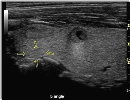
38 Acquisition Technologies B-Flow Color summary Clinical Advantages Dynamic Flow appearance High Frame rate Clear Background tissue with vessel hemodynamics Clinical Uses High Grade stenosis Soft Plaque Early Thrombus formation Aneurysm Access graft Perfusion in Placenta, Spleen, and Kidney Challenges Tissue vibration, Penetration Limits ROI angle, BFC is less angle dependent than PDI or CF All above comparisons are to LOGIQ E9 BT11 49

39 Acquisition Technologies CrossXBeam What it is: Multiple co-planar images from different angles combined into a single image in real time Why it works: Successive frames help average out noise and smooth borders 50
40 Acquisition Technologies CrossXBeam Results: Helps improve Border definition Helps improve Contrast resolution Helps reduce Angular dependence of border or edge Helps reduce Speckle / Clutter Helps increase visualization of biopsy needles 51

41 Acquisition Technologies CrossXBeam Benefits 52


42 Acquisition Technologies CrossXBeam Visualizing Transmit Straight Fire on straight structure Echo Reaches probe Straight Fire on Angled structure Echo Misses probe Angled Fire on Angled structure Echo Reaches probe 53

43 Acquisition Technologies CrossXBeam off 3 angles 7 angles 54
44 Acquisition Technologies Speed of Sound What it is: An additional control to help optimize image resolution In applications where tissue types are diverse, it allows the user to choose settings that are well suited for that particular patient Clinical impacts: Adjusting the speed of sound can help improve: Resolution A sharp image, especially in breast Signal-to-Noise Adjustable focusing helps improve SNR 55

45 Acquisition Technologies Speed of Sound Applications Breast Abdomen Abdomen 2 Renal 56

46 Acquisition Technologies Speed of Sound breast example 1420 m/s has enhanced contrast & resolution in this case 57
47 Acquisition Technologies Speed of Sound Breast Useful to help handle a variety of breast types Fatty tissue tends to image better at low speed of sound Dense tissue tends to image better close to 1500/1540 Default is 1500 Liver Available, but unlikely to offer significant benefit 58

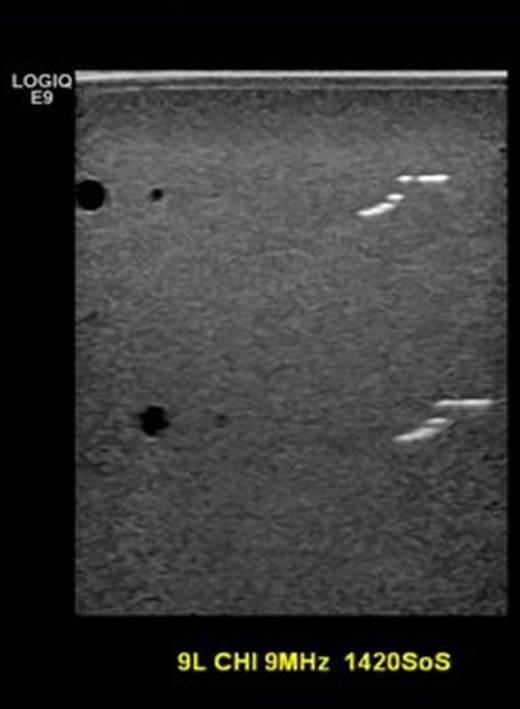
48 Acquisition Technologies Speed of Sound 59


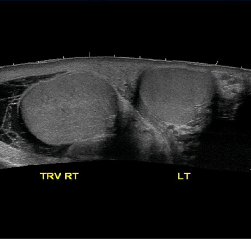
49 Acquisition Technologies Virtual Convex Expands the field of view of linear array probes Available on the B mode tab for linear array transducers 60
50 Acquisition Technologies Virtual Convex New Apex Position Probe Face Field of view Linear Probes Field of view Linear format Virtual Apex format 61


51 Acquisition Technologies LOGIQView What is it? Move the probe along the anatomy and create an image with a large field of view Benefits Enhanced demonstration of anatomical relationships Ability to measure large structures LOGIQView up to 60cm 62
52 63

53 Post-Acquisition Technologies Raw Data Speckle Reduction Imaging 64

54 Post-Acquisition Technologies Raw Data Raw data capture enables you to build a thorough exam while helping reduce scan time. This proprietary raw data format from GE Healthcare captures data earlier in the image processing chain enabling users to make changes to the data during or even after the exam has ended. Room too bright? Adjust gain later Forgot annotations? Easily add them later Delicate NICU patient? Acquire quickly then virtually rescan later Difficult vascular patient? Adjust baseline shift and sweep speed later 65
55 Post-Acquisition Technologies Raw Data Original Acoustic Data are stored before Scan Converting in a GE Raw Format to be easily accessed and re-processed any time after the exam completion. Highlights: Helps optimize sub-optimal studies. Measurements can be re-done and reports regenerated Imaging control parameters can be changed, such as: B-Mode: Gain, DR, Zoom, SRI CFM: Gain, Threshold, DualView, DR PW: Baseline, Invert, Angle, DR, Gain 66


56 Post-Acquisition Technologies Speckle reduction imaging Adaptive, real-time software algorithm min max 67

57 Post-Acquisition Technologies Speckle reduction imaging Adaptive, real-time software algorithm Preserves borders where echogenicity differences occur Smoothes the image where there is no border or edge Algorithm does not create structures but rather allows user to see the underlying anatomy 68

58 Post-Acquisition Technologies Speckle reduction imaging 69
59 Architecture of Quality Imaging Thank You! 2013 General Electric Company All rights reserved. 71 DOC
GE Healthcare LOGIQ P3. Staying ahead of the curve
 GE Healthcare LOGIQ P3 Staying ahead of the curve Ultrasound Expertise. Enhanced. GE is a trusted partner of many healthcare providers around the world. GE s TruScan technology, incorporating SmartScan,
Ultrasound Bioinstrumentation. Topic 2 (lecture 3) Beamforming
 Beamforming") Ultrasound Bioinstrumentation Topic 2 (lecture 3) Beamforming Angular Spectrum 2D Fourier transform of aperture Angular spectrum Propagation of Angular Spectrum Propagation as a Linear Spatial Filter Free
Ultrasound Bioinstrumentation Topic 2 (lecture 3) Beamforming Angular Spectrum 2D Fourier transform of aperture Angular spectrum Propagation of Angular Spectrum Propagation as a Linear Spatial Filter Free
Beyond Performance and Value
 Putting the back in ultrasound Beyond Performance and Value Esaote s new ultra-performance MyLab 9 exp ultrasound system is designed to support a full range of shared service diagnostic imaging environments.
A COMPACT SYSTEM WITH ADVANCED PERFORMANCE
 Samsung Medison is a global leading medical devices company. Founded in 1985, the company sells cutting-edge diagnostic ultrasound devices around the world in various medical fields. The company has attracted
Artifacts. Artifacts. Causes. Imaging assumptions. Common terms used to describe US images. Common terms used to describe US images
 Artifacts Artifacts Chapter 20 What are they? Simply put they are an error in imaging These artifacts include reflections that are: not real incorrect shape, size or position incorrect brightness displayed
Ultrasound Beamforming and Image Formation. Jeremy J. Dahl
 Ultrasound Beamforming and Image Formation Jeremy J. Dahl Overview Ultrasound Concepts Beamforming Image Formation Absorption and TGC Advanced Beamforming Techniques Synthetic Receive Aperture Parallel
The Script of ZST + Presentation. MIS Upstream Marketing Team [ 日期 ]
![The Script of ZST + Presentation. MIS Upstream Marketing Team [ 日期 ]](/thumbs/94/119182132.jpg "The Script of ZST + Presentation. MIS Upstream Marketing Team [ 日期 ]") 1 The Script of ZST + Presentation MIS Upstream Marketing Team [ 日期 ] 1 The Script of ZST + Presentation Since Mindray was founded to develop ultrasound business, core technology has always been the engine
1 The Script of ZST + Presentation MIS Upstream Marketing Team [ 日期 ] 1 The Script of ZST + Presentation Since Mindray was founded to develop ultrasound business, core technology has always been the engine
Introduction to Medical Engineering (Medical Imaging) Ultrasound Imaging. Ho Kyung Kim Pusan National University
 Ultrasound Imaging. Ho Kyung Kim Pusan National University") Introduction to Medical Engineering (Medical Imaging) Suetens 6 Ultrasound Imaging Ho Kyung Kim Pusan National University Sound Sonic: 20 Hz 20 khz (audible frequency) Subsonic () Ultrasound
Introduction to Medical Engineering (Medical Imaging) Suetens 6 Ultrasound Imaging Ho Kyung Kim Pusan National University Sound Sonic: 20 Hz 20 khz (audible frequency) Subsonic () Ultrasound
Physics of Ultrasound Ultrasound Imaging and Artifacts รศ.นพ.เดโช จ กราพาน ชก ล สาขาหท ยว ทยา, ภาคว ชาอาย รศาสตร คณะแพทยศาสตร ศ ร ราชพยาบาล
 Physics of Ultrasound Ultrasound Imaging and Artifacts รศ.นพ.เดโช จ กราพาน ชก ล สาขาหท ยว ทยา, ภาคว ชาอาย รศาสตร คณะแพทยศาสตร ศ ร ราชพยาบาล Diagnosis TTE TEE ICE 3D 4D Evaluation of Cardiac Anatomy Hemodynamic
Pass Ultrasound Physics Exam
 Pass Ultrasound Physics Exam Match the Answers By Mansoor Khan MBBS, RDMS, RDCS 1 Copyright 2014 Blue Cube Venture, LLC All rights reserved. The Pass Ultrasound Physics Exam Match the Answers is protected
4 Working With Scan Modes
 4 Working With Scan Modes Scan Modes Overview All of the information in this chapter pertains to live imaging. Many of the controls and functions change when you freeze the scan. For information on using
DC-6. Diagnostic Ultrasound System
 DC-6 Diagnostic Ultrasound System DC-6 is a general purpose color Doppler ultrasound system aiming at most clinical areas both in exam and research with various transducers and multi software packages
Design Your Performance
 MEDISON has been a leading name in diagnostic ultrasound since its foundation in 1985. As one of the only companies dedicated solely to ultrasound imaging, we have remained at the forefront of research
UGEO H60. Performance in Style. Features
 UGEO H60 Performance in Style The UGEO H60 implements superior performance with new design principles of simplicity and lightness. Its 10.1" touchscreen improves usability while its 18.5" LED monitor enhances
Ultrasound & Artifacts
 ISSN 2005-7881 Journal of Neurosonology 3(Suppl. 2):1-17, 2011 Ultrasound & Artifacts Siryung Han The Catholic University of Korea Artifacts False image- echoes without anatomic correlate US image dose
Photomultiplier Tube
 Nuclear Medicine Uses a device known as a Gamma Camera. Also known as a Scintillation or Anger Camera. Detects the release of gamma rays from Radionuclide. The radionuclide can be injected, inhaled or
Lesson 06: Pulse-echo Imaging and Display Modes. These lessons contain 26 slides plus 15 multiple-choice questions.
 Lesson 06: Pulse-echo Imaging and Display Modes These lessons contain 26 slides plus 15 multiple-choice questions. These lesson were derived from pages 26 through 32 in the textbook: ULTRASOUND IMAGING
COMPUTER PHANTOMS FOR SIMULATING ULTRASOUND B-MODE AND CFM IMAGES
 Paper presented at the 23rd Acoustical Imaging Symposium, Boston, Massachusetts, USA, April 13-16, 1997: COMPUTER PHANTOMS FOR SIMULATING ULTRASOUND B-MODE AND CFM IMAGES Jørgen Arendt Jensen and Peter
Chapter 4. Pulse Echo Imaging. where: d = distance v = velocity t = time
 Chapter 4 Pulse Echo Imaging Ultrasound imaging systems are based on the principle of pulse echo imaging. These systems require the use of short pulses of ultrasound to create two-dimensional, sectional
Optimisation of Image Acquisition Bordeaux 16th November J.S. McGhie W.B. Vletter R. Frowijn No disclosures
 Optimisation of Image Acquisition Bordeaux 16th November 2016 J.S. McGhie W.B. Vletter R. Frowijn No disclosures Image optimisation: The Echo machine It looks difficult to drive an echo machine!! Some
The physics of ultrasound. Dr Graeme Taylor Guy s & St Thomas NHS Trust
 The physics of ultrasound Dr Graeme Taylor Guy s & St Thomas NHS Trust Physics & Instrumentation Modern ultrasound equipment is continually evolving This talk will cover the basics What will be covered?
Resona 6 Premium Ultrasound System
 The system Resona 6 is the newly developed, unique result of the mergence of leading companies Mindray Bio-medical Electronics Co. Ltd. and ZONARE Medical Systems, Inc.. By additions to the core competencies
Ultrasound Physics. History: Ultrasound 2/13/2019. Ultrasound
 Ultrasound Physics History: Ultrasound Ultrasound 1942: Dr. Karl Theodore Dussik transmission ultrasound investigation of the brain 1949-51: Holmes and Howry subject submerged in water tank to achieve
DC-6 Expert. Diagnostic Ultrasound System
 DC-6 Expert Diagnostic Ultrasound System MINDRAY has newly released DC-6 Expert, a general purpose color Doppler ultrasound system with full ergonomic designs, supplying more accessible exams, higher imaging
160-slice CT SCANNER / New Standard for the Future
 TECHNOLOGY HISTORY For over 130 years, Toshiba has been a world leader in developing technology to improve the quality of life. Our 50,000 global patents demonstrate a long, rich history of leading innovation.
M5 Diagnostic Ultrasound System
 V0807 M5 Diagnostic Ultrasound System Mindray s ultrasound family is now introducing a new member, M5 hand-carried color Doppler system. M5, coming in a laptop size with comprehensive ergonomic design,
3. Ultrasound Imaging(2)
") 3. Ultrasound Imaging(2) Lecture 13, 14 Medical Imaging Systems Jae Gwan Kim, Ph.D. jaekim@gist.ac.kr, X 2220 Department of BioMedical Science and Engineering Gwangju Institute of Sciences and Technology
3. Ultrasound Imaging(2) Lecture 13, 14 Medical Imaging Systems Jae Gwan Kim, Ph.D. jaekim@gist.ac.kr, X 2220 Department of BioMedical Science and Engineering Gwangju Institute of Sciences and Technology
Principles of Ultrasound Imaging Image Optimization
 Principles of Ultrasound Imaging Image Optimization Robert A. Levine, MD, FACE, ECNU Thyroid Center of New Hampshire Geisel School of Medicine at Dartmouth College Disclosures: No relevant financial or
Quick Reference Guide
 siemens.com/nx3 Quick Reference Guide ACUSON NX3 Series Contents 2 System Overview 3 Getting Started 8 2D Mode and M-mode 12 Color and Spectral Doppler 24 Measurements and Calculations 38 Text, Arrows
The Physics of Echo. The Physics of Echo. The Physics of Echo Is there pericardial calcification? 9/30/13
 Basic Ultrasound Physics Kirk Spencer MD Speaker has no disclosures to make Sound Audible range 20Khz Medical ultrasound Megahertz range Advantages of imaging with ultrasound Directed as a beam Tomographic
Ultrasound Imaging Ultr Michael Dadd 2007
 Ultrasound Imaging Ultrasound Physics & Instrumentation - Recommended Reading 1. Diagnostic Ultrasound: Principles and Instruments (7th Ed) Frederick W Kremkau W B Saunders Company 2. Applied Physics &
NextGen LOGIQ e. Ultrasound. GE Healthcare. Product Description
 GE Healthcare NextGen LOGIQ e Ultrasound Product Description The NextGen LOGIQ e combines the high performance of a console system with the portability of a laptop. GE Healthcare s compact system is designed
12/26/2017. Alberto Ardon M.D.
 Alberto Ardon M.D. 1 Preparatory Work Ultrasound Physics http://www.nysora.com/mobile/regionalanesthesia/foundations-of-us-guided-nerve-blockstechniques/index.1.html Basic Ultrasound Handling https://www.youtube.com/watch?v=q2otukhrruc
Multipurpose Color Ultrasound System IMAGING SYSTEMS
 i3 Multipurpose Color Ultrasound System IMAGING SYSTEMS IMAGING SYSTEMS i3 Color Ultrasound System M The revolutionary i3 provides you outstanding 2D images and fast 4D volume images, while the streamlined
Ultrasound physical principles in today s technology
 Education Ultrasound physical principles in today s technology Brian Starkoff M.App.Sc.(Med. Ultrasound), AMS Holland Park Brisbane Queensland Australia Correspondence to email starkoff@optusnet.com.au
LOGIQ e Ultrasound. GE Healthcare. Product Description
 GE Healthcare LOGIQ e Ultrasound Product Description The LOGIQ e combines the high performance of a console system with the portability of a laptop. GE Healthcare s compact system is designed for general
Medical Imaging (EL582/BE620/GA4426)
") Medical Imaging (EL582/BE620/GA4426) Jonathan Mamou, PhD Riverside Research Lizzi Center for Biomedical Engineering New York, NY jmamou@riversideresearch.org On behalf of Prof. Daniel Turnbull Outline
Medical Imaging (EL582/BE620/GA4426) Jonathan Mamou, PhD Riverside Research Lizzi Center for Biomedical Engineering New York, NY jmamou@riversideresearch.org On behalf of Prof. Daniel Turnbull Outline
 www.hitachi-aloka.com Revolutionary Performance; Ease of Use The ProSound 6 is the next generation of compact color ultrasound systems, providing unprecedented performance with a broad range of applications.
Multi-Access Biplane Lab
 Multi-Access Biplane Lab Advanced technolo gies deliver optimized biplane imaging Designed in concert with leading physicians, the Infinix VF-i/BP provides advanced, versatile patient access to meet the
Discover the new Prestige and experience 3D/4D imaging beyond your imagination.
 3D/4D Beyond Imagination The Prestige ultrasound imaging system represents the pinnacle of more than a decade of technological advancement in 3D/4D ultrasound imaging at MEDISON. Inheriting a tradition
EVIS EUS ENDOSCOPIC ULTRASOUND CENTER EU-ME2 Dedicated ultrasound processor with versatile functionality.
 EVIS EUS ENDOSCOPIC ULTRASOUND CENTER EU-ME2 Dedicated ultrasound processor with versatile functionality. Advancing the Art of Endosonography The EU-ME2 is a high-quality compact ultrasound processor for
Nuove tecnologie per ecografia ad ultrasuoni: da 2D a 4D
 DINFO Dipartimento di Ingegneria dell Informazione Department of Information Engineering Nuove tecnologie per ecografia ad ultrasuoni: da 2D a 4D Piero Tortoli Microelectronics Systems Design Lab 1 Introduction
Key Physics and Doppler Principles
 Key Physics and Doppler Principles Robert A. Levine, MD, FACE, ECNU Thyroid Center of New Hampshire Geisel School of Medicine at Dartmouth College AACE/ACE Advanced Neck Ultrasound Training Course Disclosures:
SONOACE 6000C MT DIGITAL COLOR. Affordable PC-based Digital CFM Ultrasound System. Specifications.
 Specifications Imaging Modes Gray Scale Focusing PC Monitor Speaker Measurements Image Processing Display Functions Peripheral Devices Support Physical Dimensions Electrical Parameters Probe Types Single
Doppler in Obstetrics: book by K Nicolaides, G Rizzo, K Hecher. Chapter on Doppler ultrasound: principles and practice by Colin Deane
 Doppler in Obstetrics: book by K Nicolaides, G Rizzo, K Hecher Chapter on Doppler ultrasound: principles and practice by Colin Deane INTRODUCTION Competent use of Doppler ultrasound techniques requires
Radionuclide Imaging MII Single Photon Emission Computed Tomography (SPECT)
") Radionuclide Imaging MII 3073 Single Photon Emission Computed Tomography (SPECT) Single Photon Emission Computed Tomography (SPECT) The successful application of computer algorithms to x-ray imaging in
Radionuclide Imaging MII 3073 Single Photon Emission Computed Tomography (SPECT) Single Photon Emission Computed Tomography (SPECT) The successful application of computer algorithms to x-ray imaging in
Ultrasound Miniaturization
 Ultrasound Miniaturization Scott Smith Ultrasound Probes Lab Clinical Systems and Devices GE Global Research 2011 Joint AAPM / COMP Meeting July 31-August 4 Vancouver 1 Acknowledgements Charles Baumgartner
Vivid S5. Cardiovascular ultrasound system. GE Healthcare. Davis Medical
 GE Healthcare Vivid S5 Cardiovascular ultrasound system Versatility. It s a new design concept one that leverages our miniaturization expertise gained from the Vivid i and our performance expertise of
MR Advance Techniques. Flow Phenomena. Class II
 MR Advance Techniques Flow Phenomena Class II Flow Phenomena In this class we will explore different phenomenona produced from nuclei that move during the acquisition of data. Flowing nuclei exhibit different
Image Optimization: The Sonographer s Responsibility. Prepared by Cathy Daniels, EdD, RTR, RDMS, RDCS, RVT
 Image Optimization: The Sonographer s Responsibility Prepared by Cathy Daniels, EdD, RTR, RDMS, RDCS, RVT Image Optimization: The Sonographer s Responsibility Cathy Daniels, EdD, RTR, RDMS, RDCS, RVT Disclosure
Q5 VET. Compact, Affordable Color Doppler for VET
 Q5 VET Compact, Affordable Color Doppler for VET Key Benefits Ergonomic design for learning easily, using efficiently Professional probes and software for veterinary Abundant image mode: B, 2B,4B,C,PW,M
Introduction. Parametric Imaging. The Ultrasound Research Interface: A New Tool for Biomedical Investigations
 The Ultrasound Research Interface: A New Tool for Biomedical Investigations Shelby Brunke, Laurent Pelissier, Kris Dickie, Jim Zagzebski, Tim Hall, Thaddeus Wilson Siemens Medical Systems, Issaquah WA
Printed in Japan E318
 Diagnostic Ultrasound System MODEL PROSOUND 6 The specifications, shape and color of this product are subject to change without notice. The standard components and optional items vary depending on the
General Imaging Ultrasound in a Whole New Light. Introducing the
 General Imaging Ultrasound in a Whole New Light. Introducing the It s a Tour de Force Redefining innovation through value and performance It s the dawning of a new day in the world of compact ultrasound
Model: ESE-6 Portable Color Ultrasound System
 DATA SHEET Model: ESE-6 Portable Color Ultrasound System Ultrasound System Specifications The premium performance of the full functional Portable ESE-6 provides a fast and easy diagnosis by: Ultra-premium
Introduction to Ultrasound Physics
 Introduction to Ultrasound Physics Vassilis Sboros Medical Physics and Cardiovascular Sciences University of Edinburgh Transverse waves Water remains in position Disturbance traverse producing more wave
Cardiac MR. Dr John Ridgway. Leeds Teaching Hospitals NHS Trust, UK
 Cardiac MR Dr John Ridgway Leeds Teaching Hospitals NHS Trust, UK Cardiac MR Physics for clinicians: Part I Journal of Cardiovascular Magnetic Resonance 2010, 12:71 http://jcmr-online.com/content/12/1/71
Reconfigurable Arrays for Portable Ultrasound
 Reconfigurable Arrays for Portable Ultrasound R. Fisher, K. Thomenius, R. Wodnicki, R. Thomas, S. Cogan, C. Hazard, W. Lee, D. Mills GE Global Research Niskayuna, NY-USA fisher@crd.ge.com B. Khuri-Yakub,
Excellent Performance; Ease of Use
 Excellent Performance; Ease of Use High performance with a broad range of applications - the compact ProSound α6 is at your service. Our ProSound series has a well-established reputation in hospitals and
Retrospective Transmit Beamformation. Whitepaper. ACUSON SC2000 Volume Imaging Ultrasound System. Answers for life.
 Whitepaper Retrospective Transmit Beamformation ACUSON SC2000 Volume Imaging Ultrasound System Chuck Bradley, Ph.D. Siemens Healthcare Sector Ultrasound Business Unit Mountain View, California USA Answers
Lesson 06: Pulse-echo Imaging and Display Modes. This lesson contains 22 slides plus 15 multiple-choice questions.
 Lesson 06: Pulse-echo Imaging and Display Modes This lesson contains 22 slides plus 15 multiple-choice questions. Accompanying text for the slides in this lesson can be found on pages 26 through 32 in
Ultrasound System Specifications
 Ultrasound System Specifications The superb imaging and performance capabilities of VINNO G60 give you the confidence and efficiencies in your daily exam by: World class image quality with unmatched high-frequency
Research Support. Dual-Source CT: What is it and How Do I Test it? Cynthia H. McCollough, Ph.D.
 Dual-Source CT: What is it and How Do I Test it? Cynthia H. McCollough, Ph.D. CT Clinical Innovation Center Department of Radiology Mayo Clinic College of Medicine Rochester, MN Research Support National
Software and Hardware in CCTA. Elly Castellano PhD
 Software and Hardware in CCTA Elly Castellano PhD Outline technical requirements for coronary CTA the modern cardiac CT scanner ECG-gating technology image reconstruction algorithms 2 Technical requirements
Ziehm Vision / Ziehm Vision FD The new standard in mobile imaging. > Flat-Panel Detector. > Image Intensifier
 2 2 Ziehm Vision / Ziehm Vision FD The new standard in mobile imaging > Flat-Panel Detector > Image Intensifier 02 03 Ziehm Vision 2 18" TFT monitors provide bright, high contrast image with a wide viewing
Ultrasound System Specifications
 Ultrasound System Specifications Dedicated premium women healthcare ultrasound system VINNO M80 supports you in clinical decision-making and elevates trust in diagnostic confidence by: Premium image quality,
Brilliance in everything Philips CT products and services
 Brilliance in everything Philips CT products and services Ready for anything No one does more than Philips to help you gain the productivity you need with a comprehensive approach to CT that marries significant
Multi-Element Synthetic Transmit Aperture Method in Medical Ultrasound Imaging Ihor Trots, Yuriy Tasinkevych, Andrzej Nowicki and Marcin Lewandowski
 Multi-Element Synthetic Transmit Aperture Method in Medical Ultrasound Imaging Ihor Trots, Yuriy Tasinkevych, Andrzej Nowicki and Marcin Lewandowski Abstract The paper presents the multi-element synthetic
Ultrasound System Specifications
 Ultrasound System Specifications Advanced women healthcare features and image quality VINNO M50 increases your clinical confidence for all obstetrical, gynecological and breast exams by Innovative, cutting-edge
ENDOSCOPIC ULTRASOUND SYSTEMS
 ENDOSCOPIC ULTRASOUND SYSTEMS DISCOVER HIGH-PRECISION DIAGNOSES AND PROCEDURES NEW ENDOSCOPIC ULTRASOUND Ultrasonography revolutionized the clinical approach to patients with digestive and respiratory
The Middle East Distributor for AMBISEA Technology Corp. Electro-Medical Product Line
 The Middle East Distributor for AMBISEA Technology Corp. Electro-Medical Product Line AV-9100 Single Channel ECG 1 2 AV-9300 3-Channels ECG 3 4 5 AV-9000B Multi-Parameter Patient Monitor 6 7 8 AV-9000C
HETERONUCLEAR IMAGING. Topics to be Discussed:
 HETERONUCLEAR IMAGING BioE-594 Advanced MRI By:- Rajitha Mullapudi 04/06/2006 Topics to be Discussed: What is heteronuclear imaging. Comparing the hardware of MRI and heteronuclear imaging. Clinical applications
Session: 2A NEW ULTRASOUND SYSTEMS Chair: H. Ermert University of Bochum 2A-1 10:30 a.m.
 Session: 2A NEW ULTRASOUND SYSTEMS Chair: H. Ermert University of Bochum 2A-1 10:30 a.m. TISSUE HARMONIC IMAGING WITH IMPROVED TEMPORAL RESOLUTION D. J. NAPOLITANO*, C. H. CHOU, G. W. MCLAUGHLIN, T. L.
Model: ESE-G50 Color Ultrasound System
 DATA SHEET Model: ESE-G50 Color Ultrasound System Ultrasound System Specifications Extremely portable and exceptional performance ESE-G50 meets all your clinical needs by: Unmatched image quality All ranges
KIZLON. Sentinel House,Harvest Crescent, Ancells Business Park, Fleet Hampshire GU51 2UZ, UK Website:
 KIZLON Sentinel House,Harvest Crescent, Ancells Business Park, Fleet Hampshire GU51 2UZ, UK Email: info@kizlon.com Website: www.kizlon.com Portable Ultrasound Scanners KUS-A100 & A101 Ultrasound Scanners
ACUSON X150 Knobology & User Guide
 ACUSON X150 Knobology & User Guide ACUSON X150 CONTROL PANEL Table of Contents The ACUSON X150 knobology and user guide is the clinicians quick reference of system terms, functions and capabilities. This
Endoscopic Ultrasonography System
 Endoscopic Ultrasonic Processor SU- -H-, SU- -S- Power rating Power supply rating Current consumption(rated) Dimensions Size Weight Ultrasonography Probe types image display Scanning modes Special modes*
PERFORM Operating Document. Use and Maintenance of G.E. Ultrasound LOGIQ E
 PERFORM Operating Document Use and Maintenance of G.E. Ultrasound LOGIQ E PC-POD-IM-001-v02 Revision History Version Reason for Revision Date 01 New POD 22/May/2013 02 Minor changes only 2.2 Change responsibility
Image Quality. HTC Grid High Transmission Cellular Grid provides higher contrast images
 B R E A S T I M A G I N G S O L U T I O N S Setting the benchmark for mammography M-IV Series Innovations in breast imaging The Lorad M-IV Series exemplifies Hologic s commitment to developing advanced
S S S2 Operation Manual
 PREPARATION 1. How to create and input patient data? In the MAIN INTERFACE, press the key, to enter into the patient exam list interface. Then, click New patient item to create new patient files. Patient
GE Healthcare. Essential for life. Senographe Essential Full-Field Digital Mammography system
 GE Healthcare Essential for life Senographe Essential Full-Field Digital Mammography system Excellence in FFDM is a process. An ongoing quest, fueled by our continuing breakthroughs in breast cancer detection
SIGNA Architect. Make the unimaginable the expected. Issue Spotlight. Issue Spotlight
 SIGNA Architect Make the unimaginable the expected The potential for MR is now even more astonishing. Introducing SIGNA Architect 3.0T, the most advanced and intuitive engineering in MR technology from
12/21/2016. Siemens Medical Systems Research Agreement Philips Healthcare Research Agreement AAN and ASN Committees
 Joseph V. Fritz, PhD Nandor Pintor, MD Dent Neurologic Institute ASN 2017 Friday, January 20, 2017 Siemens Medical Systems Research Agreement Philips Healthcare Research Agreement AAN and ASN Committees
Optical coherence tomography
 Optical coherence tomography Peter E. Andersen Optics and Plasma Research Department Risø National Laboratory E-mail peter.andersen@risoe.dk Outline Part I: Introduction to optical coherence tomography
Diagnostic Ultrasound System. Operation Note
 M5 Diagnostic Ultrasound System Table of Contents System Introduction...3 Control Panel...4 Control Panel...5 Control Panel...6 Control Panel...7 Control Panel...8 Control Panel...9 Power ON / OFF the
Session: 1E CONTRAST AGENTS II Chair: K. Ferrara University of California-Davis. 1E-1 10:30 a.m.
 Session: 1E CONTRAST AGENTS II Chair: K. Ferrara University of California-Davis 1E-1 10:30 a.m. PULSE INVERSION DOPPLER FOR BLOOD FLOW DETECTION IN THE MACRO- AND MICRO-VASCULATURE WITH ULTRASOUND CONTRAST
Ultrasonic Linear Array Medical Imaging System
 Ultrasonic Linear Array Medical Imaging System R. K. Saha, S. Karmakar, S. Saha, M. Roy, S. Sarkar and S.K. Sen Microelectronics Division, Saha Institute of Nuclear Physics, 1/AF Bidhannagar, Kolkata-700064.
Point-of-Care Ultrasound in a Whole New Light. Introducing the
 Point-of-Care Ultrasound in a Whole New Light. Introducing the It s a Tour de Force for Point-of-Care Ultrasound Redefin ing innovation through value and performance It s the dawning of a new day in the
ULTRASONIC IMAGING of COPPER MATERIAL USING HARMONIC COMPONENTS
 ULTRASONIC IMAGING of COPPER MATERIAL USING HARMONIC COMPONENTS T. Stepinski P. Wu Uppsala University Signals and Systems P.O. Box 528, SE- 75 2 Uppsala Sweden ULTRASONIC IMAGING of COPPER MATERIAL USING
MULTI-FREQUENCY ULTRASOUND IMAGING: PHANTOM STUDY
 MULTI-FREQUENCY ULTRASOUND IMAGING: PHANTOM STUDY SITI NUR MASTURAH BINTI ABDUL MALEK DEPARTMENT OF DIAGNOSTIC IMAGING AND RADIOTHERAPY, KULLIYYAH OF ALLIED HEALTH SCIENCES, INTERNATIONAL ISLAMIC UNIVERSITY
GE Healthcare. Vivid S5. Cardiovascular ultrasound system
 GE Healthcare Vivid S5 Cardiovascular ultrasound system Versatility. It s a new design concept one that leverages our miniaturization expertise gained from the Vivid i and our performance expertise of
Real Time Deconvolution of In-Vivo Ultrasound Images
 Paper presented at the IEEE International Ultrasonics Symposium, Prague, Czech Republic, 3: Real Time Deconvolution of In-Vivo Ultrasound Images Jørgen Arendt Jensen Center for Fast Ultrasound Imaging,
Performing ultrasound probe quality assurance assessments: A How-to Guide
 Performing ultrasound probe quality assurance assessments: A How-to Guide A comprehensive quality assurance program has the potential to directly contribute to better patient outcomes. Regular testing
Beamforming in ultrasound
 Peter Pazmany Catholic University Faculty of Information Technology www.itk.ppke.hu Medical diagnostic systems (Orvosbiológiai képalkotó rendszerek) Beamforming in ultrasound ( Nyalábalkotás az ultrahangban)
Quality Ultrasound Systems
 Quality P11VExpert Medisono offers quality products to the medical imaging market. We offer different solutions tailored to the needs of each patient, offering the best value to every single patient. We
Invisible sophistication. Visible simplicity. CS Welcome to the simplicity of compact panoramic imaging
 Invisible sophistication. Visible simplicity. CS 8100 Welcome to the simplicity of compact panoramic imaging Introducing the CS 8100 The Carestream Dental Factor Humanized technology We keep our technology
Advanced digital image processing for clinical excellence in fluoroscopy
 Dynamic UNIQUE Digital fluoroscopy solutions Dynamic UNIQUE Advanced digital image processing for clinical excellence in fluoroscopy André Gooßen, PhD, Image Processing Specialist Dörte Hilcken, Clinical
18th World Conference on Nondestructive Testing, April 2012, Durban, South Africa. Joanna X.Qiao 1, Matthias Jobst 2
 8th World Conference on ondestructive Testing, 6-0 April 0, Durban, outh Africa An Adaptive Phased-Array Imaging ethod for Ultrasonic Testing Joanna X.Qiao, atthias Jobst GE Inspection Technologies; 50
: PT Philips Indonesia CERTIFICATE OF PRODUCTION SOLE AGENT/ DISTRIBUTOR : NA CERTIFICATE OF DISTRIBUTION (PAK)
") SOLE AGENT/ DISTRIBUTOR : PT Philips Indonesia CERTIFICATE OF PRODUCTION : NA CERTIFICATE OF DISTRIBUTION (PAK) : HK.07.Alkes/IV/625/AK.2.2012 ADDRESS : Jl. Buncit Raya Kav 99-100, Pasar Minggu, Jak-Sel
SOLE AGENT/ DISTRIBUTOR : PT Philips Indonesia CERTIFICATE OF PRODUCTION : NA CERTIFICATE OF DISTRIBUTION (PAK) : HK.07.Alkes/IV/625/AK.2.2012 ADDRESS : Jl. Buncit Raya Kav 99-100, Pasar Minggu, Jak-Sel