Advances in Computed Tomography and Digital Mammography Ruvin Deych and Sorin Marcovici Analogic Corporation Peabody, MA 01960, USA
|
|
|
- Barbra Scott
- 5 years ago
- Views:
Transcription

1 Advances in Computed Tomography and Digital Mammography Ruvin Deych and Sorin Marcovici Analogic Corporation Peabody, MA 01960, USA ISMART, November 18, 2008, Kharkov 1
2 Outline Advances in Medical X-Ray CT World market of X-Ray CT Principles of third generation CT Main performance parameters of modern CT Data Measurement System in modern X-Ray CT Future trends in X-Ray CT Advances in Digital mammography Selenium/TFT technology Performance of Selenium based mammography detectors Manufacturing Tomosynthesis 2
3 Global CT Market Note: U.S. data in orders % U.S. Decline CT Market ($US Mil) US Market ($US Mil) Rest of World Source: Philips Medical, Fuji-Kezai, Analogic 3
4 US CT Market Distribution Market Share by Product Tier (2006) Based on unit volume 20% 41% Up to 10 Slice 16 Slice 32 Slice 29% 64 Slice 10% Source: 2006 Frost & Sullivan 4

5 CT Market Pricing Trends Source: 2008 Frost & Sullivan 5
6 Evolution and Present Status of Medical X-Ray CT Imaging 6


7 Medical CT 7


8 CT Systems Medical 8

9 Analogic in CT CT Subsystems Data Management Systems (DMS) Data Acquisition Systems (DAS) Detector Arrays Gantries PowerLink Non-Contact Power Transfer Collimators Reconstruction Software Motion Control Patient Table Operator Contol Station 9


10 CT Systems Security 10
11 Milestones in X-ray CT Sequential scanning of consecutive slices: 1970s Spiral scanning-ct acquisition with continuous translation of patient: beginning of 1990s Multislice spiral CT: end of 1990s Dual source CT: 2000s Main trend: increasing speed of acquisition and axial coverage (slices) 11
12 Multislice Spiral CT Scanning Mid 1990s: 300 mm lung or abdominal examination with narrow slice requires 200 s long scan Mid 2000s: same examination takes 1-3 sec High speed of CT allows large scan within one breath hold, and to acquire images of moving organs, such as heart. 12

13 CT applications Coronary Angiography Rest phase of coronary arteries is 60 msec! Non-invasive emergency diagnosis for cause of chest pain: coronary blockage, pulmonary embolism, aortic aneurism. Alternative: 6 hour long invasive catheterization procedure with 1% risk of serious complications, including death. Courtesy of Toshiba Medical Corporation 13
14 Main drivers in X-ray CT Short scan time and large axial coverage for reliable anatomical and functional measurements of whole organs (perfusion of heart, brain, lung) Ultra-High spatial resolution Anatomical/Functional multi-modality imaging in SPECT-CT, PET-CT Patient dose reduction Is slice war over? 14
15 DMS for X-Ray CT Scan time: 0.3 sec XRT Power: 100 kw Axial coverage: mm at isocenter Spatial resolution: ~0.5 mm Remarkable progress in X- Ray CT in the past decade is largely explained by fast development of the Data Measurement System (DMS) 15
16 Main DMS Parameters Parameter Typical value Parameter Typical value X-ray energy, kvp Rotation time, sec 0.3 XRT power, kw 100 Data transfer rate, 10 GBit/sec FOV, cm 50 Operational C temperature range Isotropic resolution at Number of x-ray 300,000 isocenter, mm photons per sample at peak power Number of channels per Conversion efficiency, row el/ev Number of rows 64, 128, 320 Sampling rate, Hz 3000 Typical element size, mm Typical detector distance from XRT focal spot, mm 1x1 Data resolution, bit Dynamic range, bit
17 Detector Channels per CT High End ( slices) Mid Range (16 slices) Low End (single slice) up to 300,000 57, Analogic DAS/DMS From channels in 1993 to 300,000 channels in 2008 DAS/DMS complexity increases at constant: Cost Power consumption and almost same Mechanical Envelope 17
18 Charge Integrating CT Detector 18

19 Uniform and Adaptive Detector Configurations Uniform coverage in axial direction. Used in most 64, 128, 256 slice Scanners. Adaptive arrays have fewer septa, and and DAS channels. Used primarily in CT with in CTs 16 or fewer with 16 slices. slices. 19


20 CT Subsystems DMS 1 64 Slice+ Integrated DAS and Detector Assemblies X-ray Beamline Design 20


21 16x64 CT Detector 21
22 Requirements for Scintillators in X-Ray CT Parameter Requirement Importance Energy range kev DQE (0) >95 % Image noise, dose reduction Dose rates at detector ~0.1 Gy/s Lifetime dose 100 kgy Light output (LO) >40,000 ph/mev Image noise at high attenuation Emission spectrum, nm Used with Si photodiodes Decay time <10 s To support >10 khz DAS rates Afterglow <10 ppm at 3 ms Image artifacts Susceptibility to radiation damage <1 %/Gy Image artifacts if channels non-uniform Lifetime degradation <20 % Dynamic range reduction LO temperature coefficient Low cost, non-toxic <0.3 %/ C Image artifacts if channels non-uniform DMS cost, cost of removal 22
23 Main Scintillators in X-Ray CT Scintillator Density Thickness a to Relative Emission Primary Afterglow (g/cm 3 ) stop 99 % Light band decay (% at 3 (mm) output b maximum ( s) ms) CdWO ( 495 ) 2, 15 <0.1 Gd 2 O 3 :Eu (Y,Gd) 2 O 3 :Eu Gd 2 O 2 S:Pr,Ce, <0.1 Gd F 2 O 2 S:Tb(Ce) La 2 HfO 7 :Ti Gd 3 Ga 5 O 12 :Cr, <0.1 a Thickness to absorb 99 % of x-ray photons generated by tungsten anode x-ray tube at 140 kvp. b Relative light output measured using silicon photodiode, under 140 kvp tungsten anode XRT excitation. General Electric introduced fast Gemstone garnet based ceramic scintillator in Limited data in public domain. 23
24 Silicon Photodiodes for X-Ray CT: Main Requirements Parameter Mode of operation Elements number per chip, typ Interconnect density Pitch in x-axis, typ Pitch in z-axis, typ NEP Rise, fall time Spectral response range Photosensitivity, typ Uniformity of photosensitivity Leakage current for 1x1 mm 2 Cross-talk Terminal capacitance for 1x1 mm 2 Value P-i-n structure, Photovoltaic, 0V bias 512 >64 per pitch 1.0 mm 1.0 mm < W/ Hz <1 s nm 0.3 A/W at 500 nm +/-2% ch-to-ch 5 pa, max,@10 mv bias, 25 C, 0.1 %, max 20 pf, 10 khz 24
25 Data Acquisition Electronics: Main Requirements Parameter Performance Note Electronic Noise e (1/2),m,m = Minimum Photon Noise Digitization Interval (1/6) ( 2 + e2 ) 1/2 = Photon Noise Sampling Rate 10 khz Offset Stability 1.5 ppm FSR / C FSR = Full Scale Reading Gain Stability ± 50 ppm FSR / C Integral Non Linearity 200 ppm R / C ± 2 ppm FSR R = Reading Differential Non Linearity 30 ppm R / C ± 1 ppm FSR Power Consumption 3 mw / Channel Packaging Channels AMP/ADC ASIC 25
26 Trends in Medical CT 1. New CT scanning geometry: Dual-Source, Multi-Source, Inverse-geometry Advantages: Faster acquisition, Cone-beam artifact reduction Requires multiple DMS, or area detector, expensive 2. Energy-sensitive CT Advantages: elimination of beam hardening artifacts, material discrimination, better contrast at lower dose Solutions Dual-layered detectors Dual-source CT, kvp switching Photon Counting Detectors with multiple energy bins 3. Multimodality CT: SPECT/CT, PET/CT, Preclinical systems 26
27 Trends in Medical CT 4. Phase-contrast imaging (more distant future) Phase-contrast imaging, based on difraction is more sensitive in kev range then attenuation based imaging. Requires interferometry and difractometry detection technique. 27
28 Multiple Source CT Multiple source/detector systems-old idea becomes a desirable development Higher rotation rates require increase in X-Ray power, not achievable with present X-Ray technology Fraction of rotation is required for a full scan Issues: scatter reduction, high cost Siemens introduced commercial Dual Source CT in

29 Energy sensitive CT: Dual Energy Detector Two crystals with different emission bands are used. Radiation is hardened by the first crystal. Optical band pass filters limit diodes to see signal from only one crystal. Advantages: Simultaneous acquisition of Low and High Energy samples. High Quantum Efficiency Planar silicon PDA technology R. Deych, US Patent 7,388,208 B Incident X-ray Photons Light Photons from High Energy Scintillator Light Photons from Low Energy Scintillator 29
30 Contrast-to-Noise Model Results (Teflon detail in water background) CNR CsI:Tl(LE)/CdWO4(HE) ZnSe/CdWO4 GOS/CdWO4 GGG/CdWO4 CsI:Na/CsI:Tl ZnSe/LSO GGG/LSO LE thickness (g/cm2) 30
31 Energy sensitive CT, Single Photon Counting X-Photons Single Pixel Analog Line V SPC Threshold Noise Time SPC CI S = N S = []dt 31
32 SPC in Computed Tomography New medical applications and capabilities Contrast media removal in images Multiple contrast agents Reduction of beam hardening artifacts Patient dose reduction Requires high counting rates up to 10 9 (!) photons/sec/mm 2 32
33 Direct Conversion Detectors Dual energy CZT based detector tested in LightSpeed GE CT scanner, IEEE 2007 Pre-clinical CT scanner with 6 energy bands based on CZT technology tested by Philips, IEEE 2008 Main drawbacks: Long carrier transit time, insufficient speed Material polarization at high exposure rates 33
34 Scintillator Based SPC X-ray CT will require fast scintillators and internal gain in photodetectors Fast scintillators with Solid State PM are being proposed for CT Potential Available Fast Scintillators: LSO, LYSO, LaBr 3 New faster scintillators with 1-10 nsec decay time are required 34


35 Multimodality CT: SPECT/CT, PET/CT ECT With AC ECT NC AC Image Fusion Attenuation Map Courtesy of General Electric, Functional Imaging 35
36 Advances in X-ray CT: Conclusions and Predictions CT scanners with 320 slice acquisition in 0.3 sec are available The slice war between major medical imaging companies is over! New CT systems will include novel scanning techniques: multiple sources, inverse geometries Multi-energy CT will be needed to obtain better tissue discrimination at lower patient dose. Photon counting detection may replace charge integration X-ray CT will become new market for ultrafast nanosecond scintillators. 36
37 Advances in Digital Mammography 37
38 Stating the Problem Increasing mammography clinical diagnostic s sensitivity and specificity while optimizing patients flow and reducing operational costs. 38
39 Digital Mammography Installed Units est est. 39
40 Average Time/Patient Film-based analog mammography: minutes Se-based digital mammography: 5 6 minutes 40
41 Digital Radiography Two Step Conversion - INDIRECT X-Ray to light to electrical charge One Step Conversion - DIRECT X-Ray to electrical charge 41
42 a: Se Technologies Generation I: dielectric isolation layer deposited on top of two layer p Se structure Generation II: single Se deposition process with real time doping to create three layer pin or nip structures 42
43 Mammography Detector General Characteristics Technology: amorphous Selenium Active area: 24 cm x 30 cm Resolution: 2816 x 3584 pixels Pixel pitch: 85 μm Acquisition speed: 2 frames/second Digitization: 14 bits 43
44 Se Characteristics Atomic Number: 34 Conversion Efficiency: 50 ev / e-h Evaporation Temperature: 217 deg. C Crystallization Temperature: 60 deg. C Expansion Coefficient: 40 ppm/deg. C 44
45 a: Selenium Detector Structure X-Rays Amorphous Selenium Layer TFT Array Charge amplifier 45
46 X-Ray Absorption selenium layer Attenuated X-Ray fraction or Quantum Efficiency: 1 exp[-al] Attenuated Fraction pixel 53 kev 25 kev Tickness (mm) = a (E,Z,r) mostly photoelectric Mammography 25 kev < Ex < 40 kev RT, Radiography 40 kev < Ex < 120 kev 46
47 Charge Generation -10kV F selenium layer Number of electron-hole pair created: Ex / W +/- W +/- = W +/- (E,F) for F = 10 V/mm, W +/- ~ 50 ev 47
48 Charge Drift E selenium layer L TFT glass data line data line Gate Line Charge Collection (induction) efficiency: = μ E/L { 1-exp[-L/μ E] } For a good detector μ E >> L μ: mobility : lifetime μ E: mean free path Typical values for a-se: μ E(e) = 3-4 mm μ E(h) = 3-20 mm 48
49 TFT Pixel Architecture scan line TFT switch data line 150 um: Real-Time, GR 85 um: Mammography pixel electrode storage capacitor to charge amp Pixel pitch is larger than the pixel electrode (geometrical fill factor) 49
50 TFT Array Sequential Readout -10V data line scan line -10V +20V switch line#2 +20V -10V switch line#1 50
51 Detector Spatial Resolution selenium layer 10 μm wide Tungsten slit a Line Spread Function (LSF) FFT Sinc(a,f) = Sin(a f) (a f) 51
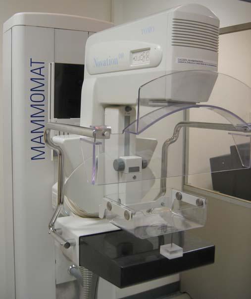
52 Detector Modulation Transfer Function (MTF) FPD14 data sinc 150um LMAM data sinc 85 um MTF frequency (lp/mm) Experimental MTF s are close to their corresponding sinc functions The first zero-crossing of each sinc function corresponds to physical pixel pitch: 6.6 lp/mm for 150 μm pixel 11.7 lp/mm for 85 μm pixel 52
53 Detector Performance: Signal to Noise Ratio (SNR) If is the number of X-Ray incident on the detector then where N e is the electronic noise SNR = N 2 e + 10 SNR FPD9 data C*sqrt The SNR curve follow a sqrt behavior above 2μR dose (ur per frame) 53
54 Detector Detective Quantum Efficiency (DQE) DQE ur 8.6 ur 6.8 ur 5.5 ur 3.9 ur 2.4 ur 1.3 ur 0.6 ur DQE remains high for high frequency values 0 DQE = Spatial frequency (lp/mm) (SNR) 2 det (SNR) 2 in frequency domain G 2 2 MTF (f) DQE(f) = Ö x NPS(f) : X-Ray fluence NPS: noise power spectrum G: conversion gain 54
55 Manufacturing Steps Deposit in vacuum amorphous Se on TFT Deposit top metal electrode on a-se the Attach high voltage contact to electrode Deposit isolation on multi-layer structure Attach peripheral ASIC electronics to TFT Assembly the detector in final enclosure Perform parametric and imaging tests 55

56 Selenium Coater 56

57 Selenium Coater 57


58 Mammography Detector Electronic sub-assembly Packaged detector 58

59 Tomosynthesis 59
60 Acknowledgements The author acknowledges the contribution of Dr. Olivier Tousignant, Anrad Corporation, Saint-Laurent, QC, Canada who made the characteristic parameters measurements of the LMAM detectors. 60
Introduction. Chapter 16 Diagnostic Radiology. Primary radiological image. Primary radiological image
 Introduction Chapter 16 Diagnostic Radiology Radiation Dosimetry I Text: H.E Johns and J.R. Cunningham, The physics of radiology, 4 th ed. http://www.utoledo.edu/med/depts/radther In diagnostic radiology
X-ray detectors in healthcare and their applications
 X-ray detectors in healthcare and their applications Pixel 2012, Inawashiro September 4th, 2012 Martin Spahn, PhD Clinical applications of X-ray imaging Current X-ray detector technology (case study radiography
Radiology Physics Lectures: Digital Radiography. Digital Radiography. D. J. Hall, Ph.D. x20893
 Digital Radiography D. J. Hall, Ph.D. x20893 djhall@ucsd.edu Background Common Digital Modalities Digital Chest Radiograph - 4096 x 4096 x 12 bit CT - 512 x 512 x 12 bit SPECT - 128 x 128 x 8 bit MRI -
Breast Tomosynthesis. Bob Liu, Ph.D. Department of Radiology Massachusetts General Hospital And Harvard Medical School
 Breast Tomosynthesis Bob Liu, Ph.D. Department of Radiology Massachusetts General Hospital And Harvard Medical School Outline Physics aspects of breast tomosynthesis Quality control of breast tomosynthesis
Mammography: Physics of Imaging
 Mammography: Physics of Imaging Robert G. Gould, Sc.D. Professor and Vice Chair Department of Radiology and Biomedical Imaging University of California San Francisco, California Mammographic Imaging: Uniqueness
Amorphous Selenium Direct Radiography for Industrial Imaging
 DGZfP Proceedings BB 67-CD Paper 22 Computerized Tomography for Industrial Applications and Image Processing in Radiology March 15-17, 1999, Berlin, Germany Amorphous Selenium Direct Radiography for Industrial
Chromatic X-Ray imaging with a fine pitch CdTe sensor coupled to a large area photon counting pixel ASIC
 Chromatic X-Ray imaging with a fine pitch CdTe sensor coupled to a large area photon counting pixel ASIC R. Bellazzini a,b, G. Spandre a*, A. Brez a, M. Minuti a, M. Pinchera a and P. Mozzo b a INFN Pisa
X-ray light valve (XLV): a novel detectors technology for digital mammography*
: a novel detectors technology for digital mammography*") X-ray light valve (XLV): a novel detectors technology for digital mammography* Sorin Marcovici, Vlad Sukhovatkin, Peter Oakham XLV Diagnostics Inc., Thunder Bay, ON P7A 7T1, Canada ABSTRACT A novel method,
X-ray light valve (XLV): a novel detectors technology for digital mammography* Sorin Marcovici, Vlad Sukhovatkin, Peter Oakham XLV Diagnostics Inc., Thunder Bay, ON P7A 7T1, Canada ABSTRACT A novel method,
COMPUTED TOMOGRAPHY 1
 COMPUTED TOMOGRAPHY 1 Why CT? Conventional X ray picture of a chest 2 Introduction Why CT? In a normal X-ray picture, most soft tissue doesn't show up clearly. To focus in on organs, or to examine the
PD233: Design of Biomedical Devices and Systems
 PD233: Design of Biomedical Devices and Systems (Lecture-8 Medical Imaging Systems) (Imaging Systems Basics, X-ray and CT) Dr. Manish Arora CPDM, IISc Course Website: http://cpdm.iisc.ac.in/utsaah/courses/
Unit thickness. Unit area. σ = NΔX = ΔI / I 0
 Unit thickness I 0 ΔI I σ = ΔI I 0 NΔX = ΔI / I 0 NΔX Unit area Δx Average probability of reaction with atom for the incident photons at unit area with the thickness of Delta-X Atom number at unit area
Current technology in digital image production (CR/DR and other modalities) Jaroonroj Wongnil 25 Mar 2016
 Jaroonroj Wongnil 25 Mar 2016") Current technology in digital image production (CR/DR and other modalities) Jaroonroj Wongnil 25 Mar 2016 Current technology in digital image production (CR/DR and other modalities) 2/ Overview Digital
Current technology in digital image production (CR/DR and other modalities) Jaroonroj Wongnil 25 Mar 2016 Current technology in digital image production (CR/DR and other modalities) 2/ Overview Digital
Data. microcat +SPECT
 Data microcat +SPECT microcat at a Glance Designed to meet the throughput, resolution and image quality requirements of academic and pharmaceutical research, the Siemens microcat sets the standard for
Pitfalls and Remedies of MDCT Scanners as Quantitative Instruments
 intensity m(e) m (/cm) 000 00 0 0. 0 50 0 50 Pitfalls and Remedies of MDCT Scanners as Jiang Hsieh, PhD GE Healthcare Technology University of Wisconsin-Madison Root-Causes of CT Number Inaccuracies Nature
Detector technology in simultaneous spectral imaging
 Computed tomography Detector technology in simultaneous spectral imaging Philips IQon Spectral CT Z. Romman, I. Uman, Y. Yagil, D. Finzi, N. Wainer, D. Milstein; Philips Healthcare While CT has become
HISTORY. CT Physics with an Emphasis on Application in Thoracic and Cardiac Imaging SUNDAY. Shawn D. Teague, MD
 CT Physics with an Emphasis on Application in Thoracic and Cardiac Imaging Shawn D. Teague, MD DISCLOSURES 3DR- advisory committee CT PHYSICS WITH AN EMPHASIS ON APPLICATION IN THORACIC AND CARDIAC IMAGING
Seminar 8. Radiology S8 1
 Seminar 8 Radiology Medical imaging. X-ray image formation. Energizing and controlling the X-ray tube. Image detectors. The acquisition of analog and digital images. Digital image processing. Selected
Setting up digital imaging department!
 Outline Setting up digital imaging department! From screen/film to digital radiography PACS/Tele radiology Setting up digital department Digital Imaging Napapong Pongnapang, Ph.D. Department of Radiological
7/24/2014. Image Quality for the Radiation Oncology Physicist: Review of the Fundamentals and Implementation. Disclosures. Outline
 Image Quality for the Radiation Oncology Physicist: Review of the Fundamentals and Implementation Image Quality Review I: Basics and Image Quality TH-A-16A-1 Thursday 7:30AM - 9:30AM Room: 16A J. Anthony
PERFORMANCE CHARACTERIZATION OF AMORPHOUS SILICON DIGITAL DETECTOR ARRAYS FOR GAMMA RADIOGRAPHY
 12 th A-PCNDT 2006 Asia-Pacific Conference on NDT, 5 th 10 th Nov 2006, Auckland, New Zealand PERFORMANCE CHARACTERIZATION OF AMORPHOUS SILICON DIGITAL DETECTOR ARRAYS FOR GAMMA RADIOGRAPHY Rajashekar
DALLA LUCE VISIBILE AI RAGGI X: NUOVI RIVELATORI DI IMMAGINI PER RAGGI X A DISCRIMINAZIONE IN ENERGIA ED APPLICAZIONI
 DALLA LUCE VISIBILE AI RAGGI X: NUOVI RIVELATORI DI IMMAGINI PER RAGGI X A DISCRIMINAZIONE IN ENERGIA ED APPLICAZIONI D. Pacella ENEA - Frascati LIMS, Frascati 14-15 ottobre 2015 Come per la fotografia:
Mammography is a radiographic procedure specially designed for detecting breast pathology Approximately 1 woman in 8 will develop breast cancer over
 Mammography is a radiographic procedure specially designed for detecting breast pathology Approximately 1 woman in 8 will develop breast cancer over a lifetime Breast cancer screening programs rely on
LaBr 3 :Ce, the latest crystal for nuclear medicine
 10th Topical Seminar on Innovative Particle and Radiation Detectors 1-5 October 2006 Siena, Italy LaBr 3 :Ce, the latest crystal for nuclear medicine Roberto Pani On behalf of SCINTIRAD Collaboration INFN
PET/CT Instrumentation Basics
 / Instrumentation Basics 1. Motivations for / imaging 2. What is a / Scanner 3. Typical Protocols 4. Attenuation Correction 5. Problems and Challenges with / 6. Examples Motivations for / Imaging Desire
Research Support. Dual-Source CT: What is it and How Do I Test it? Cynthia H. McCollough, Ph.D.
 Dual-Source CT: What is it and How Do I Test it? Cynthia H. McCollough, Ph.D. CT Clinical Innovation Center Department of Radiology Mayo Clinic College of Medicine Rochester, MN Research Support National
10/3/2012. Study Harder
 This presentation is a professional collaboration of development time prepared by: Rex Christensen Terri Jurkiewicz and Diane Kawamura Study Harder CR detection is inefficient, inferior to film screen
10/26/2015. Study Harder
 This presentation is a professional collaboration of development time prepared by: Rex Christensen Terri Jurkiewicz and Diane Kawamura Study Harder CR detection is inefficient, inferior to film screen
Software and Hardware in CCTA. Elly Castellano PhD
 Software and Hardware in CCTA Elly Castellano PhD Outline technical requirements for coronary CTA the modern cardiac CT scanner ECG-gating technology image reconstruction algorithms 2 Technical requirements
QC Testing for Computed Tomography (CT) Scanner
 Scanner") QC Testing for Computed Tomography (CT) Scanner QA - Quality Assurance All planned and systematic actions needed to provide confidence on a structure, system or component. all-encompassing program, including
QC Testing for Computed Tomography (CT) Scanner QA - Quality Assurance All planned and systematic actions needed to provide confidence on a structure, system or component. all-encompassing program, including
X-rays. X-rays are produced when electrons are accelerated and collide with a target. X-rays are sometimes characterized by the generating voltage
 X-rays Ouch! 1 X-rays X-rays are produced when electrons are accelerated and collide with a target Bremsstrahlung x-rays Characteristic x-rays X-rays are sometimes characterized by the generating voltage
Fundamentals of CMOS Image Sensors
 CHAPTER 2 Fundamentals of CMOS Image Sensors Mixed-Signal IC Design for Image Sensor 2-1 Outline Photoelectric Effect Photodetectors CMOS Image Sensor(CIS) Array Architecture CIS Peripherals Design Considerations
Acceptance Testing of a Digital Breast Tomosynthesis Unit
 Acceptance Testing of a Digital Breast Tomosynthesis Unit 2012 AAPM Spring Clinical Meeting Jessica Clements, M.S., DABR Objectives Review of technology and clinical advantages Acceptance Testing Procedures
Medical Images Analysis and Processing
 Medical Images Analysis and Processing - 25642 Emad Course Introduction Course Information: Type: Graduated Credits: 3 Prerequisites: Digital Image Processing Course Introduction Reference(s): Insight
X-ray Imaging. PHYS Lecture. Carlos Vinhais. Departamento de Física Instituto Superior de Engenharia do Porto
 X-ray Imaging PHYS Lecture Carlos Vinhais Departamento de Física Instituto Superior de Engenharia do Porto cav@isep.ipp.pt Overview Projection Radiography Anode Angle Focal Spot Magnification Blurring
Distributed source x-ray tube technology for tomosynthesis imaging
 Distributed source x-ray tube technology for tomosynthesis imaging Authors: F. Sprenger a*, X. Calderon-Colon b, Y. Cheng a, K. Englestad a, J. Lu b, J. Maltz c, A. Paidi c, X. Qian b, D. Spronk a, S.
New spectral benefi ts, proven low dose
 New spectral benefi ts, proven low dose Philips MicroDose mammography SI, technical data sheet Philips MicroDose SI with single-shot spectral imaging is a fullfi eld digital mammography solution that delivers
Characterization of photon counting CZT detectors for medical x-ray imaging and spectroscopy
 Louisiana State University LSU Digital Commons LSU Doctoral Dissertations Graduate School 2011 Characterization of photon counting CZT detectors for medical x-ray imaging and spectroscopy Shannon Fritz
NM Module Section 2 6 th Edition Christian, Ch. 3
 NM 4303 Module Section 2 6 th Edition Christian, Ch. 3 Gas Filled Chamber Voltage Gas filled chamber uses Hand held detectors cutie pie Geiger counter Dose calibrators Cutie pie Chamber voltage in Ionization
X-RAY IMAGING EE 472 F2017. Prof. Yasser Mostafa Kadah
 X-RAY IMAGING EE 472 F2017 Prof. Yasser Mostafa Kadah www.k-space.org Recommended Textbook Stewart C. Bushong, Radiologic Science for Technologists: Physics, Biology, and Protection, 10 th ed., Mosby,
X-Ray Medical Imaging and Pixel detectors
 X-Ray Medical Imaging and Pixel detectors PIXEL 2000 Genova, June 5-8 th 2000 J.P.Moy, TRI XELL, Moirans, France 1 OUTLINE - X-ray medical imaging. The requirements, some particular features - Present
CHAPTER 8 GENERIC PERFORMANCE MEASURES
 GENERIC PERFORMANCE MEASURES M.E. DAUBE-WITHERSPOON Department of Radiology, University of Pennsylvania, Philadelphia, Pennsylvania, United States of America 8.1. INTRINSIC AND EXTRINSIC MEASURES 8.1.1.
TOPICS: CT Protocol Optimization over the Range of Patient Age & Size and for Different CT Scanner Types: Recommendations & Misconceptions
 CT Protocol Optimization over the Range of Patient Age & Size and for Different CT Scanner Types: Recommendations & Misconceptions TOPICS: Computed Tomography Quick Overview CT Dosimetry Effects of CT
Designing an MR compatible Time of Flight PET Detector Floris Jansen, PhD, Chief Engineer GE Healthcare
 GE Healthcare Designing an MR compatible Time of Flight PET Detector Floris Jansen, PhD, Chief Engineer GE Healthcare There is excitement across the industry regarding the clinical potential of a hybrid
Wide-Detector CT for TAVR Planning:
 Wide-Detector CT for TAVR Planning: Impact on Iodine Dose, Radiation Dose, and Image Quality SCBTMR 2015 Annual Course Thursday, October 8 William P. Shuman MD FSCBTMR Department of Radiology University
Hardware for High Energy Applications 30 October 2009
 Paper No. 003 09 Hardware for High Energy Applications 30 October 2009 This document was created by the Federal Working Group on Industrial Digital Radiography. Reproduction is authorized. Federal Working
Radiographic sensitivity improved by optimized high resolution X -ray detector design.
 DIR 2007 - International Symposium on Digital industrial Radiology and Computed Tomography, June 25-27, 2007, Lyon, France Radiographic sensitivity improved by optimized high resolution X -ray detector
I. PERFORMANCE OF X-RAY PRODUCTION COMPONENTS FLUOROSCOPIC ACCEPTANCE TESTING: TEST PROCEDURES & PERFORMANCE CRITERIA
 FLUOROSCOPIC ACCEPTANCE TESTING: TEST PROCEDURES & PERFORMANCE CRITERIA EDWARD L. NICKOLOFF DEPARTMENT OF RADIOLOGY COLUMBIA UNIVERSITY NEW YORK, NY ACCEPTANCE TESTING GOALS PRIOR TO 1st CLINICAL USAGE
Features and Weaknesses of Phantoms for CR/DR System Testing
 Physics testing of image detectors Parameters to test Features and Weaknesses of Phantoms for CR/DR System Testing Spatial resolution Contrast resolution Uniformity/geometric distortion Dose response/signal
Preliminary Modulation Transfer Function Study on Amorphous Silicon Flat Panel System for Industrial Digital Radiography
 ECNDT 26 - Poster 17 Preliminary Modulation Transfer Function Study on Amorphous Silicon Flat Panel System for Industrial Digital Radiography Khairul Anuar MOHD SALLEH, Ab. Razak HAMZAH and Mohd Ashhar
Simulation of High Resistivity (CMOS) Pixels
 Pixels") Simulation of High Resistivity (CMOS) Pixels Stefan Lauxtermann, Kadri Vural Sensor Creations Inc. AIDA-2020 CMOS Simulation Workshop May 13 th 2016 OUTLINE 1. Definition of High Resistivity Pixel Also
Simulation of High Resistivity (CMOS) Pixels Stefan Lauxtermann, Kadri Vural Sensor Creations Inc. AIDA-2020 CMOS Simulation Workshop May 13 th 2016 OUTLINE 1. Definition of High Resistivity Pixel Also
APD Quantum Efficiency
 APD Quantum Efficiency Development of a 64-channel APD Detector Module with Individual Pixel Readout for Submillimeter Spatial Resolution in PET Philippe Bérard a, Mélanie Bergeron a, Catherine M. Pepin
X-ray Detectors: What are the Needs?
 X-ray Detectors: What are the Needs? Sol M. Gruner Physics Dept. & Cornell High Energy Synchrotron Source (CHESS) Ithaca, NY 14853 smg26@cornell.edu 1 simplified view of the Evolution of Imaging Synchrotron
PET Detectors. William W. Moses Lawrence Berkeley National Laboratory March 26, 2002
 PET Detectors William W. Moses Lawrence Berkeley National Laboratory March 26, 2002 Step 1: Inject Patient with Radioactive Drug Drug is labeled with positron (β + ) emitting radionuclide. Drug localizes
Explain what is meant by a photon and state one of its main properties [2]
![Explain what is meant by a photon and state one of its main properties [2]](/thumbs/80/82516318.jpg "Explain what is meant by a photon and state one of its main properties [2]") 1 (a) A patient has an X-ray scan taken in hospital. The high-energy X-ray photons interact with the atoms inside the body of the patient. Explain what is meant by a photon and state one of its main properties....
1 (a) A patient has an X-ray scan taken in hospital. The high-energy X-ray photons interact with the atoms inside the body of the patient. Explain what is meant by a photon and state one of its main properties....
CR Basics and FAQ. Overview. Historical Perspective
 Page: 1 of 6 CR Basics and FAQ Overview Computed Radiography is a term used to describe a system that electronically records a radiographic image. Computed Radiographic systems use unique image receptors
Thermionic x-ray. Alternative technologies. Electron Field Emission. CNT Based Field Emission X-Ray Source
 Energy Level (ev) Multi-beam x-ray source array based on carbon nanotube field emission O. Zhou, JP Lu, X. Calderon-Colon, X. Qian, G. Yang, G. Cao, E. Gidcumb, A. Tucker, J. Shan University of North Carolina
X-ray Tube and Generator Basic principles and construction
 X-ray Tube and Generator Basic principles and construction Dr Slavik Tabakov - Production of X-rays OBJECTIVES - X-ray tube construction - Anode - types, efficiency - X-ray tube working characteristics
12/21/2016. Siemens Medical Systems Research Agreement Philips Healthcare Research Agreement AAN and ASN Committees
 Joseph V. Fritz, PhD Nandor Pintor, MD Dent Neurologic Institute ASN 2017 Friday, January 20, 2017 Siemens Medical Systems Research Agreement Philips Healthcare Research Agreement AAN and ASN Committees
Gas scintillation Glass GEM detector for high-resolution X-ray imaging and CT
 Gas scintillation Glass GEM detector for high-resolution X-ray imaging and CT Takeshi Fujiwara 1, Yuki Mitsuya 2, Hiroyuki Takahashi 2, and Hiroyuki Toyokawa 2 1 National Institute of Advanced Industrial
Mammography Solution. AMULET Innovality. The new leader in the AMULET series. Tomosynthesis, 3D mammography and biopsy are all available.
 Mammography Solution AMULET Innovality The new leader in the AMULET series. Tomosynthesis, 3D mammography and biopsy are all available. FUJIFILM supports the Pink Ribbon Campaign for early detection of
Acquisition, Processing and Display
 Acquisition, Processing and Display Terri L. Fauber, R.T. (R)(M) Department of Radiation Sciences School of Allied Health Professions Virginia Commonwealth University Topics Image Characteristics Image
Photomultiplier Tube
 Nuclear Medicine Uses a device known as a Gamma Camera. Also known as a Scintillation or Anger Camera. Detects the release of gamma rays from Radionuclide. The radionuclide can be injected, inhaled or
High Energy Digital Radiography & 3D-CT for Industrial Systems
 DIR 2007 - International Symposium on Digital industrial Radiology and Computed Tomography, June 25-27, 2007, Lyon, France High Energy Digital Radiography & 3D-CT for Industrial Systems Non-Destructive
Strategies to improve the signal and noise performance of active matrix, flat-panel imagers for diagnostic x-ray applications
 Strategies to improve the signal and noise performance of active matrix, flat-panel imagers for diagnostic x-ray applications L. E. Antonuk, a) K.-W. Jee, Y. El-Mohri, M. Maolinbay, S. Nassif, X. Rong,
Basis of Computed Radiography & PACS
 Basis of Computed Radiography & PACS Slavik Tabakov Computed Radiography (CR) refers to new types of X-ray detectors (i.e. replaces the X-ray Film) The CR output media is a digital image, which can be
RANDY W. ALKIRE, GEROLD ROSENBAUM AND GWYNDAF EVANS
 S-94,316 PATENTS-US-A96698 BEAM POSITION MONITOR RANDY W. ALKIRE, GEROLD ROSENBAUM AND GWYNDAF EVANS CONTRACTUAL ORIGIN OF THE INVENTION The United States Government has rights in this invention pursuant
Chiara Secco. PET Performance measurements of the new LSO-Based Whole Body PET/CT. Scanner biograph 16 HI-REZ using the NEMA NU Standard.
 Chiara Secco PET Performance measurements of the new LSO-Based Whole Body PET/CT Scanner biograph 16 HI-REZ using the NEMA NU 2-2001 Standard. INTRODUCTION Since its introduction, CT has become a fundamental
1. Patient size AEC. Large Patient High ma. Small Patient Low ma
 Comparison of the function and performance of CT AEC systems CTUG meeting by Emily Field Trainee clinical scientist 14 th th Breakdown CT Automatic Exposure Control (AEC) Background Project Description
Medical Imaging. X-rays, CT/CAT scans, Ultrasound, Magnetic Resonance Imaging
 Medical Imaging X-rays, CT/CAT scans, Ultrasound, Magnetic Resonance Imaging From: Physics for the IB Diploma Coursebook 6th Edition by Tsokos, Hoeben and Headlee And Higher Level Physics 2 nd Edition
Lunar Technology Advantages
 Lunar Technology Advantages DXA stands for Dual-Energy X-ray Absorptiometry. It is a measurement method that uses the differences in the absorption of high energy and low energy X-ray photons by different
CT Basics: Data Acquisition Module 3
 Module 3 Transcript For educational and institutional use. This transcript is licensed for noncommercial, educational inhouse or online educational course use only in educational and corporate institutions.
Marten Bosma 1, Alex Fauler 2, Michael Fiederle 2 en Jan Visser Nikhef, Amsterdam, The Netherlands 2. FMF, Freiburg, Germany
 Marten Bosma 1, Alex Fauler 2, Michael Fiederle 2 en Jan Visser 1 1. Nikhef, Amsterdam, The Netherlands 2. FMF, Freiburg, Germany Digital Screen film Digital radiography advantages: Larger dynamic range
Future directions in Nuclear Medicine Instrumentation
 Future directions in Nuclear Medicine Instrumentation Where are we going - and why? First, the disclosure list My group at the University of Washington has research support from: NIH DOE General Electric
Diffraction-enhanced X-ray Imaging (DEXI) Medical Solutions. More information using less radiation
 Medical Solutions. More information using less radiation") Diffraction-enhanced X-ray Imaging (DEXI) Medical Solutions More information using less radiation Medical Small Animal Security NDE/NDT Diffraction-Enhanced X-ray Imaging Medical Solutions Safe non-invasive
Diffraction-enhanced X-ray Imaging (DEXI) Medical Solutions More information using less radiation Medical Small Animal Security NDE/NDT Diffraction-Enhanced X-ray Imaging Medical Solutions Safe non-invasive
Overview. Professor Roentgen was a Physicist!!! The Physics of Radiation Oncology X-ray Imaging
 The Physics of Radiation Oncology X-ray Imaging Charles E. Willis, Ph.D. DABR Associate Professor Department of Imaging Physics The University of Texas M.D. Anderson Cancer Center Houston, Texas Overview
IBEX TECHNOLOGY APPLIED TO DIGITAL RADIOGRAPHY
 WHITE PAPER: IBEX TECHNOLOGY APPLIED TO DIGITAL RADIOGRAPHY IBEX Innovations Ltd. Registered in England and Wales: 07208355 Address: Discovery 2, NETPark, William Armstrong Way, Sedgefield, UK Patents:
Maximizing clinical outcomes
 Maximizing clinical outcomes Digital Tomosynthesis Dual Energy Subtraction Automated Long Length Imaging Improved image quality at a low dose Xray Xray Patented ISS capture technology promotes high sensitivity
Image Quality and Dose. Image Quality and Dose. Image Quality and Dose Issues in MSCT. Scanner parameters affecting IQ and Dose
 Image Quality and Dose Issues in MSCT Image Quality and Dose Image quality Image noise Spatial resolution Contrast Artefacts Speckle and sharpness S. Edyvean St. George s Hospital London SW17 0QT Radiation
LSO PET/CT Pico Performance Improvements with Ultra Hi-Rez Option
 LSO PET/CT Pico Performance Improvements with Ultra Hi-Rez Option Y. Bercier, Member, IEEE, M. Casey, Member, IEEE, J. Young, Member, IEEE, T. Wheelock, Member, IEEE, T. Gremillion Abstract-- Factors which
Digital Detector Array Image Quality for Various GOS Scintillators
 Digital Detector Array Image Quality for Various GOS Scintillators More info about this article: http://www.ndt.net/?id=22768 Brian S. White 1, Mark E. Shafer 2, William H. Russel 3, Eric Fallet 4, Jacques
LaBr 3 :Ce scintillation gamma camera prototype for X and gamma ray imaging
 8th International Workshop on Radiation Imaging Detectors Pisa 2-6 July 2006 LaBr 3 :Ce scintillation gamma camera prototype for X and gamma ray imaging Roberto Pani On behalf of SCINTIRAD Collaboration
New Technology in Nuclear Medicine
 New Technology in Nuclear Medicine Reed G. Selwyn, PhD, DABR Vice Chair of Research & Imaging Sciences Associate Professor and Chief, Medical Physics Dept. of Radiology, University of New Mexico Objectives
radiography detector
 Clinical evaluation of a full field digital projection radiography detector Gary S. Shaber'1, Denny L. Leeb, Jeffrey Belib, Gregory Poweii1', Andrew D.A. Maidment'1 a Thomas Jefferson University Hospital,
Initial Certification
 Initial Certification Nuclear Medical Physics (NMP) Study Guide Part 2 Content Guide and Sample Questions The content of all ABR exams is determined by a panel of experts who select the items based on
Development of Solid-State Detector for X-ray Computed Tomography
 Proceedings of the Korea Nuclear Society Autumn Meeting Seoul, Korea, October 2001 Development of Solid-State Detector for X-ray Computed Tomography S.W Kwak 1), H.K Kim 1), Y. S Kim 1), S.C Jeon 1), G.
Maximum Performance, Minimum Space
 TECHNOLOGY HISTORY For over 130 years, Toshiba has been a world leader in developing technology to improve the quality of life. Our 50,000 global patents demonstrate a long, rich history of leading innovation.
Electronic Instrumentation for Radiation Detection Systems
 Electronic Instrumentation for Radiation Detection Systems January 23, 2018 Joshua W. Cates, Ph.D. and Craig S. Levin, Ph.D. Course Outline Lecture Overview Brief Review of Radiation Detectors Detector
X-rays in medical diagnostics
 X-rays in medical diagnostics S.Dolanski Babić 2017/18. History W.C.Röntgen (1845-1923) discovered a new type of radiation Nature, Jan. 23. 1896.; Science, Feb.14. 1896. X- rays: Induced the ionization
2/14/2019. Nuclear Medicine Artifacts. Symmetric energy windows
 Nuclear Medicine Artifacts SCPMG Medical Imaging Technology & Informatics Medical Physics Group Brian Helbig, MS, DABR 1 2 Symmetric energy windows 3 1 Dynamic clinical study Energy peak shift Electrical
Computed Tomography. The Fundamentals of... THE FUNDAMENTALS OF... Jason H. Launders, MSc. Current Technology
 The Fundamentals of... Computed Tomography Computed Tomography (CT) systems use x-rays to produce images of slices through a patient s anatomy. Despite having lower spatial resolution than other x-ray
Today s Outline - January 25, C. Segre (IIT) PHYS Spring 2018 January 25, / 26
 PHYS Spring 2018 January 25, / 26") Today s Outline - January 25, 2018 C. Segre (IIT) PHYS 570 - Spring 2018 January 25, 2018 1 / 26 Today s Outline - January 25, 2018 HW #2 C. Segre (IIT) PHYS 570 - Spring 2018 January 25, 2018 1 / 26 Today
Today s Outline - January 25, 2018 C. Segre (IIT) PHYS 570 - Spring 2018 January 25, 2018 1 / 26 Today s Outline - January 25, 2018 HW #2 C. Segre (IIT) PHYS 570 - Spring 2018 January 25, 2018 1 / 26 Today
COMPUTED RADIOGRAPHY CHAPTER 4 EFFECTIVE USE OF CR
 This presentation is a professional collaboration of development time prepared by: Rex Christensen Terri Jurkiewicz and Diane Kawamura New Technology https://www.youtube.com/watch?v=ptkzznazb 7U COMPUTED
CT Basics: Image Quality Module 6
 Module 6 For educational and institutional use. This transcript is licensed for noncommercial, educational inhouse or online educational course use only in educational and corporate institutions. Any broadcast,
X-ray Tube and Generator Basic principles and construction
 X-ray Tube and Generator Basic principles and construction Dr Slavik Tabakov - Production of X-rays and Patient Dose OBJECTIVES - X-ray tube construction - Anode - types, efficiency - Classical X-ray generator
Digital Radiographic Inspection replacing traditional RT and 3D RT Development
 Digital Radiographic Inspection replacing traditional RT and 3D RT Development Iploca Novel Construction Meeting 27&28 March 2014 Geneva By Jan van der Ent Technical Authority International Contents Introduction
Investigation of Effective DQE (edqe) parameters for a flat panel detector
 parameters for a flat panel detector") Investigation of Effective DQE (edqe) parameters for a flat panel detector Poster No.: C-1892 Congress: ECR 2013 Type: Authors: Keywords: DOI: Scientific Exhibit D. Bor 1, S. Cubukcu 1, A. Yalcin 1, O.
Investigation of Effective DQE (edqe) parameters for a flat panel detector Poster No.: C-1892 Congress: ECR 2013 Type: Authors: Keywords: DOI: Scientific Exhibit D. Bor 1, S. Cubukcu 1, A. Yalcin 1, O.
Radionuclide Imaging MII Single Photon Emission Computed Tomography (SPECT)
") Radionuclide Imaging MII 3073 Single Photon Emission Computed Tomography (SPECT) Single Photon Emission Computed Tomography (SPECT) The successful application of computer algorithms to x-ray imaging in
Radionuclide Imaging MII 3073 Single Photon Emission Computed Tomography (SPECT) Single Photon Emission Computed Tomography (SPECT) The successful application of computer algorithms to x-ray imaging in
Dose Reduction and Image Preservation After the Introduction of a 0.1 mm Cu Filter into the LODOX Statscan unit above 110 kvp
 Dose Reduction and Image Preservation After the Introduction of a into the LODOX Statscan unit above 110 kvp Abstract: CJ Trauernicht 1, C Rall 1, T Perks 2, G Maree 1, E Hering 1, S Steiner 3 1) Division
Automated dose control in multi-slice CT. Nicholas Keat Formerly ImPACT, St George's Hospital, London
 Automated dose control in multi-slice CT Nicholas Keat Formerly ImPACT, St George's Hospital, London Introduction to presentation CT contributes ~50+ % of all medical radiation dose Ideally all patients
160-slice CT SCANNER / New Standard for the Future
 TECHNOLOGY HISTORY For over 130 years, Toshiba has been a world leader in developing technology to improve the quality of life. Our 50,000 global patents demonstrate a long, rich history of leading innovation.