MECOS-C2 microscopy systems
|
|
|
- Jayson Johns
- 5 years ago
- Views:
Transcription


1 MECOS-C2 microscopy systems
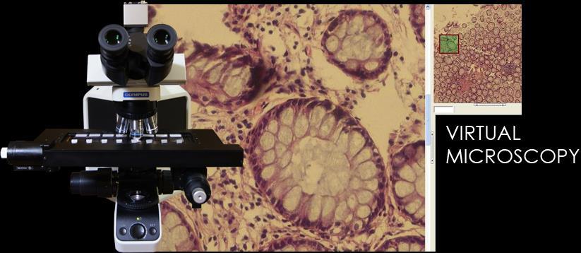
2 Microscopy systems of the MECOS-C2 family production LLC "Medical computer Systems (MECOS)" belong to a class of scanning microscopes-analyzers and are intended for: Increase of speed, accuracy, sensitivity, diagnostic significance of the group of well-known methods of analysis; Improvement of labour conditions and productivity; Creation of archives of digital copies of specimens, telemedical consultations, remote quality control and training; Automation of laborious operations of microscopy and measurements for research methods. Systems MECOS-C2 are robots, performing the loading and identification of specimens, moving the field of view in the 3d space of the specimen with navigation, selection of representative areas of analysis, detection, recognition, shooting, sorting, measurement, collection of samples of objects of analysis of the given types, formation of representative digital copy of the specimen, record of results in a database and rendering of means of visualization and remote access. Consumers: Laboratories medical, sanitary-epidemiological, research, forensic medical examination, production, veterinary.


3 Platform 1-М5 Platform 8-О9 Platform 200-О9 MECOS-C2 platforms for offline servicing party of 1/4/8/50/200/400 slides: 1- computer, 2 - monitor, 3 - control unit, 4 - motorized focus, 5 - motorized stage, 6 - digital camera, 7 - microscope, 8 - motorized turret, 9 - barcode scanner, 10 - power supply, 11 - motorized oiler, 13 - glasses loader
4 Automatic Scanner MECOS-C2-MECO-SCAN Characteristics: Microscopy of bright field or fluorescent; Objectives of the entire range of apertures; Automated production of the slide Map to select the scan area; Automated production of 2d or 3d digital copies of specimens - virtual slides (VS) - for viewing on a monitor without loss of resolution and with navigation by Map. Types of specimens and VS: 2D VS of thin specimens (cytological slides increase up to 100x; histological slides increase up to 40x); 3D VS of thick specimens with a layered shooting of each field of view at several levels of focus depth (histological slides increase more 40x; wet specimens such as sediments of urine, feces, saliva, sperm, spinal fluid, blood under cover glass or in cameras; thick blood drop smear for malaria tests; some types of prints).

; Auto scan on specified coordinates for specimens with a")
.")

5 Analysis region selection functions: With automatic generation of the Map of the slide, with selection on the Map outer boundaries of the stained areas/cover glass or areas by specified features (optical density/brightness, color, morphology); Auto scan on specified coordinates for specimens with a fixed location (liquid based cytology (LBC), biochips, slide tablets, slide-cameras). Speed and digital resolution: 2D VS: Increase mm sq/sec μm/pixel 4х 48; 1,72 10х 38; 0,7 20х 15/7 0,5/0,2 40х 6; 0,22 60х 3,9; 0,17 100х 1,2; 0,07 3D VS at 15 x 15 mm, 20 x, 8 layers of depth of focus: scan time 240 seconds, 0,5 μm/pixel.


6 Scheme of the autonomous manufacturing by MECO-SCAN of virtual slides for party of glasses with automatic selection of the scan areas Glasses Virtual slides Data base Maps with borders of scanning


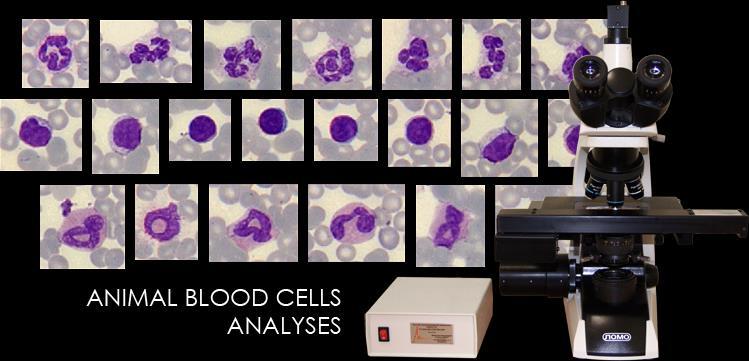
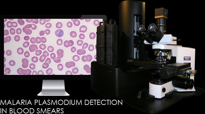
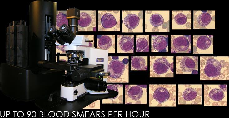
7 Automatic Scanner-Analyzer MECOS-C2- MECO-HEMO for analyses of blood smears 1. On the lens 4x is searching for the working zone of the smear. Automatically applied a drop of immersion oil. 2. On the lens 20x oil in the working area is searching for and collecting a sample of WBC specified amount. 3. Samples collecting of RBC and PL is carried at 100x oil in the region of working area with unbroken RBC morphology. 4. Repeated shooting WBC on lens 100x oil by sample coordinates before found. Blood smears of manual or automatic manufacturing. Capacity up to 90 blood smears per hour.

 to collect cells under their")
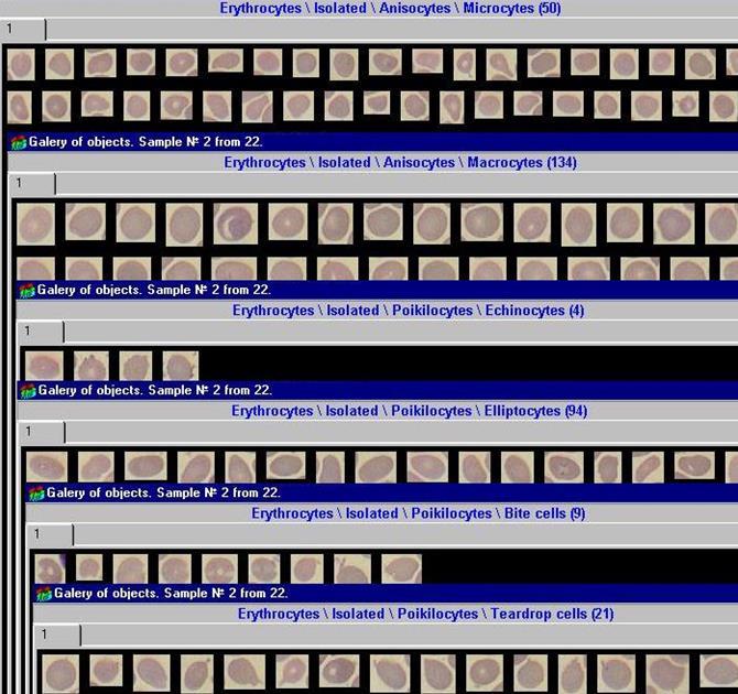
8 Automatic sorting of RBC, WBC with cell type galleries in the database. Ergonomic visual control of automatic sorting with interactive adjustment much faster and more efficient than manual microscopy. Routine analysis of large samples of cells (WBC to 400, RBC to 2000) to collect cells under their low concentration, to increase sensitivity to rare types of cells. The automatic cell morphometry.
9

10 Integration of external data, leukogram, categorical and quantitative evaluation morphology of RBC population, platelets histogram and relative concentration to RBC.

11 MECO-HEMO can be used for analyses of animal blood smears. Self-learning function of MECO-HEMO Analyzer is available to adapt automatic WBC sorting for animals of a specified types.

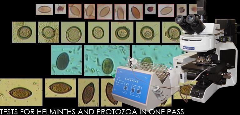
12 Automatic Analyzer-Scanner MECOS-C2-MECO-PARAS for analysis of biomaterials for protozoa and helminths MECO-PARAS performs automatic generation of 3D virtual slide (VS) of the specimen and automatic detection of helminthiasis. Types of specimens: a) specimens of feces, swabs, soil, sewage, water, fish, shellfish, crustaceans, amphibians, reptiles and their products under cover glass; b) specimens of feces from filtering test tubes in slide-camera of Fe-5 motorized station. Scanning objectives: from 20x to 100x oil. When the 20x/0.75 is applied the digital resolution of 3D VS allows to view as eggs of helminthes as majority of protozoa. Scan rate: 15 x 15 mm cover glass from 2.5 minutes; Slide-camera of the Fe-5 from 1 minute.

13 Not available for manual microscopy completeness and speed of Visual analysis using MECO-PARAS digital 3D VS. Viewing of the entire area on the full depth at different scale to search for objects of different types, with the formation of the Galleries of the detected objects.


14 Automated Search and Sorting eggs of helminths in VS as a "second opinion". A quick visual check and adjust the results of automatic detection and sorting using built-in Atlas of helminthes and protozoa.
 for ergonomic Visual analysis of the smear; Increase in")
15 Automatic Scanner-Analyzer MECOS-C2-MECO-MYCOBACT: Cytological smears on micobacterium coloured by Ziehl-Neelsen for microscopy in transmitted light or by fluorochromes for fluorescence microscopy; Mapping of the slide, automatic or interactive selection of analysis region of a given size; Automatic generation of digital virtual slide (VS) for ergonomic Visual analysis of the smear; Increase in analysis sensitivity by combining Visual and automatic detection of Mycobacteria; Informatics, database, statistics, filling out forms; Telemedicine and telemicroscopy; Automatic service up to 1/4/8/200/400 smears at different platforms; Capacity up to 30 smears per hour; Maintenance by junior staff.

16 Automatic Scanner-Analyzer MECOS-C2-MECO-MALARIA: Mapping of the slide, automatic or interactive selection of analysis region of a given size; Creation of digital copies of slides with thick and thin drops of blood, not conceding on the representativeness and the resolution of images in the microscope tubes; Increase in analysis sensitivity by more material being analyzed and collaborative automatic and Visual detection of malarial protozoa; Informatics, database, statistics, filling out forms; Telemedicine and telemicroscopy; Automatic service up to 1/4/8/200/400 smears at different platforms; Capacity up to 30 smears per hour; Maintenance by junior staff.
 or high (40x) magnification; Formation in scanning area 3D virtual slide for ergonomic viewing on a monitor without losing of")
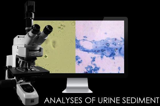
17 Automatic Scanner MECOS-C2-MECO-URIN to analyze urine and others biological liquid sediments: Automatic selection of the scan areas under cover glass, in a slide-tablet or in slide-camera; Automatic scanning of natural or stained specimens with standard (10x, 20x) or high (40x) magnification; Formation in scanning area 3D virtual slide for ergonomic viewing on a monitor without losing of resolution; Informatics including database, presentation of results in the forms, telemedicine and telemicroscopy; Loading and automatic maintenance up to 8 specimens.

18 View of 3D VS with visualization the focus shift during dive to the depth of the specimen.


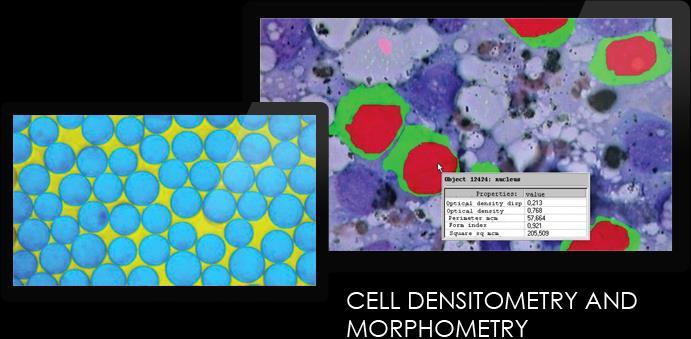
19 The MECO-DMM analyzer provides means of quantitative analysis of morphology and optical density. An example of automatic segmentation An example of interactive segmentation MECO-DMM provides convenient Viewer of objects segmented and measured.

. An example with parameters G and Hue is shown.")
. Step 3.")
20 The MECO-HYSTOCHEMICAL analyzer implements known Positive Pixel Count methodology to quantify the intensity of histo-(cyto)-chemical markers. Step 1. Interactive selection of the analysis area and 2 color parameters of every pixel of the area. The area can be any, color parameters may be R, G, B, Hue, Saturation, optical densities. Step 2. 2D distribution of color parameters of pixels appears in a scatter plotter (left). An example with parameters G and Hue is shown. Interactive selection of field of color in scatter plotter (left, hatch) allows to segment markers of the desired type ( Positive Pixels (PPs), right, allocated white color). Step 3. To graduate PPs the Histogram of the distribution of PPs on any color parameter is used (left, example of parameter Saturation). PPs of the area that match the specified percentage of the Histogram are shown with different colors (right) for Visual evaluation.
21 Automatic Scanner-analyzer MECOS-C2-MECO-METAPHASE to collect sample of metaphase plates Standard preparation of a cytological smear; Formation of digital Map of a slide on objective 10x; Searching and positioning of metaphase plates (MP) on the Map, taking into account the degree of disclosure and compact arrangement of chromosomes; Drawing of immersion oil, changing the objectives, focusing, shooting pictures of the MP on the objective with an aperture of up to 1.3 according to coordinates on the Map; Fixing high resolution MP images in the database in standardized formats for Visual analysis or use external programs of cytogenetic analysis; Performance up to 20 glasses per hour; Offline automatic servicing of the party until 1/4/8/200/400 glasses depending on MECOS-C2 platform; Maintenance of junior staff; Telemedicine and telemicroscopy.

22 Automatic detection of compact metaphase plates during a quick scan of the smear at 10x


23 bd bd bd bd Automatic shooting at 100x oil and fixing images of MP in database

24 Telemedicine MECOS-C2 tools with remote access to virtual slides and analyses results for laboratories across the country
25 MEСOS-C2 microscopy system Made in Russia Manufacturer Medical computer systems (MECOS) Company Registration certificate No 2011/10003 License for the production and maintenance No FS Ugreshskaya st. 2, building 72, Moscow, , Russia Tel. +7(495) Web:
Automated Digitization of Gram Stains. Centralized Reading. Decentralized Assessment. Improved Quality Management.
 Automated Digitization of Gram Stains Centralized Reading. Decentralized Assessment. Improved Quality Management. A GROWING DEMAND Gram staining is the rapid, easy, and inexpensive method for the assessment
EXC500p-- PATHOLOGY MICROSCOPE. EXC500hd -- HD DIGITAL PATHOLOGY MICROSCOPE. EXC500r -- RESEARCH MICROSCOPE EXC500-LABORATORY SCOPE
 EXC500p-- PATHOLOGY MICROSCOPE EXC500hd -- HD DIGITAL PATHOLOGY MICROSCOPE EXC500r -- RESEARCH MICROSCOPE EXC500-LABORATORY SCOPE The EXC500 Pathology and Laboratory Microscope is the most optically advanced
AUTOMATED MALARIA PARASITE DETECTION BASED ON IMAGE PROCESSING PROJECT REFERENCE NO.: 38S1511
 AUTOMATED MALARIA PARASITE DETECTION BASED ON IMAGE PROCESSING PROJECT REFERENCE NO.: 38S1511 COLLEGE : BANGALORE INSTITUTE OF TECHNOLOGY, BENGALURU BRANCH : COMPUTER SCIENCE AND ENGINEERING GUIDE : DR.
Computational approach for diagnosis of malaria through classification of malaria parasite from microscopic image of blood smear.
 Biomedical Research 2018; 29 (18): 3464-3468 ISSN 0970-938X www.biomedres.info Computational approach for diagnosis of malaria through classification of malaria parasite from microscopic image of blood
Color aspects and Color Standardization in Digital Microscopy
 Color aspects and Color Standardization in Digital Microscopy Yukako Yagi, PhD yyagi@partners.org Director of the MGH Pathology Imaging & Communication Technology Center Assistant Professor of Pathology,
VIDEOTEST-KARYO 3.1 SPECIFICATION
 VIDEOTEST-KARYO 3.1 System for automatic karyotyping the human chromosomes with an ability of training for automatic karyotyping the animal chromosomes (with the software for Windows 98, 2000, XP) developed
Stereotopix Research. Precision Pathology. Highthroughput. pathology. powered by newcast. Advantages of Stereotopix : RUO
 Precision Pathology Highthroughput pathology Stereotopix Research powered by newcast RUO Researchers use quantitative microscopy in many ways with the goal of producing high-quality, quantitative results
Estimating malaria parasitaemia in images of thin smear of human blood
 CSIT (March 2014) 2(1):43 48 DOI 10.1007/s40012-014-0043-7 Estimating malaria parasitaemia in images of thin smear of human blood Somen Ghosh Ajay Ghosh Sudip Kundu Received: 3 April 2014 / Accepted: 4
GALILEO TMA CK 4500 HTS Tissue Microarray Platform
 GALILEO TMA CK 4500 HTS Tissue Microarray Platform Tissue Microarray (TMA) A Block Of Samples From Hundreds Of Blocks (S. M. Hewitt, M.D., Ph.D., Tissue Array Research Program, LP, CCR, NCI, NIH) TMA technology
Colony Imaging with powerful Analysis Software
 TM Imaging with powerful Analysis Software TM Accurate Compact Fast We re not going to interpret your results, but we ll do everything to get you there From image acquisition to data visualisation, straight
Observing Microorganisms through a Microscope LIGHT MICROSCOPY: This type of microscope uses visible light to observe specimens. Compound Light Micros
 PHARMACEUTICAL MICROBIOLOGY JIGAR SHAH INSTITUTE OF PHARMACY NIRMA UNIVERSITY Observing Microorganisms through a Microscope LIGHT MICROSCOPY: This type of microscope uses visible light to observe specimens.
Using a Microscope. Year Group: BVSc1 + Document number: CSL_L07
 Year Group: BVSc1 + Document number: CSL_L07 Equipment list: Equipment for this station: Microscope Power supply and a level surface to work on Gloves The sample to examine Marker or pencil for labelling
CLEMEX intelligent microscopy
 CLEMEX intelligent microscopy Vision PE 5.0 Advanced Image Analysis Experience in Image Analysis Research and Quality Control Solutions With Vision PE, Clemex provides a powerful image analysis solution
Media Cybernetics White Paper Spherical Aberration
 Media Cybernetics White Paper Spherical Aberration Brian Matsumoto, University of California, Santa Barbara Introduction Digital photomicrographers assume that lens aberrations are corrected by the microscope
Exercise 2-A MICROSCOPIC TECHNIQUE & EXAMINATION OF MICROORGANISMS
 Exercise 2-A MICROSCOPIC TECHNIQUE & EXAMINATION OF MICROORGANISMS Introduction to Microscopic Technique Microbiology is the science or study of living organisms too small to be seen with the naked eye.
Enhanced Identification of Malarial Infected Objects using Otsu Algorithm from Thin Smear Digital Images
 International Journal of Latest Research in Science and Technology Vol.1,Issue 2 :Page No159-163,July-August(2012) http://www.mnkjournals.com/ijlrst.htm ISSN (Online):2278-5299 Enhanced Identification
Exercise 2-A MICROSCOPIC TECHNIQUE & EXAMINATION OF MICROORGANISMS
 Exercise 2-A MICROSCOPIC TECHNIQUE & EXAMINATION OF MICROORGANISMS Introduction to Microscopic Technique Microbiology is the science or study of living organisms too small to be seen with the naked eye.
COMPUTERIZED HEMATOLOGY COUNTER
 , pp.-190-194. Available online at http://www.bioinfo.in/contents.php?id=39 COMPUTERIZED HEMATOLOGY COUNTER KHOT S.T.* AND PRASAD R.K. Bharati Vidyapeeth (Deemed Univ.) Pune- 411 030, MS, India. *Corresponding
DMETRIX S (FUTURE) PERSPECTIVES ON DIGITAL IMAGING & DIGITAL PATHOLOGY SYSTEMS
 PERSPECTIVES ON DIGITAL IMAGING & DIGITAL PATHOLOGY SYSTEMS") Michael R. Descour, Ph.D., DMetrix, Inc., & University of Arizona Lloyd J. LaComb, Jr., Ph.D., DMetrix, Inc. DMETRIX S (FUTURE) PERSPECTIVES ON DIGITAL IMAGING & DIGITAL PATHOLOGY SYSTEMS Outline of presentation
Michael R. Descour, Ph.D., DMetrix, Inc., & University of Arizona Lloyd J. LaComb, Jr., Ph.D., DMetrix, Inc. DMETRIX S (FUTURE) PERSPECTIVES ON DIGITAL IMAGING & DIGITAL PATHOLOGY SYSTEMS Outline of presentation
Internal Medicine Imaging Core Emory University Department of Medicine
 Internal Medicine Imaging Core Emory University Department of Medicine 1 OPERATION OF THE ZEISS LSM 510 META YOU MUST SIGN UP TO USE THE MICROSCOPE OR COMPUTER EVERY TIME NO EXCEPTIONS Before attempting
OPELCO OPtical ELements COrporation LB Objective Series for Biological Use
 LB Objective Series for Biological Use 105 Executive Drive Suite 100 Dulles, VA 20166-9558 Tel: (703) 471-0080 S PLAN APOCHROMAT OBJECTIVES These objectives compensate for three wavelength of chromatic
FRAUNHOFER INSTITUTE FOR INTEGRATED CIRCUITS IIS. MANUAL PANORAMIC MICROSCOPY WITH istix
 FRAUNHOFER INSTITUTE FOR INTEGRATED CIRCUITS IIS MANUAL PANORAMIC MICROSCOPY WITH istix CLINICAL DIAGNOSTICS AND MATERIAL SCIENCES IMPROVED BY DIGITAL MICROSCOPY B A C K G R O U N D Due to a high grade
 Microscopy http://www.microscopyu.com/articles/phasecontrast/phasemicroscopy.html http://micro.magnet.fsu.edu/primer/anatomy/anatomy.html 2005, Dr. Jack Ikeda & Dr. Gail Grabner 9 Nikon Labophot (Question
Microscopy Techniques that make it easy to see things this small.
 Microscopy Techniques that make it easy to see things this small. What is a Microscope? An instrument for viewing objects that are too small to be seen easily by the naked eye. Dutch spectacle-makers Hans
Morphologi. Advanced image analysis for high sensitivity particle characterization. Particle size. Particle shape
 Particle size Particle shape Morphologi detailed specification sheets from www.malvern.co.uk Introducing a new concept in image analysis The Morphologi high sensitivity particle analyzer is more than just
MAKE SURE YOUR SLIDES ARE CLEAN (TOP & BOTTOM) BEFORE LOADING DO NOT LOAD SLIDES DURING SOFTWARE INITIALIZATION
 BEFORE LOADING DO NOT LOAD SLIDES DURING SOFTWARE INITIALIZATION") Olympus VS120-L100 Slide Scanner Standard Operating Procedure Startup 1) Red power bar switch (behind monitor) 2) Computer 3) Login: UserVS120 account (no password) 4) Double click: WAIT FOR INITIALIZATION
Olympus VS120-L100 Slide Scanner Standard Operating Procedure Startup 1) Red power bar switch (behind monitor) 2) Computer 3) Login: UserVS120 account (no password) 4) Double click: WAIT FOR INITIALIZATION
contents TABLE OF The SECOM platform Applications - sections Applications - whole cells Features Integrated workflow Automated overlay
 S E C O M TABLE OF contents The SECOM platform 4 Applications - sections 5 Applications - whole cells 8 Features 9 Integrated workflow 12 Automated overlay ODEMIS - integrated software Specifications 13
Microbiology Laboratory 2
 Microbiology Laboratory 2 Microscopy Background Microorganisms are too small to be seen with the naked eye. Thus a microscope is used to magnify objects so they can be observed. A lens consists of one
Digital Pathology and Tissue-based Diagnosis. How do they differ?
 Digital Pathology and Tissue-based Diagnosis. How do they differ? P. Hufnagl Institute of Pathology (Rudolf-Virchow-Haus). Humboldt University, Berlin? 10.12.2014 1 Structure of the talk Possible workflow
Operation Guide for the Leica SP2 Confocal Microscope Bio-Imaging Facility Hunter College October 2009
 Operation Guide for the Leica SP2 Confocal Microscope Bio-Imaging Facility Hunter College October 2009 Introduction of Fluoresence Confocal Microscopy The first confocal microscope was invented by Princeton
Quick and simple installation and no maintenance needed. 3 Times More affordable Than a normal SEM. Obtaining results in less than 4 minutes
 INTRODUCTION We believe that every laboratory working in the field of nanotechnology needs an SEM, therefore we would like to introduce to you our IEM series of SEM. In short space of time, our device
An Image Processing Approach for Screening of Malaria
 An Image Processing Approach for Screening of Malaria Nagaraj R. Shet 1 and Dr.Niranjana Sampathila 2 1 M.Tech Student, Department of Biomedical Engineering, Manipal Institute of Technology, Manipal University,
Teaching Digital Histology
 Teaching Digital Histology Carlos R. Morales Department of Anatomy and Cell Biology, McGill University, Montreal, Quebec, Canada The light microscope is one of the most widely used scientific instruments
Quick Operation Guide
 Quick Operation Guide Power ON Mounting specimens Set the specimen on the sample holder, and install the sample holder to the holder frame. Attach the holder frame to the XY stage. Type of holder Main
LAB 1 Introduction to Microscopy
 I. Ubiquity of Microorganisms II. Microscopy LAB 1 Introduction to Microscopy I. UBIQUITY OF MICROORGANISMS Microorganisms are ubiquitous; that is, they are present nearly everywhere. In this lab you will
Very short introduction to light microscopy and digital imaging
 Very short introduction to light microscopy and digital imaging Hernan G. Garcia August 1, 2005 1 Light Microscopy Basics In this section we will briefly describe the basic principles of operation and
The microscope is useful in making observations and collecting data in scientific experiments. Microscopy involves three basic concepts:
 AP BIOLOGY Chapter 6 NAME DATE Block MICROSCOPE LAB PART I: COMPOUND MICROSCOPE OBJECTIVES: After completing this exercise you should be able to: Demonstrate proper care and use of a compound microscope.
Leading in Desktop SEM Imaging and Analysis
 Leading in Desktop SEM Imaging and Analysis Fast. Outstanding. Reliable SEM imaging and analysis. The Phenom: World s Fastest Scanning Electron Microscope With its market-leading Phenom desktop Scanning
Chapter 2 The Study of Microbial Structure: Microscopy and Specimen Preparation
 Chapter 2 The Study of Microbial Structure: Microscopy and Specimen Preparation 1 Lenses and the Bending of Light light is refracted (bent) when passing from one medium to another refractive index a measure
Technical Aspects in Digital Pathology
 Technical Aspects in Digital Pathology Yukako Yagi, PhD yyagi@mgh.harvard.edu Director of the MGH Pathology Imaging & Communication Technology Center Assistant Professor of Pathology, Harvard Medical School
ImageXpress Micro XLS Widefield High Content Screening System. Imaging with a vision.
 ImageXpress Micro XLS Widefield High Content Screening System Imaging with a vision www.moleculardevices.com The ImageXpress Micro Widefield High Content Screening System is the ultimate combination of
FLUOLED 21 the plug-and- play microscope for TB (Mycobacterium tuberculosis), based on Olympus CX 21 microscope
, based on Olympus CX 21 microscope") FLUOLED 21 the plug-and- play microscope for TB (Mycobacterium tuberculosis), based on Olympus CX 21 microscope With fully integrated Royal Blue and White LED illumination (long life light emitting diodes)
FLUOLED 21 the plug-and- play microscope for TB (Mycobacterium tuberculosis), based on Olympus CX 21 microscope With fully integrated Royal Blue and White LED illumination (long life light emitting diodes)
ScanArray Overview. Principle of Operation. Instrument Components
 ScanArray Overview The GSI Lumonics ScanArrayÒ Microarray Analysis System is a scanning laser confocal fluorescence microscope that is used to determine the fluorescence intensity of a two-dimensional
INTRODUCTION We believe that every laboratory working in the field of nanotechnology needs an SEM, therefore we would like to introduce to you our IEM
 INTRODUCTION We believe that every laboratory working in the field of nanotechnology needs an SEM, therefore we would like to introduce to you our IEM series of SEM. In short space of time, our device
Marine Invertebrate Zoology Microscope Introduction
 Marine Invertebrate Zoology Microscope Introduction Introduction A laboratory tool that has become almost synonymous with biology is the microscope. As an extension of your eyes, the microscope is one
VISUAL PHYSICS ONLINE DEPTH STUDY: ELECTRON MICROSCOPES
 VISUAL PHYSICS ONLINE DEPTH STUDY: ELECTRON MICROSCOPES Shortly after the experimental confirmation of the wave properties of the electron, it was suggested that the electron could be used to examine objects
Microscopic Structures
 Microscopic Structures Image Analysis Metal, 3D Image (Red-Green) The microscopic methods range from dark field / bright field microscopy through polarisation- and inverse microscopy to techniques like
Studying of Reflected Light Optical Laser Microscope Images Using Image Processing Algorithm
 IRAQI JOURNAL OF APPLIED PHYSICS Fatema H. Rajab Al-Nahrain University, College of Engineering, Department of Laser and Optoelectronic Engineering Studying of Reflected Light Optical Laser Microscope Images
MIRAX SCAN The new way of looking at pathology
 Microscopy from Carl Zeiss MIRAX SCAN The new way of looking at pathology Greater reliability. Greater efficiency. Plus points for your diagnostics Better. More efficient. Quality as a factor for success
CAPTURING IMAGES ON THE HIGH-MAGNIFICATION MICROSCOPE
 University of Virginia ITC Academic Computing Health Sciences CAPTURING IMAGES ON THE HIGH-MAGNIFICATION MICROSCOPE Introduction The Olympus BH-2 microscope in ACHS s microscope lab has objectives from
Positive Pixel Count Algorithm. User s Guide
 Positive Pixel Count Algorithm User s Guide Copyright 2004, 2006 2008 Aperio Technologies, Inc. Part Number/Revision: MAN 0024, Revision B Date: December 9, 2008 This document applies to software versions
The light microscope
 What is a microscope? The microscope is an essential tool in modern biology. It allows us to view structural details of organs, tissue, and cells not visible to the naked eye. The microscope should always
Biology 29 Cell Structure and Function Spring, 2009 Springer LABORATORY 1: THE LIGHT MICROSCOPE
 Biology 29 Cell Structure and Function Spring, 2009 Springer LABORATORY 1: THE LIGHT MICROSCOPE Prior to lab: 1) Read these instructions (p 1-6) 2) Go through the online tutorial, the microscopy pre-lab
MICROSCOPE LAB. Resolving Power How well specimen detail is preserved during the magnifying process.
 AP BIOLOGY Cells ACTIVITY #2 MICROSCOPE LAB OBJECTIVES 1. Demonstrate proper care and use of a compound microscope. 2. Identify the parts of the microscope and describe the function of each part. 3. Compare
Nikon E800 Operating Instructions.
 Nikon E800 Operating Instructions. You can request electronic copies of this manual by contacting lshats@jhsph.edu Copies are also available on the JHU MMI Department web site. Please send your comments
FEATURES Industry windows paperless solutions High speed portable document scanner is well-suited for a wide variety of Window industry
 BD-S-520 High-Speed Portable HD Document Scanner FEATURES Industry windows paperless solutions High speed portable document scanner is well-suited for a wide variety of Window industry Fast scan: One second
Figure 1. Oil-immersion objectives available for use with the Lionheart FX.
 Tech Note Oil Objective Introduction The Lionheart FX automated imager is compatible with high numerical aperture oil immersion objectives. These objectives offer magnification up to 100X and significantly
White Paper Focusing more on the forest, and less on the trees
 White Paper Focusing more on the forest, and less on the trees Why total system image quality is more important than any single component of your next document scanner Contents Evaluating total system
Direct Contact Fiberoptic Plates for the Detection of Luminescent Cells
 Direct Contact Fiberoptic Plates for the Detection of Luminescent Cells Prepared for Incom, Inc. By: Dr. David W. Stowe MinoTech Engineering Dr. Michael J. Minot Incom, Inc. October 30, 2007 INCOM, Inc.
Figure 3.4 Approximate size of various types of cells. ~10 um. Red Blood Cells = mm 1500 um. Width of penny Pearson Education, Inc.
 Figure 3.4 Approximate size of various types of cells. ~10 um Red Blood Cells 1.5mm 1500 um Width of penny = 1500 Figure 4.3 The limits of resolution (and some representative objects within those ranges)
Light Microscopy. Upon completion of this lecture, the student should be able to:
 Light Light microscopy is based on the interaction of light and tissue components and can be used to study tissue features. Upon completion of this lecture, the student should be able to: 1- Explain the
TGR EDU: EXPLORE HIGH SCHOOL DIGITAL TRANSMISSION
 TGR EDU: EXPLORE HIGH SCHL DIGITAL TRANSMISSION LESSON OVERVIEW: Students will use a smart device to manipulate shutter speed, capture light motion trails and transmit their digital image. Students will
Quick Guide for Zeiss 710 Laser Scanning Confocal MGH Cancer Center
 Quick Guide for Zeiss 710 Laser Scanning Confocal MGH Cancer Center For any questions or concerns, please contact: Linda Nieman lnieman@mgh.harvard.edu Office: (617) 643-9684 Cell: (512) 565-8076 Chenyue
In our previous lecture, we understood the vital parameters to be taken into consideration before data acquisition and scanning.
 Interactomics: Protein Arrays & Label Free Biosensors Professor Sanjeeva Srivastava MOOC NPTEL Course Indian Institute of Technology Bombay Module 7 Lecture No 34 Software for Image scanning and data processing
User Manual. Digital Compound Binocular LED Microscope. MicroscopeNet.com
 User Manual Digital Compound Binocular LED Microscope Model MD82ES10 MicroscopeNet.com Table of Contents i. Caution... 1 ii. Care and Maintenance... 2 1. Components Illustration... 3 2. Installation...
The fully equipped all-round compound microscope for schools, training and laboratories
 Compound microscope OBE-1 The fully equipped all-round compound microscope for schools, training and laboratories The OBE series is a range of high-quality, fully-equipped compound microscopes, which can
CARAT 930/950. Hardness Testing & Analysis CARAT 930 / 950
 STABLE CONSTRUCTION The vibration-damped cast aluminum body comprises a robust basis for the high load-bearing Carat table with automatic X/Y axis and automatic Z axis with 8-times objective revolver (LED
Colony Imaging with powerful Analysis Software
 TM Colony Imaging with powerful Analysis Software TM Accurate Compact Fast We re not going to interpret your results, but we ll do everything to get you there From image acquisition to data visualisation,
SPOT PathSuite Solutions
 SPOT PathSuite Solutions The Perfect Fit PathStation TM Imaging system for the gross dissection in hood PathStand TM 40 imaging station for medium to large specimens PathSuite Is Easy To Use A turnkey
Match the microscope structures given in the left column with the statements in the right column that identify or describe them.
 49 Prelab for Name Match the microscope structures given in the left column with the statements in the right column that identify or describe them. Key: a. coarse adjustment knob f. turret or nosepiece
Using the Nikon TE2000 Inverted Microscope
 Wellcome Trust Centre for Human Genetics Molecular Cytogenetics and Microscopy Core Using the Nikon TE2000 Inverted Microscope Fluorescence image acquisition using Scanalytic s IPLab software and the B&W
Axio Zoom.V16 The Fluorescence Zoom Microscope for Large Fields
 Product Information Interactive PDF internet-link video/animation Release 1.0 It s About Brilliance. Because Only the Best Is Good Enough In Brief The Advantages The Applications In 1994, the molecular
Microscopes. A guide to use, general Maintenance, and repair tailored to the Olympus CX-21 microscope
 Microscopes A guide to use, general Maintenance, and repair tailored to the Olympus CX-21 microscope Topics Principles of Operation Diagrams Applications History Safety Operation Preventive Maintenance
Confocal Laser Scanning Microscopy
 Name of the Core Facility: Confocal Laser Scanning Microscopy CORE Forschungszentrum Immunologie Mainz Welcome to the CSLM Core Facility: The CLSM Core Facility enables working groups to incorporate high
Veterinary Technician s Handbook of Laboratory Procedures COPYRIGHTED MATERIAL
 Veterinary Technician s Handbook of Laboratory Procedures COPYRIGHTED MATERIAL Chapter 1 Laboratory Equipment Laboratory e quipment The variety of sophisticated laboratory equipment in a veterinary practice
QAQC LAB 589 Rappahannnock Drive White Stone Va TEL (866)
") OCCHIO Pharma CLICK FOR PRODUCT DEMO 400 OCCHIO Pharma O. O. O. O. OCCHIO Pharma 4 G 00 NANO OCCHIO 500 Occhio 500nano TECHNICAL DATASHEET Reference code: OCC023 Occhio500nano Technical specifications
OCCHIO Pharma CLICK FOR PRODUCT DEMO 400 OCCHIO Pharma O. O. O. O. OCCHIO Pharma 4 G 00 NANO OCCHIO 500 Occhio 500nano TECHNICAL DATASHEET Reference code: OCC023 Occhio500nano Technical specifications
Seeing in Biology. Resolving Power of Optical Devices. Bio 101 Laboratory 2. Microscope Intro to Cell Cycle Mitosis
 Bio 101 Laboratory 2 Microscope Intro to Cell Cycle Mitosis 1 Seeing in Biology There are many different tools that biologists/anatomists can use to see biological samples at high resolution. Some include:
Quick Guide. LSM 5 MP, LSM 510 and LSM 510 META. Laser Scanning Microscopes. We make it visible. M i c r o s c o p y f r o m C a r l Z e i s s
 LSM 5 MP, LSM 510 and LSM 510 META M i c r o s c o p y f r o m C a r l Z e i s s Quick Guide Laser Scanning Microscopes LSM Software ZEN 2007 August 2007 We make it visible. Contents Page Contents... 1
The AutoScope: An Automated Point-of-Care Urinalysis System. Sidney R. Primas and Charlie Sodini
 The AutoScope: An Automated Point-of-Care Urinalysis System Sidney R. Primas and Charlie Sodini From expensive equipment and manual cell counting to inexpensive and automated urinalysis. Background Millions
MICROSCOPE (3 x 2 hour lesson)
") MICROSCOPE (3 x 2 hour lesson) 1ST WEEK (2 HOUR): PRINCIPLE OF MICROSCOPE AND BASIC QUIZ Principle of microscope Make a simple microscope using two convex lenses to learn the principle of microscope. Identification
MICROSCOPE (3 x 2 hour lesson) 1ST WEEK (2 HOUR): PRINCIPLE OF MICROSCOPE AND BASIC QUIZ Principle of microscope Make a simple microscope using two convex lenses to learn the principle of microscope. Identification
11 Optical Microscopy
 Microscope Slides Chance Glass Chance Glass is the company that has replaced the now defunct Chance Proper. The glass used in the manufacture of all Chance Glass microscope slides is subject to rigid quality
Lab format: this lab is delivered through a combination of lab kit (LabPaq) and RWSL
 and RWSL") LAB : MITOSIS AND MEIOSIS Lab format: this lab is delivered through a combination of lab kit (LabPaq) and RWSL Relationship to theory: In the textbook (Reece et al., 9 th ed.), this lab is related to Unit
LAB : MITOSIS AND MEIOSIS Lab format: this lab is delivered through a combination of lab kit (LabPaq) and RWSL Relationship to theory: In the textbook (Reece et al., 9 th ed.), this lab is related to Unit
Microbiology: Observing Bacteria Laboratory -1. Name Date
 Microbiology: Observing Bacteria Laboratory -1 Name Date Prelab: Part 1 Introduction to the microscope- please read through this handout and label the picture on the next page before starting the lab Care
Quick Guide for Zeiss 710 Laser Scanning Confocal MGH Cancer Center
 Quick Guide for Zeiss 710 Laser Scanning Confocal MGH Cancer Center For any questions or concerns, please contact: Linda Nieman lnieman@mgh.harvard.edu Office: (617) 643-9684 Cell: (512) 565-8076 Chenyue
Light microscopy BMB 173, Lecture 14, Feb. 21, 2018
 Light microscopy The Structural Biology Continuum Next two lectures: Light microscopy Many slides taken from Scott Fraser, Murphy s Fundamentals of light microscopy, Alberts Molecular Biology of the Cell,
DIGITAL-MICROSCOPY CAMERA SOLUTIONS USB 3.0
 DIGITAL-MICROSCOPY CAMERA SOLUTIONS USB 3.0 PixeLINK for Microscopy Applications PixeLINK will work with you to choose and integrate the optimal USB 3.0 camera for your microscopy project. Ideal for use
State Library of Queensland Digitisation Toolkit: Scanning and capture guide for image-based material
 State Library of Queensland Digitisation Toolkit: Scanning and capture guide for image-based material Introduction While the term digitisation can encompass a broad range, for the purposes of this guide,
THE ULTIMATE DOCUMENT EXAMINATION SYSTEM STATE-OF-THE-ART SPECTRAL ANALYSIS FORENSIC LABS SECURITY PRINTERS IMMIGRATION AUTHORITIES
 THE ULTIMATE DOCUMENT EXAMINATION SYSTEM STATE-OF-THE-ART SPECTRAL ANALYSIS FORENSIC LABS SECURITY PRINTERS IMMIGRATION AUTHORITIES WHEN DETAILS MATTER PROJECTINA SPECTRA PRO The Ultimate Document Examination
Cellular Bioengineering Boot Camp. Image Analysis
 Cellular Bioengineering Boot Camp Image Analysis Overview of the Lab Exercises Microscopy and Cellular Imaging The purpose of this laboratory exercise is to develop an understanding of the measurements
H S P. User Manual. Cat.-No
 H S P User Manual Cat.-No. 16201 No. DATE / Rev. REVISION DESCRIPTION 1 01/2007-01 First edition R L M i ii 1 INTRODUCTION This manual is considered as a part of the instrument; it has to be at the operator
8/28/2012. Total Lab Automation in Microbiology: Much Closer than You Might Think September 6, Microbiology Yesterday
 Total Lab Automation in Microbiology: Much Closer than You Might Think September 6, 2012 Paul Bourbeau, Ph.D. Director, Microbiology Laboratory Microbiology Yesterday Microbiology too complex to automate
User Manual. Cat.-No /1
 User Manual Cat.-No. 16100/1 No. DATE / Rev. REVISION DESCRIPTION 1 01/2004-07 First edition 2 02/2006-08 Addition of Chapter 4.2.1 / Köhler Illumination; Update Specifications i ii 1 INTRODUCTION This
Y N C R O S C O P Y A DIVISION OF THE SYNOPTICS GROUP
 S Y N C R O S C O P Y A DIVISION OF THE SYNOPTICS GROUP THE PROBLEM: As a microscopist you often have to work with samples that are difficult to focus. When viewing a 3-D sample using an optical microscope
Multi-resolution Cervical Cell Dataset
 Report 37 Multi-resolution Cervical Cell Dataset Patrik Malm December 2013 Centre for Image Analysis Swedish University of Agricultural Sciences Uppsala University Uppsala 2013 Multi-resolution Cervical
Mirtec MV-3 Series Desktop AOI Systems Now with 5 and 10 MP camera!
 Mirtec MV-3 Series Desktop AOI Systems Now with 5 and 10 MP camera! MV-3 Series MIRTEC's MV-3 Series is the world's first generation of five camera Desktop AOI systems. The MV-3 Series offers advanced
Ex 1: Introduction to the microscope
 Ex 1: Introduction to the microscope So what exactly is a microorganism? Microorganisms = any living thing that is too small to be seen with the unaided eye fungus protist bacteria virus Parasitic worm
Practical work no. 3: Confocal Live Cell Microscopy
 Practical work no. 3: Confocal Live Cell Microscopy Course Instructor: Mikko Liljeström (MIU) 1 Background Confocal microscopy: The main idea behind confocality is that it suppresses the signal outside
LSM 710 Confocal Microscope Standard Operation Protocol
 LSM 710 Confocal Microscope Standard Operation Protocol Basic Operation Turning on the system 1. Switch on Main power switch 2. Switch on System / PC power button 3. Switch on Components power button 4.
PHYSICS. Chapter 35 Lecture FOR SCIENTISTS AND ENGINEERS A STRATEGIC APPROACH 4/E RANDALL D. KNIGHT
 PHYSICS FOR SCIENTISTS AND ENGINEERS A STRATEGIC APPROACH 4/E Chapter 35 Lecture RANDALL D. KNIGHT Chapter 35 Optical Instruments IN THIS CHAPTER, you will learn about some common optical instruments and
Observing Microorganisms through a Microscope
 2016/2/19 PowerPoint Lecture Presentations prepared by Bradley W. Christian, McLennan Community College CHAPTER 3 Observing Microorganisms through a Microscope 1 Figure 3.2 Microscopes and Magnification.