Y N C R O S C O P Y A DIVISION OF THE SYNOPTICS GROUP
|
|
|
- Jasper Sherman
- 6 years ago
- Views:
Transcription

1 S Y N C R O S C O P Y A DIVISION OF THE SYNOPTICS GROUP

2 THE PROBLEM: As a microscopist you often have to work with samples that are difficult to focus. When viewing a 3-D sample using an optical microscope you frequently cannot see your entire sample in focus at any one time, due to depth of field limitations. Your only option is to view a series of partially focused images. If it is critical to see your whole sample in focus, then you may have to manipulate several images with computer software, or worse still draw it by hand, to produce the picture you really want. Application areas: Entomology Histology Forensics Plant Sciences Neurology Osteology Mycology Marine Biology Cytology Fluorescence Electronics Metallurgy Mineralogy Earth Sciences Food Sciences


3 The Auto-Montage range detailed in this brochure is flexible, and includes both basic and advanced software, right through to a fully automated capture system. Whatever your application or budget, Syncroscopy has the right solution for you. Auto-Montage Pro Auto-Montage Essentials Auto-Montage Pro System Auto-Montage UNIQUE DIGITAL IMAGING > FOR PERFECTLY FOCUSED IMAGES OF 3-D SAMPLES THE SOLUTION: Auto-Montage - a software so powerful that it can combine the in-focus sections of your source images to produce one perfectly focused montage image in seconds. In addition you get the benefit of having a wide range of image manipulation functions at your fingertips to further enhance your work.

4 Auto-Montage Pro Do you: Want to quickly produce in-focus images of 3-D samples? Work with a range of different sample types? Need to make detailed measurements? Require different views of the focused image? Produce reports using images of 3-D samples? Already have an optical microscope and digital camera? Say yes to any of these questions and Auto-Montage Pro software is perfect for you.

5 FULLY FOCUSED, MONTAGE IMAGE OF A MOTH S EYE


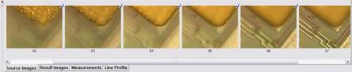
6 Simple User Interface Auto-Montage Pro features a main window with two workspaces, making the software simple to use. With viewing options presented as icons on one side of the screen and the final image on the other, you ll find it easy to track the changes you ve made to the image. The workspaces use a flow chart to show you the relationship between each image generated, as well as allowing you to quickly select which Montage or Viewing methods gives you the result you need. Producing a Focused Image One major advantage of using Auto-Montage Pro is that it can be configured to fully automate the image acquisition process, by capturing a set of source images directly from the camera. Direct acquisition into Auto-Montage Pro is available with a range of camera options. Additionally, if you are using a calibrated Z stepper, you can set up Auto-Montage Pro to automatically calculate and capture the optimum number of source images from your sample. This unique feature of Auto-Montage Pro takes the guesswork out of the capture process, by acquiring a number of images determined by the software, thus ensuring you ll produce a perfectly focused image every time - no matter how deep your sample is. Using the Scan Montage function, Auto-Montage Pro analyses the in-focus areas in each source image, and then combines them to produce a fully focused montage image. The source images used can be viewed as a film strip, positioned under the final focused image, in order that you can ensure you are using the best source images of your sample.
7

8 COLOUR RELIEF MAP OF A MOUSE EMBRYO


9 Auto-Montage Pro software offers you a wide range of algorithms (known as Montage Methods) for calculating the way in which your focused image is produced. These algorithms are unique to Syncroscopy, and ensure that whatever your application, one of these Montage Methods will guarantee you the most accurate fully focused image of your sample. Additionally, Auto-Montage Pro has a time saving preview function that allows you to see which of the software s algorithms is most suitable for your sample before processing. No other depth of field imaging software offers this level of flexibility. Viewing your Image Auto-Montage Pro offers several ways of viewing the montage image and other useful height coded data. Depth Map As part of the Scan Montage operation, Auto-Montage Pro automatically generates a powerful Depth Map image. This is a record of which source image provided the in-focus region for the fully focused montage image at each pixel location. Colour Relief Map Similar to a Depth Map, the Colour Relief Map shows in pseudo colour where each source image is positioned in the image stack. The Colour Relief Map, generated by the Scan Colour Relief function of Auto-Montage Pro provides useful colour coded depth information of a 3-D sample.

10 Enhancing your Image Scan Align If you are working with a stereo microscope, the Scan Align function compensates for the changes in position that inherently occur when capturing source images. Thus, parallax error is automatically corrected to improve the alignment of your final image. Also if your source images have been derived photographically with a macro lens and subsequently scanned in, then the Scan Align function will correct any errors in registration between frames. Viewing your Image Anaglyph Using the Scan Anaglyph feature, Auto-Montage Pro allows you to see your sample, with the aid of 3-D glasses, as a true 3-D representation. This unique function allows your samples to come to life and highlights interesting areas which might otherwise be lost. Stereo Pair The Scan Stereo Pair operation generates the stereo pair image, which when viewed correctly offers a 3-dimensional colour view of the montage image. Confidence Map Unique to Auto-Montage Pro, the View Confidence function provides a Confidence Map of your sample. The Confidence Map displays the accuracy of the final in-focus image. By shading areas of low confidence in black, and those of high confidence in white, you can quickly select the areas of highest confidence when making precise sample measurements. In addition, if you capture your source images by Scanning Electron Microscope (SEM), a newly-developed extension to the Scan Align function automatically detects and compensates for the inherent change in angular magnification as focus is moved through a very deep sample. This function will also compensate for the small change in focal length when using a modern internally-focused macro lens. Scan Enhance and Editing Auto-Montage Pro includes powerful editing and image manipulation tools. The Scan Enhance function provides controls for sharpening, brightness and contrast, as well as the ability to alter the colour balance. Auto-Montage Pro also includes comprehensive image editing functions. Both of these features save valuable time, in producing publication quality images, without the need to export image files to third party image editing software. Saving and Printing your Results All of the generated images can be printed or saved. They can also be automatically exported to other software for use in reports or presentations.

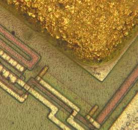
11 SCANNING ELECTRON MICROSCOPE IMAGE OF BONE


12 Syncroscopy also offers a number of options for capturing and viewing your image that can be added to your Auto-Montage Pro software. These include: Capturing your Image - Quantage If your samples have a high dynamic range - that is, if they contain some regions that are very bright and others that are very dark, then no single camera exposure setting will give you reliable information in both bright and dark areas. Quantage can help you solve this problem. Quantage is the latest patented, innovative development from the Syncroscopy team. Quantage takes control of the camera and automatically records several images using different exposure conditions, and then combines the separate images into a single resultant image. The result is a colour Quantage image, of huge dynamic range, that captures contrast from the whole range of brightness levels of the sample. Although the dynamic range is too large for the human eye to appreciate, Auto Montage can use contrast throughout the dynamic range to determine focus and therefore height information. Viewing your Image - 3-D Model Option When working with opaque samples like metals, crystals and bone, the View 3-D Model feature can be used to create a 3-D surface model of the sample that can be viewed from any angle. This view of the sample is excellent for looking at fine details, and viewing specific areas of interest such as stress fractures and surface scratches. The 3-D View model can also be exported as an image file, enabling it to be included in Word document reports or PowerPoint presentations.

13 3-D VIEW IMAGE OF FRACTURED GLASS


14 Measurements Option Auto-Montage Pro s powerful Measurements option enables you to automatically measure a range of X, Y, & Z parameters quickly and easily. Using your mouse to draw on any Montage image or source image you can perform single point measurements. Good calibration is essential for accurate measurements and by using Auto-Montage Pro you can easily calibrate your sample with a standard stage graticule, or a specimen of known size. Alternatively, if you have already calibrated your camera and microscope you can save time by selecting a calibration from the Preset Calibrations list. If you are using a calibrated Z stepper, you can use the Z-stepper property page in Auto-Montage Pro to configure your Z-stepper, thus ensuring precise automated measurements in the Z-axis. Each of your measurement results is displayed on a status bar and to save time is automatically recorded in a table under each image. The table can then be saved as a text file, or can be rapidly exported to Excel for further analysis. Using the View Line Profile feature, you can generate a graph showing the profile of the currently selected straight-line measurement. This provides useful information about the height profile of the surface of a material. Auto-Montage Pro comes complete with the following: Feature Benefit Automatic image capture from camera Saves time with file manipulation Precise, automated image reconstruction Perfectly focused image User image collection calibration Rapid, reproducible set up Interface with icons Simple to use Split image/interface viewing Effortless image manipulation Choice of Montage Methods Optimum image for each sample Range of ways to view focused image Clear, easy to measure images Optional X, Y, Z measurement Quick length, depth and volume sizing Dedicated image enhancing tools High quality images Compatible with other software Easy to export data packages
15 BEFORE AUTO-MONTAGE AFTER AUTO-MONTAGE

16 Auto-Montage Essentials Auto-Montage is also offered as an entry level version for those users who just need a basic tool to produce in-focus images of 3-D samples. Auto-Montage Essentials has limited functionality compared to Auto-Montage Pro but will enable you to improve your imaging capability and allow you to create quality images. Users of Auto-Montage Essentials can easily upgrade to Auto-Montage Pro to take advantage of all the advanced features offered by the professional package.

17 FILE CUTTING SURFACE WEAR

18 Producing a Focused Image With Auto-Montage Essentials you can open BMP, TIFF or JPEG source image files captured by any digital camera. Using the Scan Montage function, Auto-Montage Essentials analyses and selects the in-focus areas from each source image. It then combines them to produce a fully focused image. It's as simple as that! Viewing your Image As well as viewing the fully focused resultant image directly, there is also the useful Scan Anaglyph feature. This function in Auto-Montage Essentials allows you to view your sample with the aid of 3-D glasses as a true 3-D image. This provides a truly unique representation of depth data and saves time by making it easier for you to detect areas of interest within the sample. Improving your Image A poorly aligned final image is an inherent problem if you are working with a stereo microscope. This is caused by the changes in position (parallax error) that occur during the capture of source images. Using the Scan Align function in Auto-Montage Essentials you can automatically compensate for parallax error to improve the accuracy of your final image. Additionally, if your source images have been derived photographically and then scanned in, the Scan Align function will also correct any errors in registration between frames to generate an aligned image. With some single colour samples, it can be difficult to see which source image provided the detail for your resultant, fully focused, Montage image. As part of the Scan Montage operation, Auto-Montage Essentials automatically generates a Depth Map image. This is a record of which source image provided the in-focus region for the Montage image.

19 Auto-Montage Essentials also includes comprehensive image editing functions. These features save valuable time in producing publication quality images, without the need to export image files to image editing software. Saving and Printing your Results All source images, and the resultant fully focused images can be easily printed or saved in a range of file formats for use with other software such as Word for reports or PowerPoint for presentations. Auto-Montage Essentials offers you all this: Feature Uses images from any optical microscope Automatically produces in-focus images Produces an anaglyph view Range of image enhancing tools Compatible with other software packages Optional upgrade to Auto-Montage Pro Benefit Flexible and cost-effective Saves time manipulating data Accurate 3-D representation High quality images Easy to export images Extends imaging applications
20 Auto-Montage Pro System Do you: Want to quickly produce in-focus images of 3-D samples? Need to produce images and reports of many 3-D samples per day? Want integrated software, digital camera, and Z-stepper for your microscope? Want fully automated image capture? Want accurate sample measurement? Require different views of your focused image? Say yes to any of these questions and there is an Auto-Montage Pro System that is the right choice for you.

21 Here s what an Auto-Montage Pro system offers you: Auto-Montage Pro Systems offer all the benefits of the powerful Auto-Montage Pro software, plus an integrated package of an automated Z-stepper, linked to a high quality digital camera for fully automated image capture and analysis. An Auto-Montage Pro system saves time and effort in setting up and focusing a microscope. Since the Z-stepper is fully integrated it is also automatically calibrated in the Z-axis. This ensures depth measurements are accurate. As the Z-stepper focuses your microscope, the Auto-Montage Pro software automatically captures, analyses and combines all of the in-focus sections of your source images, to rapidly produce one composite, perfectly focused, montage image. Feature Integrated digital camera High resolution Automatic image reconstruction Automated Z-stepper Calibrated Z-stepper User image collection calibration Interface with icons Split image/interface viewing Choice of Montage Methods Many ways to view focused image Automated X, Y, Z measurement Dedicated image enhancing tools Compatible with other software Benefit Direct image capture to Auto-Montage Pro Quality source images Perfectly focused image Saves time with focusing Accurate depth measurements Rapid, reproducible set up Simple to use Effortless image manipulation Optimum image for each sample Clear, easy to measure images Quick length, depth and volume sizing High quality images Easy to export data

22 THICK CYTOLOGY SPECIMEN
23 Syncroscopy Researchers world-wide have chosen Auto-Montage to produce images for a wide variety of applications. Here s what some of them have to say about the performance of this exceptional software: Using Auto-Montage is helping us rapidly catalogue more than 11,000 known ant species. It is not only saving time by automating the process, but it also produces quality, in depth images, a task we have found impossible with other methods, Dr. Gary Alpert, Harvard University, USA. Before we had Auto-Montage we had to manually cut and paste in-focus sections of photos taken with a conventional camera and then re-photograph the montage. This made it tricky and time consuming to obtain a focused microfossil image. Using Auto-Montage has meant we can capture images at different focal depths in rock and can generate precise in-focus images quickly and simply. Owen Green, Department of Earth Sciences, University of Oxford, UK. We use Auto-Montage to generate important views of unique early human skeletons that are millions of years old. Producing such images of their bone and tooth tissues is significantly advancing our knowledge of human evolutionary history. Professor Timothy Bromage, Hunter College, CUNY, USA. With Auto-Montage software we no longer spend valuable time overlaying fingerprint photographs to get a complete fingerprint mark from a curved surface. One of the benefits of Auto-Montage for our fingerprint work is that we can use it to enhance images to see the fine detail, which is crucial for making fingerprint matches. Esther Neate, Wiltshire Police, UK.

24 Software features Essentials Pro Suitable for a wide range of applications Y Y Open images from another application Y Y Import your own stored images Y Y Suitable for SEM images N Y Automatic capture from camera N Y Able to use Z Stepper N Y Preview window Y Y Scan Alignment Y Y Export image Y Y Despeckle source images N Y Scan montage speed option Y Y Scan montage precision option N Y Scan enhance N Y Scan anaglyph Y Y Scan stereo pair N Y Scan colour relief N Y Montage editing Y Y Confidence map N Y Depth map Y Y Depth profile N Y Data reporting N Y Upgrades available Y Y Workspaces for smart screenshots N Y Measurements option available N Y 3D Viewer option available N Y When you have chosen an Auto-Montage software or system to fit your needs, please contact us or one of our dealers for more information and a demonstration. S Y N C R O Syncroscopy European and International Headquarters Beacon House Nuffield Road Cambridge CB4 ITF UK Tel: +44(0) Fax: +44 (0) sales@syncroscopy.com C O P Y Syncroscopy USA Headquarters 5108 Pegasus Court Suite M Frederick MD USA Tel: / Fax: ussales@syncroscopy.com Website: S A DIVISION OF THE SYNOPTICS GROUP Picture acknowledgements: Front cover image and bone image courtesy of Professor Alan Boyde, Queen Mary University of London, UK. Mouse embryo image courtesy of Professor Tim Bromage, Hunter College, CUNY, USA. Back cover image courtesy of Dr. Gary Alpert, Harvard University, USA. All trademarks acknowledged. For a full range of options, see our current price lists. C.0008.A
DIGITAL-MICROSCOPY CAMERA SOLUTIONS USB 3.0
 DIGITAL-MICROSCOPY CAMERA SOLUTIONS USB 3.0 PixeLINK for Microscopy Applications PixeLINK will work with you to choose and integrate the optimal USB 3.0 camera for your microscopy project. Ideal for use
μscope Microscopy Software
 μscope Microscopy Software Pixelink μscope Essentials (ES) Software is an easy-to-use robust image capture tool optimized for productivity. Pixelink μscope Standard (SE) Software had added features, making
U GENIUS. Gel imaging at a touch
 U GENIUS 3 Gel imaging at a touch U:GENIUS 3 Simply Genius. Designed to make your gel imaging simple, quick and easy. No set up, no external computer - just a complete imaging system for all your 1D needs.
T:GENIUS GEL IMAGING AT A TOUCH
 T:GENIUS GEL IMAGING AT A TOUCH The T:Genius is an integrated system for DNA and protein analysis and gel documentation. Based on the successful Syngene gel documentation range, the T:Genius features an
U:GENIUS S Y N G E N E. Gel imaging at a touch A DIVISION OF THE SYNOPTICS GROUP
 U:GENIUS Gel imaging at a touch S Y N G E N E A DIVISION OF THE SYNOPTICS GROUP U:GENIUS Simply Genius. Designed to make your gel imaging simple, quick and easy. No set up, no external computer - just
DEDICATED CHEMILUMINESCENCE IMAGING SYSTEM DELIVERS SIMPLICITY AND SENSITIVITY
 DEDICATED CHEMILUMINESCENCE IMAGING SYSTEM DELIVERS SIMPLICITY AND SENSITIVITY COMBINING SIMPLICITY WITH SENSITIVITY Despite chemiluminescence blotting being a widely used technique to separate and detect
Gel imaging at a touch S Y N G E N E A DIVISION OF THE SYNOPTICS GROUP
 Gel imaging at a touch S Y N G E N E A DIVISION OF THE SYNOPTICS GROUP Use the large colour touch screen to navigate your way through the functions of. The icon driven menu is both intuitive and easily
Redefining Gel and Blot Imaging
 Redefining Gel and Blot Imaging PXi AND PXi TOUCH Gel and blot imaging made easy Syngene imaging systems are recognised world-wide as high quality, high performance instruments for the capture and analysis
INGENIUS 3. Low cost, high performance gel documentation and analysis
 INGENIUS 3 Low cost, high performance gel documentation and analysis INGENIUS 3 When simplicity and budget matter. The InGenius 3 gel documentation and analysis system is compact, easy to use and offers
INGENIUS 3 LOW COST, HIGH PERFORMANCE GEL DOCUMENTATION AND ANALYSIS
 INGENIUS 3 LOW COST, HIGH PERFORMANCE GEL DOCUMENTATION AND ANALYSIS The InGenius 3 uses a high performance 3m pixel camera. The darkroom assembly is easily connected to a PC. GeneSys image acquisition
bioteknika T:GENIUS GEL IMAGING AT A TOUCH
 bioteknika T:GENIUS GEL IMAGING AT A TOUCH The T:Genius is an integrated system for DNA and protein analysis and gel documentation. Based on the successful Syngene gel documentation range, the T:Genius
SCIENTIFIC INSTRUMENT NEWS. Introduction. Design of the FlexSEM 1000
 SCIENTIFIC INSTRUMENT NEWS 2017 Vol. 9 SEPTEMBER Technical magazine of Electron Microscope and Analytical Instruments. Technical Explanation The FlexSEM 1000: A Scanning Electron Microscope Specializing
Camera Overview. Digital Microscope Cameras for Material Science: Clear Images, Precise Analysis. Digital Cameras for Microscopy
 Digital Cameras for Microscopy Camera Overview For Materials Science Microscopes Digital Microscope Cameras for Material Science: Clear Images, Precise Analysis Passionate about Imaging: Olympus Digital
Technical Benefits of the
 innovation in microvascular assessment Technical Benefits of the Moor Instruments moorflpi-2 moorflpi-2 More Info: Measurement Principle laser speckle contrast analysis Measurement 85nm Laser Wavelength
G BOX. Gel Documentation and Analysis Automated imaging
 G BOX Gel Documentation and Analysis Automated imaging GEL IMAGING AND ANALYSIS Automated imaging for all your applications Syngene imaging systems are recognised world-wide as high quality, high performance
Camera Overview. Digital Microscope Cameras for Material Science: Clear Images, Precise Analysis. Digital Cameras for Microscopy
 Digital Cameras for Microscopy Camera Overview For Materials Science Microscopes Digital Microscope Cameras for Material Science: Clear Images, Precise Analysis Passionate about Imaging: Olympus Digital
Huvitz Digital Microscope HDS-5800
 Huvitz Digital Microscope HDS-5800 Dimensions unit : mm Huvitz Digital Microscope HDS-5800 HDS-MC HDS-SS50 The world s first, convert the magnification from 50x to 5,800x with a zoom lens HDS-TS50 Huvitz
BioSpectrum Imaging System
 BioSpectrum Imaging System Imaging Made Easy for Chemiluminescence Bioluminescence Colorimetric Fluorescence MegaCam 810 Camera OptiChemi 610 Camera BioChemi 510 Camera GelCam 310 Camera 8.1 megapixel
-f/d-b '') o, q&r{laniels, Advisor. 20rt. lmage Processing of Petrographic and SEM lmages. By James Gonsiewski. The Ohio State University
 o, q&r{laniels, Advisor. 20rt. lmage Processing of Petrographic and SEM lmages. By James Gonsiewski. The Ohio State University") lmage Processing of Petrographic and SEM lmages Senior Thesis Submitted in partial fulfillment of the requirements for the Bachelor of Science Degree At The Ohio State Universitv By By James Gonsiewski
lmage Processing of Petrographic and SEM lmages Senior Thesis Submitted in partial fulfillment of the requirements for the Bachelor of Science Degree At The Ohio State Universitv By By James Gonsiewski
TECHNICAL SPECIFICATION SCHEDULE
 LAMPIRAN Q5 MEMBEKAL, MENGHANTAR, MEMASANG DAN KOMISYEN METALLURGICAL MICROSCOPE WITH COMPUTER ATTACHMENT KE P.P.KEJ. B AHAN & SUMBER MINERAL, UNIVERSITI SAINS MALAYSIA, KAMPUS KEJURUTERAAN Quotation No:
By: Louise Brown, PhD, Advanced Engineered Materials Group, National Physical Laboratory.
 NPL The Olympus LEXT - A highly flexible tool Confocal Metrology at the NPL By: Louise Brown, PhD, Advanced Engineered Materials Group, National Physical Laboratory. www.npl.co.uk louise.brown@npl.co.uk
Quick Operation Guide
 Quick Operation Guide Power ON Mounting specimens Set the specimen on the sample holder, and install the sample holder to the holder frame. Attach the holder frame to the XY stage. Type of holder Main
MODULE No. 34: Digital Photography and Enhancement
 SUBJECT Paper No. and Title Module No. and Title Module Tag PAPER No. 8: Questioned Document FSC_P8_M34 TABLE OF CONTENTS 1. Learning Outcomes 2. Introduction 3. Cameras and Scanners 4. Image Enhancement
ScanArray Overview. Principle of Operation. Instrument Components
 ScanArray Overview The GSI Lumonics ScanArrayÒ Microarray Analysis System is a scanning laser confocal fluorescence microscope that is used to determine the fluorescence intensity of a two-dimensional
contents TABLE OF The SECOM platform Applications - sections Applications - whole cells Features Integrated workflow Automated overlay
 S E C O M TABLE OF contents The SECOM platform 4 Applications - sections 5 Applications - whole cells 8 Features 9 Integrated workflow 12 Automated overlay ODEMIS - integrated software Specifications 13
Caenotec Prof. Ralf Schnabel
 Caenotec Prof. Ralf Schnabel Kleine Dorfstr. 9 38312 Börßum, Ph: 0049 151 11653356 r.schnabel@tu-bs.de Mehr braucht man nicht A simple and convenient program to document microscope pictures by Christian
Zeiss AxioImager.Z2 Brightfield Protocol
 Zeiss AxioImager.Z2 Brightfield Protocol 1) System Startup Please note put sign-up policy. You must inform the facility at least 24 hours beforehand if you can t come; otherwise, you will receive a charge
Tribometrics. Version 2.11
 Tribometrics Version 2.11 Table of Contents Tribometrics... 1 Version 2.11... 1 1. About This Document... 4 1.1. Conventions... 4 2. Introduction... 5 2.1. Software Features... 5 2.2. Tribometrics Overview...
Stereo Viewing Systems
 Stereo Viewing Systems for a wide range of inspection and material rework tasks Patented optical technology for fatigue-free viewing and superb image quality Wide range of magnification options (to x20)
Extending the Dynamic Range of Film
 Written by Jonathan Sachs Copyright 1999-2003 Digital Light & Color Introduction Limited dynamic range is a common problem, especially with today s fine-grained slide films. When photographing contrasty
Nikon. King s College London. Imaging Centre. N-SIM guide NIKON IMAGING KING S COLLEGE LONDON
 N-SIM guide NIKON IMAGING CENTRE @ KING S COLLEGE LONDON Starting-up / Shut-down The NSIM hardware is calibrated after system warm-up occurs. It is recommended that you turn-on the system for at least
Automated Imaging Technology to Simplify Your Workflow!
 Automated Imaging Technology to Simplify Your Workflow! BioSpectrum Imaging System Imaging Made Easy for Chemiluminescence Bioluminescence Colorimetric Fluorescence MegaCam 810 Camera OptiChemi 600 Camera
ICOM CIDOC Dresden 2014 Short Paper. Documentation Photography: An Integrated Process
 ICOM CIDOC Dresden 2014 Short Paper Submitted by: Suzanne Petersen McLean, BSc Collections Manager Bata Shoe Museum, Toronto www.batashoemuseum.ca sznnpetersen@gmail.com keywords: Photography, metadata,
Bringing Answers to the Surface
 3D Bringing Answers to the Surface 1 Expanding the Boundaries of Laser Microscopy Measurements and images you can count on. Every time. LEXT OLS4100 Widely used in quality control, research, and development
Histograms& Light Meters HOW THEY WORK TOGETHER
 Histograms& Light Meters HOW THEY WORK TOGETHER WHAT IS A HISTOGRAM? Frequency* 0 Darker to Lighter Steps 255 Shadow Midtones Highlights Figure 1 Anatomy of a Photographic Histogram *Frequency indicates
Before you start, make sure that you have a properly calibrated system to obtain high-quality images.
 CONTENT Step 1: Optimizing your Workspace for Acquisition... 1 Step 2: Tracing the Region of Interest... 2 Step 3: Camera (& Multichannel) Settings... 3 Step 4: Acquiring a Background Image (Brightfield)...
Ordering Information & Specifications. VisionWorksLS Capabilities. Image Analysis Capabilities
 Ordering Information & Specifications VisionWorksLS Capabilities Each system includes: Camera and lens, darkroom with motorized or manual platform, three emission filters, white light illuminator, choice
User Manual. Copyright 2010 Lumos. All rights reserved
 User Manual The contents of this document may not be copied nor duplicated in any form, in whole or in part, without prior written consent from Lumos. Lumos makes no warranties as to the accuracy of the
Clemex Vision Lite. Capture, Measure, Analyze. the image analysis people
 Clemex Vision Lite Capture, Measure, Analyze the image analysis people Acquire, Measure, Analyze with Clemex Vision Lite Clemex Vision Lite is a flexible yet affordable software solution that enables
Agilent Cary 610/620 FTIR microscopes and imaging systems RESOLUTION FOR EVERY APPLICATION
 Agilent Cary 610/620 FTIR microscopes and imaging systems RESOLUTION FOR EVERY APPLICATION AGILENT CARY 610/620 FTIR MICROSCOPES ADVANCING FTIR MICROSCOPY AND IMAGING Agilent s 610/620 FTIR microscopes
ChemiDoc-It Imaging System
 ChemiDoc-It Imaging System Ultra dark chamber and highly sensitive, scientific-grade CCD camera for chemiluminescence imaging ChemiDoc-It darkroom is light tight creating optimum imaging conditions for
Zeiss 780 Training Notes
 Zeiss 780 Training Notes Turn on Main Switch, System PC and Components Switches 780 Start up sequence Do you need the argon laser (458, 488, 514 nm lines)? Yes Turn on the laser s main power switch and
it.med.harvard.edu/ris UMAX PowerLook 1120 Flatbed Scanner WQGF
 it.med.harvard.edu/ris UMAX PowerLook 1120 WQGF Research Imaging Solutions RIS@hms.harvard.edu it.med.harvard.edu/ris Beth Beighlie Digital Imaging Coordinator Research Imaging Solutions Information Technology
9/19/16. A Closer Look. Danae Wolfe. What We ll Cover. Basics of photography & your camera. Technical. Macro & close-up techniques.
 A Closer Look Danae Wolfe What We ll Cover Basics of photography & your camera Technical Macro & close-up techniques Creative 1 What is Photography? Photography: the art, science, & practice of creating
It makes sense to read this section first if new to Silkypix... How to Handle SILKYPIX Perfectly Silkypix Pro PDF Contents Page Index
 It makes sense to read this section first if new to Silkypix... How to Handle SILKYPIX Perfectly...145 Silkypix Pro PDF Contents Page Index 0. 0.Overview and Introduction...9 0.1. Section Names...9 0.1.1.
Advanced Technology and Manufacturing Institute. Zygo ZeScope
 Advanced Technology and Manufacturing Institute Zygo ZeScope Created by Andrew Miller ATAMI Oregon State University Revision Date Description Curator 0 8/31/2018 New Document Andrew Miller Zygo ZeScope
2D Protein Gel Image Capture and Analysis
 2D Protein Gel Image Capture and Analysis the way you want it S Y N G E N E A DIVISION OF THE SYNOPTICS GROUP 2D protein gel generation, image capture and analysis systems Syngene has a worldwide reputation
GEL IMAGING AT A TOUCH
 GEL IMAGING AT A TOUCH NUGENIUS NuGenius is a new generation, low cost, integrated system for DNA and protein analysis and gel documentation. Continuing the Genius range, the NuGenius features an integrated
Technology Learning Activity: Multimedia CIMC. Student Edition TE8135
 IMC Technology Learning Activity: Multimedia Student Edition www.okcimc.com 800-654-4502 CIMC TE8135 Technology Learning Activity This TLA covers information on digital imaging sources and uses, photography
Contents STARTUP MICROSCOPE CONTROLS CAMERA CONTROLS SOFTWARE CONTROLS EXPOSURE AND CONTRAST MONOCHROME IMAGE HANDLING
 Operations Guide Contents STARTUP MICROSCOPE CONTROLS CAMERA CONTROLS SOFTWARE CONTROLS EXPOSURE AND CONTRAST MONOCHROME IMAGE HANDLING Nikon Eclipse 90i Operations Guide STARTUP Startup Powering Up Fluorescence
Camera Overview. Olympus Digital Cameras for Materials Science Applications: For Clear and Precise Image Analysis. Digital Cameras for Microscopy
 Digital Cameras for Microscopy Camera Overview For Materials Science Microscopes Olympus Digital Cameras for Materials Science Applications: For Clear and Precise Image Analysis Passionate about Imaging
GlobiScope Analysis Software for the Globisens QX7 Digital Microscope. Quick Start Guide
 GlobiScope Analysis Software for the Globisens QX7 Digital Microscope Quick Start Guide Contents GlobiScope Overview... 1 Overview of home screen... 2 General Settings... 2 Measurements... 3 Movie capture...
Compare and Contrast. Contrast Methods in Industrial Inspection Microscopy. Application Note. We explain how to
 Application Note Compare and Contrast Contrast Methods in Industrial Inspection Microscopy We explain how to E nhance materials inspection microscopy workflows Reveal surface and sub-surface imperfections
Introduction of New Products
 Field Emission Electron Microscope JEM-3100F For evaluation of materials in the fields of nanoscience and nanomaterials science, TEM is required to provide resolution and analytical capabilities that can
OLYMPUS Digital Cameras for Materials Science Applications: Get the Best out of Your Microscope
 Digital Cameras for Microscopy Camera Overview For Materials Science Microscopes OLYMPUS Digital Cameras for Materials Science Applications: Get the Best out of Your Microscope Passionate About Imaging
Gel Documentation and Analysis the way you want it S Y N G E N E A DIVISION OF THE SYNOPTICS GROUP
 Gel Documentation and Analysis the way you want it S Y N G E N E A DIVISION OF THE SYNOPTICS GROUP Syngene Gel Documentation and Analysis Syngene has long been associated with innovations in gel documentation.
Stereo Viewing Systems
 Includes NEW image capture option Stereo Viewing Systems Superior imaging for a wide range of inspection & rework tasks Patented optical technology for fatigue-free viewing and superb image quality Wide
METIS EDS GAMMA. Metis desktop professional scanner : great quality, fast and easy to use!
 Metis desktop professional scanner : great quality, fast and easy to use! The V-Table design The integrates an innovative V-Table design supported by specific software tools in order to hold the originals
I NEED A SIMPLE AND RELIABLE EDGING SOLUTION FOR MY DAILY WORKFLOW.
 I NEED A SIMPLE AND RELIABLE EDGING SOLUTION FOR MY DAILY WORKFLOW. RACE THROUGH WORKLOADS WITH THE NEXT-GENERATION KAPPA WHEN IT COMES TO QUALITY AND PRODUCTIVITY, YOUR EXPECTATIONS ARE HIGH. WITH NEKSIA
Produce stunning. Pro photographer Chris Humphreys guides you through HDR and how to create captivating natural-looking images
 Masterclass: In association with Produce stunning HDR images Pro photographer Chris Humphreys guides you through HDR and how to create captivating natural-looking images 8 digital photographer 45 masterclass4produce
Leading in Desktop SEM Imaging and Analysis
 Leading in Desktop SEM Imaging and Analysis Fast. Outstanding. Reliable SEM imaging and analysis. The Phenom: World s Fastest Scanning Electron Microscope With its market-leading Phenom desktop Scanning
G7 System Certification Application Data Sheet
 ! G7 System Certification Application Data Sheet!! The IDEAlliance Print Properties Working Group has established a certification process for G7 Systems. In accordance with this process The G7 System Certification
Color aspects and Color Standardization in Digital Microscopy
 Color aspects and Color Standardization in Digital Microscopy Yukako Yagi, PhD yyagi@partners.org Director of the MGH Pathology Imaging & Communication Technology Center Assistant Professor of Pathology,
Image optimization guide
 Image Optimization guide for Image Submittal Images can play a crucial role in the successful execution of a book project by enhancing the text and giving the reader insight into your story. Although your
TRIAXES STEREOMETER USER GUIDE. Web site: Technical support:
 TRIAXES STEREOMETER USER GUIDE Web site: www.triaxes.com Technical support: support@triaxes.com Copyright 2015 Polyakov А. Copyright 2015 Triaxes LLC. 1. Introduction 1.1. Purpose Triaxes StereoMeter is
BioSpectrum Imaging System
 BioSpectrum Imaging System Imaging Made Easy for Chemiluminescence Bioluminescence Colorimetric Fluorescence MegaCam 800 Camera OptiCam 600 Camera BioChemi 500 Camera ChemiCam 410 Camera GelCam 310 Camera
Zeiss Deconvolution Microscope: A Quick Guide
 Zeiss Deconvolution Microscope: A Quick Guide Start-up Uncover microscope. Do not put dust cover on the floor. Plug in both cameras. The default camera is the AxioCam HRm (monochrome camera) for fluorescence
Transmission Electron Microscopy 9. The Instrument. Outline
 Transmission Electron Microscopy 9. The Instrument EMA 6518 Spring 2009 02/25/09 Outline The Illumination System The Objective Lens and Stage Forming Diffraction Patterns and Images Alignment and Stigmation
Cast Iron Analysis. Nodular cast iron analysis
 Cast Iron Analysis This dhs Image Data Base module analyzes the microstructure of cast iron. The graphite particles actual make-up is crucial for material properties such as elasticity and fracture behaviour.
User Manual. cellsens 1.16 LIFE SCIENCE IMAGING SOFTWARE
 User Manual cellsens 1.16 LIFE SCIENCE IMAGING SOFTWARE Any copyrights relating to this manual shall belong to OLYMPUS CORPORATION. We at OLYMPUS CORPORATION have tried to make the information contained
Reikan FoCal Aperture Sharpness Test Report
 Focus Calibration and Analysis Software Test run on: 26/01/2016 17:02:00 with FoCal 2.0.6.2416W Report created on: 26/01/2016 17:03:39 with FoCal 2.0.6W Overview Test Information Property Description Data
User Manual Veterinary
 Veterinary Acquisition and diagnostic software Doc No.: Rev 1.0.1 Aug 2013 Part No.: CR-FPM-04-022-EN-S 3DISC, FireCR, Quantor and the 3D Cube are trademarks of 3D Imaging & Simulations Corp, South Korea,
New Benchmark of Molecular Image Software Package
 Molecular Imager New Benchmark of Molecular Image Software Package Magic software package was developed under the division of the Wealtec imaging family group. It is a professional and powerful 1D package
CLEMEX intelligent microscopy
 CLEMEX intelligent microscopy Vision PE 5.0 Advanced Image Analysis Experience in Image Analysis Research and Quality Control Solutions With Vision PE, Clemex provides a powerful image analysis solution
BLIPS perfectly aligned cleaned by any impurity Gently press the lens
 BLIPS - TIPS & TRICKS BLIPS, the thinnest Macro and Micro lenses for smartphones in the world, can guarantee an excellent picture quality for photos and videos, but they need some simple tricks to get
Image Processing Tutorial Basic Concepts
 Image Processing Tutorial Basic Concepts CCDWare Publishing http://www.ccdware.com 2005 CCDWare Publishing Table of Contents Introduction... 3 Starting CCDStack... 4 Creating Calibration Frames... 5 Create
Photo Editing Workflow
 Photo Editing Workflow WHY EDITING Modern digital photography is a complex process, which starts with the Photographer s Eye, that is, their observational ability, it continues with photo session preparations,
MAGNIFIERS & TASK LAMPS FOR INDUSTRY. presented by
 2017 MAGNIFIERS & TASK LAMPS FOR INDUSTRY presented by 1 Daylight Catalogue Professional Magnifiers & Task Lamps 16 great reasons to use Daylight Daylight Simulation Daylight 6,000 K colour temperature
AgilEye Manual Version 2.0 February 28, 2007
 AgilEye Manual Version 2.0 February 28, 2007 1717 Louisiana NE Suite 202 Albuquerque, NM 87110 (505) 268-4742 support@agiloptics.com 2 (505) 268-4742 v. 2.0 February 07, 2007 3 Introduction AgilEye Wavefront
STEM Spectrum Imaging Tutorial
 STEM Spectrum Imaging Tutorial Gatan, Inc. 5933 Coronado Lane, Pleasanton, CA 94588 Tel: (925) 463-0200 Fax: (925) 463-0204 April 2001 Contents 1 Introduction 1.1 What is Spectrum Imaging? 2 Hardware 3
nanovea.com PROFILOMETERS 3D Non Contact Metrology
 PROFILOMETERS 3D Non Contact Metrology nanovea.com PROFILOMETER INTRO Nanovea 3D Non-Contact Profilometers are designed with leading edge optical pens using superior white light axial chromatism. Nano
Reikan FoCal Aperture Sharpness Test Report
 Focus Calibration and Analysis Software Reikan FoCal Sharpness Test Report Test run on: 26/01/2016 17:14:35 with FoCal 2.0.6.2416W Report created on: 26/01/2016 17:16:16 with FoCal 2.0.6W Overview Test
Planmeca Romexis. quick guide. Viewer EN _2
 Planmeca Romexis Viewer quick guide EN 10029550_2 TABLE OF CONTENTS 1 START-UP OF PLANMECA ROMEXIS VIEWER...1 1.1 Selecting the interface language... 1 1.2 Selecting images...1 1.3 Starting the Planmeca
Jeol JEM Responsible personell: Endy ( ) Online booking is compulsory!
 Online booking is compulsory!") Jeol JEM 1230 Responsible personell: Endy (45279377) Online booking is compulsory! After training you will have access to working alone on the instrument. All insertion of samples is done by responsible
Jeol JEM 1230 Responsible personell: Endy (45279377) Online booking is compulsory! After training you will have access to working alone on the instrument. All insertion of samples is done by responsible
Introduction to 2-D Copy Work
 Introduction to 2-D Copy Work What is the purpose of creating digital copies of your analogue work? To use for digital editing To submit work electronically to professors or clients To share your work
How to Make 3D Images for Viewing with No Glasses
 By James Bruce Believe it or not, you don t actually need 3D glasses to experience convincingly realistic 3D images (or movies). You just need to make yourself go cross-eyed. Essentially, you look at two
One Week to Better Photography
 One Week to Better Photography Glossary Adobe Bridge Useful application packaged with Adobe Photoshop that previews, organizes and renames digital image files and creates digital contact sheets Adobe Photoshop
A Rapid Image Acquisition Method for Focus Stacking in Microscopy
 A Rapid Image Acquisition Method for Focus Stacking in Microscopy Douglas Clark* and Brian Brown Paedia LLC, 327 Lake Street, San Francisco, CA 94118 * dclark@paedia.com Introduction Modern image acquisition
printcontrol Printing process control at a click of the mouse
 printcontrol Printing process control at a click of the mouse Printing process control with GMG PrintControl and GMG RapidCheck The significance of process control in the graphic arts industry has grown
Contents Foreword 1 Feedback 2 Legal information 3 Getting started 4 Installing the correct Capture One version 4 Changing the version type 5 Getting
 Contents Foreword 1 Feedback 2 Legal information 3 Getting started 4 Installing the correct Capture One version 4 Changing the version type 5 Getting to know Capture One Pro 6 The Grand Overview 6 The
PASS4TEST. IT Certification Guaranteed, The Easy Way! We offer free update service for one year
 PASS4TEST IT Certification Guaranteed, The Easy Way! \ We offer free update service for one year Exam : 9A0-125 Title : Adobe Photoshop Lightroom 2 ACE Exam Vendors : Adobe Version : DEMO Get Latest &
Technical Aspects in Digital Pathology
 Technical Aspects in Digital Pathology Yukako Yagi, PhD yyagi@mgh.harvard.edu Director of the MGH Pathology Imaging & Communication Technology Center Assistant Professor of Pathology, Harvard Medical School
Adding scale bars to images
 Adding scale bars to images ImageJ/FIJI IVision Nikon Nis- Elements Imaris 8 Imagej/FIJI 1. If you are viewing images acquired in Micro- Manager and you selected the lens before acquiring the images then
Reikan FoCal Aperture Sharpness Test Report
 Focus Calibration and Analysis Software Reikan FoCal Sharpness Test Report Test run on: 10/02/2016 19:57:05 with FoCal 2.0.6.2416W Report created on: 10/02/2016 19:59:09 with FoCal 2.0.6W Overview Test
Machinery HDR Effects 3
 1 Machinery HDR Effects 3 MACHINERY HDR is a photo editor that utilizes HDR technology. You do not need to be an expert to achieve dazzling effects even from a single image saved in JPG format! MACHINERY
ImageJ, A Useful Tool for Image Processing and Analysis Joel B. Sheffield
 ImageJ, A Useful Tool for Image Processing and Analysis Joel B. Sheffield Temple University Dedicated to the memory of Dan H. Moore (1909-2008) Presented at the 2008 meeting of the Microscopy and Microanalytical
Digital Imaging - Photoshop
 Digital Imaging - Photoshop A digital image is a computer representation of a photograph. It is composed of a grid of tiny squares called pixels (picture elements). Each pixel has a position on the grid
Reikan FoCal Aperture Sharpness Test Report
 Focus Calibration and Analysis Software Reikan FoCal Sharpness Test Report Test run on: 27/01/2016 00:35:25 with FoCal 2.0.6.2416W Report created on: 27/01/2016 00:41:43 with FoCal 2.0.6W Overview Test
Invisible sophistication. Visible simplicity. CS Welcome to the simplicity of compact panoramic imaging
 Invisible sophistication. Visible simplicity. CS 8100 Welcome to the simplicity of compact panoramic imaging Introducing the CS 8100 The Carestream Dental Factor Humanized technology We keep our technology