Technical Aspects in Digital Pathology
|
|
|
- Kerrie Bailey
- 6 years ago
- Views:
Transcription

1 Technical Aspects in Digital Pathology Yukako Yagi, PhD Director of the MGH Pathology Imaging & Communication Technology Center Assistant Professor of Pathology, Harvard Medical School Affiliate Faculty, Wellman Center for Photomedicine, MGH
2 Today s topics: Contents How we use image analysis in Pathology :examples Issues in current WSI for image analysis Importance of Standardized Quality in Digital Pathology Summary of slide, image quality and color quality evaluation Data accuracy


3 Automation Plan of Histology Laboratory Image Analysis
4 Imaging Failure Pre Capture Slide Breakage Robot Breakage Slide/Specimen ID Error Cover slip Error Staining Error Tissue Processing Error Bubbles Tissue Tears Tissue Folds Tissue Thickness Error Processing Speed Image Capture Tissue Finding Errors Focusing Errors Focus Plane Errors Optical Image Formation Inappropriate Sampling Period Dynamic Range / Contrast Capture Speed Post Capture Slide Breakage Scanner Breakage Compression Image ID Error Storage Failure Display Errors Network Failure Data Integration Failure Pathologist Interpretation
5 Standardized Quality in Digital Pathology is important
6 Challenges in Digital Pathology Management Required Quality is different per situation or equipment. Consistent not necessary the best quality through out the network. It includes Image data accuracy, Image quality, color consistency, color accuracy, speed and etc.

7 How to manage Standardized Quality? Evaluate each aspect


8 Evaluate Aspects Data Quality/Accuracy Slide Quality Evaluation Image Quality Evaluation Stain Standardization Color Standardization Human Interface Network (system structure)
9 Evaluate Aspects Data Quality/Accuracy Slide Quality Evaluation Image Quality Evaluation Stain Standardization Color Standardization Human Interface Network (system structure)
10 Data accuracy
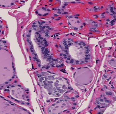
11 Data accuracy To producing accurate data (size, color, figure, measurement) is must in WSI application. Otherwise, analyzed data and developed decision support system cannot be useful. It is very difficult to notice for the users. It might be difficult to notice for the vendors too(?) It is difficult to know which one or who is causing the error scanner? viewer? software (image analysis software or browser)? user? Viewer Accuracy/reliability
12 Measurement & Appearance A part of same WSIs viewed by different viewers provided by a scanner vendor. Both 40x (clicked 40x) view. Both squares show 400um2. Size is also a little bit different.


13 Scanner A Scanner B


14 A slide was scanned by different 2 scanners. Resolutions are is 0.23um/pixel and 0.25um/pixel at 40x. 40x images was viewed on same monitor and a viewer provided each vendor. part of same WSIs viewed by different viewers provided by each scanner vendor. Possible causes are: User changed magnification accidentally by rolling the mouse wheel Scanner s resolution viewer
15 Evaluate Aspects Data Quality/Accuracy Slide Quality Evaluation Image Quality Evaluation Stain Standardization Color Standardization Human Interface Network (system structure)


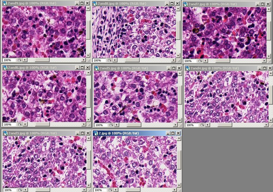
16 Tissue artifacts 50% or more of the tissue slides we deal contains fold Whole slide image Tissue fold Blurry areas Tissue fold The presence of tissue artifacts such as folds or bubbles may produce blur or unfocused areas in the scanned images especially around the artifact areas. Could we still have a good image quality even in the presence of tissue artifacts such as folds????

17 Image quality is affected by tissue artifacts in most cases

18 Image quality is affected by tissue artifacts in most cases

19 Slide quality evaluation Slide quality control

20 40x in original Results After the adjustment We can see more details in image scanned by manual mode


21 Tissue artifacts Detection for Application K-means clustering plus morphological filtering Manual segmentation αs-βv method (New) Result of the proposed method is closer to the result of manual segmentation


22 Fold segmentation by the proposed method αs-βv #1 #2 #3 fold fold #4 #5 #6 fold The folds were segmented appropriately Some of the nuclei areas are labeled as folds, especially those which are very dark and lumped together fold fold #7 #8 #9 fold fold fold fold Mislabeling of nuclei as folds explains the lower specificity of the proposed method compared to clustering
23 Tissue Sectioning: Manual vs Automation
24 If we can create Thin, uniform thickness across entire specimen.. Could improve image quality of WSI Could reduce # of points for auto focus Could reduce scanning time
25 Automated Sectioning System

26 Manual Automation
27 Manual Automation
28 Manual Automation

29 Thickness of Specimen & Staining Thicker sections are stained more by the automated staining machine 3um 3um 3um 6um 3um 7um 5um 8um 7um 6um 5um 4um
30 Tissue Sectioning: Manual vs Automation The automated sectioning machine could section tissues with better consistency and quality for most of types of organs. However, there are still some issues for clinical usage such as speed and workflow integration. We have been using automated sectioning machine for all research materials such as for 3D Imaging. * Film/tape type of automated cover slipper

31 Thicker ->more noise
32 9um 2um


33 Sakura Smart Section Dainippon Seiki AS410


34 Image Quality Scanner 1 Scanner 2 Scanner 3 Scanner 4 Scanner 5


35 Commercial Slides Available test target slides are not for WSI or too expensive Target: too easy Thickness of slide: does not fit to some of scanners
36 Image Quality Evaluation Algorithm Image Quality Multiple regression analysis Definitive evaluation index, is calculated by q s n q,, are derived from training data.


37 Original Image Image Quality Evaluation Algorithm Result of Evaluation Image Quality problem were detected in dark regions. Darker->lower quality
38 Real Time Image Quality Assessment for WSI Yukako Yagi, PhD & Noriaki Hashimoto, PhD After we experimented evaluating following 10 parameters, we have decided 4 parameters were most efficient (1), (3), (7), (8) (1) Average width of edge [1] (2) Average ratio of height to width of edge [2] (3) Independence metric from surrounding pixels [1] (4) Low frequency component in DFT image (5) HH component in DWT image (6) HL and LH component in DWT image (7) Blockiness factor [3] (8) Difference within block [3] (9) Zero-crossing rate [3] (10) Ratio of blockiness factor to subtraction within block


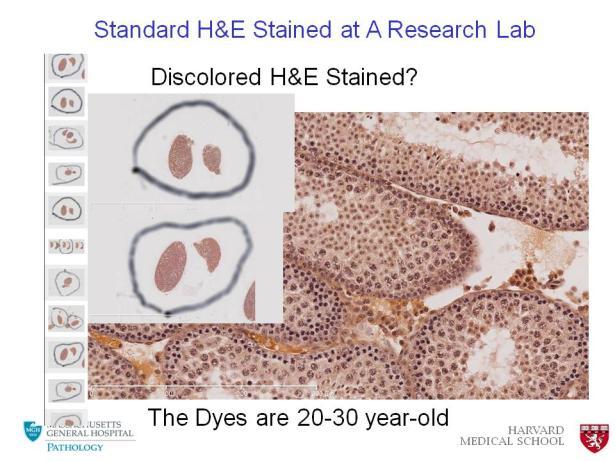
39 Intensity Canny edge detection Suppress noises and detect only edges Gaussian filtering for noise reduction Sharpness evaluation Edge detection by differential Sharpness evaluation Width of detected edge is calculated The average value in the all edges is defined as an evaluation index for sharpness s If s is large, the image is blurred Assumption: Noise appears independently Pixel


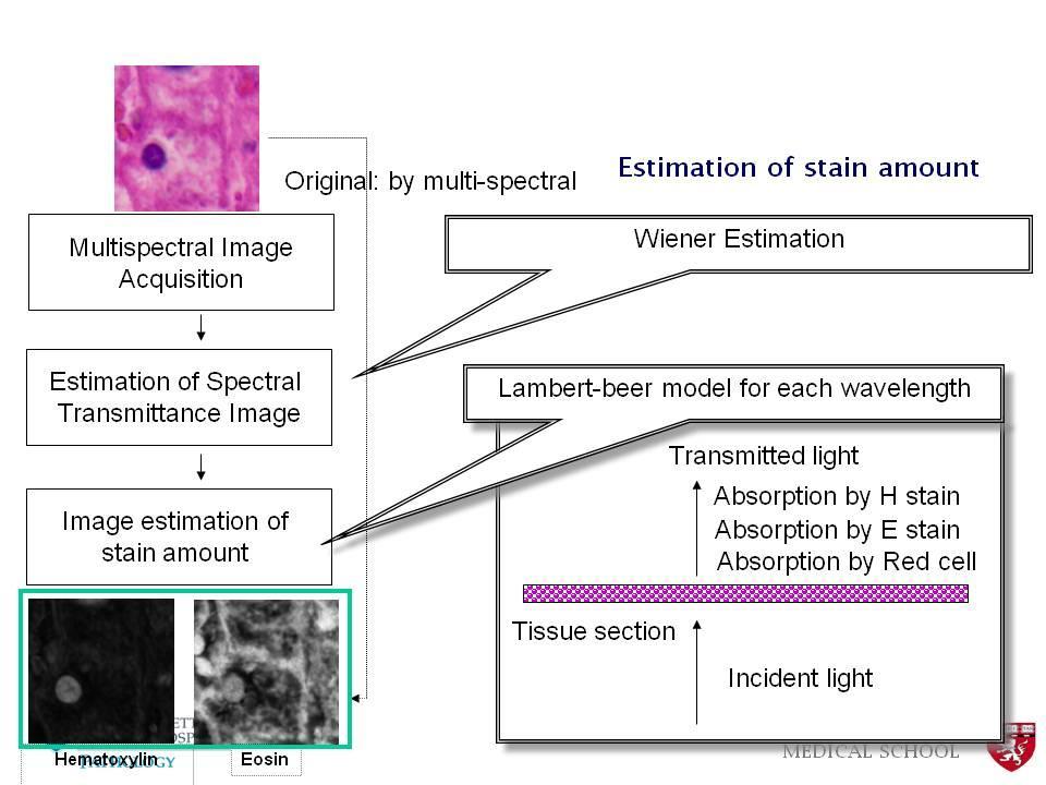
40 Application of the method to WSI Algorithm is processed for every 512x512 pixel block Visualized as a pseudo-color image


41 Comparison of images Images from excellent and poor regions Image quality = 4.66 Image quality =
42 Application to decide threshold Worked with pathologists to decide, we have decided what score to reject and accept

43 WSI Quality Evaluation Algorithm


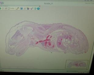
44 Application to decide threshold: already used with pathologists Worked with pathologists to decide, we have decided what score to reject and accept

45 Color


46 The causes of color variation in WSI Thickness of Specimen Staining Scanner or Scanning process Viewer Software Display Display is important for annotation and diagnosis


47 Color Standardization in WSI: From Staining to Display Multispectral Imaging application Staining Scanning Viewer software Display

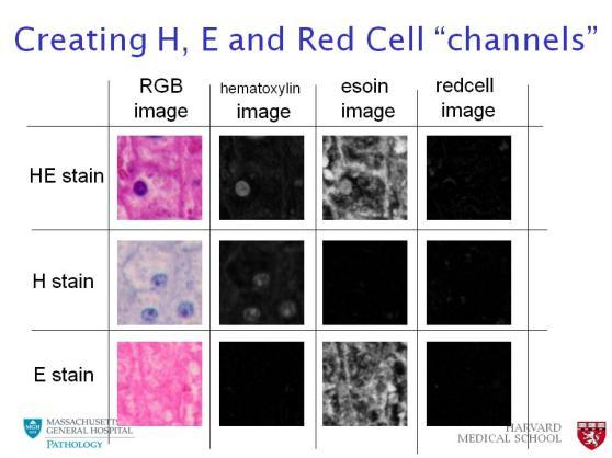

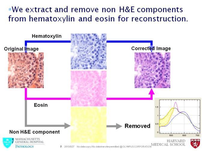

48 Staining color standardization


49 Scanning Color Standardization in WSI: Scanner Review Display The Imaging web site has the colors of the Calibration slide. Compare the displayed colors of the calibration slide to their actual colors to understand the difference VS The Imaging web site has Calibration slide.

50 Polynomial transformation Color of the patches as produced by a particular scanner Reference color of the color patches Color transformation matrix will be stored for used in color standardization Each scanner will have its own Color transformation matrix MEDICAL SCHOOL

51 WSI scanners and Color Imaging Whole slide scanner 1 (WSI 1) Whole slide scanner 2 (WSI 2) The same tissue slide was scanned with different scanners. Use the mouse embryo slide to confirm the effectiveness of the color transformation matrix H&E stained images
52 Liver Scanner A Scanner B Without color correction
 A 50 80 110140170200230 Red channel Without color correction")
53 blue channel Scanner A Scanner B Liver Nu (Orig) A Red channel Without color correction
54 Liver Scanner A Scanner B Result of color correction


55 Blue channel Scanner A Scanner B Liver Nu (Corrected) A B Red channel Result of color correction
56 Slide Quality can be evaluated to improve WSI quality Slide Quality can be improved Image Quality can be evaluated if it appropriate for diagnosis, Image analysis, or/and Education. Color of WSIs can be standardized.


57 Quality Control of WSI system Image Quality & color Color Optics Using the two calibration slides, the evaluation of image quality and color standardization could be done. Monitor


58 Tissue Processing Accurate Results require Good Images, Good images require good slide, good slides require good block Paraffin block Summary Scanning Digital Pathology can improve the patient care globally. Digital Pathology has unlimited possibility. We just to need to think what we want to do.. Sectioning Staining Cover glass Digital Stains Stained slides
59 Thank you!
Color aspects and Color Standardization in Digital Microscopy
 Color aspects and Color Standardization in Digital Microscopy Yukako Yagi, PhD yyagi@partners.org Director of the MGH Pathology Imaging & Communication Technology Center Assistant Professor of Pathology,
Digital Pathology at Johns Hopkins Practical Research and Clinical Considerations
 Digital Pathology at Johns Hopkins Practical Research and Clinical Considerations July 10, 2017 Alexander Baras, MD, PhD Assistant Professor of Pathology, Urology, and Oncology Associate Director of Pathology
DICOM-compatible compression of WSI and diagnostic evaluation
 of WSI and diagnostic evaluation R. Zwönitzer, H. Hofmann, A. Roessner, T. Kalinski 2nd European Workshop in Tissue Imaging and Analysis June 25-26, 2010 - Heidelberg 1 GPWL / GP-PPS Introduction Overview
Anatomic and Computational Pathology Diagnostic Artificial Intelligence at Scale
 Anatomic and Computational Pathology Diagnostic Artificial Intelligence at Scale John Gilbertson MD Department of Pathology Massachusetts General Hospital Partners Healthcare System Harvard Medical School
Multispectral Enhancement towards Digital Staining
 Multispectral Enhancement towards Digital Staining The Harvard community has made this article openly available. Please share how this access benefits you. Your story matters. Citation Published Version
Digital Pathology and Tissue-based Diagnosis. How do they differ?
 Digital Pathology and Tissue-based Diagnosis. How do they differ? P. Hufnagl Institute of Pathology (Rudolf-Virchow-Haus). Humboldt University, Berlin? 10.12.2014 1 Structure of the talk Possible workflow
Multilayer scanning enhances sensitivity of artificial intelligence-aided Mycobacterium tuberculosis detection
 Multilayer scanning enhances sensitivity of artificial intelligence-aided Mycobacterium tuberculosis detection Yan Xiong Peking University First Hospital, China. yanxiong1109@163.com Ao Hou ao_sure@foxmail.com
THEORY AND APPROACHES TO AUTOMATED IMAGE ANALYSIS IN DIGITAL PATHOLOGY
 THEORY AND APPROACHES TO AUTOMATED IMAGE ANALYSIS IN DIGITAL PATHOLOGY Kyle Takayama, MS Charles River Laboratories EVERY STEP OF THE WAY EVERY STEP OF THE WAY MORPHOMETRY Measurements or counts performed
InScape: Making Virtual Pathology a Reality
 InScape: Making Virtual Pathology a Reality Sally S. Agersborg, M.D., Ph.D. Quest Diagnostics, Nichols Institute San Juan Capistrano, CA Company Overview Quest Diagnostics, Nichols Institute the world
IHE Anatomic Pathology Redesign. Sardinia, Italy Nov , 2017
 IHE Anatomic Pathology Redesign Sardinia, Italy Nov. 13-15, 2017 Specimen Workflow in a Nutshell (variations likely depending on context, e.g. collection location) Create Encounter Generate Results Order
Yagi Digital Microscope Calibration
 Yagi Digital Microscope Calibration Method summary, assessment and suggestions for improvement W Craig Revie, International Color Consortium Introduction In the area of pathology, a type of digital microscope
MAKE SURE YOUR SLIDES ARE CLEAN (TOP & BOTTOM) BEFORE LOADING DO NOT LOAD SLIDES DURING SOFTWARE INITIALIZATION
 BEFORE LOADING DO NOT LOAD SLIDES DURING SOFTWARE INITIALIZATION") Olympus VS120-L100 Slide Scanner Standard Operating Procedure Startup 1) Red power bar switch (behind monitor) 2) Computer 3) Login: UserVS120 account (no password) 4) Double click: WAIT FOR INITIALIZATION
Olympus VS120-L100 Slide Scanner Standard Operating Procedure Startup 1) Red power bar switch (behind monitor) 2) Computer 3) Login: UserVS120 account (no password) 4) Double click: WAIT FOR INITIALIZATION
Digital Pathology Update
 Digital Pathology Update J. Mark Tuthill, MD Division Head, Pathology Informatics Department of Pathology & Laboratory Medicine Henry Ford Hospital Detroit, MI mtuthil1@hfhs.org ASCT Webinar, January 2016
MIRAX SCAN The new way of looking at pathology
 Microscopy from Carl Zeiss MIRAX SCAN The new way of looking at pathology Greater reliability. Greater efficiency. Plus points for your diagnostics Better. More efficient. Quality as a factor for success
Digital Microscopy: New Paradigm's for Teaching Microscopic Anatomy and Pathology
 Digital Microscopy: New Paradigm's for Teaching Microscopic Anatomy and Pathology Michael Feldman, MD, PhD Assistant Dean IT Assistant Professor Pathology University of Pennsylvania Health System Feldmanm@mail.med.upenn.edu
Positive Pixel Count Algorithm. User s Guide
 Positive Pixel Count Algorithm User s Guide Copyright 2004, 2006 2008 Aperio Technologies, Inc. Part Number/Revision: MAN 0024, Revision B Date: December 9, 2008 This document applies to software versions
FRAUNHOFER INSTITUTE FOR INTEGRATED CIRCUITS IIS. MANUAL PANORAMIC MICROSCOPY WITH istix
 FRAUNHOFER INSTITUTE FOR INTEGRATED CIRCUITS IIS MANUAL PANORAMIC MICROSCOPY WITH istix CLINICAL DIAGNOSTICS AND MATERIAL SCIENCES IMPROVED BY DIGITAL MICROSCOPY B A C K G R O U N D Due to a high grade
DIGITAL PHOTOGRAPHY Camera and image capture
 DIGITAL PHOTOGRAPHY Camera and image capture The higher the number of pixels, the better the resolution. Your camera should be able to capture images of at least 1200 x 900 pixels which is equivalent to
SECTION I - CHAPTER 2 DIGITAL IMAGING PROCESSING CONCEPTS
 RADT 3463 - COMPUTERIZED IMAGING Section I: Chapter 2 RADT 3463 Computerized Imaging 1 SECTION I - CHAPTER 2 DIGITAL IMAGING PROCESSING CONCEPTS RADT 3463 COMPUTERIZED IMAGING Section I: Chapter 2 RADT
Machine Vision for the Life Sciences
 Machine Vision for the Life Sciences Presented by: Niels Wartenberg June 12, 2012 Track, Trace & Control Solutions Niels Wartenberg Microscan Sr. Applications Engineer, Clinical Senior Applications Engineer
Second Announcement Call for Participation. (Evaluation Criteria added)
") Second Announcement Call for Participation 2 nd International Scanner Contest (ISC) (Evaluation Criteria added) P. Hufnagl 1, T. Schrader 1, 2, M.G. Rojo 3, A. Laurinavicius 4, G. Kayser 5, Y. Yagi 6 1
Second Announcement Call for Participation 2 nd International Scanner Contest (ISC) (Evaluation Criteria added) P. Hufnagl 1, T. Schrader 1, 2, M.G. Rojo 3, A. Laurinavicius 4, G. Kayser 5, Y. Yagi 6 1
Introduction Approach Work Performed and Results
 Algorithm for Morphological Cancer Detection Carmalyn Lubawy Melissa Skala ECE 533 Fall 2004 Project Introduction Over half of all human cancers occur in stratified squamous epithelia. Approximately one
Stereotopix Research. Precision Pathology. Highthroughput. pathology. powered by newcast. Advantages of Stereotopix : RUO
 Precision Pathology Highthroughput pathology Stereotopix Research powered by newcast RUO Researchers use quantitative microscopy in many ways with the goal of producing high-quality, quantitative results
Digital Microscopy. Past, Present, and Future. Cyrus V. Hedvat, MD, PhD
 Digital Microscopy Past, Present, and Future Cyrus V. Hedvat, MD, PhD N Context. Digital viewing of histologic images is moving from presentations and publications to incorporation into the daily work
Arcturus XT Laser Capture Microdissection System AutoScanXT Software Module. User Manual
 Arcturus XT Laser Capture Microdissection System AutoScanXT Software Module User Manual For Research Use Only. Not intended for any animal or human therapeutic or diagnostic use. Information in this document
MetaXpress Software: Cell Scoring Module
 MetaXpress Software: Cell Scoring Module Cell Scoring Module Overview The Cell Scoring module can be used to analyze cells imaged in 2 wavelengths W1 should be a stain for all nuclei (e.g. DAPI, Hoechst,
Teaching Digital Histology
 Teaching Digital Histology Carlos R. Morales Department of Anatomy and Cell Biology, McGill University, Montreal, Quebec, Canada The light microscope is one of the most widely used scientific instruments
Classification Accuracies of Malaria Infected Cells Using Deep Convolutional Neural Networks Based on Decompressed Images
 Classification Accuracies of Malaria Infected Cells Using Deep Convolutional Neural Networks Based on Decompressed Images Yuhang Dong, Zhuocheng Jiang, Hongda Shen, W. David Pan Dept. of Electrical & Computer
Digital Pathology and Image Analysis. Queen s University Department of Pathology and Molecular Medicine Shakeel Virk
 Digital Pathology and Image Analysis Queen s University Department of Pathology and Molecular Medicine Shakeel Virk Outline Digital Pathology and Image Analysis capabilities at Queen s Laboratory for Molecular
Y N C R O S C O P Y A DIVISION OF THE SYNOPTICS GROUP
 S Y N C R O S C O P Y A DIVISION OF THE SYNOPTICS GROUP THE PROBLEM: As a microscopist you often have to work with samples that are difficult to focus. When viewing a 3-D sample using an optical microscope
Leica DMi8A Quick Guide
 Leica DMi8A Quick Guide 1 Optical Microscope Quick Start Guide The following instructions are provided as a Quick Start Guide for powering up, running measurements, and shutting down Leica s DMi8A Inverted
DMETRIX S (FUTURE) PERSPECTIVES ON DIGITAL IMAGING & DIGITAL PATHOLOGY SYSTEMS
 PERSPECTIVES ON DIGITAL IMAGING & DIGITAL PATHOLOGY SYSTEMS") Michael R. Descour, Ph.D., DMetrix, Inc., & University of Arizona Lloyd J. LaComb, Jr., Ph.D., DMetrix, Inc. DMETRIX S (FUTURE) PERSPECTIVES ON DIGITAL IMAGING & DIGITAL PATHOLOGY SYSTEMS Outline of presentation
Michael R. Descour, Ph.D., DMetrix, Inc., & University of Arizona Lloyd J. LaComb, Jr., Ph.D., DMetrix, Inc. DMETRIX S (FUTURE) PERSPECTIVES ON DIGITAL IMAGING & DIGITAL PATHOLOGY SYSTEMS Outline of presentation
Computing for Engineers in Python
 Computing for Engineers in Python Lecture 10: Signal (Image) Processing Autumn 2011-12 Some slides incorporated from Benny Chor s course 1 Lecture 9: Highlights Sorting, searching and time complexity Preprocessing
Illumination Correction tutorial
 Illumination Correction tutorial I. Introduction The Correct Illumination Calculate and Correct Illumination Apply modules are intended to compensate for the non uniformities in illumination often present
Bowen Hills Histopathology
 Bowen Hills Histopathology Histology s Bowen Hills Challenges Relocate and amalgamate Taringa and Indooroopilly Labs into a single complex histology laboratory Change workflow, staff structure, install
Building a Business Case for Digital Pathology: The Time is Now! Drazen M. Jukic, MD, PhD
 Building a Business Case for Digital Pathology: The Time is Now! Drazen M. Jukic, MD, PhD Business Case Telepathology vs. Digital Pathology Telepathology as a term was misunderstood in the past; It denotes
Enhanced Functionality of High-Speed Image Processing Engine SUREengine PRO. Sharpness (spatial resolution) Graininess (noise intensity)
 Graininess (noise intensity)") Vascular Enhanced Functionality of High-Speed Image Processing Engine SUREengine PRO Medical Systems Division, Shimadzu Corporation Yoshiaki Miura 1. Introduction In recent years, digital cardiovascular
Vascular Enhanced Functionality of High-Speed Image Processing Engine SUREengine PRO Medical Systems Division, Shimadzu Corporation Yoshiaki Miura 1. Introduction In recent years, digital cardiovascular
A Study On Preprocessing A Mammogram Image Using Adaptive Median Filter
 A Study On Preprocessing A Mammogram Image Using Adaptive Median Filter Dr.K.Meenakshi Sundaram 1, D.Sasikala 2, P.Aarthi Rani 3 Associate Professor, Department of Computer Science, Erode Arts and Science
GALILEO TMA CK 4500 HTS Tissue Microarray Platform
 GALILEO TMA CK 4500 HTS Tissue Microarray Platform Tissue Microarray (TMA) A Block Of Samples From Hundreds Of Blocks (S. M. Hewitt, M.D., Ph.D., Tissue Array Research Program, LP, CCR, NCI, NIH) TMA technology
User Reference Manual
 User Reference Manual IN SITU HYBRIDIZATION QUANTIFICATION (for research purposes only) Contents Overview... 3 System Requirements... 3 Compatible File Formats... 3 Input Parameters... 4 Output Parameters...
Image Database and Preprocessing
 Chapter 3 Image Database and Preprocessing 3.1 Introduction The digital colour retinal images required for the development of automatic system for maculopathy detection are provided by the Department of
Axioscan - Startup. 1. Turn on the Axioscan (button to the left) and turn on the computer. 2. Log on and start the ZEN Blue software from the desktop
 and turn on the computer. 2. Log on and start the ZEN Blue software from the desktop") Axioscan - Startup 1. Turn on the Axioscan (button to the left) and turn on the computer 2. Log on and start the ZEN Blue software from the desktop 3. Press ZEN slidescan and Start System 4. Start by changing
Axioscan - Startup 1. Turn on the Axioscan (button to the left) and turn on the computer 2. Log on and start the ZEN Blue software from the desktop 3. Press ZEN slidescan and Start System 4. Start by changing
Instruction Manual T Binocular Acromat Research Scope T Trinocular Acromat Research Scope
 Research Scope Instruction Manual T-29031 Binocular Acromat Research Scope T-29041 Trinocular Acromat Research Scope T-29032 Binocular Semi-Plan Research Scope T-29042 Trinocular Semi-Plan Research Scope
SPOT PathSuite Solutions
 SPOT PathSuite Solutions The Perfect Fit PathStation TM Imaging system for the gross dissection in hood PathStand TM 40 imaging station for medium to large specimens PathSuite Is Easy To Use A turnkey
Training school in the use of open source software V June 2009 Ancaster Hall, University Park
 Training school in the use of open source software V6 24-26 June 2009 Ancaster Hall, University Park Target Audience: (1) Pathologists who are newcomers to image analysis (i.e. with little or no experience)
Calibration Slide for Histopathology task force Teleconference 20 February :00 (UK) / 10:00 (EST)
 / 10:00 (EST)") Calibration Slide for Histopathology task force Teleconference 20 February 2014 15:00 (UK) / 10:00 (EST) The meeting was called to order at 10:00 am (EST) by Craig Revie, chair of MIWG, with the following
Calibration Slide for Histopathology task force Teleconference 20 February 2014 15:00 (UK) / 10:00 (EST) The meeting was called to order at 10:00 am (EST) by Craig Revie, chair of MIWG, with the following
Operating Procedures for MICROCT1 Nikon XTH 225 ST
 Operating Procedures for MICROCT1 Nikon XTH 225 ST Ensuring System is Ready (go through to ensure all windows and tasks below have been completed either by you or someone else prior to mounting and scanning
CoE4TN4 Image Processing. Chapter 3: Intensity Transformation and Spatial Filtering
 CoE4TN4 Image Processing Chapter 3: Intensity Transformation and Spatial Filtering Image Enhancement Enhancement techniques: to process an image so that the result is more suitable than the original image
GenePix Application Note
 GenePix Application Note Biological Relevance of GenePix Results Shawn Handran, Ph.D. and Jack Y. Zhai, Ph.D. Axon Instruments, Inc. 3280 Whipple Road, Union City, CA 94587 Last Updated: Aug 22, 2003.
Physics 2310 Lab #5: Thin Lenses and Concave Mirrors Dr. Michael Pierce (Univ. of Wyoming)
") Physics 2310 Lab #5: Thin Lenses and Concave Mirrors Dr. Michael Pierce (Univ. of Wyoming) Purpose: The purpose of this lab is to introduce students to some of the properties of thin lenses and mirrors.
Physics 2310 Lab #5: Thin Lenses and Concave Mirrors Dr. Michael Pierce (Univ. of Wyoming) Purpose: The purpose of this lab is to introduce students to some of the properties of thin lenses and mirrors.
Module 3: Video Sampling Lecture 18: Filtering operations in Camera and display devices. The Lecture Contains: Effect of Temporal Aperture:
 The Lecture Contains: Effect of Temporal Aperture: Spatial Aperture: Effect of Display Aperture: file:///d /...e%20(ganesh%20rana)/my%20course_ganesh%20rana/prof.%20sumana%20gupta/final%20dvsp/lecture18/18_1.htm[12/30/2015
Digital Portable Overhead Document Camera LV-1010
 Digital Portable Overhead Document Camera LV-1010 Instruction Manual 1 Content I Product Introduction 1.1 Product appearance..3 1.2 Main functions and features of the product.3 1.3 Production specifications.4
Centre for Computational and Numerical Studies, Institute of Advanced Study in Science and Technology 2. Dept. of Statistics, Gauhati University
 Cervix Cancer Diagnosis from Pap Smear Images Using Structure Based Segmentation and Shape Analysis 1 Lipi B. Mahanta, 2 Dilip Ch. Nath, 1 Chandan Kr. Nath 1 Centre for Computational and Numerical Studies,
Project: Sudoku solver
 Project: Sudoku solver Write a program that finds the sudoku square in the image, detects the 81 fields, and identifies the number in the fields that have a number. The output should be a 9x9 matrix with
ANSWER KEY Lab 2 (IGB): Bright Field and Fluorescence Optical Microscopy and Sectioning
: Bright Field and Fluorescence Optical Microscopy and Sectioning") Phys598BP Spring 2016 University of Illinois at Urbana-Champaign ANSWER KEY Lab 2 (IGB): Bright Field and Fluorescence Optical Microscopy and Sectioning Location: IGB Core Microscopy Facility Microscope:
Phys598BP Spring 2016 University of Illinois at Urbana-Champaign ANSWER KEY Lab 2 (IGB): Bright Field and Fluorescence Optical Microscopy and Sectioning Location: IGB Core Microscopy Facility Microscope:
Image Enhancement in spatial domain. Digital Image Processing GW Chapter 3 from Section (pag 110) Part 2: Filtering in spatial domain
 Part 2: Filtering in spatial domain") Image Enhancement in spatial domain Digital Image Processing GW Chapter 3 from Section 3.4.1 (pag 110) Part 2: Filtering in spatial domain Mask mode radiography Image subtraction in medical imaging 2 Range
Image Enhancement in spatial domain Digital Image Processing GW Chapter 3 from Section 3.4.1 (pag 110) Part 2: Filtering in spatial domain Mask mode radiography Image subtraction in medical imaging 2 Range
AUTOMATED MALARIA PARASITE DETECTION BASED ON IMAGE PROCESSING PROJECT REFERENCE NO.: 38S1511
 AUTOMATED MALARIA PARASITE DETECTION BASED ON IMAGE PROCESSING PROJECT REFERENCE NO.: 38S1511 COLLEGE : BANGALORE INSTITUTE OF TECHNOLOGY, BENGALURU BRANCH : COMPUTER SCIENCE AND ENGINEERING GUIDE : DR.
Quick Operation Guide
 Quick Operation Guide Power ON Mounting specimens Set the specimen on the sample holder, and install the sample holder to the holder frame. Attach the holder frame to the XY stage. Type of holder Main
Segmentation of Microscopic Bone Images
 International Journal of Electronics Engineering, 2(1), 2010, pp. 11-15 Segmentation of Microscopic Bone Images Anand Jatti Research Scholar, Vishveshvaraiah Technological University, Belgaum, Karnataka
Segmentation of Liver CT Images
 Segmentation of Liver CT Images M.A.Alagdar 1, M.E.Morsy 2, M.M.Elzalabany 3 1,2,3 Electronics And Communications Department-.Faculty Of Engineering Mansoura University, Egypt. Abstract In this paper we
Image Smoothening and Sharpening using Frequency Domain Filtering Technique
 Volume 5, Issue 4, April (17) Image Smoothening and Sharpening using Frequency Domain Filtering Technique Swati Dewangan M.Tech. Scholar, Computer Networks, Bhilai Institute of Technology, Durg, India.
MECOS-C2 microscopy systems
 MECOS-C2 microscopy systems Microscopy systems of the MECOS-C2 family production LLC "Medical computer Systems (MECOS)" belong to a class of scanning microscopes-analyzers and are intended for: Increase
Instructions. To run the slideshow:
 Instructions To run the slideshow: Click: view full screen mode, or press Ctrl +L. Left click advances one slide, right click returns to previous slide. To exit the slideshow press the Esc key. Optical
White Paper Focusing more on the forest, and less on the trees
 White Paper Focusing more on the forest, and less on the trees Why total system image quality is more important than any single component of your next document scanner Contents Evaluating total system
Microarray Data Pre-processing. Ana H. Barragan Lid
 Microarray Data Pre-processing Ana H. Barragan Lid Hybridized Microarray Imaged in a microarray scanner Scanner produces fluorescence intensity measurements Intensities correspond to levels of hybridization
Image Viewing. with ImageScope
 Image Viewing with ImageScope ImageScope Components Use ImageScope to View These File Types: ScanScope Virtual Slides.SVS files created when the ScanScope scanner scans glass microscope slides. JPEG files
Chapter 6: TVA MR and Cardiac Function
 Chapter 6 Cardiac MR Introduction Chapter 6: TVA MR and Cardiac Function The Time-Volume Analysis (TVA) optional module calculates time-dependent behavior of volumes in multi-phase studies from MR. An
Optimization and enhancement of H&E stained microscopical images by applying bilinear interpolation method on lab color mode
 Kuru Theoretical Biology and Medical Modelling 2014, 11:9 RESEARCH Open Access Optimization and enhancement of H&E stained microscopical images by applying bilinear interpolation method on lab color mode
Practical Image and Video Processing Using MATLAB
 Practical Image and Video Processing Using MATLAB Chapter 1 Introduction and overview What will we learn? What is image processing? What are the main applications of image processing? What is an image?
Light Microscopy. Upon completion of this lecture, the student should be able to:
 Light Light microscopy is based on the interaction of light and tissue components and can be used to study tissue features. Upon completion of this lecture, the student should be able to: 1- Explain the
AUTOMATIC DETECTION OF HEDGES AND ORCHARDS USING VERY HIGH SPATIAL RESOLUTION IMAGERY
 AUTOMATIC DETECTION OF HEDGES AND ORCHARDS USING VERY HIGH SPATIAL RESOLUTION IMAGERY Selim Aksoy Department of Computer Engineering, Bilkent University, Bilkent, 06800, Ankara, Turkey saksoy@cs.bilkent.edu.tr
An Image Processing Approach for Screening of Malaria
 An Image Processing Approach for Screening of Malaria Nagaraj R. Shet 1 and Dr.Niranjana Sampathila 2 1 M.Tech Student, Department of Biomedical Engineering, Manipal Institute of Technology, Manipal University,
Using Binary Layers with NIS-Elements
 Using Binary Layers with NIS-Elements Overview This technical note describes the usage of Binary Layers with NIS-Elements. Binary layers form an extension of simple intensity thresholding technique, allowing
Overview of Digital Pathology s Current State: Technologies, Systems, Capabilities, Limitations, and Opportunities
 Overview of Digital Pathology s Current State: Technologies, Systems, Capabilities, Limitations, and Opportunities David McClintock, MD Executive War College Post-Conference Workshop Digital Pathology
Using Adobe Photoshop to enhance the image quality. Assistant course web site:
 Using Adobe Photoshop to enhance the image quality Assistant course web site: http://www.arches.uga.edu/~skwang/edit6170/course.htm Content Introduction 2 Unit1: Scan images 3 Lesson 1-1: Preparations
TOPAZ Vivacity V1.3. User s Guide. Topaz Labs LLC. Copyright 2005 Topaz Labs LLC. All rights reserved.
 TOPAZ Vivacity V1.3 User s Guide Topaz Labs LLC www.topazlabs.com Copyright 2005 Topaz Labs LLC. All rights reserved. TABLE OF CONTENTS Introduction 2 Before You Start 3 Suppress Image Noises 6 Reduce
6.A44 Computational Photography
 Add date: Friday 6.A44 Computational Photography Depth of Field Frédo Durand We allow for some tolerance What happens when we close the aperture by two stop? Aperture diameter is divided by two is doubled
Image Processing - License Plate Localization and Letters Extraction *
 OpenStax-CNX module: m33156 1 Image Processing - License Plate Localization and Letters Extraction * Cynthia Sung Chinwei Hu Kyle Li Lei Cao This work is produced by OpenStax-CNX and licensed under the
White Paper. Scanning the Perfect Page Every Time Take advantage of advanced image science using Perfect Page to optimize scanning
 White Paper Scanning the Perfect Page Every Time Take advantage of advanced image science using Perfect Page to optimize scanning Document scanning is a cornerstone of digital transformation, and choosing
IDENTIFICATION OF FISSION GAS VOIDS. Ryan Collette
 IDENTIFICATION OF FISSION GAS VOIDS Ryan Collette Introduction The Reduced Enrichment of Research and Test Reactor (RERTR) program aims to convert fuels from high to low enrichment in order to meet non-proliferation
STRUCTURE OF THE MICROSCOPE
 STRUCTURE OF THE MICROSCOPE Use the word list to label the microscope below: Light Source Coarse adjustment knob Diaphragm Stage Clips Objectives Fine Adjustment Knob Base Stage Stage Clips Arm Revolving
DIGITAL-MICROSCOPY CAMERA SOLUTIONS USB 3.0
 DIGITAL-MICROSCOPY CAMERA SOLUTIONS USB 3.0 PixeLINK for Microscopy Applications PixeLINK will work with you to choose and integrate the optimal USB 3.0 camera for your microscopy project. Ideal for use
Image processing for gesture recognition: from theory to practice. Michela Goffredo University Roma TRE
 Image processing for gesture recognition: from theory to practice 2 Michela Goffredo University Roma TRE goffredo@uniroma3.it Image processing At this point we have all of the basics at our disposal. We
Hematoxylin and Eosin Stained Tissue
 A p p l i c a t i o n N o t e Hematoxylin and Eosin Stained Tissue Using Color Brightfield Imaging with the Cytation 5 to Image Fixed and Stained Tissue Paul Held Ph. D., Laboratory Manager, Applications
DRX Plus Detectors: Going from Good to Great
 DRX Plus Detectors: Going from Good to Great Authors: Karin Töpfer, Tim Wojcik Introduction Carestream s introduction in 2009 of the world s first portable, wireless, cassette-sized detector the CARESTREAM
8/28/2012. Total Lab Automation in Microbiology: Much Closer than You Might Think September 6, Microbiology Yesterday
 Total Lab Automation in Microbiology: Much Closer than You Might Think September 6, 2012 Paul Bourbeau, Ph.D. Director, Microbiology Laboratory Microbiology Yesterday Microbiology too complex to automate
TORNIER BLUEPRINT. 3D Planning + PSI SCAN PROTOCOL
 TORNIER BLUEPRINT 3D Planning + PSI SCAN PROTOCOL Contents 3 Introduction 3 Patient preparation 3 Scanning instructions 4 Image instructions 5 Scanning parameters 6 Technical instructions 2 BLUEPRINT 3D
Computational approach for diagnosis of malaria through classification of malaria parasite from microscopic image of blood smear.
 Biomedical Research 2018; 29 (18): 3464-3468 ISSN 0970-938X www.biomedres.info Computational approach for diagnosis of malaria through classification of malaria parasite from microscopic image of blood
DICOM Correction Proposal
 Tracking Information - Administration Use Only DICOM Correction Proposal Correction Proposal Number Status CP-1713 Letter Ballot Date of Last Update 2018/01/23 Person Assigned Submitter Name David Clunie
CSE 564: Scientific Visualization
 CSE 564: Scientific Visualization Lecture 5: Image Processing Klaus Mueller Stony Brook University Computer Science Department Klaus Mueller, Stony Brook 2003 Image Processing Definitions Purpose: - enhance
Slide 1. Slide 2. Slide 3 ACR CT Accreditation. Multi-Slice CT Artifacts and Quality Control. What are the rules or recommendations for CT QC?
 Slide 1 Multi-Slice CT Artifacts and Quality Control Dianna Cody, Ph.D. Chief, Radiologic Physics UT MD Anderson Cancer Center Houston, TX Slide 2 What are the rules or recommendations for CT QC? AAPM
COMPARISON OF DIFFERENT METHODS FOR TISSUE SEGMENTATION IN HISTOPATHOLOGICAL WHOLE-SLIDE IMAGES
 COMPARISON OF DIFFERENT METHODS FOR TISSUE SEGMENTATION IN HISTOPATHOLOGICAL WHOLE-SLIDE IMAGES Péter Bándi, Rob van de Loo, Milad Intezar, Daan Geijs, Francesco Ciompi, Bram van Ginneken, Jeroen van der
Image Analysis for Fluorescence
 Image Analysis for Fluorescence Terminology Table Image Analysis Macro Colocalization Intensity Dye AFI The extraction of meaningful information from digital images by means of digital image processing
BACKGROUND SEGMENTATION IN MICROSCOPY IMAGES
 BACKGROUND SEGMENTATION IN MICROSCOPY IMAGES J.J. Charles, L.I. Kuncheva School of Computer Science, University of Wales, Bangor, LL57 1UT, United Kingdom jjc@informatics.bangor.ac.uk B. Wells Conwy Valley
Invisible sophistication. Visible simplicity. CS Welcome to the simplicity of compact panoramic imaging
 Invisible sophistication. Visible simplicity. CS 8100 Welcome to the simplicity of compact panoramic imaging Introducing the CS 8100 The Carestream Dental Factor Humanized technology We keep our technology
Scanning Procedure Using SilverFast Ai Software (6.6.1r2b) 1
 1") last modified 6.16.2015 Scanning Procedure Using SilverFast Ai Software (6.6.1r2b) 1 PREP In scanner notebook, fill out Incoming Checklist column on a Scanner Equipment Checklist sheet. Use flash drive
last modified 6.16.2015 Scanning Procedure Using SilverFast Ai Software (6.6.1r2b) 1 PREP In scanner notebook, fill out Incoming Checklist column on a Scanner Equipment Checklist sheet. Use flash drive
A Practical Guide to Frozen Section Technique
 A Practical Guide to Frozen Section Technique Editor A Practical Guide to Frozen Section Technique Editor University of Medicine and Dentistry of New Jersey New Jersey Medical School Newark, NJ USA petepath@yahoo.com
MICROSCOPY and CELL STRUCTURE
 MICROSCOPY and CELL STRUCTURE Readings: Review pp. 69-71, and Fig. 4.1 on p. 65 in your text (POHS, 5 th ed.). Introduction: Biologists rely on many different types of microscopic techniques to find out
Introduction to Image Analysis with
 Introduction to Image Analysis with PLEASE ENSURE FIJI IS INSTALLED CORRECTLY! WHAT DO WE HOPE TO ACHIEVE? Specifically, the workshop will cover the following topics: 1. Opening images with Bioformats
In our previous lecture, we understood the vital parameters to be taken into consideration before data acquisition and scanning.
 Interactomics: Protein Arrays & Label Free Biosensors Professor Sanjeeva Srivastava MOOC NPTEL Course Indian Institute of Technology Bombay Module 7 Lecture No 34 Software for Image scanning and data processing