IHE Anatomic Pathology Redesign. Sardinia, Italy Nov , 2017
|
|
|
- Jerome Stevens
- 5 years ago
- Views:
Transcription
1 IHE Anatomic Pathology Redesign Sardinia, Italy Nov , 2017
 Create Encounter Generate Results Order Procedure APW?")
2 Specimen Workflow in a Nutshell (variations likely depending on context, e.g. collection location) Create Encounter Generate Results Order Procedure APW? Perform Procedure & Related Tasks Patient Registration Receive Specimen in Lab Collect Specimen
3 Digital APW Use Case #1 Create digital copies of glass slides to preclude exhausting tissue block for outside slide reviews Request for case review from outside facility or patient All glass slides reviewed by pathologist and key slides identified Selected key slides digitally imaged Original key slides sent out for review In future, digital version can be submitted for review If additional review request comes in, can reference digital versions and/or wait for original slides Usually blocks not sent out by policy
4 Digital APW Use Case #2 Creating digital copies of immunohistochemistry positive control slides to preclude the need for creating multiple positive control slides for distribution to pathologists Request for IHC stain processed as usual Only one IHC positive control run per batch IHC positive control slides imaged and saved to network folder Positive controls NOT distributed ($$$ savings) Glass IHC slides reviewed by pathologist but same positive control reviewed digitally by all pathologists for a given IHC (i.e. only a single cytokeratin positive control slide even if requested across 10 different patient samples)
5 Digital APW Use Case #3 Creating digital copies of all glass slides for primary diagnosis Specimen collected and transported Specimen gross exam with possible digital imaging and annotation Specimen processing Glass slides produced as usual All glass slides fed into high volume automated digital scanner Scanner tags images requiring manual intervention Digital images deposited in network share, VNA, or PACS Interface message to LIS sent as each barcode read off slide Acknowledgment from LIS indicates case is valid and ready for association with digital slide assets Additional message sent when slide digitization completed Interface message sent every time slide viewed or annotated
6 Digital APW Use Case #4 Conversion from a legacy information system Transfer of existing electronic data to include text and images (WSI also) on specimens that have already been evaluated and resulted Legacy accession # needs to be considered against goforward accessioning schema
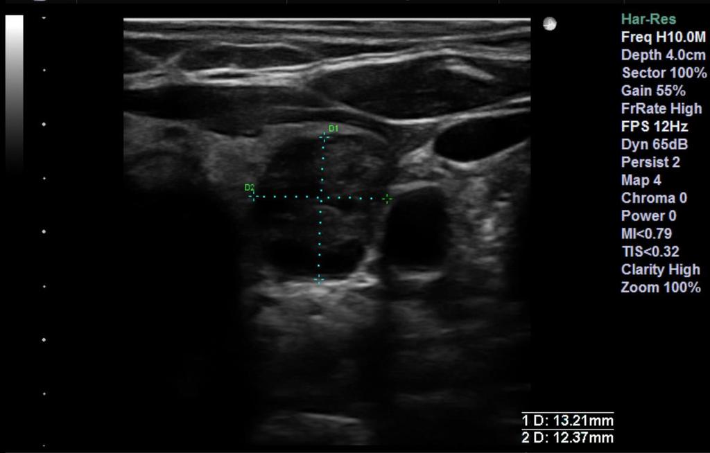
7 APW Scope Start with a tissue specimen received in laboratory? Proposal: common profile for order management and result management for all of PALM (check Berlin F2F notes) More likely to change ordered procedure, need to specify site (laterality is important) Another input as consult from another laboratory ILW (Lab-35, Lab-36) Result: ORU vs APSRv2 document (messaging profile, can have both) Can leverage LCC (Lab-6, Lab-7) Lab-1, Lab-2, Lab-3 exist today and can be leveraged In scope: creation of glass slides that have been digitized and manage those digital assets for presentation and interpretation Actors: viewer, acquisition device, storage device, order filler, analyzer manager, analyzer Separate profiles for: creation, interpretation, (and possibly viewing as a separate profile) Out of scope: FNA ultrasound images (as this is covered by existing radiology profile), Uncertain: specimen radiograph, in vivo microscopy Clinical tissue specimen workflow only? Research, teaching, tumor board out of scope Our scope is to provide infrastructure to support a future profile that to manage use cases Every digital asset has a parent? root is patient? Should or should not cover tissue microarrays (many patients on one slide)?
8
9
10
11

12


13 The Promise of Machine Learning / AI
14 Current APW Diagram Daniel et al., Arch Pathol Lab Med Vol 133, November 2009
15 One step at a time Most EHR systems will have a single order for submitting a tissue specimen to an LIS Order questions may include: Source (e.g. skin), procedure (e.g. shave biopsy), site (e.g. rt cheek) Other data elements include: Patient name, MRN Collector ID, date/time stamp Barcoding 1D for clinical lab automation 2D for small containers 2016 College of American Pathologists. All rights reserved.
 2016 College of American Pathologists. All rights reserved.")
16 One step at a time The result can be the final pathology report OR a set of digital assets (we should decide) 2016 College of American Pathologists. All rights reserved.
17 One step at a time The LIS needs to manage the manufacturing process of the digital image(s) and if in scope, the text report, including structured and possibly unstructured data The LIS could create work orders similar for the clinical laboratory Histologic events tracked by an LIS are largely manual or barcode driven today but we should expect greater use and granularity in the future 2016 College of American Pathologists. All rights reserved.
18 One step at a time Once a digital slide has been created we can explore the other half of the original APW An LIS may not use a PACS An image manager need not be linked to an image archive Image display is best driven by the LIS 2016 College of American Pathologists. All rights reserved.
19 Tissue Processing Workflow Receive Specimen Accession Gross Bench (Tissue Block Creation) Block Transport Block Process Block Embed Block Cut (Glass Slide) Slide Stain Slide Image Glass Slide Manufacturing Process
20 Workflow steps (transactions) for APW v2 1. EHR sends case order with one or more specimens 2. LIS sends case results (diagnosis) 3. LIS requests stored image(s) for specimen 4. Archive returns image(s) 5. EHR requests all images for case (not likely?) EHR Evidence Creator (WSI scanner & other imaging modalities) 6. Archive returns image(s) 7. Creator sends images for storage Archive acknowledges image(s) stored 9. LIS receives events as creator acquires, completes, modifies digital asset LIS 3 4 PACS / VN Archive 10.LIS acknowledges / approves creator transaction
21 Actors 2016 College of American Pathologists. All rights reserved.
22
23 LTW Overview
24 LTW
25 LTW
26 LDA Overview
27 LAW Overview
28 The EHR and LIS Connection The EHR focuses on the integrated care of a single patient The AP LIS focuses on the production, storage and conveyance of the diagnostic interpretation of stained tissue AP LIS modules are becoming part of larger EHR systems There is more to imaging than WSI We need to consider analyzers that work synchronously or asynchronously using machine learning for feature / pattern recognition and image analysis, likely managed by future LIS
29 Anatomic Pathology: Typical Digital Assets In Vivo Imaging (non-diagnostic) FNA ultrasound for needle placement Ex vivo Imaging Gross specimen radiograph (non-diagnostic) Glass slide (diagnostic) H&E Papanicolaou, Wright stain IHC FISH Instrument output (e.g. HPV DNA result) Digital assets include whole slide images but also other assets
30
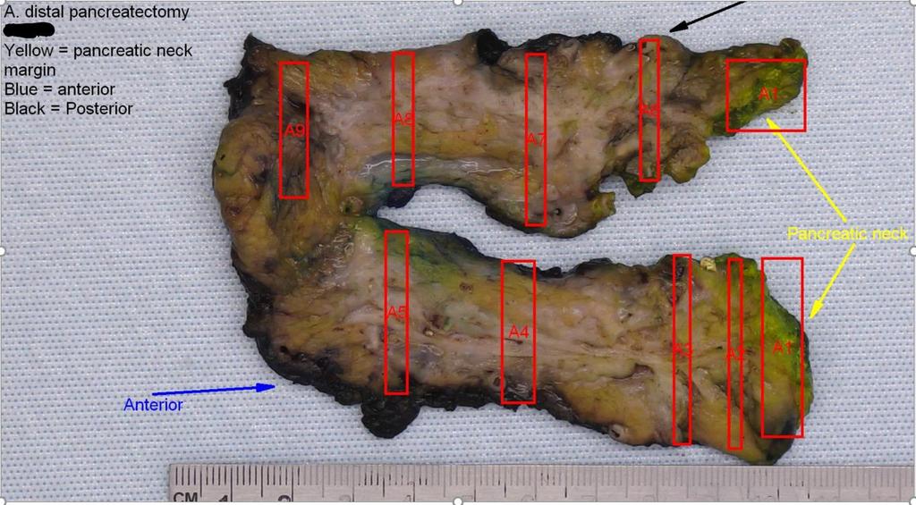
31 Gross Specimen Imaging Prior to case accessioning ID with MRN, patient name, date/time stamp Annotations Block designations, clip designation, biopsy site, calcifications
32
33 The Glass Slide Label Identifiers Barcode, 2D vs 3D Control tissue Diagnostic tissue Multiple fragments Coded fragments (e.g. 2 LNs, 1 bisected and inked) Tissue microarrays
34 Metadata Data about data (Classify as Localized to Instrument, APLIS, or EHR) Slide scanning order(s) Magnification, Z-stacking, digital filters Slide received in machine Slide scanning started Slide scanning completed Slide scanning errors / warnings Slide manually retouched Operator ID, date/time stamps begin/end, audit trail of functions applied
35 Metadata Data about data (Classify as Localized to Instrument, APLIS, or EHR) Slide received/available in AP LIS / PACS Slide viewing started Viewer ID Start time, End time Audit trail of X-Y-Z at Mag M Audit trail of digital filters applied at timepoint Tissue Annotations Margin (designate), distance to margin, benign neoplasia, dysplasia, in situ malignancy, invasive malignancy, infectious finding, inflammatory finding (acute, chronic, specified, unspecified), cell classification, structure classification, uncertain finding (ROI not otherwise classified), tumor size (with axis designations), tissue floater, mitotic figure, mitotic hot spot ROI, capsule invasion, lymph node metastasis (size, extranodal) Mark up coordinates relative to slide origin or ROI origin Slide Annotations Stain issues (too pink), cutting issues (too thick, fragmented), visibility issues (frozen section artifact, air dry artifact)
36 Metadata Data about data (Classify as Localized to Instrument, APLIS, or EHR) Slide viewing completed Slide viewing inquiry Viewer ID (years in practice, area of specialty) Start time, End time Slide ID to include stain (H&E vs IHC etc) Case type (breast, GI, lung, b9 vs neoplastic dz etc) Percent of tissue not viewed Percent of tissue not viewed twice Percent of tissue not viewed at higher than 10x mag Size of tissue on slide (area of polygon)
37 Next Steps
38 EHR (HL7 order placer) (HL7 result tracker) Acquisition Manager (Generate WOS for acquisition devices) DICOM Acquistion Modality (WSI scanner) Automation Manager (Generate WOS, e.g for evidence creators) Acquistion Modality (Macroscopic Imager) Image Manager (DICOM) LIS (HL7 order filler) Image Display (DICOM) (Digital) Evidence Creator Image Archive (DICOM, VNA / PACS)
39
40 INCLUDED FOR REFERENCE ONLY Digital Pathology Standards Integration Committee IHE PaLM Change Proposal & DICOM considerations Berlin, May 25th 2016
41 Digital Pathology Standards Integration Committee Initiated by Visiopharm (2015) Identifying and resolving practical issues around use of standards for DP International group of vendors and users involved 41
42 Activities Meetings Kick-off October 2015 DICOM and IHE training 2016 F2F meeting April 2016 Berlin meeting May 25 th Change proposal submitted to IHE for APW profile IHE proposal approved DICOM suppl. 145 suggestions 42
43 Proposal Improve and expand profile with more recent experience from using DP for Tight workflow integration (primary diagnostics!) Image processing Practical implementations 43
44 Proposal Review APW profile (w/focus on DP) Make APW actor-transaction diagram consistent with SWF Extend APW with following use cases: Pathology reporting (comparable to RAD reporting) Quality control around using WSI Adjust Pathology General Workflow with post processing 44
45 Current actors/transactions APW 45
46 Use case: Pathology reporting IHE Radiology Technical Framework, Volume 1 (RAD TF-1): Integration Profiles See RAD vol. 1, chapter 13 DSS/ Order Filler RAD-4: Procedure Scheduled RAD-13: Procedure Update RAD-42: Performed Work Status Update Report Manager RAD-46: Query Reporting Worklist RAD-38: Workitem Claimed RAD-41: Workitem Completed RAD-39: Workitem PPS in Progress RAD-40: Workitem PPS Completed Report Creator Report Reader RAD-7: Modality PS Completed Performed Procedure Step Manager Image Manager Image Archive RAD-11: Image Availability Query 46
47 Pathology Current manual QC workflow Utrecht, 2 Feb 2016, update April 28 th 2016 Use case: quality control using WSI UROLOGIST Op room / theatre TRANSPORT BOOKING CLERK or TECHNICIAN Laboratory TECHNICIAN Laboratory Image(s) created Sampling Send sample Receive sample Grossing Acquire gross image Check order Accept Review gross image Place order Y N Check order OK OK? Y N Order Placer (EMR/EHR) HL7 order OML^O21 Order filler (LIS / LIMS) Reject Fixation & Tissue processing HL7 cancellation OML^O21 Slide prep & acquistion N Check sample against gross image OK? Y Embedding Sectioning Staining Slide making Optional N Acquisition modality (Slide scanner) Acquire whole slide image Y All OK? Optical check Image(s) created Automated Quality Control N OK? (per slide) Y Human review Optional (if scanner computed high probability of success) Is prep bad? Y Image Manager/ Image Archive (VNA) N Y Is acquisition bad? N PATHOLOGIST Laboratory / Office Review each slide Is prep bad? Y Y Is acquisition bad? N 47 Use image Y All needed views present? N N N Go back to sectioning
48 Use case: quality control using WSI Sectioning Staining Slide making N Acquisition modality (Slide scanner) Acquire whole slide image Y All OK? Optical check Image(s) created Automated Quality Control Image Manager/ Image Archive N OK? (per slide) Y Human review Optional (if scanner computed high probability of success) Is prep bad? N Y TECHNICIAN Laboratory Y Is acquisition bad? N PATHOLOGIST Laboratory / Office Review each slide Is prep bad? Y Y Is acquisition bad? N Use image Y All needed views present? N N N Go back to sectioning


49 Pathology General Workflow with post processing Current use case: 49
50 Pathology General Workflow with post processing Extended use case: PATHOLOGIST or Lab technican Laboratory / Office Order additional stains Optional Slide prep & acquisition Select slides for image processing N Y Tune algorithm parameters? N Select different slides? Y N Image Processing algorithm(s) OK? Y Inform LIS Tune parameters N: retry image processing algorithm (different tuning) 50 Store results as DICOM SR and annotations/markups Make report
51 Other IHE APW proposed changes Replace RAD-43 with RAD-10 RAD-16 (retrieve image) Fetching selected number of frames PaLM-16: DICOM 2011 PS3.4, Annex Y instead of Annex C Webservice version (QIDO/WADO) Specimen preparation information not always available at scanner, how to add later to image? Relation to LTW, LAW and/or LDA 51
52 DICOM issues Annotations / markups Store using real-world coordinates? Potentially 1000s polygons in one level / Z plane / colour plane, multiplied by planes stored within instance For computational purposes and zooming not feasible to store as overlay How to retrieve for tiles displayed on screen? Overlay object Per frame? Per depth of field level / colour plane? Per instance? Per study? 52
53 DICOM issues Virtual double-staining (applied by CAD software) How to register and store? How to standardise? Blending like PET-CT? 53
54 DICOM recommendations Optical path per image instead of frame Same tile size for all images (for all layers) Handling of empty tiles (fixing background colour?) Lens power shouldn t be Type 3 but Type 1 Tag needed for number of pyramid levels (at study level) Z-plane can have 1 n tiles in it, large file size, multiple bit depths Mix of bit depth could be a problem, especially with move to multispectral (50-60 channels) Consider dictating maximums, or when to create a new separate object Limit amount of transfer syntaxes 54

55
DICOM-compatible compression of WSI and diagnostic evaluation
 of WSI and diagnostic evaluation R. Zwönitzer, H. Hofmann, A. Roessner, T. Kalinski 2nd European Workshop in Tissue Imaging and Analysis June 25-26, 2010 - Heidelberg 1 GPWL / GP-PPS Introduction Overview
Digital Pathology at Johns Hopkins Practical Research and Clinical Considerations
 Digital Pathology at Johns Hopkins Practical Research and Clinical Considerations July 10, 2017 Alexander Baras, MD, PhD Assistant Professor of Pathology, Urology, and Oncology Associate Director of Pathology
Technical Aspects in Digital Pathology
 Technical Aspects in Digital Pathology Yukako Yagi, PhD yyagi@mgh.harvard.edu Director of the MGH Pathology Imaging & Communication Technology Center Assistant Professor of Pathology, Harvard Medical School
Color aspects and Color Standardization in Digital Microscopy
 Color aspects and Color Standardization in Digital Microscopy Yukako Yagi, PhD yyagi@partners.org Director of the MGH Pathology Imaging & Communication Technology Center Assistant Professor of Pathology,
Overview of Digital Pathology s Current State: Technologies, Systems, Capabilities, Limitations, and Opportunities
 Overview of Digital Pathology s Current State: Technologies, Systems, Capabilities, Limitations, and Opportunities David McClintock, MD Executive War College Post-Conference Workshop Digital Pathology
THEORY AND APPROACHES TO AUTOMATED IMAGE ANALYSIS IN DIGITAL PATHOLOGY
 THEORY AND APPROACHES TO AUTOMATED IMAGE ANALYSIS IN DIGITAL PATHOLOGY Kyle Takayama, MS Charles River Laboratories EVERY STEP OF THE WAY EVERY STEP OF THE WAY MORPHOMETRY Measurements or counts performed
IHE Radiology Technical Framework Supplement. Stereotactic Mammography Image (SMI) Trial Implementation
 Trial Implementation") Integrating the Healthcare Enterprise 5 IHE Radiology Technical Framework Supplement 10 Stereotactic Mammography Image (SMI) 15 Trial Implementation 20 25 Date: June 11, 2013 Author: IHE Radiology Technical
Integrating the Healthcare Enterprise 5 IHE Radiology Technical Framework Supplement 10 Stereotactic Mammography Image (SMI) 15 Trial Implementation 20 25 Date: June 11, 2013 Author: IHE Radiology Technical
MIRAX SCAN The new way of looking at pathology
 Microscopy from Carl Zeiss MIRAX SCAN The new way of looking at pathology Greater reliability. Greater efficiency. Plus points for your diagnostics Better. More efficient. Quality as a factor for success
Digital Pathology and Tissue-based Diagnosis. How do they differ?
 Digital Pathology and Tissue-based Diagnosis. How do they differ? P. Hufnagl Institute of Pathology (Rudolf-Virchow-Haus). Humboldt University, Berlin? 10.12.2014 1 Structure of the talk Possible workflow
SPOT PathSuite Solutions
 SPOT PathSuite Solutions The Perfect Fit PathStation TM Imaging system for the gross dissection in hood PathStand TM 40 imaging station for medium to large specimens PathSuite Is Easy To Use A turnkey
Digital Pathology Update
 Digital Pathology Update J. Mark Tuthill, MD Division Head, Pathology Informatics Department of Pathology & Laboratory Medicine Henry Ford Hospital Detroit, MI mtuthil1@hfhs.org ASCT Webinar, January 2016
Stereotopix Research. Precision Pathology. Highthroughput. pathology. powered by newcast. Advantages of Stereotopix : RUO
 Precision Pathology Highthroughput pathology Stereotopix Research powered by newcast RUO Researchers use quantitative microscopy in many ways with the goal of producing high-quality, quantitative results
InScape: Making Virtual Pathology a Reality
 InScape: Making Virtual Pathology a Reality Sally S. Agersborg, M.D., Ph.D. Quest Diagnostics, Nichols Institute San Juan Capistrano, CA Company Overview Quest Diagnostics, Nichols Institute the world
DMETRIX S (FUTURE) PERSPECTIVES ON DIGITAL IMAGING & DIGITAL PATHOLOGY SYSTEMS
 PERSPECTIVES ON DIGITAL IMAGING & DIGITAL PATHOLOGY SYSTEMS") Michael R. Descour, Ph.D., DMetrix, Inc., & University of Arizona Lloyd J. LaComb, Jr., Ph.D., DMetrix, Inc. DMETRIX S (FUTURE) PERSPECTIVES ON DIGITAL IMAGING & DIGITAL PATHOLOGY SYSTEMS Outline of presentation
Michael R. Descour, Ph.D., DMetrix, Inc., & University of Arizona Lloyd J. LaComb, Jr., Ph.D., DMetrix, Inc. DMETRIX S (FUTURE) PERSPECTIVES ON DIGITAL IMAGING & DIGITAL PATHOLOGY SYSTEMS Outline of presentation
Digital Microscopy. Past, Present, and Future. Cyrus V. Hedvat, MD, PhD
 Digital Microscopy Past, Present, and Future Cyrus V. Hedvat, MD, PhD N Context. Digital viewing of histologic images is moving from presentations and publications to incorporation into the daily work
GE Healthcare. Senographe 2000D Full-field digital mammography system
 GE Healthcare Senographe 2000D Full-field digital mammography system Digital has arrived. The Senographe 2000D Full-Field Digital Mammography (FFDM) system gives you a unique competitive advantage. That
Bowen Hills Histopathology
 Bowen Hills Histopathology Histology s Bowen Hills Challenges Relocate and amalgamate Taringa and Indooroopilly Labs into a single complex histology laboratory Change workflow, staff structure, install
Second Announcement Call for Participation. (Evaluation Criteria added)
") Second Announcement Call for Participation 2 nd International Scanner Contest (ISC) (Evaluation Criteria added) P. Hufnagl 1, T. Schrader 1, 2, M.G. Rojo 3, A. Laurinavicius 4, G. Kayser 5, Y. Yagi 6 1
Second Announcement Call for Participation 2 nd International Scanner Contest (ISC) (Evaluation Criteria added) P. Hufnagl 1, T. Schrader 1, 2, M.G. Rojo 3, A. Laurinavicius 4, G. Kayser 5, Y. Yagi 6 1
PACS Fundamentals. By: Eng. Valentino T. Mvanga Ministry of Health and Social Welfare Tanzania
 PACS Fundamentals By: Eng. Valentino T. Mvanga Ministry of Health and Social Welfare Tanzania 1 Learning Goals To Understand the importance of PACS To Understand PACS infrastructure requirement Introduction
FRAUNHOFER INSTITUTE FOR INTEGRATED CIRCUITS IIS. MANUAL PANORAMIC MICROSCOPY WITH istix
 FRAUNHOFER INSTITUTE FOR INTEGRATED CIRCUITS IIS MANUAL PANORAMIC MICROSCOPY WITH istix CLINICAL DIAGNOSTICS AND MATERIAL SCIENCES IMPROVED BY DIGITAL MICROSCOPY B A C K G R O U N D Due to a high grade
Scopis Hybrid Navigation with Augmented Reality
 Scopis Hybrid Navigation with Augmented Reality Intelligent navigation systems for head surgery www.scopis.com Scopis Hybrid Navigation One System. Optical and electromagnetic measurement technology. As
Digital Pathology and Image Analysis. Queen s University Department of Pathology and Molecular Medicine Shakeel Virk
 Digital Pathology and Image Analysis Queen s University Department of Pathology and Molecular Medicine Shakeel Virk Outline Digital Pathology and Image Analysis capabilities at Queen s Laboratory for Molecular
Anatomic and Computational Pathology Diagnostic Artificial Intelligence at Scale
 Anatomic and Computational Pathology Diagnostic Artificial Intelligence at Scale John Gilbertson MD Department of Pathology Massachusetts General Hospital Partners Healthcare System Harvard Medical School
COST EFFECTIVE FLAT PANEL DIGITAL RADIOGRAPHY UPGRADE SOLUTIONS
 COST EFFECTIVE FLAT PANEL DIGITAL RADIOGRAPHY UPGRADE SOLUTIONS DRive is a digital imaging DR hardware & Software solution designed for General Radiography of anatomy. It intended to replace film/screen
How to introduce virtual microscopy (VM) in routine diagnostic pathology: constraints, ideas, and solutions
 in routine diagnostic pathology: constraints, ideas, and solutions") How to introduce virtual microscopy (VM) in routine diagnostic pathology: constraints, ideas, and solutions Klaus Kayser, Stephan Borkenfeld, Gian Kayser History Workflow Design of virtual microscopy Constraints
How to introduce virtual microscopy (VM) in routine diagnostic pathology: constraints, ideas, and solutions Klaus Kayser, Stephan Borkenfeld, Gian Kayser History Workflow Design of virtual microscopy Constraints
BRINGING DEEP LEARNING TO ENTERPRISE IMAGING CLINICAL PRACTICE
 BRINGING DEEP LEARNING TO ENTERPRISE IMAGING CLINICAL PRACTICE Esteban Rubens Global Enterprise Imaging Principal Pure Storage @pureesteban AI IN HEALTHCARE What is Artificial Intelligence (AI)? How is
Definiens. Tissue Studio 4.2. Tutorial 1: Composer and Nuclear Markers
 Definiens Tissue Studio 4.2 Tutorial 1: Composer and Nuclear Markers Tutorial 1: Composer and Nuclear Markers Imprint and Version Copyright 2015 Definiens AG. All rights reserved. This document may be
DICOM Educational Conference Brisbane, Australia
 DICOM Educational Conference Brisbane, Australia SEPTEMBER 27-28, 2018 DICOM OVERVIEW & PROCESS KEVIN O DONNELL, CHAIR, DICOM WG10 (STRATEGIC) PAST-CHAIR, DICOM STANDARDS CMTE CANON MEDICAL RESEARCH USA
Touch Screen Technology. Classic WAIV. with Touch Screen Technology. Legendary Reputation...Sensitivity to your budget
 Touch Screen Technology Classic WAIV with Touch Screen Technology Legendary Reputation...Sensitivity to your budget Streamline Workflow For Higher Productivity And Patient Throughput. The CRescendo Classic
EasyGrab R1. Image based in-or radiology consultation 93/42/EEC
 EasyGrab R1 Image based in-or radiology consultation 93/42/EEC Medical Image Management 2.0 Get a firm grip on image based medical documentation Image management In today s healthcare performance, accuracy
Incorporating novel image processing methods in a hospital-wide PACS
 International Congress Series 1281 (2005) 1016 1021 www.ics-elsevier.com Incorporating novel image processing methods in a hospital-wide PACS Erwin Bellon a, T, Michel Feron a, Paul Neyens a, Klaas Peeters
DICOM Conformance. DICOM Detailed Specification for Diagnostic Labs and Radiology Center Connectivity
 DICOM Detailed Specification for Diagnostic Labs and Radiology Center Connectivity Authored by Global Engineering Team, Health Gorilla April 10, 2014 Table of Contents About Health Gorilla s Online Healthcare
Digital Imaging in Anatomic Pathology
 your lab focus CE update [generalist histology] Digital Imaging in Anatomic Pathology Ted F. Beals, MD From the Department of Veteran Affairs, Veterans Health Administration, and Department of Pathology,
PathStation TM Fits Your Lab
 PathStation TM Fits Your Lab PathStation hood Compact In-hood Imaging system. Supports in-process documentation PathStation Key Features 20 MP Image Capture Great HD Video 8.33 X Optical zoom 4X Digital
Building a Business Case for Digital Pathology: The Time is Now! Drazen M. Jukic, MD, PhD
 Building a Business Case for Digital Pathology: The Time is Now! Drazen M. Jukic, MD, PhD Business Case Telepathology vs. Digital Pathology Telepathology as a term was misunderstood in the past; It denotes
DICOM Conformance Statement
 Rogan-Delft B.V. Customer Information Bulletin Title DICOM Conformance Statement Scope Rogan OnLine XS Archiver Target Group Service Engineers Page 2 of 33 How To Contact Rogan-Delft BV Wiltonstraat 41
egross Ergonomic, mobile grossing workcenter with digital documentation of surgical specimens MILESTONE
 egross Ergonomic, mobile grossing workcenter with digital documentation of surgical specimens MILESTONE H E L P I N G P A T I E N T S The egross This all-in-one grossing workstation has been specifically
Digital Imaging and Communications in Medicine (DICOM) Supplement 39: Add Stored Print Media Storage - Retire Normalized Print Media Storage
 Supplement 39: Add Stored Print Media Storage - Retire Normalized Print Media Storage") Digital Imaging and Communications in Medicine (DICOM) Supplement 39: Add Stored Print Media Storage - Retire Normalized Print Media Storage DICOM Standards Committee, Working Group 300 N. 7th Street Rosslyn,
Digital Imaging and Communications in Medicine (DICOM) Supplement 39: Add Stored Print Media Storage - Retire Normalized Print Media Storage DICOM Standards Committee, Working Group 300 N. 7th Street Rosslyn,
Kretztechnik AG. Voluson 730 Ultrasound System
 Kretztechnik Voluson 730 Ultrasound System DICOM Conformance Statement Rev 1.02 Voluson 730 DICOM Coformance Rev 1.02 2001-07-18 Kretztechnik AG Zipf/Austria KRETZTECHNIK AG TIEFENBACH 15 Telefon: +43
A Practical Overview of the Clinical and Operational Impact of Computed Radiography(CR) Implementations. Shirley Weddle, RT(R)(M), CIIP, BBA
 Implementations. Shirley Weddle, RT(R)(M), CIIP, BBA") A Practical Overview of the Clinical and Operational Impact of Computed Radiography(CR) Implementations Shirley Weddle, RT(R)(M), CIIP, BBA OBJECTIVES Define Computed Radiography (CR) Discuss CR vendor
A Practical Overview of the Clinical and Operational Impact of Computed Radiography(CR) Implementations Shirley Weddle, RT(R)(M), CIIP, BBA OBJECTIVES Define Computed Radiography (CR) Discuss CR vendor
DigiMam Conformance Statement for DICOM V3.0
 DigiMam Conformance Statement for DICOM V3.0 Copyright 2004 by I.M.S. s.r.l. DOCUMENT VERSIONS Version Date Author Changes 1.00 15-Feb-05 IMS s.r.l. First Version DOCUMENT VERSIONS Page 2 of 29 TABLE OF
34X ZOOM. PathStand TM Routine. Superior Image Quality. PathStand Key Features. 1X Zoom. 8.33X Zoom
 PathStand TM Routine Complete stand-alone Imaging system. Supports in-process documentation. PathStand Key Features 20 MP Image Capture 1080p Live Video 34X zoom Metal Construction All-in-one Computer
34X ZOOM. PathStand TM Routine. Superior Image Quality. PathStand Key Features. 1X Zoom. 8.33X Zoom
 PathStand TM Routine Complete stand-alone Imaging system. Supports in-process documentation. PathStand Key Features 20 MP Image Capture 720p Live Video 34X zoom Metal Construction All-in-one Computer LED
Automated Digitization of Gram Stains. Centralized Reading. Decentralized Assessment. Improved Quality Management.
 Automated Digitization of Gram Stains Centralized Reading. Decentralized Assessment. Improved Quality Management. A GROWING DEMAND Gram staining is the rapid, easy, and inexpensive method for the assessment
Solutions Page Content ImagePilot. Primary keywords: Digital radiography Computed radiography Image viewing and storage
 Solutions Page Content Primary keywords: Digital radiography Computed radiography Image viewing and storage Solution Description For facilities with medium volume imaging, Konica Minolta s original all-in-one
Teaching Digital Histology
 Teaching Digital Histology Carlos R. Morales Department of Anatomy and Cell Biology, McGill University, Montreal, Quebec, Canada The light microscope is one of the most widely used scientific instruments
Hematoxylin and Eosin Stained Tissue
 A p p l i c a t i o n N o t e Hematoxylin and Eosin Stained Tissue Using Color Brightfield Imaging with the Cytation 5 to Image Fixed and Stained Tissue Paul Held Ph. D., Laboratory Manager, Applications
IMPAX 6 DISPLAY TOOL LIST
 IMPAX 6 DISPLAY TOOL LIST IMPAX 6.0 TOOLS INDEX A Advance by Image Allows you to scroll from one image or frame to the next Advance by Page Pages through images in a large series, one screen at a time
In our previous lecture, we understood the vital parameters to be taken into consideration before data acquisition and scanning.
 Interactomics: Protein Arrays & Label Free Biosensors Professor Sanjeeva Srivastava MOOC NPTEL Course Indian Institute of Technology Bombay Module 7 Lecture No 34 Software for Image scanning and data processing
Best and Worst Practices and Software Development Tools DICOM
 DICOMWeb 2015 Conference & Hands-on Workshop Best and Worst Practices and Software Development Tools DICOM David Clunie (dclunie@dclunie.com) PixelMed Publishing Background & Disclosures l Owner, PixelMed
Multilayer scanning enhances sensitivity of artificial intelligence-aided Mycobacterium tuberculosis detection
 Multilayer scanning enhances sensitivity of artificial intelligence-aided Mycobacterium tuberculosis detection Yan Xiong Peking University First Hospital, China. yanxiong1109@163.com Ao Hou ao_sure@foxmail.com
(12) Patent Application Publication (10) Pub. No.: US 2007/ A1
 Patent Application Publication (10) Pub. No.: US 2007/ A1") (19) United States US 200701 18100A1 (12) Patent Application Publication (10) Pub. No.: US 2007/0118100 A1 Mahesh et al. (43) Pub. Date: (54) SYSTEM AND METHOD FOR IMPROVED ABLATION OF TUMIORS (75) Inventors:
(19) United States US 200701 18100A1 (12) Patent Application Publication (10) Pub. No.: US 2007/0118100 A1 Mahesh et al. (43) Pub. Date: (54) SYSTEM AND METHOD FOR IMPROVED ABLATION OF TUMIORS (75) Inventors:
Digital Microscopy: New Paradigm's for Teaching Microscopic Anatomy and Pathology
 Digital Microscopy: New Paradigm's for Teaching Microscopic Anatomy and Pathology Michael Feldman, MD, PhD Assistant Dean IT Assistant Professor Pathology University of Pennsylvania Health System Feldmanm@mail.med.upenn.edu
HIGH-RESOLUTION DIGITAL PHOTOSPOT SYSTEM years, CMT s technology is today proven in more than 4,500 clinical installations PRODUCT SPECIFICATION
 CMT A Medical Imaging Leader SmartSPOT PrimaX RF CMT Medical Technologies is a market leader providing high-resolution digital X-ray imaging solutions for General Radiography, R&F rooms and Angiography
Performance and care. all in one
 Performance and care all in one INNOVATION IS WHAT DRIVES US THINKING ABOUT THE FUTURE Preventive diagnostics remains an essential weapon in defeating breast cancer. Metaltronica s forward-thinking design
This document is a preview generated by EVS
 TECHNICAL REPORT ISO/TR 28380-2 First edition 2014-02-15 Health informatics IHE global standards adoption Part 2: Integration and content profiles Informatique de santé Adoption des normes globales IHE
CR Basics and FAQ. Overview. Historical Perspective
 Page: 1 of 6 CR Basics and FAQ Overview Computed Radiography is a term used to describe a system that electronically records a radiographic image. Computed Radiographic systems use unique image receptors
DICOM Conformance Statement
 GE Medical Systems Kretz Ultrasound DICOM Conformance Statement 105952 Revision 0 VOLUSON 730Expert/Pro V. 4.0.x 0123 Copyright 2000, 2001, 2002, 2003, 2004 by GE Medical Systems Kretztechnik GmbH & Co
DICOM3.0 Conformance Statement
 DICOM3.0 Conformance Statement INTRODUCTION 1 IMPLEMENTATION MODEL 2 AE Specifications 3 DIRECT DIGITIZER Communication Profiles 4 Extensions/Specializations/Privatizations 5 Configuration 6 0197 Support
IDEXX-PACS * 4.0. Imaging Software. Quick Reference Guide
 4 IDEXX-PACS * 4.0 Imaging Software Quick Reference Guide Capturing Images Before you begin: Adjust the collimation properly. Make sure the body part you are imaging matches the exam type you have selected.
DICOM Correction Proposal
 Tracking Information - Administration Use Only DICOM Correction Proposal Correction Proposal Number Status CP-1713 Letter Ballot Date of Last Update 2018/01/23 Person Assigned Submitter Name David Clunie
2 nd generation TOMOSYNTHESIS
 2 nd generation TOMOSYNTHESIS 2 nd generation DBT true innovation in breast imaging synthesis graphy Combo mode Stereotactic Biopsy Works in progress: Advanced Technology, simplicity and ergonomics Raffaello
User Manual Veterinary
 Veterinary Acquisition and diagnostic software Doc No.: Rev 1.0.1 Aug 2013 Part No.: CR-FPM-04-022-EN-S 3DISC, FireCR, Quantor and the 3D Cube are trademarks of 3D Imaging & Simulations Corp, South Korea,
Digital Image Management: the Basics
 Digital Image Management: the Basics Napapong Pongnapang, Ph.D. Department of Radiological Technology Faculty of Medical Technology Mahidol University Outline From screen/film to digital radiography PACS/Tele
Positive Pixel Count Algorithm. User s Guide
 Positive Pixel Count Algorithm User s Guide Copyright 2004, 2006 2008 Aperio Technologies, Inc. Part Number/Revision: MAN 0024, Revision B Date: December 9, 2008 This document applies to software versions
Module 1A: Record images of ledger/card or catalog/field notes (materials not stored with specimens)
") Module 1: Imaging objects (Fluid-preserved) Module 1A: Record images of ledger/card or catalog/field notes (materials not stored with specimens) Task ID Task Name Explanations and Comments Resources T1
Module 1: Imaging objects (Fluid-preserved) Module 1A: Record images of ledger/card or catalog/field notes (materials not stored with specimens) Task ID Task Name Explanations and Comments Resources T1
MacroPATH The new line of Digital Imaging Systems for Grossing
 MILESTONE H E L P I N G P A T I E N T S MacroPATH The new line of Digital Imaging Systems for Grossing If the dimensions of the specimen are not recorded, the key section not taken, and the proper special
Regulatory Forum. Society of Toxicologic Pathology Position Paper on Pathology Image Data: Compliance with 21 CFR Parts 58 and 11
 Regulatory Forum Toxicologic Pathology, 35:450 455, 2007 Copyright C by the Society of Toxicologic Pathology ISSN: 0192-6233 print / 1533-1601 online DOI: 10.1080/01926230701284509 Society of Toxicologic
Center for Microscopy and Image Analysis Axio Scan.Z1 Operating Manual
 No index entries found. Center for Microscopy and Image Analysis Axio Scan.Z1 Operating Manual Table of contents 1. Starting procedure... 3 1.1. Turn on hardware... 3 1.2. Starting ZEN blue... 4 2. Load
Digital Imaging Outline
 Digital Microscopy and Imaging Update Michael Feldman, MD, PhD Associate Professor of Pathology Assistant Dean for Information Technology University of Pennsylvania School of Medicine (Feldmanm@mail.med.upenn.edu)
FCR XL-2 and FCR XC-2. High-quality digital x-ray that's perfect for your private practice.
 F U J I F I L M D I G I T A L X - R A Y FCR XL-2 and FCR XC-2. High-quality digital x-ray that's perfect for your private practice. So small, yet so powerful. With a compact footprint of less than 2.5
Clinical and management aspects of digital imaging and PACS
 Clinical and management aspects of digital imaging and PACS พญ.จามร เช อเพชระโสภณ นายกร งส ว ทยาสมาคมแห งประเทศไทย chamareec@gmail.com www.radiologythailand.org Digital imaging Abbreviation and terminology
A Module for Visualisation and Analysis of Digital Images in DICOM File Format
 A Module for Visualisation and Analysis of Digital Images in DICOM File Format Rumen Rusev Abstract: This paper deals with design and realisation of software module for visualisation and analysis of digital
DICOM Correction Item
 DICOM Correction Item Correction Number CP-564 Log Summary: Type of Modification Correction Name of Standard PS 3.3, PS 3.6, PS 3.17 2004 Rationale for Correction A mammography CAD system often prefers
High-sensitivity. optical molecular imaging and high-resolution digital X-ray. In-Vivo Imaging Systems
 High-sensitivity optical molecular imaging and high-resolution digital X-ray In-Vivo Imaging Systems In vivo imaging solutions available in several packages Carestream Molecular Imaging offers a selection
Virtual Microscopy: Potential Applications in Medical Education and Telemedicine in Countries with Developing Economies
 Virtual Microscopy: Potential Applications in Medical Education and Telemedicine in Countries with Developing Economies Paul Fontelo 1, Ernest DiNino 2, Krista, Johansen 2, Ashraf Khan 2, Michael Ackerman
Virtual 3D Microscopy Using Multiplane Whole Slide Images in Diagnostic Pathology
 Anatomic Pathology / Diagnostic Virtual Pathology Virtual 3D Microscopy Using Multiplane Whole Slide Images in Diagnostic Pathology Thomas Kalinski, MD, 1 Ralf Zwönitzer, 2 Saadettin Sel, MD, 1 Matthias
The Fastest, Easiest, Most Accurate Way To Compare Parts To Their CAD Data
 210 Brunswick Pointe-Claire (Quebec) Canada H9R 1A6 Web: www.visionxinc.com Email: info@visionxinc.com tel: (514) 694-9290 fax: (514) 694-9488 VISIONx INC. The Fastest, Easiest, Most Accurate Way To Compare
Photo Digitization. Pre-Digitization (including planning) Digitization. Post-Digitization
 Digitization. Post-Digitization") Photo Digitization A photo digitization projects begin prior to the first scan and extend beyond the completion of the actual digitization. The following is a breakdown of pre-, during, and post- digitization
Photo Digitization A photo digitization projects begin prior to the first scan and extend beyond the completion of the actual digitization. The following is a breakdown of pre-, during, and post- digitization
SmartRAD. Advanced Digital Radiography System
 SmartRAD Advanced Digital Radiography System SmartRAD Expanding The Horizons Of Digital Radiography CMT introduces the SmartRAD Digital Radiography system, featuring an integrated flat panel digital detector
LSM 800 Confocal Microscope Standard Operation Protocol
 LSM 800 Confocal Microscope Standard Operation Protocol Turning on the system 1. Switch on the Main switch (labeled 1 and 2 ) mounted on the wall. 2. Turn the Laser Key (labeled 3 ) 90 clockwise for power
Building of a composite virtual slide from contiguous tissue samples
 PROCEEDINGS Building of a composite virtual slide from contiguous tissue samples Open Access Benoît Plancoulaine 1,3, Myriam Oger 1,2*, Nicolas Elie 1,3, Philippe Belhomme 1,3, Paulette Herlin 1,2, Abir
Digital Imaging started in the 1972 with Digital subtraction angiography Clinical digital imaging was employed from the 1980 ~ 37 years ago Amount of
 Digital Imaging started in the 1972 with Digital subtraction angiography Clinical digital imaging was employed from the 1980 ~ 37 years ago Amount of radiation to the population due to Medical Imaging
Chapter 3. 3D Lung Pathology Imaging
 A version of this chapter has been published in Anat Rec (Hoboken), vol. 290, pp. 1377-87, 2007. We cannot solve our problems with the same thinking we used when we created them Albert Einstein Chapter
The Versatile and Powerful ACLxy. ACLxy
 The Versatile and Powerful ACLxy ACLxy Rolling into a Clinic, Imaging Center and Hospital Near You! COMPUTED RADIOGRAPHY (CR) IS RAPIDLY THE BEGINNING. THE OREX CR SOLUTION DRA- BECOMING A DRIVING FORCE
Digital Imaging CT & MR
 Digital Imaging CT & MR January 22, 2008 Digital Radiography, CT and MRI generate images in a digital format What is a Digital Image? A digital image is made up of picture elements, pixels row by column
Tmypacs. DICOM Conformance Statement 1.0. Document Version: Tmypacs. Product Name(s): 2.0. Release:
: 2.0. Release:") Tmypacs DICOM Conformance Statement Document Version: Product Name(s): Release: 1.0 Tmypacs 2.0 Date: January 19, 2014 1. COMFORMANCE STATEMENT OVERVIEW The TmypacsServer is a DICOM server. The ImageServer
Tmypacs DICOM Conformance Statement Document Version: Product Name(s): Release: 1.0 Tmypacs 2.0 Date: January 19, 2014 1. COMFORMANCE STATEMENT OVERVIEW The TmypacsServer is a DICOM server. The ImageServer
DELWORKS DR MEDICAL. take the next step
 DELWORKS DR MEDICAL take the next step DELWORKS MEDICAL DR If you are thinking of taking the next step to digital radiography, consider a DelWorks Medical DR Retrofit Package, the easy and affordable way
Eleva Workspot 1.x - Training. Advanced User Functions. State: April 2007
 Eleva Workspot 1.x - Training Advanced User Functions State: April 2007 Purpose of this Presentation General Get an overview about all "" functions in Eleva Workspot 1.x These are available in the System
Digital Imaging and Communications in Medicine (DICOM) Supplement 56: Ultrasound Waveform
 Supplement 56: Ultrasound Waveform") Digital Imaging and Communications in Medicine (DICOM) Supplement 56: Ultrasound Waveform DICOM Standards Committee, Working Group 12 - Ultrasound 1300 N. 17 th Street, Suite 1847 Rosslyn, Virginia 22209
Digital Imaging and Communications in Medicine (DICOM) Supplement 56: Ultrasound Waveform DICOM Standards Committee, Working Group 12 - Ultrasound 1300 N. 17 th Street, Suite 1847 Rosslyn, Virginia 22209
Code.No Specimen Turn over Time Fixative Used/Acceptance Criteria 501A Eyeball 4 DAYS 10%Neutral buffered formalin
 HISTOPATHOLOGY Tests(Specimens) under Scope of NABL Code.No Specimen Turn over Time Fixative Used/Acceptance Criteria 501A Eyeball 4 DAYS 10%Neutral buffered formalin 502A Exenterated specimen 4 DAYS 10%Neutral
Stack or Trash? Quality assessment of virtual slides. Anatomic pathology & collaboration
 11th European Congress on Telepathology 5th International Congress on Virtual Microscopy 6-9 June 2012, Venice, Italy Stack or Trash? Quality assessment of virtual slides David Ameisen Christophe Deroulers,
Virtual Electron Microscopy update after one year of routine use
 The Hong Kong College of Pathologists, Incorporated in Hong Kong with Limited Liability Volume 7, Issue 2 July 2012 Editorial note: Many of us who have tried operating an electron microscope would share
4.0 How to Turn On the Selenia Dimensions
 Chapter 2 System Controls and Indicators How to Turn On the Selenia Dimensions 4.0 How to Turn On the Selenia Dimensions 4.1 Preparation 1. Reset all three Emergency Off switches. Emergency Off Switches
PharmaClik Rx 1.1 New Features
 PharmaClik Rx 1.1 New Features 2013, McKesson Canada. All rights reserved. The information contained in this document is proprietary to McKesson Canada. Table of Contents Patient Waive Preferences... 1
DICOM Image And Data Management For Nuclear Medicine, Physiological Measurements, Radiotherapy And Ultrasound READ ONLINE
 DICOM Image And Data Management For Nuclear Medicine, Physiological Measurements, Radiotherapy And Ultrasound READ ONLINE radiology information system (RIS) solutions, workstations for acquisition, non-dicom
Examion. New. Image Acquisition and Diagnostics. R a d i o D i g i t a l e. examion aqs veterinray software.
 Examion New V191303 V191304 V1913041 V191305 examion aqs veterinray software Image Acquisition and Diagnostics Z.I. de Noville-les-Bois 1 rue de la Tour (Bât. 31) B-5380 Fernelmont (Belgium) Tél. : +32
Image Analysis for Fluorescence
 Image Analysis for Fluorescence Terminology Table Image Analysis Macro Colocalization Intensity Dye AFI The extraction of meaningful information from digital images by means of digital image processing
A Practical Guide to Frozen Section Technique
 A Practical Guide to Frozen Section Technique Editor A Practical Guide to Frozen Section Technique Editor University of Medicine and Dentistry of New Jersey New Jersey Medical School Newark, NJ USA petepath@yahoo.com
ECOVIEW 9 / ECOVIEW 9 PLUS Digital Radiographic System
 ECORAY Co., LTD. 3F, Urbanlight B/D, 630, Eonju-ro, Gangnam-gu, Seoul, Korea 135-832 Factory at 621-14, Dochun-dong, Gwangsan-gu, Gwangju, Korea 506-301 TEL. : +82-70-7510-3400 FAX. : +82-70-8630-3420