High-sensitivity. optical molecular imaging and high-resolution digital X-ray. In-Vivo Imaging Systems
|
|
|
- Douglas Russell
- 5 years ago
- Views:
Transcription


1 High-sensitivity optical molecular imaging and high-resolution digital X-ray In-Vivo Imaging Systems


 to deliver precise anatomical localization of molecular and cellular biomarkers.")


2 In vivo imaging solutions available in several packages Carestream Molecular Imaging offers a selection of KODAK In-Vivo Imaging Systems so you can choose one that best meets your particular imaging needs. Each combines high-sensitivity optical molecular imaging and high-resolution digital X-ray in a single multimodal system. In-Vivo Imaging Systems F and FX KODAK In-Vivo Imaging Systems F and FX provide high performance optical molecular imaging of near-ir fluorescent, radioisotopic and luminescent labels in small animals. They feature cooled CCD technology, selectable multi-wavelength illumination, and the In-Vivo FX (pictured here) includes an X-ray module for sensitive, quantitative X-ray imaging enabling precise anatomical localization of biomarkers of interest. In-Vivo Imaging Systems F and FX Pro The NEW KODAK In-Vivo Imaging System FX Pro combines high-sensitivity optical molecular imaging and high resolution digital X-ray (In-Vivo FX Pro only) to deliver precise anatomical localization of molecular and cellular biomarkers. New full precision automation simplifies complex multimodal imaging protocols and takes sensitivity, throughput, and ease of use to an entirely new level. Digital X-ray Specimen System 4000 and 4000 Pro In-Vivo Multispectral Imaging System FX The KODAK In-Vivo Multispectral Imaging System FX combines multispectral imaging with high-resolution X-ray imaging. The fully automated system s powerful multispectral analysis software identifies and separates multiple fluorchromes which are spatially co-registered on the image. In addition, the system is capable of detecting luminescence and radioisotopic signals. KODAK Digital X-ray Specimen (DXS) Systems are ideal for small animal X-rays, plants, and more. The cabinet-style systems feature energy ranging from kvp and a radiographic phosphor screen, generating images with outstanding 25 line pair per millimeter resolution. The DXS 4000 Pro features automated controls and filters for enhanced workflow.


3 In-Vivo Imaging Systems Sets the Standard for Multimodal Molecular Imaging KODAK In-Vivo Imaging Systems combine high-sensitivity optical molecular imaging and highresolution digital X-ray in a single multimodal system. Whether you re performing multi-wavelength fluorescent, radioisotopic, luminescent, X-ray, or a combination of all of these imaging modalities, there s a KODAK In-Vivo Imaging System to meet your needs. Unmatched Imaging Versatility Quantitative imaging of multi-wavelength fluorescent, luminescent, and radioisotopic labeled biomolecules in combination with X-ray imaging Selectable multi-wavelength excitation from 385 to 770 nm allows for quantitative imaging of a wide range of fluorochromes and label multiplexing Anatomical localization of molecular biomarkers with precise co-registration of optical molecular images with X-rays Longer excitation wavelengths green to near-ir improve the penetration of light into tissue, enabling whole body, optical in vivo molecular imaging Accommodates in vitro assay formats including blots, plates, and gels Superior Image Quality Advanced camera electronics and cooled CCD technology allow long exposure times and image integration, ideal for luminescent and radioisotopic labels Up to 16-pixel symmetrical and asymmetrical X- and/or Y- binning options allow for up to a 256-fold increase in detection sensitivity Closed optical path image (COPI) chamber design maximizes sensitivity and resolution by minimizing the distance from the subject to the lens Visualize and accurately quantify bright and faint signals across >4.0 orders of magnitude in a single image Patented wide angle emission filters eliminate image artifacts to enhance detection sensitivity and image quality Fast, Convenient Workflow Excitation light is optimized to ensure high-quality images and time-saving throughput Live preview and parfocal optical design facilitate easy subject positioning and focusing Standard, time-lapse, and progressive exposure options execute multiple imaging protocols Save your preferred exposure routines for one-click access
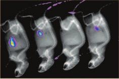
4 Fluorescence X-ray Merged Fluorescence imaging X-ray imaging Images are merged using KODAK Molecular Imaging Software, allowing precise co-registration of the fluorescent molecular biomarker with the anatomical X-ray High performance Enables accurate quantitation of biomolecules of interest in basic research, drug discovery, drug development, and therapeutic monitoring applications utilizing small animals Improves understanding of imaging agent s biodistribution through combined use of time-lapse molecular imaging and digital X-ray imaging Safe in-lab operation the In-Vivo FX System complies with federal safety regulations for cabinet X-ray imaging systems Complete System Includes animal management center, ports, and thermal controls to facilitate imaging of small animals KODAK Molecular Imaging Software provides accurate quantitative analysis, comparative intensity, geometry and positional data. The software also provides annotation capabilities and powerful database tools. KODAK In-Vivo Imaging System F The KODAK In-Vivo Imaging System F provides all the features of the KODAK In-Vivo FX System with the exception of X-ray imaging capabilities. Automated Control: the Pro Series KODAK In-Vivo Systems are available in the Pro configuration, providing fully automated controls that enable reproducibility of protocols and increase workflow efficiencies Automated computer-controlled configuration minimizes set-up time, maximizing efficiency and throughput of measurements The highly accurate automated lens system records the precise f-stop, zoom, and focal plane every time, helping to ensure reproducibility and traceability Smart digital positioning technology operates 15 excitation (In-Vivo FX Pro only) and four emission filters to deliver precision alignment Automated aluminum filters enable control of X-ray wavelengths for optimal X-ray imaging of soft tissue or bone KODAK In-Vivo Multispectral Imaging System FX The system s new computer controlled multispectral tuning of excitation light provides enhanced sensitivity allowing for the identification and separation of multiple fluorochromes and the removal of autofluorescence background. The KODAK In-Vivo Multispectral System automatically generates multispectral fluorochrome image cubes with spatially co-registered X-ray and white light images for improved localization of biological markers in vivo. A wide range of excitation wavelengths, from optical through near-infrared, enable optimum imaging of a wide range of fluorochromes and biomarkers. Sophisticated software algorithms remove autofluorescence for improved signal-to-noise and detection Powerful software identifies fluorochromes through excitationbased signature by modeling of data and providing unmixing of the fluorochromes New image capture control software allows complex imaging protocols to be easily established, stored and repeated Automated excitation and emission filter systems with 29 excitation filter positions and four patented wide angle emission filter positions deliver outstanding fluorescent imaging sensitivity and flexibility
5 Specifications In-Vivo F In-Vivo F Pro In-Vivo FX In-Vivo FX Pro In-Vivo Multispectral DXS 4000 DXS 4000 Pro Camera CCD Monochrome interlined CCD Pixel Density 2048 x 2048 pixels Cooling 29 C absolute, thermoelectrically cooled Lens 10x zoom, mm, ƒ2.8 Manual Automated Manual Automated Automated Manual Automated Illumination Source 150W Halogen (standard), 175W Xenon (optional) Xenon Fluorescence Selectable multi-wavelength, epi-illumination, Halogen Manual 6 position Manual 6 position Selectable multi-wavelength, epi-illumination, Xenon Optional with manual 6 position Automated 15 position filter wheel Optional 6 position Automated 15 position filter wheel Automated 29 position filter wheel White Light Epi-illumination Transillumination Digital X-ray Energy Range Approximately kvp Maximum Current Approximately 150 ua Spot Size < 50 U Target Material Tungsten Window Filtration Beryllium Cone of Illumination >33 degrees Filtration Aluminum 2 filters 4 automated filters 4 automated filters 2 filters 4 automated filters Excitation Filters Included w/system 18 mm (ex465, ex535, ex625, and ex720) 25 mm (ex390, ex430, ex470, ex510, ex530, ex550, ex590, ex610, ex630, ex670, ex690, ex710, ex730, ex770) 25 mm (ex390, ex410, ex420, ex430, ex440, ex450, ex460, ex470, ex480, ex490, ex500, ex510, ex520, ex530, ex540, ex550, ex560, ex570, ex590, ex600, ex610, ex630, ex650, ex670, ex690, ex710, ex730, ex770) Available Filters, 18mm ex385, ex415, ex430, ex465, ex475, ex515, ex535, ex545, ex555, ex610, ex625, ex635, ex710, ex720, ex 730, ex765 Available Filters, 25mm 10 nm increments from 390 nm to 770 nm Emission Filters Included w/system em535wa, em600wa, em700wa, em790wa em535wa, em600wa, em700wa, em790wa, ex750, ex830 Standard Accessory em440wa, em535wa, em570wa, em600wa, em670wa, em700wa, em750wa, em790wa, em830wa Performance Specifications Imaging Area 2 x 2 cm to 20 x 20 cm, continuous zoom Resolution 10 micron/pixel Pixel Size 7.4 µm Data Acquisition 16-bit single capture n-bit data acquistion Dark Current Noise e-pixels/sec Read Noise <9-rms (nominal) Dynamic Range >4.0 orders of magnitude Binning 1x2, 2x2, 1x4, 2x4, 4x4, 1x8, 2x8, 4x8, 8x8, 16x16 Exposure Modes Single Capture: sec 100 min (X-ray min sec) Multiple Capture: sec 100 min, 32 accumulations max Progressive Exposure: sec 100 min per frame, minimum increment = 0.01 sec Time Lapse Exposure: sec 100 min per exposure, minimum interval = 0.1 sec Animal Management Animal Management Chambers Thermal Control Module Atmospheric Ports System Requirements Interface IEEE 1394 (FIREWIRE) Operating Systems WINDOWS 2000/XP MACINTOSH OS X Power Requirements 120 VAC, 7A 230 VAC, 3.5A
 18F Radioisotopic image co-registered with X-ray image Product")


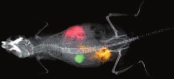
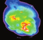
6 Imaging capabilities include: Multispectral (fluorochrome unmixing) Fluorescent optical image co-registered with white light mouse image X-ray of mouse knee joint (zoomed) Three fluorescent images co-registered on X-ray image of a mouse Luminescence in a 96 well plate (zoomed) 18F Radioisotopic image co-registered with X-ray image Product Selection Chart White Light Imaging Luminescence Multi-wavelength Fluorescence nm Multispectral Imaging (Fluorochrome unmixing) Radioisotopic Imaging In-Vivo F Series In-Vivo FX Series In-Vivo Multispectral DXS Systems X-ray Imaging While KODAK Image Stations and KODAK In-Vivo Systems can be used for in vivo and in vitro molecular imaging of materials, researchers should be aware that the methods of preparing and viewing the materials for molecular imaging may be subject to various patent rights. All images were captured using KODAK Molecular Imaging System Technology. Find out more For more information, to request pricing, an in-lab demo, or to place an order, call , exp. code 7. Outside the U.S.: mi.carestreamhealth.com Carestream Health, Inc. 4 Science Park New Haven, CT Carestream is a trademark of Carestream Health. The Kodak trademark and trade dress are used under license from Kodak. Carestream Molecular Imaging is a division of Carestream Health, Inc. Printed in U.S.A. 3/08 Carestream Health, Inc., 2008
In-Vivo IMAGING SYSTEMS. A complete line of high resolution optical & X-ray systems for pre-clinical imaging
 In-Vivo IMAGING SYSTEMS A complete line of high resolution optical & X-ray systems for pre-clinical imaging In-Vivo Imaging Systems Carestream is a strong, successful, multi-billion dollar, international
KODAK Image Station In-Vivo FX
 KODAK Image Station In-Vivo FX User s Guide IB5410113 07/06 Eastman Kodak Company, 2005-2006. All rights are reserved. No section of this manual may be photocopied, reproduced, translated to another language,
Multi-species Optical and X-ray Imaging System
 IVIS Lumina XRMS Series III P R O D U C T N O T E Pre-clinical in vivo imaging Key Features Optical and X-ray imaging Multi-species imaging including mice and rats High resolution, low dose digital X-ray
Automated Imaging Technology to Simplify Your Workflow!
 Automated Imaging Technology to Simplify Your Workflow! BioSpectrum Imaging System Imaging Made Easy for Chemiluminescence Bioluminescence Colorimetric Fluorescence MegaCam 810 Camera OptiChemi 600 Camera
BioSpectrum Imaging System
 BioSpectrum Imaging System Imaging Made Easy for Chemiluminescence Bioluminescence Colorimetric Fluorescence MegaCam 810 Camera OptiChemi 610 Camera BioChemi 510 Camera GelCam 310 Camera 8.1 megapixel
Ordering Information & Specifications. VisionWorksLS Capabilities. Image Analysis Capabilities
 Ordering Information & Specifications VisionWorksLS Capabilities Each system includes: Camera and lens, darkroom with motorized or manual platform, three emission filters, white light illuminator, choice
FluorChem M MultiFluor System
 FluorChem M MultiFluor System Advancing Effortless Multiplex Western Blot Imaging Multiplex Western Analysis FluorChem M Imaging System FluorChem M sets a new standard for quantitative multiplex Western
Improving the Collection Efficiency of Raman Scattering
 PERFORMANCE Unparalleled signal-to-noise ratio with diffraction-limited spectral and imaging resolution Deep-cooled CCD with excelon sensor technology Aberration-free optical design for uniform high resolution
KODAK Image Station 4000R
 KODAK Image Station 4000R User s Guide IB5426004 04/07 Carestream Health, Inc., 2005-2007. All rights are reserved. No section of this manual may be photocopied, reproduced, translated to another language,
Imagers- Molecular, Cell Standard Operating Procedures
 Bio-Rad ChemiDoc XRS and Image Lab Software Jump to Export Images to other Apps Floid cell imaging station Life technologies Jump to Chemi-luminescence Protocol Imagers- Molecular, Cell Standard Operating
BioSpectrum Imaging System
 BioSpectrum Imaging System Imaging Made Easy for Chemiluminescence Bioluminescence Colorimetric Fluorescence MegaCam 800 Camera OptiCam 600 Camera BioChemi 500 Camera ChemiCam 410 Camera GelCam 310 Camera
In-Vivo Imaging: IVIS Lumina XR. William R. Anderson IVIS Product Specialist
 In-Vivo Imaging: IVIS Lumina XR William R. Anderson IVIS Product Specialist 1 What will be covered? Introduction Principles of optical In Vivo Imaging Key IVIS Hardware components Overview of Living Image
INGENIUS 3. Low cost, high performance gel documentation and analysis
 INGENIUS 3 Low cost, high performance gel documentation and analysis INGENIUS 3 When simplicity and budget matter. The InGenius 3 gel documentation and analysis system is compact, easy to use and offers
INGENIUS 3 LOW COST, HIGH PERFORMANCE GEL DOCUMENTATION AND ANALYSIS
 INGENIUS 3 LOW COST, HIGH PERFORMANCE GEL DOCUMENTATION AND ANALYSIS The InGenius 3 uses a high performance 3m pixel camera. The darkroom assembly is easily connected to a PC. GeneSys image acquisition
Seishi IKAMI* Takashi KOBAYASHI** Yasutake TANAKA* and Akira YAMAGUCHI* Abstract. 2. System configuration. 1. Introduction
 Development of a Next-generation CCD Imager for Life Sciences Research Seishi IKAMI* Takashi KOBAYASHI** Yasutake TANAKA* and Akira YAMAGUCHI* Abstract We have developed a next-generation CCD-based imager
FUSION SOLO S CHEMILUMINESCENCE & OPTIONAL FLUORESCENCE IMAGING WESTERN BLOT IMAGING
 CHEMILUMINESCENCE & OPTIONAL FLUORESCENCE IMAGING WESTERN BLOT IMAGING ULTRA SENSITIVE IMAGING The Fusion Solo is a high-end ultrasensitive scientific optical system, designed to extract the lowest level
Fluorescent Imaging. Description and Theory of Operation. System Components
 Concept Tech Note 4 Fluorescent Imaging Description and Theory of Operation System Components The IVIS Spectrum, IVIS 200 Series Imaging System, and IVIS Lumina offer built-in fluorescence imaging capability
Opterra II Multipoint Scanning Confocal Microscope. Innovation with Integrity
 Opterra II Multipoint Scanning Confocal Microscope Enabling 4D Live-Cell Fluorescence Imaging through Speed, Sensitivity, Viability and Simplicity Innovation with Integrity Fluorescence Microscopy The
X-ray phase-contrast imaging
 ...early-stage tumors and associated vascularization can be visualized via this imaging scheme Introduction As the selection of high-sensitivity scientific detectors, custom phosphor screens, and advanced
ChemiDoc-It Imaging System
 ChemiDoc-It Imaging System Ultra dark chamber and highly sensitive, scientific-grade CCD camera for chemiluminescence imaging ChemiDoc-It darkroom is light tight creating optimum imaging conditions for
Data. microcat +SPECT
 Data microcat +SPECT microcat at a Glance Designed to meet the throughput, resolution and image quality requirements of academic and pharmaceutical research, the Siemens microcat sets the standard for
Redefining Gel and Blot Imaging
 Redefining Gel and Blot Imaging PXi AND PXi TOUCH Gel and blot imaging made easy Syngene imaging systems are recognised world-wide as high quality, high performance instruments for the capture and analysis
F U S I O N F X 7 S A L E S & M A R K E T I N G T O O L S
 F U S I O N F X 7 S A L E S & M A R K E T I N G T O O L S 1/19 S A L E S & M A R K E T I N G T O O L S 1- Sales tools 1.1 Product introduction 1.2 Specifications 1.3 Market positioning 1.4 Tender unique
BIO IMAGING. Choose your application of STELLA 2000 STELLA 3200 STELLA BIO Image. light source. light source. light source.
 www.raytest.com Choose your application of BIO IMAGING STELLA 2000 STELLA 3200 STELLA 8300 CCD-camera 2048 x 2048 2184 x 1472 pixel pixel 3326 x 2505 pixel light source uv-light table uv-light table uv-light
Camera Overview. Digital Microscope Cameras for Material Science: Clear Images, Precise Analysis. Digital Cameras for Microscopy
 Digital Cameras for Microscopy Camera Overview For Materials Science Microscopes Digital Microscope Cameras for Material Science: Clear Images, Precise Analysis Passionate about Imaging: Olympus Digital
Development of a Next-Generation Laser-Scanner System for Life Science Research
 Development of a Next-Generation Laser-Scanner System for Life Science Research Masaki TAKAMATSU* Yasutake TANAKA* Takashi KOBAYASHI* Hiromi ISHIKAWA* and Akira YAMAGUCHI* Abstract We developed a next-generation
Camera Overview. Digital Microscope Cameras for Material Science: Clear Images, Precise Analysis. Digital Cameras for Microscopy
 Digital Cameras for Microscopy Camera Overview For Materials Science Microscopes Digital Microscope Cameras for Material Science: Clear Images, Precise Analysis Passionate about Imaging: Olympus Digital
C A P A B I L I T I E S 4 I The range 6 I Performance and innovations 8 I Advanced technologies 10 I Applications 12 I Software
 C A P A B I L I T I E S 4 I The range 6 I Performance and innovations 8 I Advanced technologies 10 I Applications 12 I Software S Y S T E M S 16 I Fusion Spectra 18 I Fusion FX 20 I Fusion SL 22 I Fusion
SMALL ANIMAL IMAGING SYSTEM OV110
 SMALL ANIMAL IMAGING SYSTEM OV110 Olympus in vivo fluorescence molecular imaging systems: enhanced performance for translational research. Low magnification A high performance fluorescence imaging From
Synergy NEO HTS Multi-Mode Microplate Reader
 MICROPLATE READERS Synergy NEO HTS Multi-Mode Microplate Reader The smart alternative for today s assay development and screening applications. The Smarter HTS Reader Synergy NEO is designed for today
T:GENIUS GEL IMAGING AT A TOUCH
 T:GENIUS GEL IMAGING AT A TOUCH The T:Genius is an integrated system for DNA and protein analysis and gel documentation. Based on the successful Syngene gel documentation range, the T:Genius features an
CAMAG TLC VISUALIZER 2
 CAMAG TLC VISUALIZER 2 Professional Imaging and Documentation System for TLC/HPTLC Chromatograms with a new Digital CCD Camera, connected by USB 3.0 WORLD LEADER IN PLANAR CHROMATOGRAPHY Visualization,
GE Healthcare. Senographe 2000D Full-field digital mammography system
 GE Healthcare Senographe 2000D Full-field digital mammography system Digital has arrived. The Senographe 2000D Full-Field Digital Mammography (FFDM) system gives you a unique competitive advantage. That
BioSpectrum Imaging System
 BioSpectrum Imaging System Advanced and automated high resolution system for chemiluminescent, bioluminescent, fluorescent and colorimetric imaging Scientific Grade Cameras are housed in the top of the
AxioCam MRc 5 A World of Digital Possibilities
 Microscopy from Carl Zeiss AxioCam MRc 5 A World of Digital Possibilities More flexibility and more performance in microscope camera technology Impressive Performance A trend setter in digital microscopy,
Gel Documentation and Analysis Automated imaging
 Gel Documentation and Analysis Automated imaging GEL IMAGING AND ANALYSIS Automated imaging for all your Syngene imaging systems are recognised world-wide as high quality, high performance instruments
OLYMPUS Digital Cameras for Materials Science Applications: Get the Best out of Your Microscope
 Digital Cameras for Microscopy Camera Overview For Materials Science Microscopes OLYMPUS Digital Cameras for Materials Science Applications: Get the Best out of Your Microscope Passionate About Imaging
Visibility of Detail
 Visibility of Detail Radiographic Quality Quality radiographic images represents the, and information is for diagnosis. The of the anatomic structures and the accuracy of their ( ) determine the overall
G BOX. Gel Documentation and Analysis Automated imaging
 G BOX Gel Documentation and Analysis Automated imaging GEL IMAGING AND ANALYSIS Automated imaging for all your applications Syngene imaging systems are recognised world-wide as high quality, high performance
GEL DOCUMENTATION AND ANALYSIS Automated imaging
 GEL DOCUMENTATION AND ANALYSIS Automated imaging GEL IMAGING AND ANALYSIS Automated imaging for all your applications Syngene imaging systems are recognised world-wide as high quality, high performance
Pixel shift in fluorescence microscopy
 Pixel shift in fluorescence microscopy 1. Introduction Multicolor imaging in fluorescence microscopy is typically performed by sequentially acquiring images of different colors. An overlay of these images
MULTI FLUORESCENCE AND CHEMILUMINESCENCE IMAGING SYSTEM DETECTION WITH A DIFFERENCE
 MULTI FLUORESCENCE AND CHEMILUMINESCENCE IMAGING SYSTEM DETECTION WITH A DIFFERENCE REAL IMAGING FOR REAL SCIENTISTS Western blot and gel imaging remain the cornerstones of life science research. With
AxioCam HR Success Through Performance
 Microscopy from Carl Zeiss AxioCam HR Success Through Performance The high-resolution camera for digital documentation Superior performance for research and routine work brilliant quality documentation
Redefining gel and blot imaging
 Redefining gel and blot imaging PXi AND PXi TOUCH Gel and blot imaging made easy Syngene imaging systems are recognised world-wide as high quality, high performance instruments for the capture and analysis
Compatible with Windows 8/7/XP, and Linux; Universal programming interfaces for easy custom programming.
 NIRvana: 640LN The NIRvana: 640LN from Princeton Instruments is a scientific-grade, deep-cooled, large format InGaAs camera for low-light scientific SWIR imaging and spectroscopy applications. The camera
We attempted to separate the two dyes by acquiring images using a single excitation wavelength and just two emission wavelengths.
 TN437: Spectral Separation of monochrome images using Volocity 4.0 Introduction Spectral Separation is a technique that allows the user to separate images containing data from more than one fluorochrome
Life Science Instrumentation. New Generation. Light Sheet Fluorescence Microscope. Alph
 Life Science Instrumentation Light Sheet Fluorescence Microscope New Generation Alph Modular Light Sheet Microscope Alpha 3 is a new generation of light sheet fluorescence microscope addressing the needs
Multispectral. imaging device. ADVANCED LIGHT ANALYSIS by. Most accurate homogeneity MeasureMent of spectral radiance. UMasterMS1 & UMasterMS2
 Multispectral imaging device Most accurate homogeneity MeasureMent of spectral radiance UMasterMS1 & UMasterMS2 ADVANCED LIGHT ANALYSIS by UMaster Ms Multispectral Imaging Device UMaster MS Description
Practical work no. 3: Confocal Live Cell Microscopy
 Practical work no. 3: Confocal Live Cell Microscopy Course Instructor: Mikko Liljeström (MIU) 1 Background Confocal microscopy: The main idea behind confocality is that it suppresses the signal outside
Scanning Electron Microscopy. EMSE-515 F. Ernst
 Scanning Electron Microscopy EMSE-515 F. Ernst 1 2 Scanning Electron Microscopy Max Knoll Manfred von Ardenne Manfred von Ardenne Principle of Scanning Electron Microscopy 3 Principle of Scanning Electron
High Resolution BSI Scientific CMOS
 CMOS, EMCCD AND CCD CAMERAS FOR LIFE SCIENCES High Resolution BSI Scientific CMOS Prime BSI delivers the perfect balance between high resolution imaging and sensitivity with an optimized pixel design and
2D Protein Gel Image Capture and Analysis
 2D Protein Gel Image Capture and Analysis the way you want it S Y N G E N E A DIVISION OF THE SYNOPTICS GROUP 2D protein gel generation, image capture and analysis systems Syngene has a worldwide reputation
Very short introduction to light microscopy and digital imaging
 Very short introduction to light microscopy and digital imaging Hernan G. Garcia August 1, 2005 1 Light Microscopy Basics In this section we will briefly describe the basic principles of operation and
Image Capture TOTALLAB
 1 Introduction In order for image analysis to be performed on a gel or Western blot, it must first be converted into digital data. Good image capture is critical to guarantee optimal performance of automated
Chemiluminescence- and Fluorescence-Imager Celvin S
 Chemiluminescence- and Fluorescence-Imager Chemiluminescence on Western Blots Fluorescent staining of proteins Fluorescent staining of DNA Chemiluminescence/Fluorescence in multi-well plates Colorimetrically-stained
Specifications Summary 1. Array Size (pixels) Pixel Size. Sensor Size. Pixel Well Depth (typical) 95,000 e - 89,000 e -
 Pixel Size. Sensor Size. Pixel Well Depth (typical) 95,000 e - 89,000 e -") Apogee Alta Series System Features 1 High Resolution Sensor 1.0 Megapixel sensor with 13 mm pixels delivers a large field of view with high resolution. Programmable TE cooling down to 50 o C below ambient
Apogee Alta Series System Features 1 High Resolution Sensor 1.0 Megapixel sensor with 13 mm pixels delivers a large field of view with high resolution. Programmable TE cooling down to 50 o C below ambient
Maximizing clinical outcomes
 Maximizing clinical outcomes Digital Tomosynthesis Dual Energy Subtraction Automated Long Length Imaging Improved image quality at a low dose Xray Xray Patented ISS capture technology promotes high sensitivity
Camera Overview. Olympus Digital Cameras for Materials Science Applications: For Clear and Precise Image Analysis. Digital Cameras for Microscopy
 Digital Cameras for Microscopy Camera Overview For Materials Science Microscopes Olympus Digital Cameras for Materials Science Applications: For Clear and Precise Image Analysis Passionate about Imaging
Gel Documentation and Analysis the way you want it S Y N G E N E A DIVISION OF THE SYNOPTICS GROUP
 Gel Documentation and Analysis the way you want it S Y N G E N E A DIVISION OF THE SYNOPTICS GROUP Syngene Gel Documentation and Analysis Syngene has long been associated with innovations in gel documentation.
IMAGE SENSOR SOLUTIONS. KAC-96-1/5" Lens Kit. KODAK KAC-96-1/5" Lens Kit. for use with the KODAK CMOS Image Sensors. November 2004 Revision 2
 KODAK for use with the KODAK CMOS Image Sensors November 2004 Revision 2 1.1 Introduction Choosing the right lens is a critical aspect of designing an imaging system. Typically the trade off between image
Light Microscopy. Upon completion of this lecture, the student should be able to:
 Light Light microscopy is based on the interaction of light and tissue components and can be used to study tissue features. Upon completion of this lecture, the student should be able to: 1- Explain the
Multifluorescence The Crosstalk Problem and Its Solution
 Multifluorescence The Crosstalk Problem and Its Solution If a specimen is labeled with more than one fluorochrome, each image channel should only show the emission signal of one of them. If, in a specimen
BD LSRFortessa X-20. Special Order Product. Designed for limited space and boundless potential
 BD LSRFortessa X-2 Special Order Product Designed for limited space and boundless potential Next generation high performance cell analyzer The BD LSRFortessa X-2 cell analyzer delivers high performance,
Invisible sophistication. Visible simplicity. CS Welcome to the simplicity of compact panoramic imaging
 Invisible sophistication. Visible simplicity. CS 8100 Welcome to the simplicity of compact panoramic imaging Introducing the CS 8100 The Carestream Dental Factor Humanized technology We keep our technology
TEM Cameras. Digital Cameras for Electron Microscopy
 Digital Imaging Solutions TEM Cameras Side- and bottom-mounted TEM cameras Digital Cameras for Electron Microscopy IMAGING SOLUTIONS FOR ELECTRON MICROSCOPY. BASED ON OPTO-DIGITAL KNOW-HOW. DESIGNED BY
ProX Intraoral X-ray. PLANMECA is proud to introduce a new intraoral X-ray unit to its comprehensive collection of imaging products- the ProX.
 The premium intraoral X-ray unit... ProX Intraoral X-ray PLANMECA is proud to introduce a new intraoral X-ray unit to its comprehensive collection of imaging products- the ProX. This advanced unit provides
Products - Microarray Scanners - Laser Scanners - InnoScan 900 Series and MAPIX Software
 Products - Microarray Scanners - Laser Scanners - InnoScan 900 Series and MAPIX Software Arrayit offers the world s only next generation microarray scanning technology, with proprietary rotary motion control,
Section lll: SM Series Spectrometer. ometers SPECTRAL PRODUCTS
 Section lll: SM Series ometers SPECTROMETERS SM200 OEM Packaged Fiber Optic CCD SM240 Hand-Held CCD SM241 Near Infrared Enhanced CCD AD300 Back Thinned CCD Tunable, TE Cooled SM300 Fluorencenced/Raman
New Benchmark of Molecular Image Software Package
 Molecular Imager New Benchmark of Molecular Image Software Package Magic software package was developed under the division of the Wealtec imaging family group. It is a professional and powerful 1D package
SpectraMax i3x Multi-Mode Detection Platform. Explore a wealth of applications in one future-ready system
 SpectraMax i3x Multi-Mode Detection Platform Explore a wealth of applications in one future-ready system Benefits User-upgradeable application modules including cellular imaging Sensitivity across spectrum
ODYSSEY Fc IMAGING SYSTEM
 ODYSSEY Fc IMAGING SYSTEM One System, Two Detection Methods Infrared Fluorescence and Chemiluminescence Quantitative, Two-Color Western Blots Chemiluminescence Detection wide Linear Dynamic Range no Film
Why and How? Daniel Gitler Dept. of Physiology Ben-Gurion University of the Negev. Microscopy course, Michmoret Dec 2005
 Why and How? Daniel Gitler Dept. of Physiology Ben-Gurion University of the Negev Why use confocal microscopy? Principles of the laser scanning confocal microscope. Image resolution. Manipulating the
HoloMonitor M4. For powerful discoveries in your incubator
 HoloMonitor M4 For powerful discoveries in your incubator HoloMonitor offers unique imaging capabilities that greatly enhance our understanding of cell behavior, previously unachievable by other technologies
Luminescent Background Sources and Corrections
 Concept Tech Note 1 Luminescent Background Sources and Corrections The background sources of light from luminescent images are inherently very low. This appendix discusses sources of background and how
Minimizes reflection losses from UV to IR; No optical losses due to multiple optical surfaces; Optional AR coating and wedge windows available.
 SOPHIA: 2048B The SOPHIA : 2048B camera from Princeton Instruments (PI) is fully integrated, ultra-low noise 2048 x 2048, 15 µm pixel CCD camera designed expressly for the most demanding quantitative scientific
Automated Cellular Imaging and Analysis System
 Automated Cellular Imaging and Analysis System CELLULAR IMAGING AND ANALYSIS FOR SCREENING AUTOMATED ACQUISITION AUTOMATED ANALYSIS HIGH RESOLUTION The ImageXpress TM 5000A automated cellular imaging and
Nikon. King s College London. Imaging Centre. N-SIM guide NIKON IMAGING KING S COLLEGE LONDON
 N-SIM guide NIKON IMAGING CENTRE @ KING S COLLEGE LONDON Starting-up / Shut-down The NSIM hardware is calibrated after system warm-up occurs. It is recommended that you turn-on the system for at least
1. Editorial. N 9 June Content
 N 9 June 2010 Content 1. Editorial 2. Timelapse: news and updates 3. n vivo rodent imaging setup available in Epalinges 4. 2010 Workshops 5. Spotlight on mage Stitching 1. Editorial We welcome new and
NPTEL VIDEO COURSE PROTEOMICS PROF. SANJEEVA SRIVASTAVA
 HANDOUT LECTURE-31 MICROARRAY WORK-FLOW: IMAGE SCANNING AND DATA PROCESSING Slide 1: This module contains the summary of the discussion with Mr. Pankaj Khanna, an application specialist from Spinco Biotech,
BIOMOLECUL AR IMAGING CHEMILUMINESCENCE BIOLUMINESCENCE FLUORESCENCE MULTIPLEXING VISIBLE IMAGING
 BIOMOLECUL AR IMAGING CHEMILUMINESCENCE BIOLUMINESCENCE FLUORESCENCE MULTIPLEXING VISIBLE IMAGING Content About us p. 4 Our range p. 6 Advanced systems p. 8 Alliance Chroma p. 10 Alliance 7 series p. 12
bioteknika T:GENIUS GEL IMAGING AT A TOUCH
 bioteknika T:GENIUS GEL IMAGING AT A TOUCH The T:Genius is an integrated system for DNA and protein analysis and gel documentation. Based on the successful Syngene gel documentation range, the T:Genius
U GENIUS. Gel imaging at a touch
 U GENIUS 3 Gel imaging at a touch U:GENIUS 3 Simply Genius. Designed to make your gel imaging simple, quick and easy. No set up, no external computer - just a complete imaging system for all your 1D needs.
Camera Overview. Digital Microscope Cameras for Material Science: Clear Images, Precise Analysis. Digital Cameras for Microscopy
 Digital Cameras for Microscopy Camera Overview For Materials Science Microscopes Digital Microscope Cameras for Material Science: Clear Images, Precise Analysis Passionate about Imaging: Olympus Digital
QE65000 Spectrometer. Scientific-Grade Spectroscopy in a Small Footprint. now with. Spectrometers
 QE65000 Spectrometer Scientific-Grade Spectroscopy in a Small Footprint QE65000 The QE65000 Spectrometer is the most sensitive spectrometer we ve developed. Its Hamamatsu FFT-CCD detector provides 90%
Overview. About other software. Administrator password. 58. UltraVIEW VoX Getting Started Guide
 Operation 58. UltraVIEW VoX Getting Started Guide Overview This chapter outlines the basic methods used to operate the UltraVIEW VoX system. About other software Volocity places great demands on the computer
contents TABLE OF The SECOM platform Applications - sections Applications - whole cells Features Integrated workflow Automated overlay
 S E C O M TABLE OF contents The SECOM platform 4 Applications - sections 5 Applications - whole cells 8 Features 9 Integrated workflow 12 Automated overlay ODEMIS - integrated software Specifications 13
The Bruker In-Vivo Xtreme II System for Flexible and Sensitive Multimodal Preclinical Imaging
 The Bruker In-Vivo Xtreme II System for Flexible and Sensitive Multimodal Preclinical Imaging Todd A. Sasser 1, Joshua McHattan 2, Jens Waldeck 3. Author Information: 1 Bruker Biospin Inc., 44 Manning
Precision Performance Power
 ODYSSEY HF SERIES ULTRA High Frequency X-Ray Technology Precision Performance Power Innovations in Digital Imaging. TM STEP 1 Select anatomical region STEP 2 Select anatomical view STEP 3 Ready for exposure
ODYSSEY HF SERIES. ULTRA High Frequency X-Ray Technology. Precision... Performance... Power. Innovations in Digital Imaging.
 ODYSSEY HF SERIES ULTRA High Frequency X-Ray Technology Precision... Performance... Power Innovations in Digital Imaging. TM STEP 1 Select anatomical region STEP 2 Select anatomical view STEP 3 Ready for
Ideal for display mura (nonuniformity) evaluation and inspection on smartphones and tablet PCs.
 evaluation and inspection on smartphones and tablet PCs.") 2D Color Analyzer 8 Ideal for display mura (nonuniformity) evaluation and inspection on smartphones and tablet PCs. Accurately and easily measures the distribution of luminance and chromaticity. Advanced
2D Color Analyzer 8 Ideal for display mura (nonuniformity) evaluation and inspection on smartphones and tablet PCs. Accurately and easily measures the distribution of luminance and chromaticity. Advanced
3. are adherent cells (ie. cells in suspension are too far away from the coverslip)
") Before you begin, make sure your sample... 1. is seeded on #1.5 coverglass (thickness = 0.17) 2. is an aqueous solution (ie. fixed samples mounted on a slide will not work - not enough difference in refractive
Before you begin, make sure your sample... 1. is seeded on #1.5 coverglass (thickness = 0.17) 2. is an aqueous solution (ie. fixed samples mounted on a slide will not work - not enough difference in refractive
Proudly serving laboratories worldwide since 1979 SPECIFICATIONS
 www.ietltd.com Proudly serving laboratories worldwide since 1979 SPECIFICATIONS Scan RDI Specifications System Components Main analytical console Laser Module CRT Printer Data Manager Motorized stage (option)
Technical Benefits of the
 innovation in microvascular assessment Technical Benefits of the Moor Instruments moorflpi-2 moorflpi-2 More Info: Measurement Principle laser speckle contrast analysis Measurement 85nm Laser Wavelength
Oriel MS260i TM 1/4 m Imaging Spectrograph
 Oriel MS260i TM 1/4 m Imaging Spectrograph MS260i Spectrograph with 3 Track Fiber on input and InstaSpec CCD on output. The MS260i 1 4 m Imaging Spectrographs are economical, fully automated, multi-grating
U:GENIUS S Y N G E N E. Gel imaging at a touch A DIVISION OF THE SYNOPTICS GROUP
 U:GENIUS Gel imaging at a touch S Y N G E N E A DIVISION OF THE SYNOPTICS GROUP U:GENIUS Simply Genius. Designed to make your gel imaging simple, quick and easy. No set up, no external computer - just
MS260i 1/4 M IMAGING SPECTROGRAPHS
 MS260i 1/4 M IMAGING SPECTROGRAPHS ENTRANCE EXIT MS260i Spectrograph with 3 Track Fiber on input and InstaSpec IV CCD on output. Fig. 1 OPTICAL CONFIGURATION High resolution Up to three gratings, with
Spectrum 400. FT-IR and FT-NIR Spectrometer. There is only one answer.
 Spectrum 400 FT-IR and FT-NIR Spectrometer There is only one answer. The latest innovation in PerkinElmer s long history of IR technology leadership For over 60 years, PerkinElmer has been the world leader
Low Voltage Electron Microscope
 LVEM 25 Low Voltage Electron Microscope fast compact powerful Delong America FAST, COMPACT AND POWERFUL The LVEM 25 offers a high-contrast, high-throughput, and compact solution with nanometer resolutions.
LVEM 25. Low Voltage Electron Mictoscope. fast compact powerful
 LVEM 25 Low Voltage Electron Mictoscope fast compact powerful FAST, COMPACT AND POWERFUL The LVEM 25 offers a high-contrast, high-throughput, and compact solution with nanometer resolutions. All the benefits
Specifications. Offers the best spatial resolution for multi-stripe spectroscopy. Provides the user the choice of either high accuracy slit mechanism
 SpectraPro Series Monochromators and Spectrographs The PI/Acton SpectraPro Series imaging spectrographs and monochromators represent the latest advance in the industry-standard SpectraPro family. The SpectraPro