PACS Fundamentals. By: Eng. Valentino T. Mvanga Ministry of Health and Social Welfare Tanzania
|
|
|
- Aldous Casey
- 6 years ago
- Views:
Transcription

1 PACS Fundamentals By: Eng. Valentino T. Mvanga Ministry of Health and Social Welfare Tanzania 1
2 Learning Goals To Understand the importance of PACS To Understand PACS infrastructure requirement Introduction to other clinical information system 2
3 Objectives Introduction to picture archiving and communication systems (PACS). Compare and contrast the various types of PACS display workstations. Differentiate between the different types of digital imaging workflow. Define system architecture and recognize the three major models. Summarize the common functions found on a PACS workstation. Describe the situations and users that might use advanced PACS workstation functions. 3
4 Key Terms Archive Client/server-based system DICOM Display workstation Distributed system File room workstation Hanging protocol Navigation functions PACS QC station Reading station Review station Soft copy System architecture Teleradiology Web-based system Workflow 4
5 Introduction PACS Picture Archiving Communication Systems 5
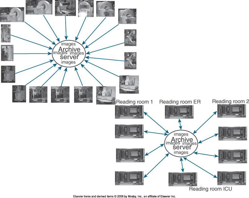
6 Fundamentals PACS consists of the following: Digital acquisition (Picture) Display workstations Storage devices (Archiving) Components are interconnected through an intricate network. (Communication) PACS is the electronic version of the radiologist s reading room and the file room. 6

7 Typical PACS Design 7
8 Fundamentals First PACS Early 1980s Served one single modality Large research institutions Most developed by scientists in those institutions Later Vendors became more involved. Proprietary systems were developed. Standardization 8
9 DICOM First version was completed in DICOM Digital imaging and communications in medicine. Universally accepted standard Laid the groundwork for the future development of integrated PACSs. Now every modality and PACS communicates via DICOM. Each vendor and modality boasts DICOM compatibility. Each DICOM statement must be read carefully to determine the extent of the compatibility. 9

10 Components PACS can be broken into three fundamental parts: 10
11 Image Acquisition Images are acquired in a digital format: Ultrasound Early ultrasound mini-pacs networks became a norm in many hospitals Computed Tomography (CT) As the images sets increased in number, this necessitated the transition to soft-copy reading Magnetic Resonance Imaging (MRI) As with CT as the numbers of images increased, reading on a monitor became a good alternative. Nuclear medicine Computed radiography Mammography 11

12 Display Workstations Display workstation is any computer used to view a digital image. Most interactive part of a PACS. Used inside and outside of the radiology department. 12
13 Display Workstations Display station. Receives images from archive or various radiology modalities Presents images to be viewed Workstation has some sort of PACS application software. Some may have advanced software with additional image processing capabilities. 13

14 Archive Servers File room of the PACS Consists of the following: Database server or image manager Short-term and long-term storage Workflow manager Central part of the PACS Houses all of the historic and current data May also serve as the centralized node that receives all images before interpretation 14
15 Workflow Workflow How a process is done step by step or how a task is completed How one completes an exam from order entry to transcribed report Exact workflow different in every radiology department 15
16 Generic Workflow Digital is similar but still different from film-based system. 16
17 System Architecture American National Standards Institute and the Institute of Electrical and Electronics Engineers Definition of system architecture: Basic organization of a system come to life in its components, their associations to each other and the environment, and the principles leading its design and development 17
18 System Architecture In other words, System architecture is the hardware and software infrastructure of the systems workflow. In PACS the system architecture normally consists of the following: Acquisition devices Storage and display workstations Image management system 18
19 PACS Architecture Three common PACS architectures Client/server-based Distributed systems Web-based systems 19
20 Client/Server-Based Systems Images are sent to archive server Display workstation functions as a client of the archive server Accesses images based on a centralized worklist. Person at the client chooses a name from the list. Archive server sends the image data to the client. The image data is only on the client while viewing. Most systems allow basic image manipulation at the client. Changes are saved on the archive server. 20
21 Client/Server-Based Systems 21
22 Client/Server-Based Systems Advantages: Any exam sent to the PACS is available anywhere without other interventions. Only one person can open the study with the intent to read it. Others that open the study receive a message that the study is open and being read. There is no need to pull or send historic images to a particular workstation because the old studies are available with the new on the archive. 22
23 Client/Server-Based Systems Disadvantages: The archive server is seen as a single point of failure. If the archive goes down, the entire system is down and no image movement can take place. Newly acquired images must remain at the modality until the archive is up and can receive the images. System is network dependent. Images are flying back and forth between the archive and the workstations. Network can become bogged down. Archive server is handling many requests at once and can become bottlenecked. 23

24 Distributed Systems Distributed systems are also known as distributed or stand-alone models. Acquisition modalities send the images to a designated reading station and possibly to review stations. In some systems the images are sent from the modality to the archive server, and the archive server distributes the images to the designated workstation. 24
25 Distributed Systems 25
26 Distributed Systems Reading station designations may be designed on radiologist s reading preferences. Example: MRI may send to one station CT sends to another Or all cross-sectional neurologic images may be sent to one station, whereas all body imaging may be sent to another Designation is decided after extensive workflow observations. 26
27 Distributed Systems Workstations can query and retrieve images from the archive. All images are locally stored. Images are then sent to the archive server once the images have been read. Images remain on local hard drive of workstation until they are deleted by user or system rules. 27
28 Distributed Systems Advantages: If the archive server goes down, local reading at the workstations is not interrupted. After archive comes back up, the images that have been changed and signed off are automatically forwarded to the archive to be saved. PACS data is less likely to be lost because of multiple copies in various locations. System is less dependent on network for speed. User can work on one exam while workstation is pulling next exam to be read. Workstation can fetch historic images based on rules set up by the user. Fetching can be done while other exams are being read. 28
29 Distributed Systems Disadvantages: Personnel rely heavily on system to perform image distribution correctly. If distribution is wrong, then prefetching of historic exams will be wrong. Each workstation has a different worklist; only one person at a time can work on that list. It can be inconvenient to read additional studies. Radiologist would have to move to another workstation to read the images. Users must depend on query and retrieve function when nonscheduled exams arrive at workstation. It is possible for two radiologists to be reading the same exam and not know it until they try to start dictation. Paper requisition becomes an important piece of information. 29
30 Web-Based Systems Web-based systems are similar to a client/server systems. Biggest differences are that images and application software are held centrally and loads to the client display. Only images are held at the archive. 30

31 Web-Based Systems 31
32 Web-Based Systems Advantages: Hardware at the client can be anything that will support an appropriate web browser. This condition allows for greater flexibility with hardware. This can be a disadvantage because low-end PCs can be used and the image displays (monitors) may not be of diagnostic quality. The same application can be used on-site and at home in teleradiology situations. Teleradiology is the reading of images from outside the hospital walls. 32
33 Web-Based Systems Disadvantages: System functionality may be limited because of software not being locally installed. Bandwidth of the network connection limits the amount of data that can be transmitted. Some programs are large and cannot be transmitted via a network. Network is the biggest obstacle to performance. 33
34 Display Workstations Most interactive part of PACS Hands-on component Consists of the following: Monitor Computer with a mouse and keyboard Different hardware requirements for each system 34


35 Display Workstations Conventional film/screen radiography uses large multiviewer lightboxes. With early PACS, radiologists thought that they needed four to six monitors. Now, as radiologists have become more comfortable, the number of monitors has dropped to an average of two. Drop can be attributed to continued development of viewing software and better hardware. 35

 Liquid crystal display (LCD)")
36 Monitor Display Workstations One of the most important elements Several types of monitors Cathode ray tube (CRT) Liquid crystal display (LCD) 36
37 Display Workstations Monitor, continued CRT Heavy Puts off a lot of heat Very bright (good thing) Can view from most any angle 37
38 Display Workstations Monitor, continued LCD Dropped in price and has risen in quality. LCD will soon take over PACS display market because of its size, resolution, and lack of heat production. LCD requires less maintenance. LCD gives more light. LCD can be used in areas with a high amount of ambient light. 38

 and digital radiography (DR) images are read")
39 Display Workstations Resolution and orientation of the monitor is a factor in determining which type of monitor is to be used. Most cross-sectional imaging is read on a 1K square monitor. Most computed radiography (CR) and digital radiography (DR) images are read on at least a 2K portrait monitor. 39
40 Display Workstations Number of pixels contained on a display is known as its resolution. More pixels: The higher the resolution, the more information that can be displayed. Resolution also is defined as the process or capability of distinguishing between individual parts of an image that are adjacent. 40
41 Display Workstations Pixels are arranged in a matrix. Common screen resolutions found on today s monitors are the following: (1K) (2K) (3K) (5K) 41
42 Display Workstations Medical displays are generally higher quality than displays for other applications. Radiologists use highest-resolution monitors available for the modality that is being read: Mammography requires a 5K or 5-megapixel resolution. Cross-sectional image only requires a 1K monitor. If referring physician is not the primary doctor reading the exams, a 1K monitor would be sufficient. 42
43 Display Workstations Workstations can be categorized by use: Primary reading stations for radiologists Review stations for referring physicians Technologist quality control (QC) station for technologist review of images Image management station for the file room personnel Each has a specific, main purpose. Workstations are placed in strategic areas near the enduser of that particular need. Workstations may be made up of different hardware depending on demand and need of user and requirements of the software that will be used. 43

44 Radiologist Reading Stations Station is used by a radiologist to make a primary diagnosis. Station will have the highestquality hardware, including best monitor. Computer hardware used depends on the needs of the PACS vendor but usually is robust, with little downtime. 44

45 Radiologist Reading Stations Keyboard and mouse can be customized to needs of department. Many different styles of mice are available. Access to the RIS is nearby. 45
46 Radiologist Reading Stations Dictation system is near or is connected to PACS station. Many systems are integrating the RIS and dictation system within the PACS software. Grouping allows a more seamless workflow with little to no paper. Station streamlines the completion of the study. 46

47 Physician Review Stations Station is a step-down model of the radiologist s reading station. Station may have the same level of software but may reduce some advanced functions. One of the most important features is ability to view current and previous reports with images. Many vendors are integrating the RIS functions with PACS software. 47
48 Physician Review Stations Most referring physicians want to read radiologist s report along with viewing images. Many times, report is more important to them than the images. Software may be loaded on a stand-alone station that is dedicated to viewing images. Or, software may be delivered over a web browser on any personal computer within an office or floor. 48

49 Physician Review Stations In high-volume areas such as the emergency room and intensive care unit, there are dedicated PACS workstations strictly for image viewing. These may have the higher-end monitors, but many may have a lower-end monitor because of the costs. One of the greatest advantages of a PACS is the ability to view the same set of images in multiple locations at one time. Referring physician can pull up the patient s images in the office and read the radiologist s report and then call the radiologist on the phone and consult while both parties are viewing the same set of images. Continuity and speed of patient care has shown improvement with the use of PACS. 49

50 Technologist QC Stations Used to review images after acquisition but before sending them to the radiologist May be used to improve or adjust image-quality characteristics May be used to verify patient demographic information Placed between the CR and DR acquisition modalities as a pass-through to ensure that the images have met the departmental quality standard 50
51 Technologist QC Station Generally has a 1K monitor. Does not have the resolution capabilities of the radiologist s reading station. Care required of technologist when manipulating images not to change the appearance too much from original acquired image. Technologist should consult frequently with the radiologist to ensure quality. Station can also be used to query and retrieve historic images to check previous pathologic conditions or body characteristics. Station can help with the selection of technical factors or procedural protocol. QC station can afford same benefit as pulling the film jacket. 51

52 The File Room Workstation Before PACS, file room was a large open room with endless rows of shelves full of film jackets. Today, it may be as simple as several computers and a dry laser to make copies for outside needs. Workstation may be used to look up exams for a physician or to print copies of images for the patient to take to an outside physician. Many hospitals are moving away from printing films because of the cost. Hospitals are moving toward burning compact disks (CDs) with the patient s images. CDs can be made quickly and at a reduced cost compared with film. 52
53 Common Functions Navigation functions Image manipulation and enhancement functions Image management functions Advanced workstation functions 53

54 Navigation Functions Used to move through images, series, studies, and patients Worklist used to navigate through patient files Customizable for the user 54
55 Navigation Functions Modern PACS software conforms to the windows look and feel: Use of grab bars on the right hand side of windows to scroll through a list Activation of the scroll wheel on the mouse to scroll through the list 55
56 Navigation Functions Mouse is a useful navigation tool. Right mouse button offers many shortcut features in a menu of frequently used tasks and applications. Hanging protocols are available: Each user has the ability to set up a custom hanging protocol. Protocol is defined as how a set of images will be displayed on the monitor. 56
57 Hanging protocols Navigation Functions Example: CT exam is selected. Can be viewed four images on each monitor. CR image is selected. Can be viewed as one image on each monitor. Protocol can also be specified to show the previous exam on one monitor and the current exam on the other. Once set, the most efficient study navigation is determined. 57
58 Study Navigation A study in PACS is the current or previous exam being viewed. Study may consist of two or three single images such as the case with CR and DR. Study may contain several series of images such as the case with MRI. Images can be simply paged through with the scroll wheel or arrows on the keyboard. 58
59 Study Navigation Images can be run through in stacks. Stack mode of scrolling through images made is called cine. Cine means to move through frame by frame of the series of images. Study may have an automatic setting that will run through the images at a preset pace. Cine function is used most often in cross-sectional imaging. 59
60 Study Navigation Icon may be available for the following: To move between a patient s various studies To open the next unread patient file in the worklist To close patient or study icon Closes the active patient or study Pulls up the worklist or moves to the next unread patient Customizable rules can be set up per user to optimize workflow. 60

61 Image Manipulation and Enhancement Functions Tools Window/level May be a default function of the left mouse button. By depressing and holding down the mouse button and moving the mouse up and down and left and right, the window and level can be adjusted. Window represents the range of gray values. Level represents the center value of the range. A change in the window and level appears to change the brightness and contrast of the image. 61

62 Image Manipulation and Enhancement Functions Tools, continued Annotations Annotations are NOT to be used to label left or right to indicate the patient s side. Annotations are used to indicate prone or supine, 30 minutes, upright or flat. Any other image information is appropriate. Radiologist will place arrows or circles around pathologic or questionable areas. 62

63 Image Manipulation and Enhancement Functions Tools, continued Flip and rotate Tool is used to orient the image in the correct anatomic hanging position. Tool is usually a left-to-right flip and a 90-degree clockwise and counterclockwise icon. Use of lead markers is important to ensure that the radiologist is reading the correct side. Digital R and L may not be upheld in court during a legal case because of the ability to mark anywhere on the image and flip and rotate the image into any layout on the screen. 63

64 Image Manipulation and Enhancement Functions Tools, continued Pan, zoom, and magnify Tools are used primarily by the radiologist to increase the size of an area on the image. Magnify usually magnifies a square area of the image. Square can be moved around the image to quickly see various areas magnified. Pan and zoom functions are usually used together. Image is first zoomed up to the desired magnification level. Pan icon is activated. Zoomed image can be moved around to view the different areas of the image. 64

65 Image Manipulation and Enhancement Functions Tools, continued Measurements Various measurement functions are found on a PACS station. Most common is the distance measurement. Size of a pixel is a known measurement, so the software has the ability to measure structures on the image based on this. Another common measurement is an angle measurement. Can give an angle measurement between two structures Commonly used when reading spine studies 65
66 Image Manipulation and Enhancement Functions Tools, continued Measurements Region of interest Measurement tool determines the pixel intensity of a certain area. Each type of tissue or fluid has a different intensity of reading. Radiologist can make a determination whether something is solid or fluid. 66

67 Patient demographics Image Management Functions Patient demographics must be correct. If demographics are not correct at the archive level, the images could be lost. Changes should only be made when the information is absolutely known to be wrong. Many hospitals allow only certain persons the access to change demographics just to keep the errors to a minimum. 67

68 Query/retrieve icon Image Management Functions Used to retrieve on demand any studies from the archive Allows user to query a study on multiple fields Patient s name or identification Date of service Modality Diagnosis code or comment field 68
69 Image Management Functions CD burning option Feature may only be available in the file room to control the CDs that are sent out. HIPAA (Health Insurance Portability and Accountability Act) compliance must also be maintained. Copy and paste Function is used with the web-based systems when creating presentations for conferences. Patient information must be removed from the image before it is placed into a presentation. 69
70 Image Management Functions Print films Printing is usually only done in the file room so that control can be maintained over the printed films for HIPAA purposes and cost reasons. Workstations may be connected to paper printers for quick consults and for medical records. 70
71 Advanced Workstation Functions Advanced functions are usually placed on specialty workstations for the radiologist, and some are found on the technologist QC station to further enhance the images. The following is a bulleted list of some of the most common advanced functions: Multiplanar reconstruction (MPR) Maximum intensity projection and minimum intensity projection (MIP and MinIP) Volume rendering technique Shaded surface display Stitching 71

72 Reading Station Advanced Functions Multiplanar reconstruction (MPR) MPR is one of the most commonly used three-dimensional rendering techniques. When doing a CT scan of a patient, thin axial slices can be acquired of a volume of tissue. Slices can then be loaded into the MPR software, and a reconstruction in another plane can be produced. Most common application is producing coronal images from the axial set to reduce radiation to the patient and scan time at the modality. 72
. Function is commonly performed after injection of contrast medium on CT and MRI studies.")
73 Reading Station Advanced Functions Maximum intensity projection and minimum intensity projection (MIP and MinIP) Function is used to visualize vessels (MIP) and air-filled structures (MinIP). Function is commonly performed after injection of contrast medium on CT and MRI studies. Contrast medium shows areas of strictures and blockages within the vessels. 73

74 Reading Station Advanced Functions Volume rendering technique Function is similar to MIP. Function allows user to assign colors based on the intensity of the tissue. Bone, contrast medium, and organs can be visualized using various colors. Function uses a histogram-type graph to differentiate the various structures. 74

75 Reading Station Advanced Functions Shaded surface display Using a threshold of pixel intensity values, everything below the threshold is removed and everything above is assigned a color and is shown as a threedimensional object. 75

76 Technologist QC Station Advanced Functions Stitching Stitching is used when multiple images need to be put together in one image. Most common application is for full-spine radiographs or a scoliosis series.» Exam was traditionally performed on a 3- foot film and was processed. CR manufacturers have developed a 3- foot CR cassette that contains multiple image plates (IPs).» Each of the IPs is scanned through the reader, and the individual images are sent to the QC workstation.» Software interpolates images and connects them using known markers from the IPs.» Technologist has the ability to adjust the connection of the images. Long leg images are used for leg-length discrepancy. If the special 3-foot cassettes are not available, a radiopaque ruler can be used to ensure that the images are stitched at the right area. 76
77 Image Postprocessing Many other advanced workstation functions are available to be added to the PACS workstation. This is a growing field with advancements coming each year. Specific information about how to perform these procedures can be found in the vendor s user manual. 77
78 PACS integration to CDIS CDIS(Clinical Devices Information System) Medical image/data other than PACS ECG, Patient monitor, Ventilator, defibrillator, Ophthalmology, Function test, Clinical pathology HL7, XML, RS232C and DICOM are the communication formats in use. 78
79 Summary A PACS consists of digital acquisition, display workstations, and storage devices interconnected through an intricate network. Digital imaging and communications in medicine (DICOM) is a universally accepted standard for exchanging medical images between the modality, viewing stations, and the archive. A display workstation is any computer that a health care worker uses to view a digital image, and it is the most interactive part of a PACS. The archive is the central part of the PACS and houses all of the historic data along with the current data being generated. Workflow is how a process is done step by step or how a task is completed. 79
80 Summary System architecture is the basic organization of a system, come to life in its components, their associations to each other and the environment, and the principles leading its design and development, or in other words, system architecture can be defined as the hardware and software infrastructure of the systems workflow. Common system architectures found with a PACS are client/server-based systems, distributed or stand-alone systems, and webbased systems. 80
81 Summary Display stations can be categorized by their means of use, such as primary reading stations for radiologists, review stations for referring physicians, technologist QC stations for technologist review of images, and image management stations for the file room personnel. Many functions are available on a PACS workstation, and each set of functions can be broken down into four categories: navigation functions, image manipulation and enhancement functions, image management functions, and advanced workstation functions. 81
82 Thanking you for your attention 82
COST EFFECTIVE FLAT PANEL DIGITAL RADIOGRAPHY UPGRADE SOLUTIONS
 COST EFFECTIVE FLAT PANEL DIGITAL RADIOGRAPHY UPGRADE SOLUTIONS DRive is a digital imaging DR hardware & Software solution designed for General Radiography of anatomy. It intended to replace film/screen
Touch Screen Technology. Classic WAIV. with Touch Screen Technology. Legendary Reputation...Sensitivity to your budget
 Touch Screen Technology Classic WAIV with Touch Screen Technology Legendary Reputation...Sensitivity to your budget Streamline Workflow For Higher Productivity And Patient Throughput. The CRescendo Classic
SECTION I - CHAPTER 1 DIGITAL RADIOGRAPHY: AN OVERVIEW OF THE TEXT. Exam Content Specifications 8/22/2012 RADT 3463 COMPUTERIZED IMAGING
 RADT 3463 - COMPUTERIZED IMAGING Section I: Chapter 1 RADT 3463 Computerized Imaging 1 SECTION I - CHAPTER 1 DIGITAL RADIOGRAPHY: AN OVERVIEW OF THE TEXT RADT 3463 COMPUTERIZED IMAGING Section I: Chapter
FCR XL-2 and FCR XC-2. High-quality digital x-ray that's perfect for your private practice.
 F U J I F I L M D I G I T A L X - R A Y FCR XL-2 and FCR XC-2. High-quality digital x-ray that's perfect for your private practice. So small, yet so powerful. With a compact footprint of less than 2.5
The Flash IIP Console is the heart of every FCR system. It s designed to maximize productivity in the busiest environments.
 Choose the FCR system that best fits your practice. The FCR XL-2. Perfect for higher-volume environments. It can process up to 94 images per hour yet it fits right into small exam rooms or offices where
IMPAX 6 DISPLAY TOOL LIST
 IMPAX 6 DISPLAY TOOL LIST IMPAX 6.0 TOOLS INDEX A Advance by Image Allows you to scroll from one image or frame to the next Advance by Page Pages through images in a large series, one screen at a time
CR30-ORACLE CR. with Office Viewer. A Revelation in Computed Radiography. Innovations in Digital Imaging. Solution for Quantum Medical Imaging
 CR30-ORACLE CR with Office Viewer A Revelation in Computed Radiography Innovations in Digital Imaging Solution for Quantum Medical Imaging CR30-ORACLE CR with Office Viewer The CR30-ORACLE CR System Package
A Practical Overview of the Clinical and Operational Impact of Computed Radiography(CR) Implementations. Shirley Weddle, RT(R)(M), CIIP, BBA
 Implementations. Shirley Weddle, RT(R)(M), CIIP, BBA") A Practical Overview of the Clinical and Operational Impact of Computed Radiography(CR) Implementations Shirley Weddle, RT(R)(M), CIIP, BBA OBJECTIVES Define Computed Radiography (CR) Discuss CR vendor
A Practical Overview of the Clinical and Operational Impact of Computed Radiography(CR) Implementations Shirley Weddle, RT(R)(M), CIIP, BBA OBJECTIVES Define Computed Radiography (CR) Discuss CR vendor
GE Healthcare. Senographe 2000D Full-field digital mammography system
 GE Healthcare Senographe 2000D Full-field digital mammography system Digital has arrived. The Senographe 2000D Full-Field Digital Mammography (FFDM) system gives you a unique competitive advantage. That
Get more from your images with Symphony Image Processing
 DIRECT RADIOGRAPHY The user-friendly DelWorks image acquisition and processing software possesses a wide range of tools for a variety of image manipulations. Its user interface simplifies every step of
Get more from your images with Symphony Image Processing
 DIRECT RADIOGRAPHY The user-friendly DelWorks image acquisition and processing software provides a wide range of tools for a variety of image enhancements. Its user interface simplifies every step of the
CR35-SP+ Computed Radiography (CR) System. Innovations in Digital Imaging
 System. Innovations in Digital Imaging") CR35-SP+ Computed Radiography (CR) System Innovations in Digital Imaging Solution for Quantum Medical Imaging CR35-SP+ Package An Agfa Healthcare Solution for Quantum Medical Imaging The CR35-SP+ Package
CR35-SP+ Computed Radiography (CR) System Innovations in Digital Imaging Solution for Quantum Medical Imaging CR35-SP+ Package An Agfa Healthcare Solution for Quantum Medical Imaging The CR35-SP+ Package
Clinical and management aspects of digital imaging and PACS
 Clinical and management aspects of digital imaging and PACS พญ.จามร เช อเพชระโสภณ นายกร งส ว ทยาสมาคมแห งประเทศไทย chamareec@gmail.com www.radiologythailand.org Digital imaging Abbreviation and terminology
Grubstr- 6-8 D Vörstetten Tel.: +49(0) Fax: +49(0) Steinhart Medizinsysteme GmbH
 Fax: +49(0) Steinhart Medizinsysteme GmbH") Hipax Diagnostic Workstation Table of Contents 1. PRODUCT DESCRIPTION... 2 2. HIPAX DW MODULES... 2 2.1 DW BASE MODULE... 2 2.1.1 DW BASE MODULE STANDARD (05-010)... 2 2.1.2 DW BASE MODULE LIGHT (05-020)...
Hipax Diagnostic Workstation Table of Contents 1. PRODUCT DESCRIPTION... 2 2. HIPAX DW MODULES... 2 2.1 DW BASE MODULE... 2 2.1.1 DW BASE MODULE STANDARD (05-010)... 2 2.1.2 DW BASE MODULE LIGHT (05-020)...
SECTION I - CHAPTER 2 DIGITAL IMAGING PROCESSING CONCEPTS
 RADT 3463 - COMPUTERIZED IMAGING Section I: Chapter 2 RADT 3463 Computerized Imaging 1 SECTION I - CHAPTER 2 DIGITAL IMAGING PROCESSING CONCEPTS RADT 3463 COMPUTERIZED IMAGING Section I: Chapter 2 RADT
DELWORKS DR MEDICAL. take the next step
 DELWORKS DR MEDICAL take the next step DELWORKS MEDICAL DR If you are thinking of taking the next step to digital radiography, consider a DelWorks Medical DR Retrofit Package, the easy and affordable way
COMPUTED RADIOGRAPHY (CR)
") COMPUTED RADIOGRAPHY (CR) Moving with the time Avi Avner BVSc BSc CVR DVDI MRCVS CR-Basics A five step process: 1. X-ray image received on phosphor plate 2. Image extracted from phosphor plate by Laser
COMPUTED RADIOGRAPHY (CR) Moving with the time Avi Avner BVSc BSc CVR DVDI MRCVS CR-Basics A five step process: 1. X-ray image received on phosphor plate 2. Image extracted from phosphor plate by Laser
Digital Image Management: the Basics
 Digital Image Management: the Basics Napapong Pongnapang, Ph.D. Department of Radiological Technology Faculty of Medical Technology Mahidol University Outline From screen/film to digital radiography PACS/Tele
Things you may want to know
 Things you may want to know 1 / 9 Last updated 18-Nov-05 Why medical monitors are special? Unlike commercial displays, Medical displays are used in the hospital and they are displaying contain life critical
Small Animal Radiographic Techniques and Positioning COPYRIGHTED MATERIAL
 Small Animal Radiographic Techniques and Positioning COPYRIGHTED MATERIAL Section 1 Theory and Equipment 1 Introduction to Digital Imaging Small animal radiography has changed dramatically in the past
DICOM Conformance. DICOM Detailed Specification for Diagnostic Labs and Radiology Center Connectivity
 DICOM Detailed Specification for Diagnostic Labs and Radiology Center Connectivity Authored by Global Engineering Team, Health Gorilla April 10, 2014 Table of Contents About Health Gorilla s Online Healthcare
IDEXX-PACS * 4.0. Imaging Software. Quick Reference Guide
 4 IDEXX-PACS * 4.0 Imaging Software Quick Reference Guide Capturing Images Before you begin: Adjust the collimation properly. Make sure the body part you are imaging matches the exam type you have selected.
SmartRAD. Advanced Digital Radiography System
 SmartRAD Advanced Digital Radiography System SmartRAD Expanding The Horizons Of Digital Radiography CMT introduces the SmartRAD Digital Radiography system, featuring an integrated flat panel digital detector
Digital Imaging CT & MR
 Digital Imaging CT & MR January 22, 2008 Digital Radiography, CT and MRI generate images in a digital format What is a Digital Image? A digital image is made up of picture elements, pixels row by column
China Resources Wandong Medical Equipment Co., Ltd. High Frequency 50kW Digital RF System - HF51-5
 China Resources Wandong Medical Equipment Co., Ltd. High Frequency 50kW Digital RF System - HF51-5 #3, No.9, Jiuxianqiaodong Road, Chaoyang District, Beijing 100015, P.R. China E-mail: international@wandong.com.cn
SIMPLE. VERSATILE. VERSATILE PRINTING ON FILM OR PAPER. DRYVIEW CHROMA Imaging System
 DRYVIEW CHROMA Imaging System VERSATILE PRINTING ON FILM OR PAPER One of the most trusted names in medical printing now offers a highly versatile printer that empowers physicians and other healthcare professionals
See what you need to see, and see it anywhere.
 See what you need to see, and see it anywhere. Fujifilm Computed Radiography (FCR) that makes the best use of Fujifilm s unique image processing technology has become even more convenient. In addition
Digital Imaging Considerations Computed Radiography
 Digital Imaging Considerations Digital Radiography Computed Radiography o Cassette based Direct or Indirect Digital Radiography o Cassetteless Computed Radiography 1 CR Image Acquisition Most like conventional
INNOVATION BY DESIGN. Toshiba A History of Leadership REMOTE CONTROL R/F SYSTEM
 INNOVATION BY DESIGN For over 130 years, Toshiba has led the world in developing technology to improve the quality of life. This Made for Life TM commitment is reflected in our family of leading-edge imaging
User Manual Veterinary
 Veterinary Acquisition and diagnostic software Doc No.: Rev 1.0.1 Aug 2013 Part No.: CR-FPM-04-022-EN-S 3DISC, FireCR, Quantor and the 3D Cube are trademarks of 3D Imaging & Simulations Corp, South Korea,
Examion. New. Image Acquisition and Diagnostics. R a d i o D i g i t a l e. examion aqs veterinray software.
 Examion New V191303 V191304 V1913041 V191305 examion aqs veterinray software Image Acquisition and Diagnostics Z.I. de Noville-les-Bois 1 rue de la Tour (Bât. 31) B-5380 Fernelmont (Belgium) Tél. : +32
CR Basics and FAQ. Overview. Historical Perspective
 Page: 1 of 6 CR Basics and FAQ Overview Computed Radiography is a term used to describe a system that electronically records a radiographic image. Computed Radiographic systems use unique image receptors
ZView ver.2 DR Console Software. User Manual. (for HUMAN) Manual Version : 2.1 Revision : 002
 Manual Version : 2.1 Revision : 002") ver.2 DR Console Software User Manual (for HUMAN) Manual Version : 2.1 Revision : 002 Table of Contents 1. Introduction... 4 2. Purpose... 4 3. System requirements... 5 3-1. Workstation... 5 3-2. Monitor...
ver.2 DR Console Software User Manual (for HUMAN) Manual Version : 2.1 Revision : 002 Table of Contents 1. Introduction... 4 2. Purpose... 4 3. System requirements... 5 3-1. Workstation... 5 3-2. Monitor...
MUSICA Nerve Center. Artificial Intelligence. Intelligent tools for your Digital Radiography workflow. Fluoroscopy. Workflow Optimization
 Image Quality Bariatric Abdomen Pediatric Imaging Diagnostic Confidence Fluoroscopy Neonatal Imaging Scatter Suppression Dental Full Leg Full Spine Exposure Control Index Artificial Intelligence General
ddraura Series Productivity meets versatility
 ddraura Series Productivity meets versatility ddraura OTC APS Easy operation The ddraura OTC with APS s touch screen console brings the control desk to the patient s side. It allows the technologist to
Quantum Medical Imaging
 Quantum Medical Imaging Pioneering technology from the premiere innovator in veterinary imaging Corporate Overview Quantum Medical Imaging is a highly innovative company which designs and manufactures
Image Display and Perception
 Image Display and Perception J. Anthony Seibert, Ph.D. Department of Radiology UC Davis Medical Center Sacramento, California, USA Image acquisition, display, & interpretation X-rays kvp mas Tube filtration
The Versatile and Powerful ACLxy. ACLxy
 The Versatile and Powerful ACLxy ACLxy Rolling into a Clinic, Imaging Center and Hospital Near You! COMPUTED RADIOGRAPHY (CR) IS RAPIDLY THE BEGINNING. THE OREX CR SOLUTION DRA- BECOMING A DRIVING FORCE
HIGH-RESOLUTION DIGITAL PHOTOSPOT SYSTEM years, CMT s technology is today proven in more than 4,500 clinical installations PRODUCT SPECIFICATION
 CMT A Medical Imaging Leader SmartSPOT PrimaX RF CMT Medical Technologies is a market leader providing high-resolution digital X-ray imaging solutions for General Radiography, R&F rooms and Angiography
Copyright 2014 SOTA Imaging. All rights reserved. The CLIOSOFT software includes the following parts copyrighted by other parties:
 2.0 User Manual Copyright 2014 SOTA Imaging. All rights reserved. This manual and the software described herein are protected by copyright laws and international copyright treaties, as well as other intellectual
DirectView CR 850 System. A fast, premium single-cassette CR system
 A fast, premium single-cassette CR system DirectView CR 850 System DirectView CR 850 System Optional Accessories V3 Software/Feature Package Image preparation entry features include a configurable text
Planmeca Romexis. quick guide. Viewer EN _2
 Planmeca Romexis Viewer quick guide EN 10029550_2 TABLE OF CONTENTS 1 START-UP OF PLANMECA ROMEXIS VIEWER...1 1.1 Selecting the interface language... 1 1.2 Selecting images...1 1.3 Starting the Planmeca
DICOM Implementations for Digital Radiography
 The Medicine Behind the Image DICOM Implementations for Digital Radiography David A. Clunie Princeton Radiology Pharmaceutical Research Disclosure & Acknowledgements CTO RadPharm Proprietor of PixelMed
2 nd generation TOMOSYNTHESIS
 2 nd generation TOMOSYNTHESIS 2 nd generation DBT true innovation in breast imaging synthesis graphy Combo mode Stereotactic Biopsy Works in progress: Advanced Technology, simplicity and ergonomics Raffaello
BENNETT RENAISSANCE SERIES. Dedicated to Quality, Innovation and the Chiropractic Profession
 BENNETT RENAISSANCE SERIES TM Dedicated to Quality, Innovation and the Chiropractic Profession About HCMI, Inc. For over 20 years, HCMI, Inc. has been providing quality equipment for the chiropractic profession.
Medici DR Systems. DR flat panel upgrade kit for existing X-ray systems for operation without cassettes. Medici DR upgrade kit.
 DR flat panel upgrade kit for existing X-ray systems for operation without cassettes Medici DR Systems with dicom PACS DX-R Software DR Retrofit Systems for the Future Medici DR upgrade kit Upgrading to
Digital radiography: Practical advantages of Digital Radiography. Practical Advantages in image quality
 Digital radiography: Digital radiography is set to become the most common form of processing radiographic images in the next 10 years. This is due to a number of practical and image quality issues. Practical
Maximum Performance, Minimum Space
 TECHNOLOGY HISTORY For over 130 years, Toshiba has been a world leader in developing technology to improve the quality of life. Our 50,000 global patents demonstrate a long, rich history of leading innovation.
Multi-Access Biplane Lab
 Multi-Access Biplane Lab Advanced technolo gies deliver optimized biplane imaging Designed in concert with leading physicians, the Infinix VF-i/BP provides advanced, versatile patient access to meet the
SYLLABUS. TITLE: Equipment Operation I. DEPARTMENT: Radiologic Technology
 CODE: RADT 156 INSTITUTE: Health Science TITLE: Equipment Operation I DEPARTMENT: Radiologic Technology COURSE DESCRIPTION: This course covers the principles of equipment operation and maintenance of radiographic
CONSOLE ADVANCE The He art of Your FDR/ FCR System
 CONSOLE ADVANCE The He art of Your FDR/ FCR System Our aim the ultimate in easy operation The culmination of over 25 years of experience following the invention of the worlds first CR system, the FCR101,
A True Innovation in Non-Destructive Testing System FUJIFILM COMPUTED RADIOGRAPHY. Series 4 CR
 A True Innovation in Non-Destructive Testing System FUJIFILM COMPUTED RADIOGRAPHY Series 4 CR Fujifilm, the absolute pioneer in digitized medical X-ray imaging advanced into the industrial inspection field
X o- System. Chiro- Systems. Chiropractic X-Ray Systems and Digital Solutions. Digital Radiography Computerized Radiography Chiropractic Systems
 X o- System Chiropractic X-Ray Systems and Digital Solutions Chiro- Systems Digital Radiography Computerized Radiography Chiropractic Systems Quantum s Chiropractic Imaging Solutions Digital Work Flow
Solutions Page Content ImagePilot. Primary keywords: Digital radiography Computed radiography Image viewing and storage
 Solutions Page Content Primary keywords: Digital radiography Computed radiography Image viewing and storage Solution Description For facilities with medium volume imaging, Konica Minolta s original all-in-one
While digital techniques have the potential to reduce patient doses, they also have the potential to significantly increase them.
 In press 2004 1 2 Guest Editorial (F. Mettler, H. Ringertz and E. Vano) Guest Editorial (F. Mettler, H. Ringertz and E. Vano) Digital radiology An appropriate analogy that is easy for most people to understand
Revosoft Operators Manual
 1 Revosoft Operators Manual Contents LAUNCHING REVOSOFT:... 2 Entering a New Patient:... 2 IMAGE VIEWER:... 6 Mouse Functions:... 6 Panning the image:... 6 Magnification:... 6 Annotations:... 7 Text and
TECHNICAL DATA. GIOTTO IMAGE SDL/W is pre-arranged for Full Field Digital Biopsy examination with the patient in prone position.
 Ver. 01/06/07 TECHNICAL DATA GIOTTO IMAGE SDL/W LOW DOSE, FULL FIELD DIGITAL MAMMOGRAPHY UNIT USING AMORPHOUS SELENIUM (a-se) TECHNOLOGY DETECTOR (pre-arranged for stereotactic biopsy with the same digital
K ODAK D IRECTV IEW CR 950 SYSTEM
 CR 950 SYSTEM Fast, central CR processing with decentralized workflow HEALTH IMAGING A BETTER V IEW OF L IFE. CR 950 SYSTEM F AST, CENTRAL CR PROCESSING WITH Maximize the productivity of centralized computed
K ODAK D IRECTV IEW CR 850 SYSTEM
 CR 850 SYSTEM The fastest single-cassette CR system HEALTH IMAGING A BETTER V IEW OF L IFE. CR 850 SYSTEM THE FASTEST SINGLE-CASSETTE CR SYSTEM Boost productivity with the quicker, easy-to-use Kodak DirectView
K ODAK D RYV IEW 8900 LASER IMAGER
 K ODAK D RYV IEW 8900 LASER IMAGER The world s only 100-megapixel dry medical imager. High-speed printing without compromise! HEALTH IMAGING A BETTER V IEW OF L IFE. K ODAK D RYV IEW 8900 LASER IMAGER
Acquisition, Processing and Display
 Acquisition, Processing and Display Terri L. Fauber, R.T. (R)(M) Department of Radiation Sciences School of Allied Health Professions Virginia Commonwealth University Topics Image Characteristics Image
efficiency ddrcompact Series
 efficiency ddrcompact Series beyond conventional DR Swissray is the pioneer and worldwide leader in the design, manufacturing and marketing of state-of-the-art Digital Radiography systems. Swissray s
VISION u UNIVERSAL AUTOPOSITIONING
 VISION u UNIVERSAL AUTOPOSITIONING HIGH PERFORMANCE DIGITAL IMAGING Vision U is a latest generation, universal digital radiography (DR) system for a wide range of general and specialist diagnostic imaging
Think Digital. ddr Modulaire A quantum leap in radiography workflow and efficiency.
 Think Digital. ddr Modulaire A quantum leap in radiography workflow and efficiency. ddrmodulaire When performance is decisive. For the most demanding requirements. The ddrmodulaire is a space efficient,
Digital radiography. bucky table and wall stand as a dual detector or wireless system. Amadeo DR Systems
 Amadeo DR Systems with dicom PACS DX-R Software X-ray Systems for the Future Digital radiography with Amadeo R-DR including bucky table and wall stand as a dual detector or wireless system High contrast
Outline ASRT Changes Impact on current curriculum Potential new courses WECM Changes Last update Resources and needs
 Change nd Annual Blinn College 2 nd Educator s Workshop For Radiologic Sciences July 28, 2007 Christi Carter, MSRS, RT(R) Outline ASRT Changes Impact on current curriculum Potential new courses WECM Changes
IHE Radiology Technical Framework Supplement. Stereotactic Mammography Image (SMI) Trial Implementation
 Trial Implementation") Integrating the Healthcare Enterprise 5 IHE Radiology Technical Framework Supplement 10 Stereotactic Mammography Image (SMI) 15 Trial Implementation 20 25 Date: June 11, 2013 Author: IHE Radiology Technical
Integrating the Healthcare Enterprise 5 IHE Radiology Technical Framework Supplement 10 Stereotactic Mammography Image (SMI) 15 Trial Implementation 20 25 Date: June 11, 2013 Author: IHE Radiology Technical
Computed Radiography
 Computed Radiography High-Capacity, Modular, CR Solutions Performance...Reliability...Ease-of-Use Widely recognized as the optimum combination of quality, performance and value in the industry, Xpress
RAD. Experiences Using the RADspeed Pro EDGE Package. 1. Hospital Description. 2. Background of Adoption. Hirohito Tanaka
 RAD Experiences Using the RADspeed Pro EDGE Package Hirohito Tanaka, R.T. Department of Radiology, Chibune General Hospital Hirohito Tanaka 1. Hospital Description Our hospital was originally established
PLD5600A High Frequency Digital Gastrointestinal &DR System(630mA)
") PLD5600A High Frequency Digital Gastrointestinal &DR System(630mA) Application: Full support perspective, gastrointestinal spot film, GI (barium meal, barium enema), orthopedic photography, pediatrics
PLD5600A High Frequency Digital Gastrointestinal &DR System(630mA) Application: Full support perspective, gastrointestinal spot film, GI (barium meal, barium enema), orthopedic photography, pediatrics
Sound Advice from our website
 Sound Advice from our website www.medical.philips.com/us Our workflow has just astronomically improved. It's really unbelievable. - Barry Friedman, Chief Radiology Technologist, Mount Auburn Hospital,
Barco medical display systems. Product catalog
 Barco medical display systems Product catalog TECHNOLOGy THAT MAkES A DIFFERENCE Backlight Output Stabilization (BLOS) Backlight Output Stabilization (BLOS) is an image stabilization system driven by a
imagespectrum ADVANCED DIGITAL IMAGE MANAGEMENT SYSTEM Get a Better Handle on the Big Picture
 ADVANCED DIGITAL IMAGE MANAGEMENT SYSTEM Get a Better Handle on the Big Picture SECURELY STREAMLINE YOUR PRACTICE WORKFLOW imagespectrum enables eye care practices, clinics, and even entire hospital departments
CR Retrofit Kit Package Configuration Integrated PACS
 CR Retrofit Kit Package Configuration Integrated PACS Description: The CR Retrofit Kit Integrated PACS makes the use of analogue x-ray films, the use of film cassettes and intensifying screens, the use
2D, 3D CT Intervention, and CT Fluoroscopy
 2D, 3D CT Intervention, and CT Fluoroscopy SOMATOM Definition, Definition AS, Definition Flash Answers for life. Siemens CT Vision Siemens CT Vision The justification for the existence of the entire medical
Classic Image Suite. Legendary Reputation...Sensitivity to your budget 'LYLVLRQ RI &DUHVWUHDP
 Classic Image Suite Legendary Reputation...Sensitivity to your budget 'LYLVLRQ RI &DUHVWUHDP Classic Image Suite Simple, Affordable, Powerful A Complete Digital Image Acquisition and Management Solution
ddraura Series Productivity meets versatility
 ddraura Series Productivity meets versatility ddraura OTC APS Easy operation The ddraura OTC APS s touch screen console brings the control desk to the patient s side. It allows the technologist to activate
Mimics inprint 3.0. Release notes Beta
 Mimics inprint 3.0 Release notes Beta Release notes 11/2017 L-10740 Revision 3 For Mimics inprint 3.0 2 Regulatory Information Mimics inprint (hereafter Mimics ) is intended for use as a software interface
DICOM Implementations for Digital Radiography
 The Medicine Behind the Image DICOM Implementations for Digital Radiography Dr. David A. Clunie, MB.,BS., FRACR Chief Technology Officer RadPharm, Inc. Disclosures David Clunie, MB.,BS., FRACR CTO, RadPharm,
Digital X-ray. in the modern medical practice. Digital X-ray 0482 FDA. Digital Image Management
 Digital Image Management R Digital X-ray in the modern medical practice 0482 FDA 510(k) application has been cleared by the FDA No. K070618 Digital X-ray Digital Image Management R Digital images and documents
C506-E064. Full digital system. Printed in Japan A-NS
 C506-E064 Full digital system Printed in Japan 6295-08807-30A-NS Full digital system Highest Image Quality in Its Class Comprehensive Full-Digital System FLEXAVISION is a full-digital R/F system equipped
DICOM Educational Conference Brisbane, Australia
 DICOM Educational Conference Brisbane, Australia SEPTEMBER 27-28, 2018 DICOM OVERVIEW & PROCESS KEVIN O DONNELL, CHAIR, DICOM WG10 (STRATEGIC) PAST-CHAIR, DICOM STANDARDS CMTE CANON MEDICAL RESEARCH USA
VISION u UNIVERSAL AUTOPOSITIONING
 VISION u UNIVERSAL AUTOPOSITIONING aec - dap HIGH PERFORMANCE DIGITAL IMAGING Vision U is a latest generation, universal digital radiography (DR) system for a wide range of general and specialist diagnostic
SUSPENSION CRITERIA FOR IMAGE MONITORS AND VIEWING BOXES.
 SUSPENSION CRITERIA FOR IMAGE MONITORS AND VIEWING BOXES. Tingberg, Anders Published in: Radiation Protection Dosimetry DOI: 10.1093/rpd/ncs302 Published: 2013-01-01 Link to publication Citation for published
ddr Compact Series Setting a new benchmark in digital radiography.
 ddr Compact Series Setting a new benchmark in digital radiography. ddrcompact When productivity and exceptional value come together. With the introduction of its newest DR system, the ddrcompact, Swissray
OTC18 OVERHEAD TUBE CRANE SYSTEM
 OTC18 OVERHEAD TUBE CRANE SYSTEM System Overview Clinical Performance Versatile and intuitive, the OTC18M System delivers enhanced patient comfort and optimized workflow. Precisely designed to withstand
Veterinary Science Preparatory Training for the Veterinary Assistant. Floron C. Faries, Jr., DVM, MS
 Veterinary Science Preparatory Training for the Veterinary Assistant Floron C. Faries, Jr., DVM, MS Radiology Floron C. Faries, Jr., DVM, MS Objectives Determine the appropriate machine settings for making
ADC COMPACT FULL-LEG/FULL-SPINE APPLICATION SOFTWARE USER MANUAL
 ADC COMPACT FULL-LEG/FULL-SPINE APPLICATION SOFTWARE USER MANUAL TABLE OF CONTENTS Full-leg/full-spine image stitching: a new and accurate CR-based imaging software 1 The conventional approach 1 CR and
Quick Reference Guide
 siemens.com/nx3 Quick Reference Guide ACUSON NX3 Series Contents 2 System Overview 3 Getting Started 8 2D Mode and M-mode 12 Color and Spectral Doppler 24 Measurements and Calculations 38 Text, Arrows
Requirements for Going Filmless
 Quality Assurance in Teleradiology and PACS: Implications for System Design and Maintenance Andrew D. A. Maidment, Ph.D. Director Radiological Imaging Physics Thomas Jefferson University Department of
(12) Patent Application Publication (10) Pub. No.: US 2007/ A1
 Patent Application Publication (10) Pub. No.: US 2007/ A1") (19) United States US 200701 18100A1 (12) Patent Application Publication (10) Pub. No.: US 2007/0118100 A1 Mahesh et al. (43) Pub. Date: (54) SYSTEM AND METHOD FOR IMPROVED ABLATION OF TUMIORS (75) Inventors:
(19) United States US 200701 18100A1 (12) Patent Application Publication (10) Pub. No.: US 2007/0118100 A1 Mahesh et al. (43) Pub. Date: (54) SYSTEM AND METHOD FOR IMPROVED ABLATION OF TUMIORS (75) Inventors:
Scalable Data Storage Analysis and Solution for Picture Archiving and Communication Systems (PACS)
") Scalable Data Storage Analysis and Solution for Picture Archiving and Communication Systems (PACS) Ayhan Akbal and Erhan Akbal II. PIURE ARCHIVING AND COMMUNICATION SYSTEM Abstract Information technologies
Scalable Data Storage Analysis and Solution for Picture Archiving and Communication Systems (PACS) Ayhan Akbal and Erhan Akbal II. PIURE ARCHIVING AND COMMUNICATION SYSTEM Abstract Information technologies
State of the art technology a nd excellent images. DR flat panel upgrade kit for existing X-ray systems for operation without cassettes
 DR flat panel upgrade kit for existing X-ray systems for operation without cassettes Medici DR Systemsvet with DX-R Acquisition Software Medici DR upgrade kit DR Retrofit Systems for the Future State of
Digital Imaging started in the 1972 with Digital subtraction angiography Clinical digital imaging was employed from the 1980 ~ 37 years ago Amount of
 Digital Imaging started in the 1972 with Digital subtraction angiography Clinical digital imaging was employed from the 1980 ~ 37 years ago Amount of radiation to the population due to Medical Imaging
Maximizing clinical outcomes
 Maximizing clinical outcomes Digital Tomosynthesis Dual Energy Subtraction Automated Long Length Imaging Improved image quality at a low dose Xray Xray Patented ISS capture technology promotes high sensitivity
4.0 How to Turn On the Selenia Dimensions
 Chapter 2 System Controls and Indicators How to Turn On the Selenia Dimensions 4.0 How to Turn On the Selenia Dimensions 4.1 Preparation 1. Reset all three Emergency Off switches. Emergency Off Switches
Breast Imaging Basics: Module 10 Digital Mammography
 Module 10 Transcript For educational and institutional use. This test bank is licensed for noncommercial, educational inhouse or online educational course use only in educational and corporate institutions.
DR _ solutions. We understand that customers don t need just products, they want. solutions
 DR _ solutions We understand that customers don t need just products, they want solutions index company profile 1974-2005 2006-2007 - 2008 ITALRAY Srl was founded in 1974 as the production branch of Marzocchi
DRX SERIES. Radiographic Systems. Division of Carestream
 Radiographic Systems Division of Carestream QUANTUM: LEADING THE WAY IN DIGITAL IMAGING Q-Rad-Digital DRX-Series fully integrated DR Systems: The newest generation of Quantum s integrated DR Systems with
The DR system in a suitcase for mobile X-ray
 Leonardo DR 1417 The DR system in a suitcase for mobile X-ray total weight only ca. 22kg The portable DR system for mobile X-ray OR Technology Digital X-ray and Imaging Solutions OR Technology has been